ATLAS Corneal Topography System - Tecnologia Dos · · 2011-12-26ATLAS Corneal Topography System...
6
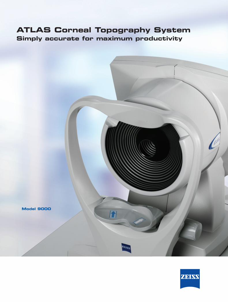
ATLAS Corneal Topography System Simply accurate for maximum productivity Model 9000
Transcript of ATLAS Corneal Topography System - Tecnologia Dos · · 2011-12-26ATLAS Corneal Topography System...
ATLAS Corneal Topography SystemSimply accurate for maximum productivity
Model 9000
ATL.
1587
SA
P 00
0000
-150
2-42
0©
2007
Car
l Zei
ss M
edite
c, In
c. A
ll rig
hts r
eser
ved.
Spe
cific
atio
ns su
bjec
t to
chan
ge. P
rinte
d in
USA
. 090
7 5M
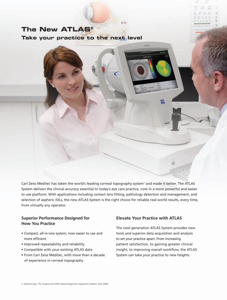
Superior Performance Designed for How You Practice
• Compact, all-in-one system, now easier to use and
more efficient
• Improved repeatability and reliability
• Compatible with your existing ATLAS data
• From Carl Zeiss Meditec, with more than a decade
of experience in corneal topography
Elevate Your Practice with ATLAS
The next-generation ATLAS System provides new
tools and superior data acquisition and analysis
to set your practice apart. From increasing
patient satisfaction, to gaining greater clinical
insight, to improving overall workflow, the ATLAS
System can take your practice to new heights.
The New ATLAS® Take your practice to the next level
Carl Zeiss Meditec has taken the world’s leading corneal topography system1 and made it better. The ATLAS
System delivers the clinical accuracy essential to today’s eye care practice, now in a more powerful and easier
to use platform. With applications including contact lens fitting, pathology detection and management, and
selection of aspheric IOLs, the new ATLAS System is the right choice for reliable real-world results, every time,
from virtually any operator.
1- Marketscope, The Surgical and Office Based Diagnostic Equipment Market, April 2006
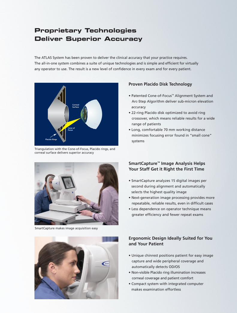
Proven Placido Disk Technology
• Patented Cone-of-Focus™ Alignment System and
Arc-Step Algorithm deliver sub-micron elevation
accuracy
• 22-ring Placido disk optimized to avoid ring
crossover, which means reliable results for a wide
range of patients
• Long, comfortable 70 mm working distance
minimizes focusing error found in “small cone”
systems
SmartCapture™ Image Analysis Helps Your Staff Get it Right the First Time
• SmartCapture analyzes 15 digital images per
second during alignment and automatically
selects the highest quality image
• Next-generation image processing provides more
repeatable, reliable results, even in difficult cases
• Less dependence on operator technique means
greater efficiency and fewer repeat exams
Ergonomic Design Ideally Suited for You and Your Patient
• Unique chinrest positions patient for easy image
capture and wide peripheral coverage and
automatically detects OD/OS
• Non-visible Placido ring illumination increases
corneal coverage and patient comfort
• Compact system with integrated computer
makes examination effortless
Proprietary Technologies Deliver Superior Accuracy
The ATLAS System has been proven to deliver the clinical accuracy that your practice requires.
The all-in-one system combines a suite of unique technologies and is simple and efficient for virtually
any operator to use. The result is a new level of confidence in every exam and for every patient.
SmartCapture makes image acquisition easy
Corneal Surface
Placido Rings
Cone ofFocus
Triangulation with the Cone-of-Focus, Placido rings, and corneal surface delivers superior accuracy
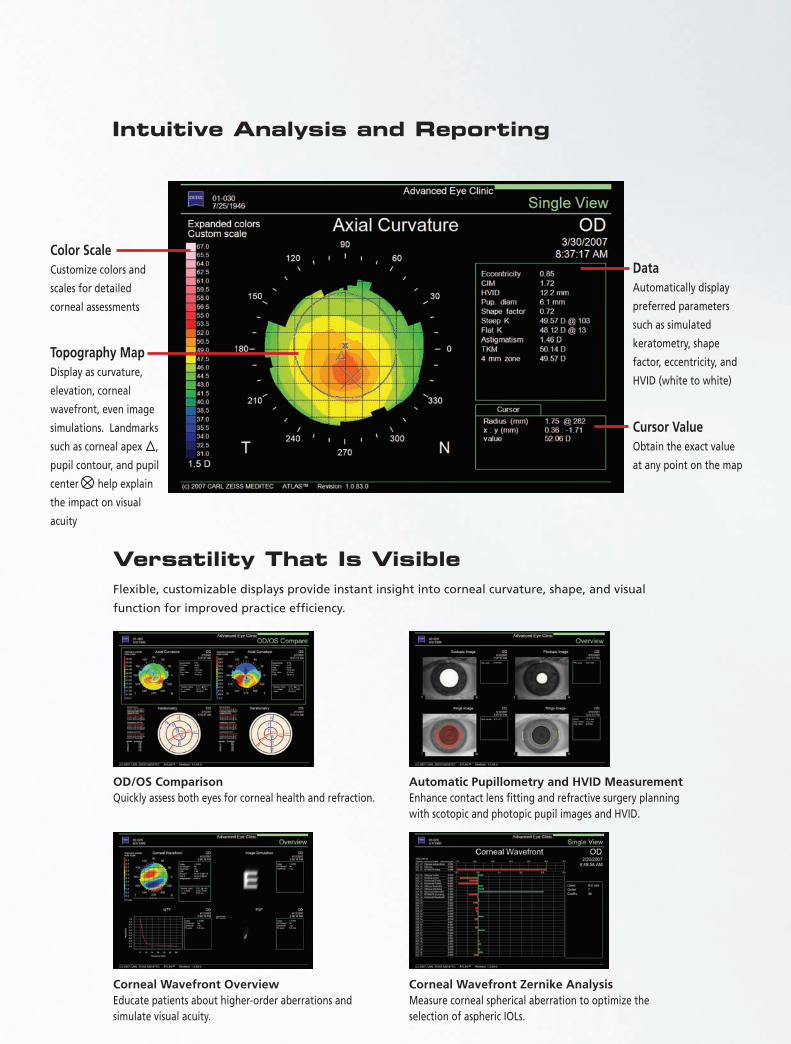
Color ScaleCustomize colors and
scales for detailed
corneal assessments
Intuitive Analysis and Reporting
Topography MapDisplay as curvature,
elevation, corneal
wavefront, even image
simulations. Landmarks
such as corneal apex ,
pupil contour, and pupil
center help explain
the impact on visual
acuity
DataAutomatically display
preferred parameters
such as simulated
keratometry, shape
factor, eccentricity, and
HVID (white to white)
Cursor ValueObtain the exact value
at any point on the map
Versatility That Is VisibleFlexible, customizable displays provide instant insight into corneal curvature, shape, and visual
function for improved practice efficiency.
OD/OS ComparisonQuickly assess both eyes for corneal health and refraction.
Automatic Pupillometry and HVID MeasurementEnhance contact lens fitting and refractive surgery planning with scotopic and photopic pupil images and HVID.
Corneal Wavefront Overview Educate patients about higher-order aberrations and simulate visual acuity.
Corneal Wavefront Zernike Analysis Measure corneal spherical aberration to optimize the selection of aspheric IOLs.
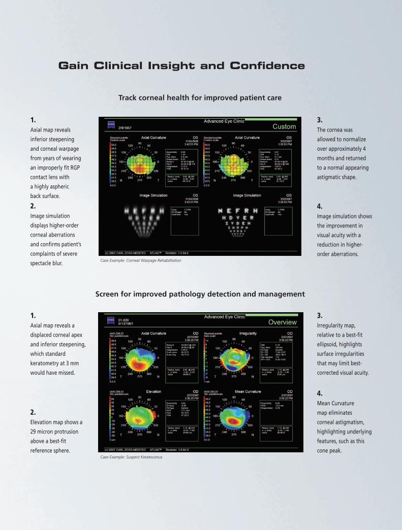
Gain Clinical Insight and Confidence
Track corneal health for improved patient care
Case Example: Corneal Warpage Rehabilitation
Screen for improved pathology detection and management
3.The cornea was
allowed to normalize
over approximately 4
months and returned
to a normal appearing
astigmatic shape.
4.Image simulation shows
the improvement in
visual acuity with a
reduction in higher-
order aberrations.
1.Axial map reveals
inferior steepening
and corneal warpage
from years of wearing
an improperly fit RGP
contact lens with
a highly aspheric
back surface.
2.Image simulation
displays higher-order
corneal aberrations
and confirms patient’s
complaints of severe
spectacle blur.
1.Axial map reveals a
displaced corneal apex
and inferior steepening,
which standard
keratometry at 3 mm
would have missed.
2.Elevation map shows a
29 micron protrusion
above a best-fit
reference sphere.
3.Irregularity map,
relative to a best-fit
ellipsoid, highlights
surface irregularities
that may limit best-
corrected visual acuity.
4.Mean Curvature
map eliminates
corneal astigmatism,
highlighting underlying
features, such as this
cone peak.Case Example: Suspect Keratoconus
When patients entrust you with their eyesight, their vision and your expertise converge. ATLAS from
Carl Zeiss Meditec empowers you with the most intuitive, advanced diagnostic solution. Along with our
dedication to clinical and technical excellence, we offer world-class training, on-site support and ongoing
educational opportunities. Partner with Zeiss for maximum productivity.
ATL.
1587
SA
P 00
0000
-150
2-42
0©
2007
Car
l Zei
ss M
edite
c, In
c. A
ll rig
hts r
eser
ved.
Spe
cific
atio
ns su
bjec
t to
chan
ge. P
rinte
d in
USA
. 090
7 5M
Technical Specifications
Working DistanceField of View Placido Rings Illumination Source Optics Curvature Measurement Range Accuracy ReproducibilityHVID (white to white) Measurement Range ResolutionPupillometry Acquired Images Measurement Range Resolution Views
Presentation Displays
Optional Software2
Computer
Dimensions/Weight(Instrument only)Electrical
70 mm17 mm X 14.5 mm22 (18 superiorly, 22 inferiorly)Non-visible infrared (950 nm) LEDDigital CMOS camera with 1280x1024 pixel resolution 15 to 95 D (3.5 to 22.5 mm)± 0.05 D (± 0.01 mm)1
± 0.10 D (± 0.02 mm)1
10.0 to 14.0 mm0.1 mmScotopic and photopic (700 nm)0.5 to 11.0 mm0.1 mm• Axial Curvature• Tangential Curvature• Elevation (Best-Fit Sphere)• Irregularity (Best-Fit Ellipsoid)• Videokeratoscopic (Rings, Scotopic, Photopic)• Keratometry• Refractive Power• Mean Curvature• Corneal Wavefront• Image Simulation• Point Spread Function (PSF)• Modulation Transfer Function (MTF)• Single View• Overview• OD/OS Comparison• Difference • Trend with Time• Custom• PathFinder™ II Corneal Analysis Software• MasterFit™ II Contact Lens Software• ATLAS™ Review Software• Windows® XP Professional• Pentium® M Processor • Internal storage: up to 35,000 exams• CD-RW/DVD-ROM• 3 Ethernet, 2 USB 2.0 ports• Integrated 12.1” color flat panel display• 52 L x 37 W x 50 H (cm)• 39 lbs. (17.7 kg)100-240V~: 50/60Hz, 2-1A
Carl Zeiss Meditec AG
Goeschwitzer Str. 51-52
07745 Jena
Germany
Phone: +49 3641220-333
Fax: +49 3641220-282
www.meditec.zeiss.com
1- To one standard deviation on a properly calibrated 42.51 D (7.94 mm) test object. 2- Available with ATLAS Software version 2.0
NOTE: All technical specifications are subject to change without notice.Windows is a registered trademark of Microsoft Corporation. Pentium is a registered trademark of Intel Corporation.
Carl Zeiss Meditec Inc.
5160 Hacienda Drive
Dublin, CA 94568
USA
Phone: +1 925 557 4100
Fax: +1 925 557 4101
www.meditec.zeiss.com
ATLAS Model 9000