
ATF6 as a Transcription Activator of the Endoplasmic Reticulum
11
MOLECULAR AND CELLULAR BIOLOGY, 0270-7306/00/$04.0010 July 2000, p. 5096–5106 Vol. 20, No. 14 Copyright © 2000, American Society for Microbiology. All Rights Reserved. ATF6 as a Transcription Activator of the Endoplasmic Reticulum Stress Element: Thapsigargin Stress-Induced Changes and Synergistic Interactions with NF-Y and YY1 MINGQING LI, PETER BAUMEISTER, BINAYAK ROY, TREVOR PHAN, DOLLY FOTI, SHENGZHAN LUO, AND AMY S. LEE* Department of Biochemistry and Molecular Biology, and the USC/Norris Comprehensive Cancer Center, Keck School of Medicine of the University of Southern California, Los Angeles, California 90089-9176 Received 11 February 2000/Returned for modification 10 March 2000/Accepted 22 March 2000 ATF6, a member of the leucine zipper protein family, can constitutively induce the promoter of glucose- regulated protein (grp) genes through activation of the endoplasmic reticulum (ER) stress element (ERSE). To understand the mechanism of grp78 induction by ATF6 in cells subjected to ER calcium depletion stress mediated by thapsigargin (Tg) treatment, we discovered that ATF6 itself undergoes Tg stress-induced changes. In nonstressed cells, ATF6, which contains a putative short transmembrane domain, is primarily associated with the perinuclear region. Upon Tg stress, the ATF6 protein level dropped initially but quickly recovered with the additional appearance of a faster-migrating form. This new form of ATF6 was recovered as soluble nuclear protein by biochemical fractionation, correlating with enhanced nuclear localization of ATF6 as revealed by immunofluorescence. Optimal ATF6 stimulation requires at least two copies of the ERSE and the integrity of the tripartite structure of the ERSE. Of primary importance is a functional NF-Y complex and a high-affinity NF-Y binding site that confers selectivity among different ERSEs for ATF6 inducibility. In addition, we showed that YY1 interacts with ATF6 and in Tg-treated cells can enhance ATF6 activity. The ERSE stimulatory activity of ATF6 exhibits properties distinct from those of human Ire1p, an upstream regulator of the mammalian unfolded protein response. The requirement for a high-affinity NF-Y site for ATF6 but not human Ire1p activity suggests that they stimulate the ERSE through diverse pathways. ATF6 was first isolated as a member of an extensive family of leucine zipper proteins able to selectively form DNA bind- ing heterodimers (5). ATF6 is a 670-amino-acid protein; how- ever, it exhibits an electrophoretic mobility of 90 kDa in so- dium dodecyl sulfate (SDS)-polyacrylamide gels (36). The physiological role of ATF6 in the control of gene expression in mammalian cells is just emerging. Intriguingly, while ATF6 was originally isolated through probing of a cDNA expression li- brary with a multimerized ATF site, specific DNA binding for ATF6 has not yet been demonstrated. Functional studies in- dicate that ATF6 can bind to the serum response factor tran- scriptional activation domain, and transfection experiments us- ing antisense vector directed against ATF6 suggest that it is required for the serum induction of the c-fos promoter (36). Further, it was discovered that ATF6, rather than the serum response factor, is the target of p38 mitogen-activated protein kinase (MAPK) phosphorylation (25). When mammalian cells are subjected to calcium depletion stress or protein glycosylation block, the transcription of a family of glucose-regulated protein (grp) genes encoding en- doplasmic reticulum (ER) chaperones is induced to high levels (11, 12). This induction process, referred to as the unfolded protein response (UPR) (10), is mediated by novel promoter regulatory motifs present in multiple copies on the promoters of ER chaperone genes (20, 33). The consensus mammalian ER stress response element (ERSE) consists of a tripartite structure CCAATN 9 CCACG, with N 9 being a strikingly GC- rich region of 9 bp. The ERSE binds multiple mammalian transcription factors including the multimeric CCAAT binding protein NF-Y (also referred to as CBF), YY1, Y-box proteins YB-1 and dbpA, and a newly discovered ER stress-inducible binding factor referred to as ERSF (14, 15, 20, 21). Using a dominant negative mutant to interfere with NF-Y function, it was demonstrated that optimal induction of ERSE requires NF-Y (35). While overexpression of YY1 has no effect on the basal activity of the grp78 promoter, it is able to stimulate the grp78 promoter activity in stressed cells (15). The various ERSEs share similar sequence motifs and can independently confer the mammalian ER stress response to heterologous promoters (13); however, there are subtle differ- ences which are evolutionarily conserved (3). In particular, ERSE-163 contained within the rat grp78 core (16) bears an unconventional CGAAT motif for NF-Y/CBF binding and the sequence CCAGC instead of the consensus CCACG motif found in ERSE-98, which is most proximal to the TATA ele- ment. Mutational analysis of ERSE-163 reveals that the unique CCAGC motif could be the binding site of a putative mam- malian homologue of the yeast Hac1 protein (yHac1p) that binds and activates the yeast UPR and that YY1 serves as a coactivator of this factor in mammalian cells (3). Using yeast one-hybrid screening, ATF6 was isolated as a putative ERSE binding protein (33). Overexpression of ATF6 in HeLa cells constitutively induced ERSE-mediated transcrip- tion. Subsequently, it was reported that in HeLa cells ATF6 is a transmembrane ER glycoprotein (6). Upon stress treatment, a fraction of the 90-kDa ATF6 underwent transient proteoly- * Corresponding author. Mailing address: Department of Biochem- istry and Molecular Biology, USC/Norris Comprehensive Cancer Cen- ter, Keck School of Medicine of the University of Southern California, 1441 Eastlake Ave., Los Angeles, CA 90089-9176. Phone: (323) 865- 0507. Fax: (323) 865-0094. E-mail: [email protected]. 5096 Downloaded from https://journals.asm.org/journal/mcb on 18 October 2021 by 211.105.41.41.
Transcript of ATF6 as a Transcription Activator of the Endoplasmic Reticulum

MOLECULAR AND CELLULAR BIOLOGY,0270-7306/00/$04.0010
July 2000, p. 5096–5106 Vol. 20, No. 14
Copyright © 2000, American Society for Microbiology. All Rights Reserved.
ATF6 as a Transcription Activator of the Endoplasmic ReticulumStress Element: Thapsigargin Stress-Induced Changes
and Synergistic Interactions with NF-Yand YY1
MINGQING LI, PETER BAUMEISTER, BINAYAK ROY, TREVOR PHAN, DOLLY FOTI,SHENGZHAN LUO, AND AMY S. LEE*
Department of Biochemistry and Molecular Biology, and the USC/Norris Comprehensive Cancer Center, Keck Schoolof Medicine of the University of Southern California, Los Angeles, California 90089-9176
Received 11 February 2000/Returned for modification 10 March 2000/Accepted 22 March 2000
ATF6, a member of the leucine zipper protein family, can constitutively induce the promoter of glucose-regulated protein (grp) genes through activation of the endoplasmic reticulum (ER) stress element (ERSE). Tounderstand the mechanism of grp78 induction by ATF6 in cells subjected to ER calcium depletion stressmediated by thapsigargin (Tg) treatment, we discovered that ATF6 itself undergoes Tg stress-induced changes.In nonstressed cells, ATF6, which contains a putative short transmembrane domain, is primarily associatedwith the perinuclear region. Upon Tg stress, the ATF6 protein level dropped initially but quickly recovered withthe additional appearance of a faster-migrating form. This new form of ATF6 was recovered as soluble nuclearprotein by biochemical fractionation, correlating with enhanced nuclear localization of ATF6 as revealed byimmunofluorescence. Optimal ATF6 stimulation requires at least two copies of the ERSE and the integrity ofthe tripartite structure of the ERSE. Of primary importance is a functional NF-Y complex and a high-affinityNF-Y binding site that confers selectivity among different ERSEs for ATF6 inducibility. In addition, we showedthat YY1 interacts with ATF6 and in Tg-treated cells can enhance ATF6 activity. The ERSE stimulatory activityof ATF6 exhibits properties distinct from those of human Ire1p, an upstream regulator of the mammalianunfolded protein response. The requirement for a high-affinity NF-Y site for ATF6 but not human Ire1p activitysuggests that they stimulate the ERSE through diverse pathways.
ATF6 was first isolated as a member of an extensive familyof leucine zipper proteins able to selectively form DNA bind-ing heterodimers (5). ATF6 is a 670-amino-acid protein; how-ever, it exhibits an electrophoretic mobility of 90 kDa in so-dium dodecyl sulfate (SDS)-polyacrylamide gels (36). Thephysiological role of ATF6 in the control of gene expression inmammalian cells is just emerging. Intriguingly, while ATF6 wasoriginally isolated through probing of a cDNA expression li-brary with a multimerized ATF site, specific DNA binding forATF6 has not yet been demonstrated. Functional studies in-dicate that ATF6 can bind to the serum response factor tran-scriptional activation domain, and transfection experiments us-ing antisense vector directed against ATF6 suggest that it isrequired for the serum induction of the c-fos promoter (36).Further, it was discovered that ATF6, rather than the serumresponse factor, is the target of p38 mitogen-activated proteinkinase (MAPK) phosphorylation (25).
When mammalian cells are subjected to calcium depletionstress or protein glycosylation block, the transcription of afamily of glucose-regulated protein (grp) genes encoding en-doplasmic reticulum (ER) chaperones is induced to high levels(11, 12). This induction process, referred to as the unfoldedprotein response (UPR) (10), is mediated by novel promoterregulatory motifs present in multiple copies on the promotersof ER chaperone genes (20, 33). The consensus mammalian
ER stress response element (ERSE) consists of a tripartitestructure CCAATN9CCACG, with N9 being a strikingly GC-rich region of 9 bp. The ERSE binds multiple mammaliantranscription factors including the multimeric CCAAT bindingprotein NF-Y (also referred to as CBF), YY1, Y-box proteinsYB-1 and dbpA, and a newly discovered ER stress-induciblebinding factor referred to as ERSF (14, 15, 20, 21). Using adominant negative mutant to interfere with NF-Y function, itwas demonstrated that optimal induction of ERSE requiresNF-Y (35). While overexpression of YY1 has no effect on thebasal activity of the grp78 promoter, it is able to stimulate thegrp78 promoter activity in stressed cells (15).
The various ERSEs share similar sequence motifs and canindependently confer the mammalian ER stress response toheterologous promoters (13); however, there are subtle differ-ences which are evolutionarily conserved (3). In particular,ERSE-163 contained within the rat grp78 core (16) bears anunconventional CGAAT motif for NF-Y/CBF binding and thesequence CCAGC instead of the consensus CCACG motiffound in ERSE-98, which is most proximal to the TATA ele-ment. Mutational analysis of ERSE-163 reveals that the uniqueCCAGC motif could be the binding site of a putative mam-malian homologue of the yeast Hac1 protein (yHac1p) thatbinds and activates the yeast UPR and that YY1 serves as acoactivator of this factor in mammalian cells (3).
Using yeast one-hybrid screening, ATF6 was isolated as aputative ERSE binding protein (33). Overexpression of ATF6in HeLa cells constitutively induced ERSE-mediated transcrip-tion. Subsequently, it was reported that in HeLa cells ATF6 isa transmembrane ER glycoprotein (6). Upon stress treatment,a fraction of the 90-kDa ATF6 underwent transient proteoly-
* Corresponding author. Mailing address: Department of Biochem-istry and Molecular Biology, USC/Norris Comprehensive Cancer Cen-ter, Keck School of Medicine of the University of Southern California,1441 Eastlake Ave., Los Angeles, CA 90089-9176. Phone: (323) 865-0507. Fax: (323) 865-0094. E-mail: [email protected].
5096
Dow
nloa
ded
from
http
s://j
ourn
als.
asm
.org
/jour
nal/m
cb o
n 18
Oct
ober
202
1 by
211
.105
.41.
41.

sis, releasing an N-terminal 50-kDa protein with activatingproperties that could be detected as a soluble nuclear proteinfollowing biochemical fractionation (6). These important ob-servations also raise several critical questions. For example,since in vitro-translated ATF6 failed to bind ERSE (33) andthus far direct DNA binding activity of ATF6 has not beendemonstrated, how does it activate ERSE activity? Since tran-scription of grp genes is sustained as long as the stress induceris present (13) whereas the appearance of p50 is transient (6,33), are there other mechanisms to sustain ATF6 activity? IsATF6 the predicted mammalian Hac1? The human and mousehomologues of yeast Irep, a transmembrane ER kinase with adual endonuclease activity that splices the yeast HAC1 mRNAinto a translatable form in response to ER stress, have beenisolated (26, 28). Overexpression of mammalian Ire1p activatesboth the grp genes and CHOP-encoding genes. Further, over-expression of the murine Ire1p also leads to the developmentof programmed cell death in transfected cells (28). This raisesthe critical issue of whether ATF6 is a downstream target ofthe mammalian Ire1p, the putative master switch of the mam-malian UPR.
In addressing these questions, we discovered that in additionto transient proteolysis, ATF6 itself undergoes specific changesin cells treated with thapsigargin (Tg), a strong inducer of themammalian UPR through perturbance of calcium homeosta-sis. The stimulation of ERSE-mediated transcription activityby ATF6 requires the integrity of the tripartite structure of theERSE, a high-affinity CCAAT binding site for NF-Y/CBF, anda functional NF-Y complex. ATF6 is an interactive partner ofYY1. In Tg-treated cells, the stimulatory activity of ATF6 can
be further enhanced by YY1 with a functional DNA bindingdomain. We showed that ATF6 exhibits properties distinctfrom those of human Ire1p (hIre1p) and yHac1p in activatingthe grp78 promoter, suggesting that they may represent diverseregulatory pathways.
MATERIALS AND METHODS
Cell culture conditions. NIH 3T3 and Cos cells were maintained in high-glucose Dulbecco’s modified Eagle’s medium containing 10% fetal bovine serum,2 mM glutamine, and 1% penicillin-streptomycin-neomycin at 35°C. For stressinduction, cells were grown to 80% confluence and treated with 300 nM Tg forvarious time intervals as indicated. For treatment with genistein, 24 h followingtransfection, the cells were treated with 140 mM genistein for different timeintervals as indicated. When the cells were treated with both genistein and Tg,genistein was added 15 min prior to Tg treatment.
Western blotting. Whole cell lysates were prepared in radioimmunoprecipita-tion (RIPA) buffer as previously described (4). For ATF6 Western blotting,50-mg aliquots of extracted proteins were separated by SDS-polyacrylamide gelelectrophoresis (SDS-PAGE) on 6 to 8% denaturing gels and transferred elec-trophoretically to Hybond ECL nitrocellulose membrane (Amersham, Piscat-away, N.J.). For detection of the ATF6 doublet protein bands, 6% gels were run.The blotted membrane was then blocked with 5% nonfat dry milk in 13 Tris-buffered saline (20 mM Tris-HCl [pH 7.5], 500 nM NaCl) for 1 h. The primaryantibody used was rabbit polyclonal ATF6 antibody directed against the c1.12domain (36) (kindly provided by Ron Prywes, Department of Biological Science,Columbia University) at a dilution of 1:800. The secondary antibody used washorseradish peroxidase-conjugated goat anti-rabbit (Sigma), at a dilution of1:5,000. For hemagglutinin epitope (HA)-tagged ATF6 Western blotting, 8 mg ofHA-tagged pCGN-ATF6 plasmid was transfected into 2 3 106 Cos cells usingSuperFect reagent (Qiagen Inc., Valencia, Calif.). Forty hours after transfectionwith or without specific drug treatment, the cells were lysed in RIPA buffer.Twenty micrograms of protein extract was separated by SDS-PAGE on 6%denaturing gels. The primary antibody used was mouse monoclonal HA antibody(Santa Cruz Biotechnology, Santa Cruz, Calif.) at a dilution of 1:500; the secondantibody used was horseradish peroxidase-conjugated goat anti-mouse (Roche,
FIG. 1. Tg stress-induced changes of ATF6. (A) Schematic drawing of the primary structure of ATF6, a 670-amino-acid (a.a.) protein. The positions of thetransactivation domain, the b-ZIP domain, and a putative transmembrane (TM) domain are in brackets. The locations of the serine-rich (SR) region, putative nuclearlocalization signal (NLS), and the subfragment c1.12 used to generate antibody against the basic region of ATF6 are also indicated. (B) Total cell lysates were preparedfrom NIH 3T3 cells treated with Tg for the indicated time and analyzed by immunoblotting using anti-ATF6 antibody. Twenty micrograms of each protein sample wasapplied to each lane. The single 90-kDa ATF6 band (at time zero) is indicated by a single closed arrowhead and the ATF6 doublet band in the later time points ismarked by the double arrowheads. The b-actin protein band (open arrowhead) in the same Western blot served as the protein loading control. (C) The level of ATF6following Tg stress was quantitated by densitometry and plotted against the kinetics of accumulation of the grp78 mRNA. The measurement of grp78 mRNA levels inTg-treated NIH 3T3 cells has been described elsewhere (13). A Western blot of ATF6 protein level after 0, 2, 8, and 12 h of Tg treatment is shown in the inset. Fiftymicrograms of each protein sample was applied to each lane. (D) Schematic drawing of HA-tagged full-length ATF6 (36). Shown below is the Western blot withanti-HA antibody performed on total cell lysate prepared from Cos cells transfected with empty vector (V) or with HA-ATF6 expression vector and treated with Tgfor the indicated time. The position of the HA-ATF6 doublet is indicated.
VOL. 20, 2000 MECHANISM OF ATF6 ACTIVATION OF THE grp78 PROMOTER 5097
Dow
nloa
ded
from
http
s://j
ourn
als.
asm
.org
/jour
nal/m
cb o
n 18
Oct
ober
202
1 by
211
.105
.41.
41.
Indianapolis, Ind.) at a dilution of 1:5,000. For the detection of GRP94, 30 mg ofHeLa nuclear extract or whole cell extract was separated by SDS-PAGE on an8% gel. The first antibody used was anti-KDEL rabbit polyclonal antibody at a1:1,000 dilution (StressGen, Victoria, British Columbia, Canada); an anti-rabbit-horseradish peroxidase conjugate was used as secondary antibody (Santa Cruz)at a 1:5,000 dilution. For the detection of YY1, the membrane was incubated ina 1:1,000 dilution of anti-mouse monoclonal YY1 antibody (Santa Cruz) and ata 1:5,000 dilution of goat anti-mouse horseradish peroxidase-conjugated anti-body (Santa Cruz). The protein bands were visualized by enhanced chemilumi-nescence (Amersham). The intensity of the protein bands was quantitated byscanning the autoradiographs with a Bio-Rad GS-700 densitometer.
Plasmids. The construction of grp promoter-chloramphenicol acetyltrans-ferase (CAT) fusion genes 2457, 2154, 2130, 2104, and 285 have been de-scribed elsewhere (29). The construction of (2169/2135)MCAT, (2159/2110)MCAT, and (2109/274)MCAT has been described elsewhere (13).Mutants of (2109/274)MCAT [CCAAT(m), CCACG(m1), and GGC(m)] werepreviously described (20). The (2169/2135)MCAT mutant CGAAT(m) wasconstructed similarly. The ERSE-131 mutant CCAAG(m1) was constructed us-ing a Quikchange site-directed mutagenesis kit (Stratagene, La Jolla, Calif.). Themutated bases in all constructs were confirmed by DNA sequencing. The mam-malian expression vector pCGN-ATF6 containing HA-tagged full-length ATF6driven by the cytomegalovirus (CMV) promoter was provided by Ron Prywes(Department of Biological Science, Columbia University); its construction hasbeen described elsewhere (36). Expression vectors CMV-YY1 and the DNAbinding site deletion mutant CMV-YY1D were gifts from Y. Shi (Harvard Med-ical School); their construction has been described previously (24). The expres-sion vector for hIre1p (26) was a gift from R. Kaufman (University of Michigan).The construction of pMCX-Hac1, the mammalian expression vector for the230-amino-acid yHac1 protein, has been described elsewhere (3). The expressionvector for NF-YA29 has been described elsewhere (18).
Transfection conditions. NIH 3T3 cells were seeded in six-well plates andgrown to 60 to 80% confluence. One microgram of the reporter plasmid was
cotransfected with 1 mg of pCH110, an expression vector for b-galactosidaseunder the control of the simian virus 40 (SV40) promoter, along with 1 mg ofpCGN-ATF6 or CMV empty vector, using SuperFect reagent (Qiagen). In someexperiments, 1 mg of CMV-YY1 or CMV-YY1D was also added. Transfectionswith hIre1p and yHac1p expression vectors were performed identically to that ofATF6. For stress induction, 24 h after transfection the cells were treated with 300nM Tg for 16 h prior to harvesting. Preparation of the cell lysates for CAT assaysand the quantitation of the CAT assays have been described elsewhere (15). Cellextracts corresponding to equal b-galactosidase units were used. Each transfec-tion was repeated three to five times.
Immunofluorescence staining. NIH 3T3 cells were grown to 80% confluencein chamber slides (Nalge Nunc International, Naperville, Ill.), washed with phos-phate-buffered saline (PBS), and fixed with 4% paraformaldehyde in PBS for 10min. For the detection of ATF6, the cells were stained with anti-ATF6 (c1.12)antibody (1:100 dilution) as primary antibody and fluorescein-labeled anti-rabbitimmunoglobulin G (IgG; 1:100 dilution; Vector Laboratories, Inc., Burlingame,Calif.) as secondary antibody. For the detection of YY1, cells were stained withanti-YY1 monoclonal antibody (1:100 dilution; Santa Cruz) as primary antibodyand Texas red anti-mouse IgG (1:100 dilution) as secondary antibody.
Biochemical fractionation. NIH 3T3 cells grown in duplicate in 150-mm-diameter dishes to 70% confluence were either nontreated or treated with Tg for6 h. The cells were trypsinized, washed with PBS twice, then resuspended in 600ml of hypotonic buffer (10 mM HEPES [pH 7.9], 0.1 mM EDTA, 1 mM dithio-threitol [DTT], 10 mM KCl, protease inhibitor cocktail [Roche]), and incubatedat 4°C for 10 min. The cells were lysed by the addition of 3 ml of NP-40 andvortexed for 30 s. After confirmation of cell lysis by Trypan blue uptake assay, thefree nuclei were pelleted from the cytoplasmic fraction by centrifugation at 5,000rpm and then washed twice in hypotonic buffer. For the preparation of solublenuclear protein, the nuclei were incubated successively in 300 ml of hypotonicbuffer containing 150, 250, or 500 mM NaCl and 10% glycerol at 4°C for 10 min.The supernatant from each wash was separated from the nuclei by centrifugationat 5,000 rpm. Aliquots of these supernatants were concentrated 2.5-fold byMicrocon-10 centrifugal filter devices (Millipore Corporation, Bedford, Mass.).For preparation of the membrane-bound protein, the final nucleus pellet wasincubated at room temperature with 300 ml of hypotonic buffer containing 10%glycerol and 1% SDS. Following low-speed shaking for 1 h, the sample wascentrifuged at 15,000 rpm for 30 min and the supernatant was collected. Forevaluation of protein concentration and integrity, equal volumes of the proteinsamples were incubated in Laemmli buffer at 95°C for 10 min, loaded on anSDS–8% polyacrylamide gel, and run at 50 V for 20 min and then 140 V for 45min. The gel was incubated in Coomassie blue staining solution for 1 h at roomtemperature and then destained overnight. Preparation of the highly concen-trated HeLa nuclear extract from control cells and cells treated with Tg for 6 hhas been described elsewhere (22).
EMSA. Conditions for the electrophoretic mobility shift assays (EMSAs) usingHeLa nuclear extract and synthetic ERSE as probe have been described else-where (20, 21).
GST pull-down assays. Glutathione S-transferase (GST) was used alone or asa GST-YY1 or GST-Ras fusion. In vitro-translated 35S-labeled ATF6 was pre-pared with the TNT coupled reticulocyte system from Promega (Madison, Wis.).Five microliters of the labeled ATF6 was mixed with bacterially expressed GSTproteins, and the pull-down assays were performed as previously described (31).
Coimmunoprecipitation assays. A total of 2 3 106 Cos cells were transfectedwith 8 mg of plasmid pCGN-ATF6 or pCGN using SuperFect reagent. The cellswith or without Tg treatment were harvested 48 h after transfection and lysed in300 ml of NP-40 buffer (0.5% NP-40, 50 mM HEPES [pH 7.5], 150 mM sodiumchloride). Protein extract (500 mg) from each sample was pretreated with 50 mlof protein A-Sepharose beads (Sigma) for 1 h at 4°C prior to incubation with 3ml of anti-YY1 monoclonal antibody (Santa Cruz), 2 ml of normal rabbit serum,or 2 ml of NF-Y/CBF rabbit polyclonal antibody (kindly provided by SankarMaity, University of Texas) for 30 min. Following the incubation period, 50 ml ofprotein A-Sepharose beads was added, and the mixtures were rotated at 4°Covernight. The beads were then washed successively once with buffer I (PBScontaining 0.5% NP-40 and 1 mM DTT), buffer II (100 mM Tris-HCl [pH 7.4],500 mM LiCl, 1 mM DTT), and buffer III (10 mM Tris-HCl [pH 7.4], 100 mMNaCl, 1 mM DTT). The immunoprecipitate was released from the washed beadsby the addition of 30 ml of 1 3 SDS loading buffer (50 mM Tris-HCl [pH 6.8],100 mM DTT, 2% SDS, 0.1% bromophenol blue, 10% glycerol), followed byheating at 100°C for 5 min. The supernatant obtained after centrifugation wasresolved by SDS-PAGE on an 8% gel and subjected to Western blot analysis todetect the coimmunoprecipitated protein.
RESULTS
Tg stress-induced changes of ATF6. The human cDNA se-quence of ATF6 predicts a protein of 670 amino acids (Fig.1A). As previously noted, ATF6 contains a serine-rich regionat its N terminus and a central basic region spanning fromamino acids 303 to 330 followed by a leucine zipper sequence(the b-ZIP region) (36). The transactivation domain of ATF6
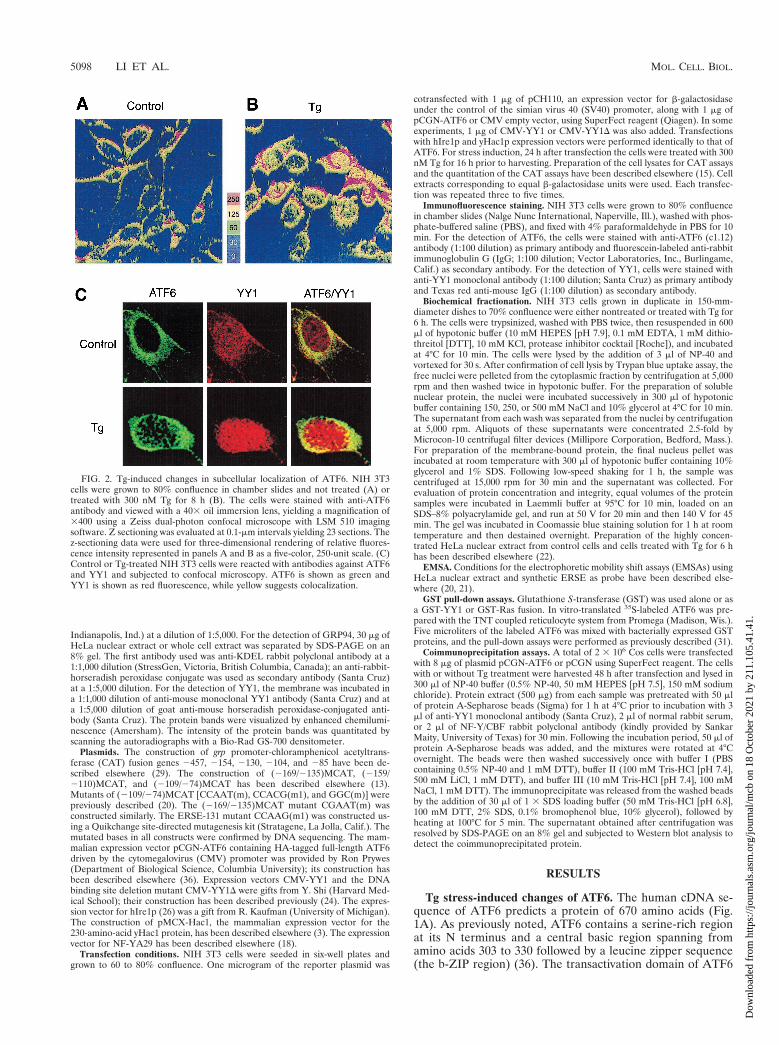
FIG. 2. Tg-induced changes in subcellular localization of ATF6. NIH 3T3cells were grown to 80% confluence in chamber slides and not treated (A) ortreated with 300 nM Tg for 8 h (B). The cells were stained with anti-ATF6antibody and viewed with a 403 oil immersion lens, yielding a magnification of3400 using a Zeiss dual-photon confocal microscope with LSM 510 imagingsoftware. Z sectioning was evaluated at 0.1-mm intervals yielding 23 sections. Thez-sectioning data were used for three-dimensional rendering of relative fluores-cence intensity represented in panels A and B as a five-color, 250-unit scale. (C)Control or Tg-treated NIH 3T3 cells were reacted with antibodies against ATF6and YY1 and subjected to confocal microscopy. ATF6 is shown as green andYY1 is shown as red fluorescence, while yellow suggests colocalization.
5098 LI ET AL. MOL. CELL. BIOL.
Dow
nloa
ded
from
http
s://j
ourn
als.
asm
.org
/jour
nal/m
cb o
n 18
Oct
ober
202
1 by
211
.105
.41.
41.

is contained within the first 273 amino acids (25). Computeranalysis of the primary sequence of ATF6 further predicts thelocation of several putative nuclear localization signals aroundamino acids 170, 330, and 380 and, strikingly, a short trans-membrane region from amino acids 378 to 398 flanked byhighly hydrophilic domains (7).
To detect ATF6 expression, Western blotting of total NIH3T3 cell lysate was performed, using an antibody with estab-lished specificity against ATF6 (36). In nonstressed NIH 3T3cells, ATF6 was constitutively expressed and migrated as asingle 90-kDa protein band in SDS-PAGE (Fig. 1B). Upon Tgtreatment, a reduction in endogenous ATF6 was observed at2 h, suggesting proteolysis as previously reported (6, 33). By4 h, the basal level of ATF6 was restored; at the same time, anew and slightly faster-migrating band was detected (Fig. 1B).This doublet band pattern persisted while the level of ATF6started to increase. By 12 h of Tg treatment, there was a four-to fivefold increase in the steady-state level of ATF6 comparedto nonstressed cells (Fig. 1C). We noted that the doublet ATF6band pattern was best resolved with low protein amountloaded on a 6% gel; when the protein amount was high, asshown in the inset of Fig. 1C, the doublet pattern merged intoone band. The kinetics of accumulation of p90 ATF6 as relatedto the increase in grp78 transcript level is summarized in Fig.1C.
To confirm that the new band was derived from ATF6, Coscells were transfected with an expression vector of HA-taggedp90 ATF6. Total cell lysate was prepared from cells treatedwith Tg for various times and subjected to Western blot anal-ysis with the anti-HA antibody (Fig. 1D). The anti-HA anti-body was highly specific for the exogenous ATF6 since theHA-ATF6 protein band was detected in cell lysate transfectedwith the ATF6 expression vector but not with the empty vector.As in the case of the endogenous ATF6 protein, a doublet wasobserved starting at 4 h after Tg treatment. In contrast to theendogenous protein, no decrease in the HA-ATF6 proteinlevel was detected following Tg treatment (Fig. 1D). For ex-
ogenously expressed ATF6, we could not detect any 50-kDaprotein of HA-ATF6 in either control or Tg-treated cells evenwhen large amounts of protein lysates were analyzed (data notshown). We also had difficulty detecting the endogenous p50ATF6 band in a consistent manner using the anti-ATF6 anti-body.
Enhanced nuclear localization of p90 ATF6 after Tg stress.To investigate the subcellular localization of endogenousATF6 in control and Tg-treated cells, immunofluorescencestudies were performed using anti-ATF6 antibody. As shownby the relative fluorescence intensity compiled by three-dimen-sional rendering of the z-sectioning data, ATF6 was detectedpredominantly in the perinuclear region in nonstressed controlcells (Fig. 2A), in agreement with a putative transmembranedomain in ATF6 (Fig. 1A). YY1, a soluble nuclear transcrip-tion factor, was primarily localized in the nucleus, while minoroverlap of YY1 and ATF6 immunofluorescence in the cyto-plasm was also observed in control cells (Fig. 2C). Upon Tgtreatment for 8 h correlating with the rapid increase in grp78transcript level (Fig. 1C), both ATF6 and YY1 showed in-creased nuclear localization while ATF6 retained perinuclearstaining (Fig. 2B and C). Further, through confocal micros-copy, the appearance of ATF6 in close proximity with YY1inside the nucleus was detected in Tg-treated but not controlcells (Fig. 2C).
The subcellular localization of p90 ATF6 was further inves-tigated through biochemical fractionation of NIH 3T3 cellextracts. Endogenous ATF6 was detected by immunoblottingas shown in Fig. 3A. Cell lysates were prepared from controlcells and cell treated with Tg for 6 h when the doublet form ofp90 ATF6 was prominent (Fig. 3B, lanes 1 and 2). Free nucleiwere isolated from the cytoplasm and subjected to successivesalt washes for the elution of soluble nuclear protein, and themembrane-bound protein was released from the nuclei pelletfollowing the final wash by resuspension in a buffer containing1% SDS. The protein samples were subjected to SDS-PAGE(6% gel) and Western blotted with anti-ATF6 antibody. In
FIG. 3. Biochemical fractionation of ATF6 in control and Tg-treated cells. (A) Representative immunoblot of whole cell extract (WCE) prepared from control NIH3T3 cells (C) and cells treated with Tg for 16 h (Tg) with the anti-ATF6 antibody. Positions of the molecular size marker are indicated on the left, and the positionof the ATF6 doublet is indicated by arrowheads. (B) Control cells and cells treated with Tg for 6 h were fractionated into cytoplasmic and nuclear fractions aftersuccessive washes with hypotonic buffer containing 0.15, 0.25, and 0.5 M NaCl. The membrane-bound protein was released from the final nucleus pellet with buffercontaining 1% SDS. WCE was prepared by lysing an aliquot of the cells directly in RIPA buffer. Equal volumes of the samples from control and Tg-treated cells wereapplied onto SDS–6% polyacrylamide gels. For the NaCl wash fractions, the supernatants were concentrated 2.5-fold prior to loading onto the gel. The upper panelshows an immunoblot with anti-ATF6 antibody. The lower panel shows Coomassie blue staining pattern of 5 ml each of WCE and 30 ml each of the other samples asindicated above. (C) Western blot of ATF6 using 30 mg of HeLa nuclear extract (NE) from control and Tg-treated cells applied onto an SDS–8% polyacrylamide gel.The same blot was reacted with anti-YY1 antibody. In panels B and C, positions of the molecular size marker are indicated. (D) Thirty-microgram aliquots of HeLaWCE or NE from control and Tg-treated cells were subjected to Western blotting for detection of GRP94.
VOL. 20, 2000 MECHANISM OF ATF6 ACTIVATION OF THE grp78 PROMOTER 5099
Dow
nloa
ded
from
http
s://j
ourn
als.
asm
.org
/jour
nal/m
cb o
n 18
Oct
ober
202
1 by
211
.105
.41.
41.

agreement with the immunofluorescence staining (Fig. 2), p90ATF6 from control cells was predominantly associated with themembrane fraction in control cells (Fig. 3B, lane 11). ForTg-treated cells, p90 ATF6 was detected also in the 0.25 and0.5 M NaCl wash fractions (Fig. 3B, lanes 8 and 10). Further,it was the faster-migrating form of ATF6 induced by Tg treat-ment that was preferentially recovered as the soluble nuclearprotein. The enrichment of the soluble nuclear protein in thesalt washes was confirmed by rehybridization of the sameWestern blot with an antibody directed against TFII-I/BAP-135, a soluble nuclear protein (32) (data not shown).
Localization of p90 ATF6 inside the nucleus is not confinedto NIH 3T3 cells. In nonstressed HeLa cells, nuclear stainingof ATF6 in addition to perinuclear staining was observed byimmunofluorescence (6). In highly concentrated HeLa nuclearextract containing both soluble and membrane-bound nuclearprotein prepared by the method of Shapiro et al. (22), p90ATF6 could be detected along with YY1 from both controlcells and cells treated with Tg for 6 h (Fig. 3C). Since the gelelectrophoresis was performed for a shorter time, the doubletform of ATF6 was not resolved under these conditions. Thesenuclear extracts were free of contamination from ER proteinssuch as GRP94 (19), which was readily detectable from theHeLa whole cell extract (Fig. 3D, lane 1) but was absent fromthe nuclear protein preparations (lanes 2 and 3).
Selective activation of ERSEs by ATF6. To map the ATF6target sites within the grp78 promoter, ATF6 expression vectorwas cotransfected with a series of 59 deletion mutants of the ratgrp78 promoter linked to the CAT reporter gene. In every
construct examined, the ability of ATF6 to stimulate the grp78promoter directly correlated with that of Tg stress. Thus, weobserved eight- and sixfold activation by ATF6 for the 2457and 2154 constructs and fourfold stimulation for the 2130construct but no stimulation with the 2104 or 285 construct(Fig. 4A). The failure to stimulate the 2104 construct showedthat ATF6 activation of the rat grp78 promoter requires at leasttwo copies of ERSE, and the failure to stimulate the 285construct ruled out the possibility that ATF6 acts through theTATA element of the grp78 promoter. When the ERSEs wereexamined individually, ERSE-98 [contained in duplicate within(2109/274) MCAT)] and ERSE-131 [contained in duplicatewithin (2159/2110) MCAT] were efficiently about five- toeightfold) activated by ATF6 and Tg (Fig. 4B). In contrast,ATF6 was not able to activate ERSE-163 [contained in dupli-cate within (2169/2135)MCAT], which responded to Tg stresswith a twofold induction. These results show that ATF6 induc-tion of the individual ERSE is selective, with ERSE-98 as themost efficient target.
ATF6 activation of the ERSE requires a strong NF-Y bind-ing site and a functional NF-Y complex. To map the sequencewithin wild-type (wt) ERSE-98 required for ATF6 stimulation,ERSE-98 with a mutated binding site for either NF-Y/CBF,ERSF, or YY1 was tested for ATF6 inducibility (Fig. 5A). Asconfirmed in EMSAs using HeLa nuclear extract, ERSE-98formed three protein complexes with the above three factors(20) (Fig. 6A). The ability to compete for YY1 and NF-Y/CBFbinding was lost upon mutation of the CCACG and CCAATsequences, respectively (Fig. 6B, lanes 3 and 4). These same
FIG. 4. Comparison of the sequence requirement for ATF6 and Tg stimulation of the rat grp78 promoter. (A) Summary of the effect of 59 deletion (2457, 2154,2130, 2104, and 285) of the rat grp78 promoter on ATF6 stimulation. The locations of the three ERSEs in the rat grp78 promoter with respect to the TATA sequencemotifs are indicated, and (11) represents the site of transcription initiation. The consensus ERSE unit is comprised of a CCAAT( ) and a CCACG (F) sequenceseparated by a 9-bp (∧) GC-rich motif. The grp-CAT constructs were transiently transfected into NIH 3T3 cells with either ATF6 expression vector or empty CMVvector. The fold stimulation by ATF6 overexpression (black bar) was compared to Tg treatment (striped bar), with standard deviations as indicated. (B) Selectivestimulation of the ERSEs by ATF6. Schematic drawings of the CAT genes driven by grp78 promoter subfragments containing the respective ERSE linked in duplicatecopies to the minimal mouse mammary tumor virus promoter are shown. The fold stimulation by ATF6 overexpression (black bar) is compared to Tg treatment (stripedbar), with standard deviations as indicated.
5100 LI ET AL. MOL. CELL. BIOL.
Dow
nloa
ded
from
http
s://j
ourn
als.
asm
.org
/jour
nal/m
cb o
n 18
Oct
ober
202
1 by
211
.105
.41.
41.

mutations also eliminated the stimulation of ERSE-98 byATF6, whereas mutation in the GGC motif which abolishedERSF binding (20) reduced the stimulation by ATF6 to abouthalf (Fig. 5A). Thus, optimal stimulation by ATF6 requires thesequence integrity of the tripartite structure of ERSE.
The importance of a strong NF-Y binding site for ATF6stimulation was further demonstrated by mutation analysis ofERSE-163, which cannot be stimulated by ATF6. ERSE-163differs from ERSE-98 in that it contains a weak NF-Y bindingsite but a stronger YY1 binding site (15). Thus, ERSE-163contains a CGAAT sequence for NF-Y binding instead of theconsensus CCAAT contained within ERSE-98 (Fig. 5B). Thisresulted in lower binding affinity for NF-Y, as confirmed by theweaker ability of ERSE-163 than of ERSE-98 to compete forNF-Y binding in EMSA (Fig. 6C, compare lanes 2 and 3 tolanes 6 and 7). Mutation of this weak CGAAT site to theconsensus CCAAT site resulted in equivalent binding affinityfor NF-Y as ERSE-98 (Fig. 6C, compare lanes 2 and 3 to lanes4 and 5). Correspondingly, the inability of ATF6 to activateERSE-163 was completely reversed when the CGAAT motif ofERSE-163 was mutated to conform to the consensus CCAATmotifs among the ERSEs (Fig. 5B). Similarly, conversion ofthe first CCAAC motif of ERSE-131 to CCAAT further in-creased its ability to respond to ATF6 and Tg induction (Fig.5C). These collective results establish high-affinity NF-Y/CBFbinding as critical for ATF6 stimulation of the ERSE. Further,the requirement of a functional complex of NF-Y/CBF forATF6 stimulation was tested through the use of NF-YA29, adominant negative mutant of NF-Y (18). NF-Y/CBF is a het-eromeric transcription factor consisting of at least three sub-units (17). NF-YA29, in which three amino acids in the DNAbinding domain of NF-YA have been mutated, forms a com-plex with NF-YB, rendering it functionally inactive as a tran-scription activator. Expression of NF-YA29 suppressed ATF6as well as Tg stimulation of ERSE-98 (Fig. 7). The sameamount of NF-YA29 was without effect on the SV40 promoter-driven CAT activity (Fig. 7).
YY1 is an interactive partner and a coactivator of ATF6 inTg-stressed cells. The lack of demonstrable binding of ATF6to the ERSE and the functional dependence of ATF6 on anintact NF-Y and YY1 binding site on ERSE-98 prompted us todetermine whether ATF6 activation is mediated by protein-protein interaction with either NF-Y, YY1, or both. To test fortheir in vivo association, Cos cells were transfected with eitherthe empty vector or the expression vector for the HA-taggedATF6. Total cell lysates prepared from nontreated or Tg-treated cells were subjected to immunoprecipitation with ei-ther anti-NF-Y, anti-YY1, or anti-HA antibody, with normalrabbit serum or preimmune serum as a negative control. Theimmunoprecipitates were then Western blotted with the anti-bodies to detect coimmunoprecipitation of the proteins. Ex-amples for these reactions are shown in Fig. 8. While we couldnot detect interaction between HA-ATF6 with NF-Y, YY1 wasable to interact with HA-ATF6 in both nonstressed (Fig. 8A)and Tg-stressed (Fig. 8B) extracts. To test for direct interactionbetween ATF6 and YY1, in vitro-translated p90 ATF6 wasmixed with GST-YY1, GST-Ras, or the GST vector alone. Ourresults showed that ATF6 could associate with recombinantYY1 in vitro but showed no affinity for either GST alone orGST-Ras (Fig. 8C).
The functional relationship between ATF6 and YY1 wasexamined by cotransfection of expression vectors for these twoproteins into NIH 3T3 cells (Fig. 9). We observed that whileATF6 itself was a strong inducer of ERSE-98 in nonstressedcells, coexpression of YY1 minimally increased the ATF6 ac-tivity, and YY1 deleted of its zinc finger DNA binding domain(YY1D) was without effect (Fig. 9). In contrast, under theTg-stressed conditions, YY1 enhanced ATF6 stimulation ofERSE-98. Although the magnitude of stimulation was only inthe range of 1.5- to 2-fold, probably due to the high abundanceof endogenous YY1, YY1D was not able to stimulate ATF6activity. We noted in these experiments that addition of Tg tothe ATF6-overexpressing cells further increased the promoteractivity from 10- to 35-fold (Fig. 9). This additional increase in
FIG. 5. Target sites for ATF6 and Tg stimulation within ERSE. (A) Summary of effect of mutation of the NF-Y binding site [CCAAT(m)], ERSF site [GGC(m)],and YY1 site [CCACG(m1)] within ERSE-98 on ATF6 and Tg stimulation. The locations of these sites within ERSE-98 are shown, with the mutated bases highlightedin bold lowercase. Transient transfections into NIH 3T3 cells were performed. The fold stimulation by ATF6 overexpression (black bar) is compared to Tg treatment(striped bar), with standard deviations as indicated. (B) Effect of mutation of the NF-Y binding site on ERSE-163 on ATF6 and Tg stimulation. The single base changewithin (2169/2135)MCAT is as indicated. (C) Effect of mutation of the NF-Y binding site on ERSE-131 on ATF6 and Tg stimulation. The single base change within(2159/2110)MCAT is indicated.
VOL. 20, 2000 MECHANISM OF ATF6 ACTIVATION OF THE grp78 PROMOTER 5101
Dow
nloa
ded
from
http
s://j
ourn
als.
asm
.org
/jour
nal/m
cb o
n 18
Oct
ober
202
1 by
211
.105
.41.
41.

promoter activity following Tg treatment was consistently ob-served even in transfectants with maximum dosage for ATF6overexpression (data not shown), suggesting that additionalmechanisms were activated to maximize grp78 promoter activ-ity under Tg stress conditions.
Distinct activating properties of hIre1p and ATF6. With theidentification of Ire1p as an upstream activator of the mam-malian UPR (9, 26, 28), we tested whether ATF6 is a down-stream target of hIre1p. Also, since yHac1p is an activator ofmammalian grp78 and grp94 promoter activities (3), we askedwhether ATF6 is the mammalian counterpart of yHac1 bycomparing the activating properties of ATF6 and yHac1. Thestress induction of the grp78 promoter has been shown to besensitive to the tyrosine kinase inhibitor genistein, which hasno effect on the basal grp78 promoter activity (1, 35). All threeproteins, hIre1p, ATF6, and yHac1p, were able to stimulatethe grp78 promoter three- to sixfold in the absence of ER stress(Fig. 10A). If the three proteins are functionally equivalent,their levels of sensitivity to genistein may be similar. Our re-sults showed that the induction of the grp78 promoter activityby both hIre1p and yHac1p was inhibited by genistein. Incontrast, ATF6-mediated stimulation of the grp78 promoteractivity was resistant to genistein treatment. Further, genisteinexhibited no effect on the Tg stress-induced changes of ATF6
(Fig. 10B). These results indicate either that ATF6 is down-stream of the genistein inhibitory step which suppresses hIre1paction or that ATF6 and hIre1p could act through diversemechanisms.
Next we examined the effect of coexpression of hIre1p andATF6. When NIH 3T3 cells were transfected with ATF6 orhIre1p individually, we detected a 9- or 5-fold, respectively,induction of ERSE-98 activity (Fig. 11A). Coexpression ofhIre1p with ATF6 resulted in a dosage-dependent increase inERSE-98 activity up to 17-fold. A kinase-defective form ofhIre1p, referred to as hIre1p(K599A), was constructed by sub-stituting the conserved lysine at residue 599 in the putativeATP binding domain with alanine (26). When hIre1p(K599A)was coexpressed with ATF6, no additive effect was observed(Fig. 11A). The stimulatory effect of hIre1p on ATF6 wasclearly not due to increased proteolytic cleavage of p90 ATF6.In cells cotransfected with hIre1p, the levels of ATF6 in con-trol, Tg-treated, genistein-treated, or Tg- and genistein-co-treated cells were similar or slightly higher than those trans-fected with the empty vector (Fig. 11B).
Since NF-Y is a critical component for ATF6 stimulation ofERSE-directed transcription, we next examined the effect of ahigh-affinity NF-Y binding site on the stimulatory activities ofhIre1p, ATF6, and yHac1. Using wt ERSE-163/CAT and mu-
FIG. 6. Effect of sequence mutations on factor binding affinity. (A) In the EMSA reactions, radiolabeled ERSE-98 (wt) was mixed with HeLa nuclear extractprepared from control (2) or Tg-treated cells (1) (lanes 1 and 2); the GGC(m) oligomer was used as radiolabeled probe (lanes 3 and 4); anti-YY1 antibody was addedto the EMSA reaction with the wt probe (lanes 5 and 6), and anti-NF-Y antibody was added to the EMSA reaction with the wt probe (lanes 7 and 8). (B) The NF-Yand YY1 complexes formed were subjected to competition by unlabeled oligomers. No competitor (lane 1) or a 50-fold molar excess of wt ERSE-98 (lane 2) or itsmutated forms (lanes 3 and 4) as indicated at the top was added. (C) The NF-Y and YY1 complexes were subjected to competition in EMSA. Lanes contained nocompetitor (lane 1), 10- and 50-fold molar excess of wt ERSE-98 (lanes 2 and 3), CGAAT mutant (lanes 4 and 5), or wt ERSE-163 (lanes 6 and 7), and 50-fold molarexcess of a random synthetic oligomer of equal length (lane 8). The sequences of the wt and mutated forms of ERSE-98 and ERSE-163 are shown in Fig. 5. Positionsof the ERSF, NF-Y, and YY1 complexes are indicated.
5102 LI ET AL. MOL. CELL. BIOL.
Dow
nloa
ded
from
http
s://j
ourn
als.
asm
.org
/jour
nal/m
cb o
n 18
Oct
ober
202
1 by
211
.105
.41.
41.

tated ERSE-163/CAT as the test genes, we observed that onlyyHac1p was able to stimulate the wt ERSE-163/CAT (Fig.12A). The induction level was about sixfold. In contrast, bothhIre1p and ATF6 were without effect. By converting the weakNF-Y binding site CGAAT within ERSE-163 to a CCAATconsensus motif, ATF6 was able to stimulate this promoterabout 10-fold. The same mutation did not rescue hIre1p activ-ity, nor did it further enhance the yHac1p activity. Similarly,ATF6 stimulation of ERSE-131/CAT was increased from 10-to 30-fold when the CCAAC motif was mutated to the con-sensus CCAAT motif; however, this same mutation had min-imal effect on hIre1p activity (Fig. 12B). Collectively, these
experiments established that ATF6 stimulation of the grp78promoter is enhanced by a high-affinity NF-Y binding site,whereas the activity of the downstream target of hIre1p thatmediates the activation of the grp78 promoter is not dependenton a strong NF-Y binding site. We further observed that incontrast to hIre1p and ATF6, yHac1p can neither bind norstimulate ERSE-98 activity (3).
DISCUSSION
The recent identification of ATF6 as a transcription factorthat can constitutively induce the grp promoter in an ERSE-dependent manner (33) raises the question of how this stimu-lation can be achieved. One proposed mechanism is that ERstress induced proteolysis of the membrane-bound p90 ATF6,releasing a soluble p50ATF6 and leading to induced transcrip-tion in the nucleus (6). This is similar to the proteolytic mat-uration cascade of steroid regulatory element binding protein(SREBP) to regulate cholesterol and fatty acid biosynthesis, aprocess which also takes place in the ER (27, 30). However,whereas the proteolytic cleavage product of SREBP is abun-dant and stable, p50 ATF6 was observed only transiently and invery low amounts (33). Further, the ability of ATF6 to bindDNA has not yet been established.
We report here that ATF6 undergoes multiple forms ofchange upon treatment of cells with Tg which perturbs calciumhomeostasis and propose a mechanism for its activation of thegrp78 promoter through physical and functional interactionwith previously established DNA binding components of theERSE (Fig. 13). ATF6 is constitutively expressed as a 90-kDaprotein in nonstressed cells. Within 2 h of Tg treatment, therewas an initial drop of endogenous ATF6 protein level, inagreement with proteolytic cleavage as reported elsewhere(33). For exogenously expressed p90 ATF6 that is highly po-
FIG. 7. Effect of coexpression of NF-YA29 on promoter activities. ForERSE-98-mediated promoter activity, NIH 3T3 cells were transfected with(2109/74)MCAT as the reporter gene. The cells were either nontreated, treatedwith Tg, or cotransfected with empty CMV vector, pCGN-ATF6, or NF-YA29 asindicated into NIH 3T3 cells. The CAT activity in nonstressed cells transfectedwith the empty CMV vector was set at 1. pSV2CAT, used as the reporter genefor SV40-mediated promoter activity, was cotransfected with either empty vectoror NF-YA29. The relative promoter activities are shown.
FIG. 8. Interaction of ATF6 with YY1 in coimmunoprecipitation assays. (A)Cell lysates in NP-40 buffer prepared from Cos cells transfected with either theempty vector (V) or pCGN-ATF6 (ATF6) were immunoprecipitated (IP) with 2ml of anti-NF-Y (lanes 1 and 2) or anti-YY1 (lanes 3 and 4) antibody. Theimmunoprecipitates were applied to SDS–8% denaturing polyacrylamide gelsand Western blotted with anti-HA antibody. The position of HA-ATF6 is indi-cated. (B) Cos cells transfected either with the empty vector (V) or HA-ATF6(ATF6) were subjected to Tg treatment. The lysate was immunoprecipitated withanti-HA (lanes 1 and 2) or anti-YY1 (lanes 3 and 4) antibody and Westernblotted with anti-HA antibody. The position of HA-ATF6 is indicated. The sameblot (lanes 1 through 4) was washed and Western blotted with anti-YY1 anti-body. The position of YY1 is indicated. (C) GST pull-down assays. The reactionswere performed using in vitro-translated ATF6 and GST (lane 1), GST-YY1(lane 2), and GST-Ras (lane 3). Lane 4 represents 20% of input radiolabeledATF6. The protein bound onto the beads was eluted, applied to an SDS–8%polyacrylamide gel, and detected by autoradiography. Positions of the proteinsize markers are indicated.
FIG. 9. Effect of overexpression of ATF6, YY1, and YY1D on ERSE-98-mediated CAT activity. The construct (2109/274)MCAT, used as the reportergene, was cotransfected with either the empty CMV vector (V) or expressionvector for ATF6, YY1, or YY1D, alone and in combinations as indicated, intoNIH 3T3 cells. The transfected cells were either grown under normal cultureconditions (control) or treated with Tg. The CAT activity in nonstressed cellstransfected with the empty CMV vector was set at 1. Relative promoter activitiesare shown with standard deviations.
VOL. 20, 2000 MECHANISM OF ATF6 ACTIVATION OF THE grp78 PROMOTER 5103
Dow
nloa
ded
from
http
s://j
ourn
als.
asm
.org
/jour
nal/m
cb o
n 18
Oct
ober
202
1 by
211
.105
.41.
41.

tent as a transactivator for ERSE, this drop in protein level wasnot observed. Rather, for both endogenous and exogenouslyexpressed ATF6, at 4 h following Tg stress, and an additional,faster-migrating form was detected. With prolonged Tg treat-ment, as the grp78 transcript accumulated to high levels, thetotal amount of ATF6 also increased. The new form of ATF6can be recovered as a soluble nuclear protein, in support ofenhanced nuclear localization of ATF6 as detected by immu-nofluorescence. How might the new form of ATF6 be gener-ated? Since this new form is also readily detected with exoge-nous expression of p90 ATF6 tagged at its N terminus with theHA epitope, it must have been generated from p90 ATF6through either alternative splicing or posttranslational modifi-cation. The former mechanism is particularly attractive sincesplicing of the short transmembrane domain from ATF6 willbe able to convert it from a predominantly membrane-associ-ated form to a soluble form. For posttranslational modifica-tion, it is possible Tg treatment triggers phosphorylation ofATF6, which is a known target for the p38 MAPK (25), orother kinases are involved. The faster-migrating form couldalso arise from alteration of glycosylation, acetylation, or meth-ylation status of ATF6 triggered by Tg stress. However, theATF6 doublet appeared to migrate slower than the nonglyco-sylated form of ATF6 generated by tunicamycin treatment ofthe cells (data not shown). An additional mechanism isthrough proteolytic cleavage. Since the HA tag at the N ter-minus was retained in the new form, proteolytic trimminginvolving the immediate N terminus of the protein is unlikely.Future investigations are needed to resolve this.
Considering that ATF6 either has no or extremely weak
DNA binding affinity, it may require interaction with otherproteins to activate the ERSE. Likely candidates for the ATF6interactive partners in achieving ERSE activation includeNF-Y, YY1, and ERSF, proteins known to bind to ERSE (20).Mutation analysis to define the sites on ERSE-98 required forATF6 stimulation revealed that binding sites for all three pro-teins are important. Of primary importance is NF-Y binding tothe CCAAT sequence motif within all ERSEs. NF-Y is a mul-timeric CCAAT binding protein and requires at least threesubunits to bind to its DNA site (17). Here we showed thatmutation of the CCAAT binding site within the ERSE abol-ished ATF6 stimulation. Conversely, by changing the low-af-finity NF-Y binding site to the consensus CCAAT binding sitewithin ERSE-163, ATF6 inducibility was restored. Similar ef-fects were observed for ERSE-131. We further showed that afunctional NF-Y complex is required for optimal ATF6 stim-ulation. Interestingly, NF-Y, acting synergistically with SREBP,is also found to be critical for the transcriptional activation ofcholesterol biosynthesis genes (2, 8).
YY1 is a ubiquitous transcription factor unique to highereukaryotes (23). Unconventional YY1 binding sites are locatedwithin the ERSEs (3, 20). Previous studies have shown thatYY1 regulates the c-fos promoter through direct interactionwith ATF/CREB, such that the C-terminal zinc finger domain
FIG. 10. Effect of genistein on ATF6 activity and protein level. (A) Effect ofgenistein on hIre1p, ATF6, and yHac1p stimulation of the grp78 promoter. NIH3T3 cells were transiently transfected with 2154CAT and either the empty CMVvector (V) or expression vector for hIre1p, ATF6, or yHac1p as indicated. Thecells were either nontreated (2G) or treated with 140 mM genistein (1G). CATactivity in the nontreated cells transfected with the empty CMV vector was set at1. Relative promoter activities are shown with standard deviations. (B) Effect ofgenistein on ATF6 protein level. Total cell lysates were prepared from NIH 3T3cells with no drug treatment or Tg treated for 2 or 10 h, in the presence orabsence of genistein (G), as indicated at the top; 30 mg of cell lysate from eachsample was applied to an SDS–6% polyacrylamide gel and Western blotted withanti-ATF6 antibody.
FIG. 11. Effect of hIre1p coexpression on ATF6. (A) NIH 3T3 cells weretransiently transfected with (2109/274)MCAT and either the empty CMV vec-tor (V), ATF6 expression vector, or hIre1p and its dominant negative mutantK599A, alone and in combination, as indicated. The relative promoter activitiesare shown with standard deviations. (B) Plasmid pCGN-ATF6 was cotransfectedinto Cos cells with the empty CMV vector (V) (lanes 1 to 4) or expression vectorfor hIre1p (lanes 5 to 8). The transfected cells were either nontreated (lanes 1and 5) or treated with Tg (lanes 2 and 6), genistein (lanes 3 and 7), or genisteinand Tg (lanes 4 and 8). Equal amounts of cell lysate from each sample wereWestern blotted with anti-HA antibody. The position of HA-ATF6 is indicated.
5104 LI ET AL. MOL. CELL. BIOL.
Dow
nloa
ded
from
http
s://j
ourn
als.
asm
.org
/jour
nal/m
cb o
n 18
Oct
ober
202
1 by
211
.105
.41.
41.

of YY1 is necessary and sufficient for binding with the b-ZIPregion of ATF (34). Here we showed that YY1 can interactwith ATF6 both in vitro and in vivo. We further observed thatoverexpression of YY1 also enhanced ATF6 stimulation ofERSE-98, suggesting that YY1 can act as a coactivator ofATF6. Recently, we showed that YY1 can stimulate yHac1p-
mediated activation of the grp78 promoter (3). Like ATF6,yHac1p is a b-ZIP protein and is an activator of the grp pro-moters in nonstressed cells. However, ATF6 and yHac1p ap-pear to act on different target sites of the grp78 promoter, withyHac1p having the unique ability to stimulate ERSE-163 bear-ing the weak NF-Y site (3). While the magnitude of activationby YY1 is modest due to the abundance of endogenous YY1,its stimulatory effect for both yHac1 and ATF6 is dependent onthe zinc finger DNA binding domain. This suggests that YY1could enhance ATF6 action through anchoring of the proteincomplex onto the ERSE, or it could involve protein-proteininteractions, leading to maximal stimulation. It is also possiblethat the YY1 binding site within the ERSE also binds other,yet unidentified proteins capable of interacting with ATF6 andmodifying ATF6 activity. Since Tg treatment of cells alreadyexpressing high levels of ATF6 results in substantial furtherenhancement of the grp response, additional mechanisms spe-cifically activated by Tg may contribute to maximal Tg stressinduction of the grp promoter. Such mechanisms may includemodification of ATF6 to convert it to a more potent form, aswell as the activation of YY1 and other cofactors.
In defining the relationship between ATF6 and hIre1p, wediscovered different properties for the two proteins in activat-ing the grp78 promoter. First, while overexpression of bothproteins can stimulate the grp78 promoter in nonstressed cells,the enhancement mediated by hIre1p, but not ATF6, is sensi-tive to the tyrosine kinase inhibitor genistein, previously shownto suppress the grp stress response (1, 35). Second, ATF6stimulation requires a strong NF-Y binding site, whereas theactivity of the downstream target of hIre1p that mediates theactivation of the grp78 promoter does not. Last, overexpres-sion of hIre1p does not promote proteolysis of ATF6, nor doesit substantially increase its protein level. While the critical issueof whether ATF6 is a downstream target of Ire1p requiresfurther investigations, the difference in their activating prop-erties suggests diverse regulatory pathways.
FIG. 12. Distinct activating properties of ATF6, hIre1p, and yHac1. (A) Effect of mutation of ERSE-163 on hIre1p, ATF6, and yHac1p stimulation. NIH 3T3 cellswere transiently transfected with the wt construct (2169/2135)MCAT or the construct bearing the CGAAT(m) mutation and either the empty CMV vector (V) orexpression vector for hIre1p, ATF6, or yHac1p as indicated. CAT activity in cells transfected with the empty CMV vector was set at 1. Relative promoter activities areshown with standard deviations. (B) Effect of mutation of ERSE-131 on hIre1p and ATF6 stimulation. NIH 3T3 cells were transiently transfected with the wt construct(2159/2110)MCAT or the construct bearing the CCAAC(m1) mutation and either the empty CMV vector (V) or expression vector for hIre1p or ATF6 as indicated.Relative promoter activities are shown with standard deviations.
FIG. 13. Model for ATF6 induction of grp78 following Tg stress. The 90-kDaATF6 is primarily associated with the perinuclear region in nonstressed cells.Upon Tg treatment, the ATF6 level initially drops but quickly recovers with theadditional appearance of a faster-migrating form ([ATF6]f) and an increase inthe amount of ATF6. A fraction of ATF6 enters the nucleus. Through interac-tion with YY1, ATF6 becomes part of a multiprotein complex binding onto theERSE of the grp78 promoter. YY1 also enhances ATF6 activity. Other factorsthat bind to ERSE include an ERSF and the CCAAT binding protein NF-Y. Thestimulatory activity of ATF6 on the grp78 promoter depends on the integrity ofthe ERSE structure. In addition, a high-affinity NF-Y binding site and a func-tional NF-Y complex are required for optimal stimulation by ATF6. OtherTg-induced modifications of the transcription factors may also occur. This mul-tiprotein complex, acting in concert with the basal transcription machinery,stimulates grp78 transcription.
VOL. 20, 2000 MECHANISM OF ATF6 ACTIVATION OF THE grp78 PROMOTER 5105
Dow
nloa
ded
from
http
s://j
ourn
als.
asm
.org
/jour
nal/m
cb o
n 18
Oct
ober
202
1 by
211
.105
.41.
41.
ACKNOWLEDGMENTS
We are greatly indebted to Ron Prywes, Yang Shi, Randy Kaufman,Sankar Maity, and Peter Edwards for the ATF6, YY1, hIre1p, andNF-Y/CBF reagents; we thank Ebrahim Zandi, Ramachandra Reddy,and Xinke Chen for helpful discussions.
We thank David Hinton and Ernesto Barron for helpful suggestionsand assistance in microscopy, which was performed at the ElectronMicroscopy Core Facility at the Doheny Eye Institute, USC, supportedby NEI/NIH core grant EY03040 and the Norris Cancer Center. Thiswork was supported by Public Health Service grant CA27607 from theNCI, National Institutes of Health, to A.S.L.
REFERENCES1. Cao, X., Y. Zhou, and A. S. Lee. 1995. Requirement of tyrosine- and serine/
threonine kinases in the transcriptional activation of the mammalian grp78/BiP promoter by thapsigargin. J. Biol. Chem. 270:494–502.
2. Dooley, K. A., S. Millinder, and T. F. Osborne. 1998. Sterol regulation of3-hydroxy-3-methylglutaryl-coenzyme A synthase gene through a direct in-teraction between sterol regulatory element binding protein and the trimericCCAAT-binding factor/nuclear factor Y. J. Biol. Chem. 273:1349–1356.
3. Foti, D. M., A. Welihinda, R. J. Kaufman, and A. S. Lee. 1999. Conservationand divergence of the yeast and mammalian unfolded protein response.Activation of specific mammalian endoplasmic reticulum stress element ofthe grp78/BiP promoter by yeast Hac1. J. Biol. Chem. 274:30402–30409.
4. Gazit, G., J. Lu, and A. S. Lee. 1999. De-regulation of GRP stress proteinexpression in human breast cancer cell lines. Breast Cancer Res. Treat.54:135–146.
5. Hai, T. W., F. Liu, W. J. Coukos, and M. R. Green. 1989. Transcription factorATF cDNA clones: an extensive family of leucine zipper proteins able toselectively form DNA-binding heterodimers. Genes Dev. 3:2083–2090.
6. Haze, K., H. Yoshida, H. Yanagi, T. Yura, and K. Mori. 1999. Mammaliantranscription factor ATF6 is synthesized as a transmembrane protein andactivated by proteolysis in response to endoplasmic reticulum stress. Mol.Biol. Cell 10:3787–3799.
7. Hofmann, K., and W. Stoffel. 1993. TMbase—a database of membranespanning proteins segments. Biol. Chem. Hoppe-Seyler 347:166.
8. Jackson, S. M., J. Ericsson, R. Mantovani, and P. A. Edwards. 1998. Syn-ergistic activation of transcription by nuclear factor Y and sterol regulatoryelement binding protein. J. Lipid Res. 39:1712–1721.
9. Kaufman, R. J. 1999. Stress signaling from the lumen of the endoplasmicreticulum: coordination of gene transcriptional and translational controls.Genes Dev. 13:1211–1233.
10. Kozutsumi, Y., M. Segal, K. Normington, M. J. Gething, and J. Sambrook.1988. The presence of malfolded proteins in the endoplasmic reticulumsignals the induction of glucose-regulated proteins. Nature 332:462–464.
11. Lee, A. S. 1987. Coordinated regulation of a set of genes by glucose andcalcium ionophores in mammalian cells. Trends Biochem. Sci. 12:20–23.
12. Lee, A. S. 1992. Mammalian stress response: induction of the glucose-regu-lated protein family. Curr. Opin. Cell Biol. 4:267–273.
13. Li, W. W., S. Alexandre, X. Cao, and A. S. Lee. 1993. Transactivation of thegrp78 promoter by Ca21 depletion. A comparative analysis with A23187 andthe endoplasmic reticulum Ca21-ATPase inhibitor thapsigargin. J. Biol.Chem. 268:12003–12009.
14. Li, W. W., Y. Hsiung, V. Wong, K. Galvin, Y. Zhou, Y. Shi, and A. S. Lee.1997. Suppression of grp78 core promoter element-mediated stress inductionby the dbpA and dbpB (YB-1) cold shock domain proteins. Mol. Cell. Biol.17:61–68.
15. Li, W. W., Y. Hsiung, Y. Zhou, B. Roy, and A. S. Lee. 1997. Induction of themammalian GRP78/BiP gene by Ca21 depletion and formation of aberrantproteins: activation of the conserved stress-inducible grp core promoter el-ement by the human nuclear factor YY1. Mol. Cell. Biol. 17:54–60.
16. Li, W. W., L. Sistonen, R. I. Morimoto, and A. S. Lee. 1994. Stress inductionof the mammalian GRP78/BiP protein gene: in vivo genomic footprintingand identification of p70CORE from human nuclear extract as a DNA-binding component specific to the stress regulatory element. Mol. Cell. Biol.14:5533–5546.
17. Maity, S. N., and B. de Crombrugghe. 1998. Role of the CCAAT-bindingprotein CBF/NF-Y in transcription. Trends Biochem. Sci. 23:174–178.
18. Mantovani, R., X. Y. Li, U. Pessara, R. Hooft van Huisjduijnen, C. Benoist,and D. Mathis. 1994. Dominant negative analogs of NF-YA. J. Biol. Chem.269:20340–20346.
19. Reddy, R. K., J. Lu, and A. S. Lee. 1999. The endoplasmic reticulum chap-erone glycoprotein GRP94 with Ca21-binding and antiapoptotic propertiesis a novel proteolytic target of calpain during etoposide-induced apoptosis.J. Biol. Chem. 274:28476–28483.
20. Roy, B., and A. S. Lee. 1999. The mammalian endoplasmic reticulum stressresponse element consists of an evolutionarily conserved tripartite structureand interacts with a novel stress-inducible complex. Nucleic Acids Res.27:1437–1443.
21. Roy, B., W. W. Li, and A. S. Lee. 1996. Calcium-sensitive transcriptionalactivation of the proximal CCAAT regulatory element of the grp78/BiPpromoter by the human nuclear factor CBF/NF-Y. J. Biol. Chem. 271:28995–29002.
22. Shapiro, D. J., P. A. Sharp, W. W. Wahli, and M. J. Keller. 1988. A high-efficiency HeLa cell nuclear transcription extract. DNA 7:47–55.
23. Shi, Y., J.-S. Lee, and K. M. Galvin. 1997. Everything you have ever wantedto know about Yin Yang 1 . . . . Biochim. Biophys. Acta 1332:F49–F66.
24. Shi, Y., E. Seto, L. S. Chang, and T. Shenk. 1991. Transcriptional repressionby YY1, a human GLI-Kruppel-related protein, and relief of repression byadenovirus E1A protein. Cell 67:377–388.
25. Thuerauf, D. J., N. D. Arnold, D. Zechner, D. S. Hanford, K. M. DeMartin,P. M. McDonough, R. Prywes, and C. C. Glembotski. 1998. p38 Mitogen-activated protein kinase mediates the transcriptional induction of the atrialnatriuretic factor gene through a serum response element. A potential rolefor the transcription factor ATF6. J. Biol. Chem. 273:20636–20643.
26. Tirasophon, W., A. A. Welihinda, and R. J. Kaufman. 1998. A stress responsepathway from the endoplasmic reticulum to the nucleus requires a novelbifunctional protein kinase/endoribonuclease (Ire1p) in mammalian cells.Genes Dev. 12:1812–1824.
27. Wang, X., R. Sato, M. S. Brown, X. Hua, and J. L. Goldstein. 1994.SREBP-1, a membrane-bound transcription factor released by sterol-regu-lated proteolysis. Cell 77:53–62.
28. Wang, X. Z., H. P. Harding, Y. Zhang, E. M. Jolicoeur, M. Kuroda, and D.Ron. 1998. Cloning of mammalian Ire1 reveals diversity in the ER stressresponses. EMBO J. 17:5708–5717.
29. Wooden, S. K., L. J. Li, D. Navarro, I. Qadri, L. Pereira, and A. S. Lee. 1991.Transactivation of the grp78 promoter by malfolded proteins, glycosylationblock, and calcium ionophore is mediated through a proximal region con-taining a CCAAT motif which interacts with CTF/NF-I. Mol. Cell. Biol.11:5612–5623.
30. Worgall, T. S., S. L. Sturley, T. Seo, T. F. Osborne, and R. J. Deckelbaum.1998. Polyunsaturated fatty acids decrease expression of promoters withsterol regulatory elements by decreasing levels of mature sterol regulatoryelement-binding protein. J. Biol. Chem. 273:25537–25540.
31. Wu, F., and A. S. Lee. 1998. Identification of AP-2 as an interactive target ofRb and a regulator of the G1/S control element of the hamster histone H3.2promoter. Nucleic Acids Res. 26:4837–4845.
32. Yang, W., and S. Desiderio. 1997. BAP-135, a target for Bruton’s tyrosinekinase in response to B cell receptor engagement. Proc. Natl. Acad. Sci. USA94:604–609.
33. Yoshida, H., K. Haze, H. Yanagi, T. Yura, and K. Mori. 1998. Identificationof the cis-acting endoplasmic reticulum stress response element responsiblefor transcriptional induction of mammalian glucose-regulated proteins. In-volvement of basic leucine zipper transcription factors. J. Biol. Chem. 273:33741–33749.
34. Zhou, Q., R. W. Gedrich, and D. A. Engel. 1995. Transcriptional repressionof the c-fos gene by YY1 is mediated by a direct interaction with ATF/CREB. J. Virol. 69:4323–4330.
35. Zhou, Y., and A. S. Lee. 1998. Mechanism for the suppression of the mam-malian stress response by genistein, an anticancer phytoestrogen from soy.J. Natl. Cancer Inst. 90:381–388.
36. Zhu, C., F. E. Johansen, and R. Prywes. 1997. Interaction of ATF6 andserum response factor. Mol. Cell. Biol. 17:4957–4966.
5106 LI ET AL. MOL. CELL. BIOL.
Dow
nloa
ded
from
http
s://j
ourn
als.
asm
.org
/jour
nal/m
cb o
n 18
Oct
ober
202
1 by
211
.105
.41.
41.


![Endoplasmic reticulum[1]](https://static.fdocuments.in/doc/165x107/58ed5fc71a28aba1678b4611/endoplasmic-reticulum1.jpg)


