At the leading edge of three-dimensional cell...
10
Journal of Cell Science At the leading edge of three-dimensional cell migration Ryan J. Petrie* and Kenneth M. Yamada* Laboratory of Cell and Developmental Biology, National Institute of Dental and Craniofacial Research, National Institutes of Health, Bethesda, MD 20892, USA *Authors for correspondence ([email protected]; [email protected]) Journal of Cell Science 125, 5917–5926 ß 2012. Published by The Company of Biologists Ltd doi: 10.1242/jcs.093732 Summary Cells migrating on flat two-dimensional (2D) surfaces use actin polymerization to extend the leading edge of the plasma membrane during lamellipodia-based migration. This mode of migration is not universal; it represents only one of several mechanisms of cell motility in three-dimensional (3D) environments. The distinct modes of 3D migration are strongly dependent on the physical properties of the extracellular matrix, and they can be distinguished by the structure of the leading edge and the degree of matrix adhesion. How are these distinct modes of cell motility in 3D environments related to each other and regulated? Recent studies show that the same type of cell migrating in 3D extracellular matrix can switch between different leading edge structures. This mode-switching behavior, or plasticity, by a single cell suggests that the apparent diversity of motility mechanisms is integrated by a common intracellular signaling pathway that governs the mode of cell migration. In this Commentary, we propose that the mode of 3D cell migration is governed by a signaling axis involving cell–matrix adhesions, RhoA signaling and actomyosin contractility, and that this might represent a universal mechanism that controls 3D cell migration. Key words: Cell migration, Extracellular matrix, Intracellular signaling, Mechanotransduction, Pseudopodia Introduction Autonomous cell motility is found throughout the tree of life. For example, unicellular protozoa hunt their prey through mud and leaf litter along the pond bottom, whereas highly specialized immune cells seek and destroy microbial invaders within a complex three- dimensional (3D) extracellular matrix. The diversity in cell types and environments requiring 3D cell motility is associated with the wide diversity of molecular mechanisms used for migration (Allen and Allen, 1978; Friedl and Wolf, 2010; Haston et al., 1982). In vitro studies of metazoan cell migration have established that cells use actin polymerization coupled with cell–matrix adhesion to generate thin and wide lamellipodial protrusions to crawl across rigid 2D tissue culture surfaces (Abercrombie et al., 1970a; Abercrombie et al., 1970b; Dipasquale, 1975). However, imaging of cells moving in 3D models of extracellular matrix, as well as in vivo, reveal that lamellipodia-based motility is only one of multiple migration strategies (Friedl and Wolf, 2010; La ¨mmermann and Sixt, 2009; Ridley, 2011). These distinct migration strategies, or modes, are distinguished by differences in cell morphology, the extent of adhesion to the surrounding extracellular matrix and the mechanics of leading edge protrusion. Significantly, studies of both cancer cell lines and normal fibroblasts show that cells of the same type can use different modes of migration depending on the physical properties of the extracellular matrix, the degree of extracellular proteolysis and on soluble signaling factors (Petrie et al., 2012; Sanz-Moreno et al., 2011; Wolf et al., 2003). These demonstrations of mode switching, or plasticity (Friedl and Wolf, 2010), within a single cell type suggest that the apparent diversity of migration strategies might in fact be based on a common intracellular pathway that governs the switch between modes of cell motility. Although this type of regulation of cell morphology or migration mode had been suggested previously, the molecular mechanisms linking soluble signaling factors and matrix structure to the mode of 3D cell migration remained unclear (Albrecht-Buehler, 1980; Bovee, 1964; Friedl, 2004; Harris, 1994). Cells use cell–matrix adhesions in combination with contractility of the actin cytoskeleton to sense and respond to changes in rigidity of the extracellular matrix in 2D and 3D as reviewed previously (Peyton et al., 2007; Roca-Cusachs et al., 2012). The small GTPase RhoA, Rho-associated kinases (ROCK1 and ROCK2, hereafter referred to as ROCK), and the actin-binding motor protein myosin II are important components of this process of mechanotransduction. Significantly, this RhoA–ROCK– myosin-II signaling axis also has a key role in dictating the mode of 3D cell migration (Petrie et al., 2012; Sanz-Moreno and Marshall, 2010). This Commentary will first briefly review mechanotransduction and the cell motility cycle, before describing the distinct modes of 3D migration on the basis of the type of protrusion that forms the leading edge. We will then present evidence supporting the concept that the mode of 3D cell migration depends on the response of the ubiquitous RhoA– ROCK–myosin-II signaling axis to adhesion-mediated mechanical signals during cell migration. Regulation of actomyosin contractility by cell–matrix adhesion Intracellular signal transduction, or mechanotransduction, in response to elevated matrix stiffness can increase actomyosin contractility to initiate adhesion maturation, modulate gene transcription or trigger directional cell migration (Choi et al., 2008; Dupont et al., 2011; Lo et al., 2000; for recent in-depth reviews of the mechanisms of integrin-mediated mechanotransduction, see Moore et al., 2010; Roca-Cusachs et al., 2012; Schwarz and Gardel, 2012). Cells are able to sense and respond to changes in matrix rigidity by several mechanisms (Fig. 1). b1 integrins are the primary plasma membrane Commentary 5917
Transcript of At the leading edge of three-dimensional cell...
Journ
alof
Cell
Scie
nce
At the leading edge of three-dimensional cell migration
Ryan J. Petrie* and Kenneth M. Yamada*Laboratory of Cell and Developmental Biology, National Institute of Dental and Craniofacial Research, National Institutes of Health, Bethesda,MD 20892, USA
*Authors for correspondence ([email protected]; [email protected])
Journal of Cell Science 125, 5917–5926� 2012. Published by The Company of Biologists Ltddoi: 10.1242/jcs.093732
SummaryCells migrating on flat two-dimensional (2D) surfaces use actin polymerization to extend the leading edge of the plasma membraneduring lamellipodia-based migration. This mode of migration is not universal; it represents only one of several mechanisms of cellmotility in three-dimensional (3D) environments. The distinct modes of 3D migration are strongly dependent on the physical properties
of the extracellular matrix, and they can be distinguished by the structure of the leading edge and the degree of matrix adhesion. How arethese distinct modes of cell motility in 3D environments related to each other and regulated? Recent studies show that the same type ofcell migrating in 3D extracellular matrix can switch between different leading edge structures. This mode-switching behavior, or
plasticity, by a single cell suggests that the apparent diversity of motility mechanisms is integrated by a common intracellular signalingpathway that governs the mode of cell migration. In this Commentary, we propose that the mode of 3D cell migration is governed by asignaling axis involving cell–matrix adhesions, RhoA signaling and actomyosin contractility, and that this might represent a universal
mechanism that controls 3D cell migration.
Key words: Cell migration, Extracellular matrix, Intracellular signaling, Mechanotransduction, Pseudopodia
IntroductionAutonomous cell motility is found throughout the tree of life. Forexample, unicellular protozoa hunt their prey through mud and leaf
litter along the pond bottom, whereas highly specialized immunecells seek and destroy microbial invaders within a complex three-dimensional (3D) extracellular matrix. The diversity in cell types
and environments requiring 3D cell motility is associated with thewide diversity of molecular mechanisms used for migration (Allenand Allen, 1978; Friedl and Wolf, 2010; Haston et al., 1982).
In vitro studies of metazoan cell migration have established thatcells use actin polymerization coupled with cell–matrix adhesion
to generate thin and wide lamellipodial protrusions to crawl acrossrigid 2D tissue culture surfaces (Abercrombie et al., 1970a;Abercrombie et al., 1970b; Dipasquale, 1975). However, imaging
of cells moving in 3D models of extracellular matrix, as well as in
vivo, reveal that lamellipodia-based motility is only one of multiplemigration strategies (Friedl and Wolf, 2010; Lammermann and
Sixt, 2009; Ridley, 2011). These distinct migration strategies, ormodes, are distinguished by differences in cell morphology, theextent of adhesion to the surrounding extracellular matrix and the
mechanics of leading edge protrusion.
Significantly, studies of both cancer cell lines and normalfibroblasts show that cells of the same type can use different modes
of migration depending on the physical properties of theextracellular matrix, the degree of extracellular proteolysis and
on soluble signaling factors (Petrie et al., 2012; Sanz-Moreno et al.,2011; Wolf et al., 2003). These demonstrations of mode switching,or plasticity (Friedl and Wolf, 2010), within a single cell type
suggest that the apparent diversity of migration strategies might infact be based on a common intracellular pathway that governs theswitch between modes of cell motility. Although this type of
regulation of cell morphology or migration mode had beensuggested previously, the molecular mechanisms linking soluble
signaling factors and matrix structure to the mode of 3D cellmigration remained unclear (Albrecht-Buehler, 1980; Bovee,1964; Friedl, 2004; Harris, 1994).
Cells use cell–matrix adhesions in combination withcontractility of the actin cytoskeleton to sense and respond tochanges in rigidity of the extracellular matrix in 2D and 3D as
reviewed previously (Peyton et al., 2007; Roca-Cusachs et al.,2012). The small GTPase RhoA, Rho-associated kinases (ROCK1and ROCK2, hereafter referred to as ROCK), and the actin-binding
motor protein myosin II are important components of this processof mechanotransduction. Significantly, this RhoA–ROCK–
myosin-II signaling axis also has a key role in dictating themode of 3D cell migration (Petrie et al., 2012; Sanz-Moreno andMarshall, 2010). This Commentary will first briefly review
mechanotransduction and the cell motility cycle, beforedescribing the distinct modes of 3D migration on the basis of thetype of protrusion that forms the leading edge. We will then
present evidence supporting the concept that the mode of 3D cellmigration depends on the response of the ubiquitous RhoA–ROCK–myosin-II signaling axis to adhesion-mediated mechanical
signals during cell migration.
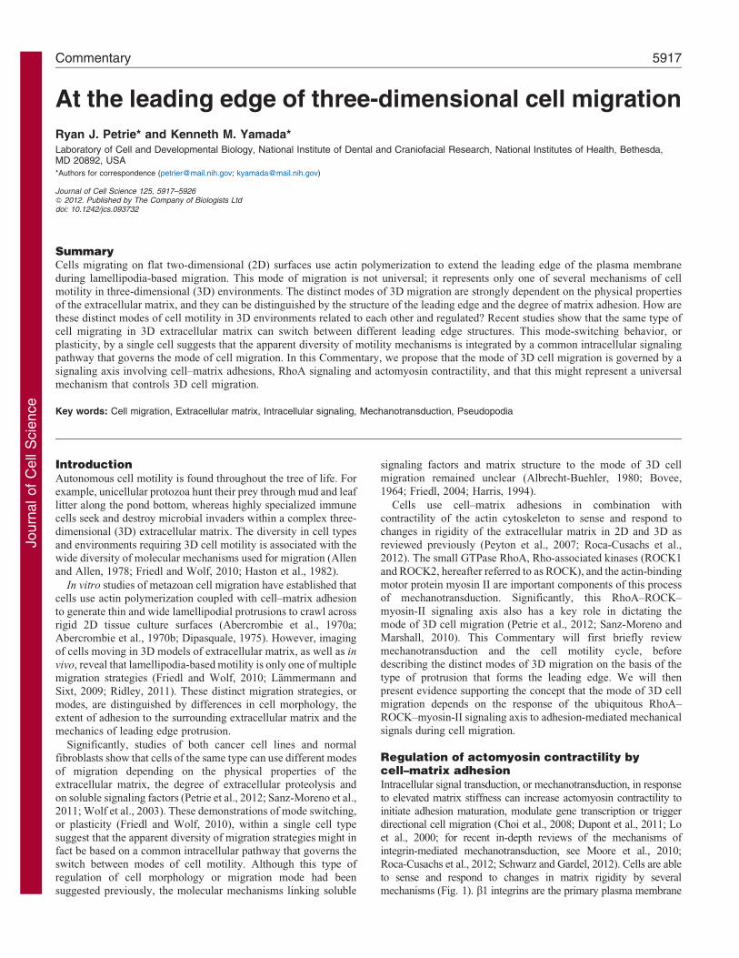
Regulation of actomyosin contractility bycell–matrix adhesionIntracellular signal transduction, or mechanotransduction, in response
to elevated matrix stiffness can increase actomyosin contractility toinitiate adhesion maturation, modulate gene transcription or triggerdirectional cell migration (Choi et al., 2008; Dupont et al., 2011; Lo
et al., 2000; for recent in-depth reviews of the mechanisms ofintegrin-mediated mechanotransduction, see Moore et al., 2010;Roca-Cusachs et al., 2012; Schwarz and Gardel, 2012). Cells are able
to sense and respond to changes in matrix rigidity by severalmechanisms (Fig. 1). b1 integrins are the primary plasma membrane
Commentary 5917
Journ
alof
Cell
Scie
nce
receptors transmitting tensional forces from the actin cytoskeleton to
the extracellular matrix (Danen et al., 2002; Guilluy et al., 2011b).
Myosin-II-mediated contractility is required for cells to actively sense
changes in the rigidity of the extracellular matrix (Engler et al., 2006;
Pelham and Wang, 1997). The action of myosin II along actin stress
fibers maintains the basal tension on the cell-matrix adhesions. This
basal tension enables mechanosensitive focal adhesion proteins to
sense the increase in resistance, which results when the basal
actomyosin tension pulls on a more rigid extracellular matrix. The
increased tension at focal adhesions can cause calcium influx through
stretch-activated calcium channels, trigger the integrin-dependent
activation of focal adhesion kinase (FAK) and of Src, and change the
conformation of certain mechanosensing proteins, such as p130Cas
(also known as BCAR1), talin and vinculin, to initiate intracellular
signaling and mechanotransduction (Moore et al., 2010).
Rho signaling also plays key roles in mechanotransduction.
Activating signals or diminished microtubule stability recruit the Rho
guanine-nucleotide-exchange factors (GEFs) GEF-H1 (also known
as ARHGEF2) and leukemia-associated Rho GEF (LARG, also
known as ARHGEF12) to b1-integin-containing focal adhesions
(Guilluy et al., 2011b; Heck et al., 2012). These GEFs bind and
activate RhoA by catalyzing the exchange of GDP for GTP. GTP-
bound RhoA can activate many downstream targets, such as ROCK
and the actin-nucleating family of mammalian homologues of
Diaphanous (mDia) proteins. mDia increases actin stress fiber
formation, and ROCK regulates phosphorylation of the regulatory
myosin light chain to further increase actomyosin contractility
(Fig. 1) (Nakano et al., 1999; Totsukawa et al., 2000). Thus, RhoA,
ROCK and myosin II activity act in a mechanical feedback loop to
respond to changes in extracellular matrix rigidity in 2D and 3D. In
addition, an important feature of this mechanical signaling network is
the contribution of extrinsic soluble factors, such as growth factors or
cytokines, which can modulate RhoA activity to increase or decrease
actomyosin contractility independently of matrix rigidity (Ridley and
Hall, 1992).
The cell motility cycleCell migration depends on a series of discrete cellular
mechanisms that function together during the cell motility
cycle (Lauffenburger and Horwitz, 1996), a process that is best
understood for metazoan cells adhering to and crawling over 2D
surfaces (Abercrombie et al., 1970a; Ridley et al., 2003).
Although the cell motility cycle and mechanotransduction share
many components (e.g. regulation by Rho family GTPases,
myosin II activity and cytoskeleton remodeling), these processes
can be considered independently when the cell is moving across a
structurally uniform surface.
The motility cycle begins when a stationary cell receives a
motogenic signal, such as the growth factors or cytokines in serum,
and becomes motile, forming distinct leading and trailing edges.
Internal polarization of both microtubules and the secretory
apparatus restrict lateral protrusions and facilitates the delivery
of vesicular cargo to the leading edge (Bergmann et al., 1983;
Gundersen and Bulinski, 1988; Kupfer et al., 1982; Vasiliev et al.,
1970). The Rho family GTPases Rac1 and Cdc42 activate the actin
nucleator Arp2/3 at the leading edge to polymerize actin and form
thin, wide lamellipodial protrusions (Nalbant et al., 2004; Wu et al.,
2012; Wu et al., 2009). The lipid second messenger
phosphatidylinositol (3,4,5)-trisphosphate [PtdIns(3,4,5)P3] is
also enriched in lamellipodia during persistent migration in a
given direction (Welf et al., 2012). Lamellipodia undergo cycles of
protrusion and retraction, but they can be stabilized by the
formation of nascent adhesions beneath the leading edge
(Giannone et al., 2004). Arp2/3-dependent actin polymerization
at the leading edge, in combination with myosin II activity, results
in actomyosin contraction and the retrograde flow of filamentous
actin (F-actin) towards the cell body (Ponti et al., 2004).
Basal actomyosin contractility
Matrixrigidity
GEF-H1 LARG
RhoA-GDP RhoA-GTP
ROCK
MLC P MLC
Nascentadhesion F-actin Myosin IIMature
adhesion
Increased actomyosin contractility
Mec
hani
cal f
eedb
ack
Potential regulation byRac–Rho crosstalk
Mechanosensingmatrix rigidity
WAVE2
ARHGAP22
NEDD9DOCK3
Rac1-GDP Rac1-GTP
Intracellularpressure
Membranetension
Rac1-GTP
FilGAP
Key
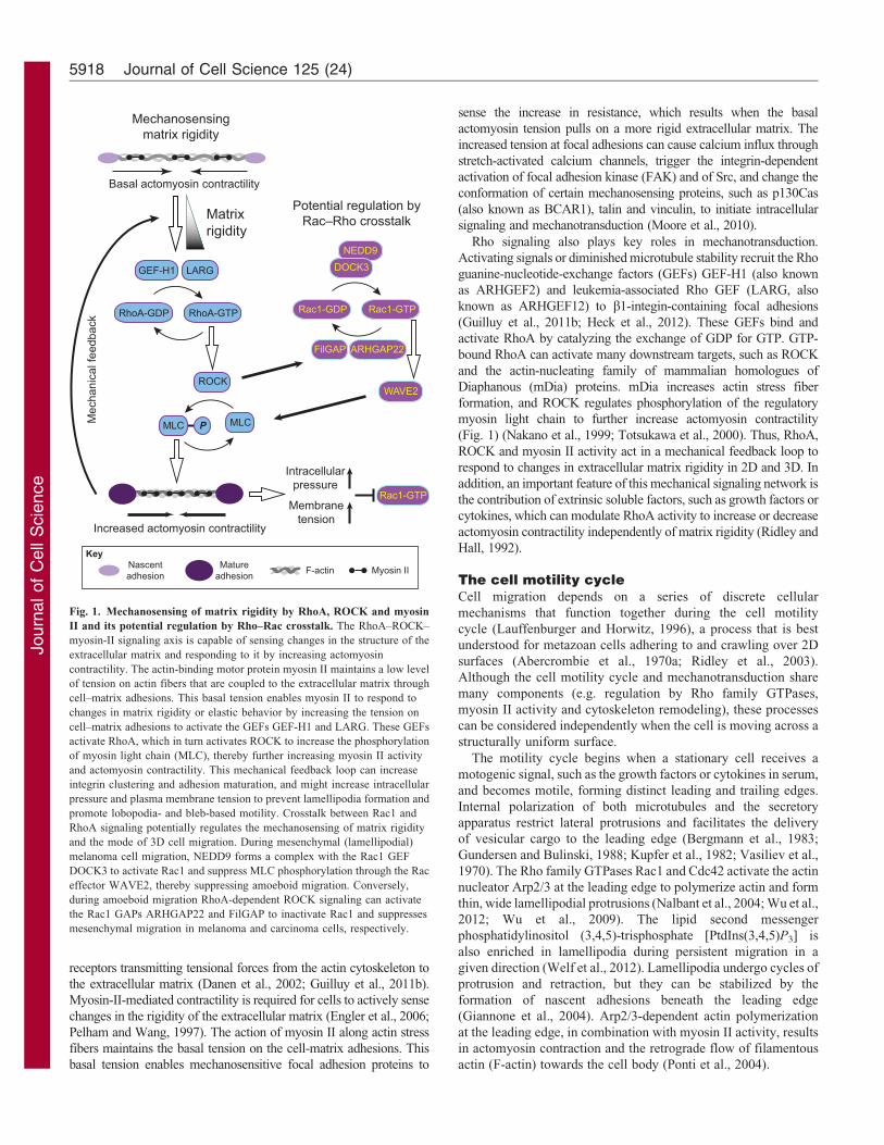
Fig. 1. Mechanosensing of matrix rigidity by RhoA, ROCK and myosin
II and its potential regulation by Rho–Rac crosstalk. The RhoA–ROCK–
myosin-II signaling axis is capable of sensing changes in the structure of the
extracellular matrix and responding to it by increasing actomyosin
contractility. The actin-binding motor protein myosin II maintains a low level
of tension on actin fibers that are coupled to the extracellular matrix through
cell–matrix adhesions. This basal tension enables myosin II to respond to
changes in matrix rigidity or elastic behavior by increasing the tension on
cell–matrix adhesions to activate the GEFs GEF-H1 and LARG. These GEFs
activate RhoA, which in turn activates ROCK to increase the phosphorylation
of myosin light chain (MLC), thereby further increasing myosin II activity
and actomyosin contractility. This mechanical feedback loop can increase
integrin clustering and adhesion maturation, and might increase intracellular
pressure and plasma membrane tension to prevent lamellipodia formation and
promote lobopodia- and bleb-based motility. Crosstalk between Rac1 and
RhoA signaling potentially regulates the mechanosensing of matrix rigidity
and the mode of 3D cell migration. During mesenchymal (lamellipodial)
melanoma cell migration, NEDD9 forms a complex with the Rac1 GEF
DOCK3 to activate Rac1 and suppress MLC phosphorylation through the Rac
effector WAVE2, thereby suppressing amoeboid migration. Conversely,
during amoeboid migration RhoA-dependent ROCK signaling can activate
the Rac1 GAPs ARHGAP22 and FilGAP to inactivate Rac1 and suppresses
mesenchymal migration in melanoma and carcinoma cells, respectively.
Journal of Cell Science 125 (24)5918

Journ
alof
Cell
Scie
nce
Actomyosin contraction leads to the enlargement and
strengthening of nascent adhesions, which, through the process
of adhesion maturation, then develop into focal adhesions (Choi
et al., 2008; Vicente-Manzanares et al., 2007). Intracellular
proteins in these maturing adhesions can act as components of a
molecular clutch, linking the retrograde flow of F-actin to the
extracellular matrix (Brown et al., 2006; Hu et al., 2007;
Mitchison and Kirschner, 1988). Engagement of the molecular
clutch slows the retrograde flow of F-actin and enables actin
polymerization at the leading edge to push the plasma membrane
forward. As the cell body and nucleus move over the mature focal
adhesions, adhesion disassembly is initiated and actomyosin
contracts the rear of the cell to form the trailing edge.
Although the basic components of the cell motility cycle
(protrusion, attachment, contraction and detachment) are likely to
be conserved among the different modes of 3D cell migration, the
underlying molecular mechanisms can vary (for reviews, see
Friedl and Wolf, 2009; Friedl and Wolf, 2010). These distinct
mechanisms manifest as differences in overall cell morphology,
the type and strength of cell–matrix adhesions, the speed of actin
retrograde flow, or the directional persistence and velocity of
migration (Danen et al., 2005; Doyle et al., 2012; Doyle et al.,
2009; Lammermann and Sixt, 2009; Renkawitz and Sixt, 2010).
It is the structure of the leading edge, however, that appears to
best define the mode of cell migration.
Protrusions define the mode of migrationThe mode of cell migration has historically been classified on the
basis of the morphology of the leading edge (Abercrombie et al.,
1970b; Bovee, 1964; Trinkaus, 1973), and molecular studies have
established that the different types of leading edge protrusions resultfrom distinct combinations of signaling and actin regulatory proteins
(recently reviewed in Charras and Paluch, 2008; Chhabra and Higgs,
2007; Ridley, 2011). Therefore, for the purposes of this
Commentary, we classify the mode of 3D cell migration primarily
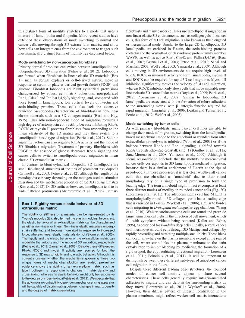
by the type of structure that forms the leading edge (Fig. 2; Table 1).
The current nomenclature of cellular protrusions associated
with cell motility is rooted in the morphological description of
unicellular protozoa (Calkins, 1933). The term pseudopodium
encompasses any dynamic structure extending from the cell body
that is capable of extension and retraction. These pseudopodia, or
more simply protrusions, occur as several functionally distinct
types. Rhizopodia are anastomosing protrusions with retrograde
transport towards the cell body, and they resemble retraction
Axopodia
Rhizopodia
Cellbody
A
B
C
D
Lamellipodia
Lobopodia
Amoeboid cancercells
Small blebs
Hemispherical blebs
Actin-enriched
2D g
lass
Vann
ella
miro
ides
3D C
DM
Am
oeba
pro
teus
2D g
lass
Fibr
obla
stK
erat
inoc
yte
2D g
lass
Fibr
osar
com
a ce
lls3D
col
lage
n
leading edge
Cortactin
3D c
olla
gen
Fibr
obla
st
F-actin
GFP–actin
GFP–actin
GFP–actin
Cortactin
LB
LB
LM
LM
LM
Fig. 2. Pseudopodium identity can define the mode of 2D and 3D
cell migration. There is a wide diversity in the types of pseudopodia,
or protrusions, that are used to extend the leading edge during cell
migration on 2D surfaces and in 3D extracellular matrix. The type of
pseudopodium can be used to define a specific mode of cell motility.
(A) Lamellipodia-based migration is used by cells that migrate on 2D
glass (upper and middle panels) and in a 3D collagen matrix (lower
panel). Lamellipodia are thin fan-shaped protrusions enriched in F-
actin and actin-binding proteins such as cortactin. (B) Lobopodia are
blunt cylindrical protrusions that might be driven by intracellular
pressure rather than actin polymerization. They are classically
associated with giant amoeba (lower panel), but are also formed by
metazoan cells migrating in linear elastic 3D material, such as cell-
derived matrix (CDM) (upper panel). (C) Round amoeboid migration
of cancer cells in 3D collagen comprises at least three distinct modes
of migration that are characterized by multiple small blebs (upper
panel), large hemispherical blebs (middle panel), or an actin-enriched
leading edge (lower panel). (D) Rhizopodia and axopodia are
pseudopodia used by certain protozoa to migrate and feed. LM,
lamellipodium; LB, lobopodium. Broken white arrows indicate the
direction of migration.
Pseudopodia and the mode of migration 5919

Journ
alof
Cell
Scie
nce
fibers found on dividing or migrating tissue culture cells (Koonce
and Schliwa, 1986; Mitchison, 1992). Axopodia are rigid radial
projections that are enriched in microtubules (Tilney, 1968). Both
rhizopodia and axopodia are used for movement and feeding by
particular classes of protozoa (Lee et al., 1985). Lobopodia are
blunt cylindrical protrusions driven by intracellular pressure,
used by lobose amoeba (Kudo, 1977), and have been recently
discovered in fibroblasts migrating in particular 3D environments
(Petrie et al., 2012). Increased intracellular pressure, acting alone
or in combination with local weakness in the cell cortex, can
trigger the rapid expansion of spherical membrane blebs (Charras
et al., 2005). These small cytoplasmic extensions are rapidly
retracted by actomyosin contraction and can be seen on moving
cells (Charras and Paluch, 2008). Lamellipodia are thin veil-like
extensions of the cytoplasm that are driven by actin
polymerization and are used by many tissue culture cells
migrating in 2D and 3D environments (Abercrombie et al.,
1970b; Petrie et al., 2012) and certain small amoeba (Lee and
Jacobson, 1997). Finally, filopodia are actin-rich finger-like
protrusions at the leading edge that act to sense the local
microenvironment (Chhabra and Higgs, 2007). Filopodia can act
alone or in combination with blebs, lobopodia or lamellipodia in
2D and 3D environments (Svitkina et al., 2003; Tomasek et al.,
1982; Trinkaus, 1973). Although not directly associated with the
leading edge, other types of pseudopodia are indirectly involved
in cell motility. Invadopodia and podosomes are actin-rich
structures, but they use integrin-dependent adhesion and
proteolysis to degrade extracellular matrix proteins and
penetrate 3D environments in normal and pathological settings
(Chen, 1989; Linder et al., 2011).
The existence of so many functionally distinct leading edge
structures might suggest that there are numerous independent
mechanisms that are capable of sustaining cell movement. Such
complexity would complicate our understanding of normal cell
migration and the molecular defects leading to cancer cell
invasion and metastasis. By contrast, individual metazoan cells
have been observed to switch rapidly between different modes of
3D cell migration. This mode-switching behavior is often
dictated by the level of activity of RhoA, ROCK and myosin
II, suggesting that these ubiquitous components help to govern
pseudopodium identity and the mode of 3D cell migration.
Mode switching during 3D cell migrationIt is well known that cytoskeleton and actomyosin contractility
can dramatically change cell morphology in response to
extracellular cues. For example, the small amoeba Vannella
miroides extends clear conical pseudopodia from a central cell
body when it is floating in liquid, but forms flattened
lamellipodia when migrating over 2D surfaces (Bovee, 1964;
Lee and Jacobson, 1997). This adaptability is also well
documented for ‘deep’ cells that migrate in the 2D space
between the epithelium and the underlying internal yolk layer
during embryonic development of the teleost Fundulus
(Trinkaus, 1973; Trinkaus and Erickson, 1983); these rapidly
migrating, weakly adherent cells use large hemispherical blebs
for migration, similar to primordial germ cells (Goudarzi et al.,
2012; Lammermann and Sixt, 2009). The blebs bulge outwards in
the direction of migration and fill with cytoplasm without the
characteristic retraction phase that is associated with smaller
blebs (Charras et al., 2005). During later developmental stages,
Table 1. The degree of adhesion and RhoA signaling can uniquely identify the mode of 3D migration
Mode of 3D migration Cell shape Mode switching AdhesionRhoA
signalingPolarizedsignaling
Rho and Raccross-talk References
Lobopodial Elongated Switch tolamellipodia
High Required No Unknown (Petrie et al.,2012)
Lamellipodial Elongated Switch tolobopodia(normal cells)and amoeboid(cancer cells)
High Not required Yes In mesenchymalcancer cellsRac1 activitysuppresses RhoAand amoeboidmigration. Rac1activity does notprevent normalfibroblasts fromswitching tolobopodialmotility.
(Petrie et al.,2012; Sahaiand Marshall,2003; Sanz-Moreno et al.,2008)
Amoeboid(cancer cells)a
Round Switch tolamellipodialmigration
Low Required PtdIns(3,4,5)P3
is not polarizedActivating Rac1 or
inactivatingRhoA willswitch cells tolamellipodialmovement.
(Lorentzen et al.,2011; Sahaiand Marshall,2003; Wolfet al., 2003)
Filopodial Round orelongated
Can be found withlamellipodia,lobopodia, andblebs
High or Low Not required No No (Nalbant et al.,2004; Svitkinaet al., 2003;Tomasek et al.,1982;Trinkaus,1973)
aThis term can refer to least three distinct modes of migration: small-bleb-based migration, hemispherical-bleb-based migration and migration with an actin-enriched leading edge. How these parameters compare amongst these different types of cancer cell migration is unclear.
Journal of Cell Science 125 (24)5920
Journ
alof
Cell
Scie
nce
this distinct form of motility switches to a mode that uses a
mixture of lamellipodia and filopodia. More recent studies have
extended these observations of mode switching to normal and
cancer cells moving through 3D extracellular matrix, and show
how cells can integrate cues from the environment to trigger such
mechanistically distinct forms of motility, as discussed below.
Mode switching by non-cancerous fibroblasts
Primary dermal fibroblasts can switch between lamellipodia- and
lobopodia-based 3D migration (Petrie et al., 2012). Lobopodia
are formed when fibroblasts in linear-elastic 3D materials (Box
1), such as dermal explants or cell-derived matrix, move in
response to serum or platelet-derived growth factor (PDGF) and
glucose. Fibroblast lobopodia are blunt cylindrical protrusions
characterized by robust cell–matrix adhesions, non-polarized
Rac1, Cdc42 and PtdIns(3,4,5)P3 signaling, and, compared with
those found in lamellipodia, low cortical levels of F-actin and
actin-binding proteins. These cells also lack the extensive
branched pseudopodia characteristic of fibroblasts in non-linear
elastic materials such as a 3D collagen matrix (Bard and Hay,
1975). This adhesion-dependent mode of migration requires a
high degree of actomyosin contractility because inhibiting RhoA,
ROCK or myosin II prevents fibroblasts from responding to the
linear elasticity of the 3D matrix and they then switch to a
lamellipodia-based type of migration (Petrie et al., 2012). Soluble
signaling factors can also regulate RhoA activity and the mode of
3D fibroblast migration. Treatment of primary fibroblasts with
PDGF in the absence of glucose decreases RhoA signaling and
causes cells to switch to lamellipodia-based migration in linear
elastic 3D extracellular matrix.
In contrast to blunt cylindrical lobopodia, 3D lamellipodia are
small fan-shaped structures at the tips of prominent pseudopodia
(Grinnell et al., 2003; Petrie et al., 2012), although the length of the
pseudopodia can vary depending on the motogen used to stimulate
migration and the mechanical properties of the 3D collagen matrix
(Kim et al., 2012). On 2D surfaces, however, lamellipodia tend to be
wide flattened protrusions (Abercrombie et al., 1970b). Primary
fibroblasts and many cancer cell lines use lamellipodial migration innon-linear elastic 3D environments, such as collagen gels. In cancer
cells, this form of 3D cell migration is also known as the elongatedor mesenchymal mode. Similar to the larger 2D lamellipodia, 3Dlamellipodia are enriched in F-actin, the actin-binding proteinscortactin and the Wiskott–Aldrich syndrome protein family member
WAVE, as well as active Rac1, Cdc42 and PtdIns(3,4,5)P3 (Basset al., 2007; Grinnell et al., 2003; Petrie et al., 2012; Sahai andMarshall, 2003; Wolf et al., 2003; Yamazaki et al., 2009). Although
cells moving in 3D environments do not require high levels ofRhoA, ROCK or myosin II activity to form lamellipodia, myosin IIand ROCK can be required for rapid 3D cell migration. Myosin II
inhibition significantly reduces the velocity of 3D cell migration,whereas ROCK inhibition only slows cells that move in pliable non-linear elastic 3D extracellular matrix (Doyle et al., 2009; Petrie et al.,2012; Provenzano et al., 2008). Similar to lobopodia, 3D
lamellipodia are associated with the formation of robust adhesionsto the surrounding matrix, with b1 integrin function required forrapid migration (Carragher et al., 2006; Deakin and Turner, 2011;
Petrie et al., 2012; Wolf et al., 2003).
Mode switching by tumor cells
As with primary fibroblasts, many cancer cell lines are able tochange their mode of migration, switching from the lamellipodia-based mesenchymal mode to the amoeboid or rounded form afterextracellular proteolysis is inhibited (Wolf et al., 2003) or if the
balance between RhoA and Rac1 signaling is shifted towardsRhoA through Rho–Rac crosstalk (Fig. 1) (Guilluy et al., 2011a;Sanz-Moreno et al., 2008; Yamazaki et al., 2009). Although it
seems reasonable to conclude that the motility of mesenchymalcancer cells corresponds to 3D lamellipodia-mediated migration,because there is a similar enrichment of F-actin at the tips of
pseudopodia in these processes, it is less clear whether all cancercells that are classified as ‘amoeboid’ due to their roundmorphology rely on a single type of protrusion to form the
leading edge. The term amoeboid might in fact encompass at leastthree distinct modes of motility in rounded cancer cells (Fig. 2C)(Lorentzen et al., 2011). The adenocarcinoma cell line MTLn3 ismorphologically round in 3D collagen, yet it has a leading edge
that is enriched in F-actin (Wyckoff et al., 2006), similar to bordercells migrating in Drosophila melanogaster egg chambers (Wanget al., 2010). Walker carcinosarcoma cells are round and protrude
large hemispherical blebs in the direction of cell movement, whichfill with cytoplasm without being retracted (Keller and Bebie,1996), as described for Fundulus deep cells. Finally, several cancer
cell lines move as round cells through 3D Matrigel and collagen byrapidly protruding and retracting multiple small blebs. These blebscan occur anywhere on the plasma membrane except at the rear of
the cell, where ezrin links the plasma membrane to the actincytoskeleton to inhibit blebbing by mediating the formation of arigid uropod, thereby facilitating directional migration (Lorentzenet al., 2011; Poincloux et al., 2011). It will be important to
distinguish between these different sub-types of amoeboid cancercell migration in the future.
Despite these different leading edge structures, the rounded
modes of cancer cell motility appear to share severalcharacteristics. These cells generally require integrin-mediatedadhesion to migrate and can deform the surrounding matrix as
they move (Lorentzen et al., 2011; Wyckoff et al., 2006).However, their diffuse pattern of integrin localization in theplasma membrane might reflect weaker cell–matrix interactions
Box 1. Rigidity versus elastic behavior of 3Dextracellular matrix
The rigidity or stiffness of a material can be represented by its
Young’s modulus (E ), also termed the elastic modulus. In contrast,
the elastic behavior of an extracellular matrix can be characterized
as either non-linear or linear. Non-linear elastic materials undergo
strain stiffening and become more rigid in response to increased
force, whereas linear elastic materials do not (Storm et al., 2005).
The rigidity and the elastic behavior of the extracellular matrix can
modulate the velocity and the mode of 3D migration, respectively
(Petrie et al., 2012; Zaman et al., 2006). Despite these differences,
RhoA, ROCK and myosin II activity are required for both the
response to 3D matrix rigidity and to elastic behavior. Although it is
currently unclear whether the mechanisms governing these two
unique forms of mechanotransduction are related, preliminary
evidence shows the rigidity of an extracellular matrix, such as
type I collagen, is responsive to changes in matrix density and
cross-linking, whereas its elastic behavior might only be responsive
to the degree of cross-linking (Petrie et al., 2012). We speculate that
the actomyosin-contractility-dependent mechanosensing apparatus
will be capable of discriminating between changes in matrix density
and the degree of matrix cross-linking.
Pseudopodia and the mode of migration 5921
Journ
alof
Cell
Scie
nce
than those in normal cells undergoing 3D lamellipodial or
lobopodial migration (Deakin and Turner, 2011; Petrie et al.,
2012; Poincloux et al., 2011; Roca-Cusachs et al., 2009; Wolf
et al., 2003). Amoeboid cancer cell migration is also strongly
dependent on RhoA and ROCK signaling, along with actomyosin
contractility (Sahai and Marshall, 2003; Sanz-Moreno et al.,
2011). Reducing RhoA, ROCK or myosin II signaling by direct
inhibition (Sahai and Marshall, 2003; Wilkinson et al., 2005) or
indirectly through Rho–Rac crosstalk increasing Rac1 activity
(Sanz-Moreno et al., 2008; Yamazaki et al., 2009) leads to a
switch whereby round amoeboid cancer cells migrate using the
elongated lamellipodial mode of 3D migration.
3D lobopodial, lamellipodial and amoeboid migration can
be uniquely identified on the basis of a combination of only
two characteristics: the degree of cell–matrix adhesion and
the requirement for RhoA, ROCK or myosin II activity
(Lammermann and Sixt, 2009) (Table 1). Interestingly, the
balance between cell–matrix adhesion and actomyosin
contractility also governs the transition between lamellipodia- and
bleb-based migration of Walker carcinosarcoma cells on 2D
surfaces (Bergert et al., 2012). Given that these properties are also
integral to the cellular response to matrix rigidity, it is possible to
propose a general model in which mechanotransduction through the
cell–matrix-adhesion–RhoA–ROCK–myosin-II axis dictates the
mode of 3D cell migration (Fig. 3). It is possible that these
distinct forms of migration are not mutually exclusive, but can be
found in the same cell under some conditions.
Regulation of the mode of 3D migration by RhoA,actomyosin contractility and Rho–Rac crosstalkTreating fibroblasts in 3D collagen with motogens triggers the
cell motility cycle and lamellipodia-based migration. During
migration in non-linear elastic 3D collagen matrix, RhoA activity
and actomyosin contractility are relatively low, allowing Rac1-
dependent signaling pathways to sustain lamellipodia-based
migration (Petrie et al., 2012; Sanz-Moreno et al., 2008). This
basal level of RhoA activity is likely to be sufficient for a
response to changes in matrix structure and for regulating Rac1
activity to facilitate lamellipodia protrusions through the matrix
(Guilluy et al., 2011b; Machacek et al., 2009). Reducing Rac1
signaling in normal (i.e. non-cancerous) fibroblasts that undergo
3D lamellipodial migration does not induce a switch to
lobopodia-based motility (Petrie et al., 2012). This finding
indicates that lamellipodial migration in normal fibroblasts
migrating in 3D collagen is not maintained by Rac1 crosstalk-
mediated inhibition of RhoA. We speculate that when fibroblasts
sense linear elastic 3D material, such as cell-derived matrix or
covalently cross-linked collagen, they respond by activating
RhoA and increasing actomyosin contractility (Box 1). Although
increased actomyosin contractility in fibroblasts on 2D surfaces
can lead to cell rounding (Chartier et al., 1991), more effective
cell–matrix adhesion in 3D environments could translate the
increased actomyosin contractility into lobopodia-based
migration by at least two mechanisms. First, increased RhoA
activity could antagonize Rac1-dependent signal transduction and
lamellipodia formation through biochemical crosstalk (for a
review, see Guilluy et al., 2011a). A second possibility is that
increased actomyosin contractility could physically alter the
plasma membrane to dampen Rac1 signaling and lamellipodia
formation. For example, localized Rac1 signaling is inhibited by
increased tension in the plasma membrane (Houk et al., 2012;
Katsumi et al., 2002). Therefore, elevated actomyosin
contractility in a cell that moves through the 3D extracellular
matrix could increase intracellular pressure and plasma
membrane tension (Raucher and Sheetz, 1999) to prevent
lamellipodia formation and lead to the cell switching to a
Matrix
elasti
c beh
avior
Growth
factor
regula
tion o
f Rho
A
3D lobopodia
Protease activity
Rho GTPase crosstalkOffOn
RhoARac1
Linea
r
Non-lin
ear
High
Low
?
Cell–matrixadhesion
RhoA–ROCK–myosin-II
3D lamellipodiaCell–matrixadhesion
RhoA–ROCK–myosin-II
3D amoeboid cancer motilityCell–matrixadhesion
RhoA–ROCK–myosin-II
Fig. 3. Mechanical control of the mode of 3D cell migration. Three modes of 3D metazoan cell motility can be uniquely identified based on only two
characteristics: their degree of cell–matrix adhesion and their requirement for RhoA, ROCK and myosin II activity. 3D lobopodia-based motility is associated with
robust cell–matrix adhesion and requires RhoA signaling. Reducing RhoA activity, through soluble signaling factors or changes in the elastic behavior of the 3D
matrix, causes adherent cells to undergo a transition to 3D lamellipodia-based motility. Cancer cells switch to the rounded low adhesion, high contractility mode of
amoeboid cancer cell motility upon inhibition of protease activity or upon modulation of Rho GTPase crosstalk. Cells that use lamellipodia might have low
membrane tension; therefore increased actomyosin contractility could elevate membrane tension and prevent lamellipodia formation during both lobopodial and
round amoeboid cancer cell migration. It remains to be determined how cells capable of lobopodia-based migration transition to round bleb-based motility
(indicated by the question mark).
Journal of Cell Science 125 (24)5922
Journ
alof
Cell
Scie
nce
lobopodia-based migration (Fig. 1). Rho protein crosstalk and
plasma membrane tension could also cooperate during 3D cellmigration to coordinate RhoA and Rac1 signaling in response tochanges in actomyosin contractility (de Kreuk and Hordijk,
2012).
The transition from lamellipodia- to bleb-based cancer cellmigration in a 3D collagen matrix also requires changes in Rac1,RhoA, ROCK and myosin II signaling (reviewed by Sanz-Moreno
and Marshall, 2010). The balance between Rho and Rac signaling,as mediated by crosstalk, governs the shift between lamellipodialand round bleb-based migration of certain cancer cells (Fig. 1)
(Sanz-Moreno et al., 2008; Yamazaki et al., 2009). In melanomacells, the Crk-associated substrate (Cas) family member NEDD9forms a complex with the GEF DOCK3 to activate Rac1 andpromote mesenchymal cancer cell migration (Ahn et al., 2012;
Sanz-Moreno et al., 2008). Active Rac1 suppresses amoeboidcancer migration through its effector WAVE2 (also known asWASF2), which acts through an unknown mechanism to reduce
myosin light chain phosphorylation and actomyosin contractility(Sanz-Moreno et al., 2008). Similarly, active RhoA and ROCK canactivate the GTPase-activating proteins (GAPs) and Rac1
antagonists ARHGAP22 (Sanz-Moreno et al., 2008) and FilGAP(also known as ARHGAP24) (Saito et al., 2012), possibly byincreasing plasma membrane tension (Sanz-Moreno and Marshall,
2009) to decrease Rac1 signaling and lamellipodia formation.Because protease inhibition can also switch cancer cells to bleb-based motility (Wolf et al., 2003), it will be important toinvestigate whether it also increases the activity of RhoA,
ROCK and myosin II.
The central role of RhoA signaling in promoting bothlobopodial and amoeboid cancer cell migration might indicate
that these two modes of 3D cell migration are closely related. Itwill be important to determine whether transformed cells utilize3D lobopodia-based migration and conversely whether cells candirectly switch between lobopodial and round-cell amoeboid 3D
migration.
The relationship between the 3D motility ofnormal and cancer cellsIdentifying how 3D cell migration becomes abnormally regulatedin cancer cells is crucial for understanding cancer invasion andmetastasis. The switch between lamellipodial and amoeboid 3D
migration of cancer cells is likely to be related to a similarmechanism in normal untransformed cells (Sanz-Moreno andMarshall, 2010). However, the degree of similarity is difficult to
establish without detailed comparisons between cancer cells andtheir normal counterparts. For example, primary fibroblasts andcancer cells share the ability to switch between different modesof 3D migration, yet N-Ras-transformed HT1080 fibrosarcoma
cells form lamellipodia in linear elastic cell-derived matrix, incontrast to normal fibroblasts, which form lobopodia under theseconditions (Petrie et al., 2012). We speculate these varying
responses could be explained by the fact that HT1080 cells havean increased protease activity and reduced mechanotransductioncompared with that in primary fibroblasts (Brenner et al., 2000;
Jones and DeClerck, 1980). The increased proteolysis associatedwith HT1080 cells could locally reduce the rigidity and changethe elastic behavior of the surrounding 3D matrix (Kirmse et al.,
2011; Kirmse et al., 2012; Petrie et al., 2012). Such a matrixmodification might diminish mechanotransduction and preventHT1080 cells from properly responding to changes in matrix
structure, thereby preventing adhesion-dependent lobopodia-based 3D migration. This hypothesis could be tested by
experimentally manipulating the level of protease activity thatis associated with normal and transformed cells and determiningwhether this resulted in a change in their mode of 3D migration.
Although the rounded hemispherical bleb-based mode of 3Dmigration is used by normal cells in some developmental settings,it remains to be established how these rounded bleb-based modes
of cell migration are regulated in matched pairs of normal andcancer cells. Motility of rounded cancer cells is associated withan increased actomyosin contraction that is coupled with
decreased integrin clustering and adhesion (Deakin and Turner,2011; Lorentzen et al., 2011; Poincloux et al., 2011). Reducedadhesion and lower levels of integrin expression have also beenassociated with oncogenic transformation (Plantefaber and
Hynes, 1989; Pylayeva-Gupta et al., 2011), and defectivefibroblast adhesion has been correlated with a rounded cellmorphology (Pouyssegur and Pastan, 1976). We speculate that
the diffuse localization of integrins in cell–matrix adhesions ofmigrating amoeboid cancer cells could be relevant to thecharacteristic behavior of their protrusions: cell extensions
might be unable to resist the pulling force associated withincreased actomyosin contractility, resulting in pseudopodiaretraction and cell rounding (Maddox and Burridge, 2003).These differences might explain why increased RhoA, ROCK
and myosin II signaling leads to motility by round cancer cells.whereas in primary fibroblasts with normal integrin expressionand function it leads to elongated lobopodia-based migration.
Matrix adhesion and cell velocityIt is important to note that cell–matrix interactions andactomyosin contractility can regulate the speed of 3D cellmigration without switching of the mode. Although leukocytes
and fibroblasts can both use 3D lamellipodia-based migration,leukocytes use integrin-independent adhesion (Schmidt andFriedl, 2010) without actomyosin contraction to interact weaklywith the surrounding matrix, whereas fibroblasts use integrins
and actomyosin contraction to interact strongly with the matrixand produce high traction forces (Renkawitz and Sixt, 2010).During fibroblast migration in fibrillar environments, myosin-II-
mediated contraction stabilizes integrin-based adhesions underthe lamella, the region adjacent to the lamellipodium, to engage amolecular clutch and slow the retrograde flow of actin more
efficiently than in fibroblasts migrating on a 2D substrate (Doyleet al., 2012). In combination, these factors enhance lamellipodialextension, stimulate forward progression of the lamella and
facilitate rapid cell migration. In contrast, 3D lamellipodia-basedleukocyte migration is associated with low actomyosincontractility, allowing for rapid integrin-independent low-traction movement (Lammermann et al., 2008; Renkawitz and
Sixt, 2010). Leukocytes can locally modulate the rate of actinpolymerization at the leading edge to compensate for subcellularchanges in matrix adhesion and increased traction forces, to
maintain their rapid 3D migration (Renkawitz et al., 2009).
What is the function of mode switching?Because mode switching occurs during 3D migration of bothnormal and cancer cells, it is likely to facilitate 3D motility
directly or to participate in other aspects of cell function that arecoupled with cell movement. For example, each mode of 3Dmigration might be particularly efficient in a given extracellular
Pseudopodia and the mode of migration 5923

Journ
alof
Cell
Scie
nce
matrix. This efficiency could manifest itself as either faster
motility or increased directional persistence in response to an
external cue. Although the functional relationship of the 3D
migration to directionality is currently unclear, particular modes
of 3D migration can be associated with more rapid migration,
such as fibroblast migration through linear elastic 3D matrix in
the absence of glucose (Petrie et al., 2012) and the migration of
certain cancer cells through collagen (Pinner and Sahai, 2008;
Sanz-Moreno et al., 2008; Wilkinson et al., 2005). However,
there are examples in which switching to a different mode of
migration is not associated with an accelerated migration.
Protease inhibitors switch HT1080 cells from a lamellipodia-
based migration to a round bleb-based amoeboid motility in loose
3D collagen without changing their velocity (Wolf et al., 2003).
Similarly, inhibiting RhoA or ROCK activity during primary
fibroblast migration in cell-derived matrix causes the cells to
switch from lobopodia- to lamellipodia-based migration without
reducing their speed (Petrie et al., 2012). If a specific mode of 3D
cell migration is not always required for efficient migration, what
other purposes might mode switching serve? There are at least
two additional possibilities. First, the mode of 3D migration
could have a role in other aspects of cell function that are linked
with motility; in the case of normal fibroblasts it could facilitate
the production and remodeling of extracellular matrix
(Pattabiraman and Rao, 2010; Zhong et al., 1998), whereas the
lamellipodia-based 3D migration of HT1080 cells might promote
matrix degradation (Friedl and Wolf, 2009). Alternatively,
rounded bleb-based and lobopodial migration could act to
maintain leading edge protrusion at times when elevated
intracellular pressure and membrane tension prevent the
formation of lamellipodia.
Conclusions and perspectivesIn this Commentary, we have reviewed accumulating evidence
for the concept that the response of cells to the elastic behavior of
the extracellular matrix and rigidity sensing by integrin-based
adhesions, combined with the coordinated activity of RhoA,
ROCK and myosin II, form the basis of a ubiquitous regulatory
pathway governing the mode of 3D cell migration (Fig. 3). This
model predicts that growth factor and cytokine regulation of
RhoA signaling will be as important as the structure of the 3D
extracellular matrix in determining the mode of cell migration.
Therefore, triggering cell migration with a defined motogen,
instead of a complex mixture of soluble factors as are found in
serum, might be helpful in further exploring how cell motility
depends on the dual influences exerted by the structure of the
extracellular matrix and intracellular signal transduction
(Grinnell and Petroll, 2010).
Precise regulation of the RhoA–ROCK–myosin-II axis and
intracellular contractility mediated by the structure of the
extracellular matrix and extrinsic soluble factors has an
important role in many cellular processes. Elucidating how this
universal mechanical signaling network governs the mode of 3D
cell migration should help to reveal how it contributes to normal
and dysregulated cell and tissue functions (Ingber, 2003; Rape
et al., 2011).
AcknowledgementsWe thank Dr Will Daley and Dr Andrew Doyle for their helpfulcomments on the manuscript.
FundingThis work of our laboratory was supported by the IntramuralResearch Program of the National Institute of Dental andCraniofacial Research [project DE000524]. Deposited in PMC forrelease after 12 months.
ReferencesAbercrombie, M., Heaysman, J. E. and Pegrum, S. M. (1970a). The locomotion of
fibroblasts in culture. I. Movements of the leading edge. Exp. Cell Res. 59, 393-398.Abercrombie, M., Heaysman, J. E. and Pegrum, S. M. (1970b). The locomotion of
fibroblasts in culture. II. ‘‘Ruffling’’. Exp. Cell Res. 60, 437-444.Ahn, J., Sanz-Moreno, V. and Marshall, C. J. (2012). The metastasis gene NEDD9
product acts through integrin b3 and Src to promote mesenchymal motility and inhibitamoeboid motility. J. Cell Sci. 125, 1814-1826.
Albrecht-Buehler, G. (1980). Autonomous movements of cytoplasmic fragments. Proc.
Natl. Acad. Sci. USA 77, 6639-6643.Allen, R. D. and Allen, N. S. (1978). Cytoplasmic streaming in amoeboid movement.
Annu. Rev. Biophys. Bioeng. 7, 469-495.Bard, J. B. and Hay, E. D. (1975). The behavior of fibroblasts from the developing
avian cornea. Morphology and movement in situ and in vitro. J. Cell Biol. 67, 400-418.
Bass, M. D., Roach, K. A., Morgan, M. R., Mostafavi-Pour, Z., Schoen, T.,
Muramatsu, T., Mayer, U., Ballestrem, C., Spatz, J. P. and Humphries, M. J.(2007). Syndecan-4-dependent Rac1 regulation determines directional migration inresponse to the extracellular matrix. J. Cell Biol. 177, 527-538.
Bergert, M., Chandradoss, S. D., Desai, R. A. and Paluch, E. (2012). Cell mechanicscontrol rapid transitions between blebs and lamellipodia during migration. Proc. Natl.
Acad. Sci. USA 109, 14434-14439.Bergmann, J. E., Kupfer, A. and Singer, S. J. (1983). Membrane insertion at the
leading edge of motile fibroblasts. Proc. Natl. Acad. Sci. USA 80, 1367-1371.Bovee, E. C. (1964). Morphological differences among Pseudopodia of various small
amebae and their functional significance. In Primitive Motile Systems in Cell Biology
(ed. R. D. Allen and N. Kamiya), pp. 189-219. New York, London: Academic Press.Brenner, K. A., Corbett, S. A. and Schwarzbauer, J. E. (2000). Regulation of
fibronectin matrix assembly by activated Ras in transformed cells. Oncogene 19,3156-3163.
Brown, C. M., Hebert, B., Kolin, D. L., Zareno, J., Whitmore, L., Horwitz, A. R.and Wiseman, P. W. (2006). Probing the integrin-actin linkage using high-resolutionprotein velocity mapping. J. Cell Sci. 119, 5204-5214.
Calkins, G. N. (1933). The Biology of the Protozoa. Philadelphia: Lea & Febiger.Carragher, N. O., Walker, S. M., Scott Carragher, L. A., Harris, F., Sawyer, T. K.,
Brunton, V. G., Ozanne, B. W. and Frame, M. C. (2006). Calpain 2 and Srcdependence distinguishes mesenchymal and amoeboid modes of tumour cell invasion:a link to integrin function. Oncogene 25, 5726-5740.
Charras, G. and Paluch, E. (2008). Blebs lead the way: how to migrate withoutlamellipodia. Nat. Rev. Mol. Cell Biol. 9, 730-736.
Charras, G. T., Yarrow, J. C., Horton, M. A., Mahadevan, L. and Mitchison, T. J.
(2005). Non-equilibration of hydrostatic pressure in blebbing cells. Nature 435, 365-369.
Chartier, L., Rankin, L. L., Allen, R. E., Kato, Y., Fusetani, N., Karaki, H., Watabe,
S. and Hartshorne, D. J. (1991). Calyculin-A increases the level of proteinphosphorylation and changes the shape of 3T3 fibroblasts. Cell Motil. Cytoskeleton
18, 26-40.Chen, W.-T. (1989). Proteolytic activity of specialized surface protrusions formed at
rosette contact sites of transformed cells. J. Exp. Zool. 251, 167-185.Chhabra, E. S. and Higgs, H. N. (2007). The many faces of actin: matching assembly
factors with cellular structures. Nat. Cell Biol. 9, 1110-1121.Choi, C. K., Vicente-Manzanares, M., Zareno, J., Whitmore, L. A., Mogilner,
A. and Horwitz, A. R. (2008). Actin and alpha-actinin orchestrate the assembly andmaturation of nascent adhesions in a myosin II motor-independent manner. Nat. Cell
Biol. 10, 1039-1050.Danen, E. H. J., Sonneveld, P., Brakebusch, C., Fassler, R. and Sonnenberg,
A. (2002). The fibronectin-binding integrins a5b1 and alphavbeta3 differentiallymodulate RhoA-GTP loading, organization of cell matrix adhesions, and fibronectinfibrillogenesis. J. Cell Biol. 159, 1071-1086.
Danen, E. H. J., van Rheenen, J., Franken, W., Huveneers, S., Sonneveld, P., Jalink,K. and Sonnenberg, A. (2005). Integrins control motile strategy through a Rho-cofilin pathway. J. Cell Biol. 169, 515-526.
de Kreuk, B.-J. and Hordijk, P. L. (2012). Control of Rho GTPase function by BAR-domains. Small GTPases 3, 45-52.
Deakin, N. O. and Turner, C. E. (2011). Distinct roles for paxillin and Hic-5 inregulating breast cancer cell morphology, invasion, and metastasis. Mol. Biol. Cell 22,327-341.
Dipasquale, A. (1975). Locomotion of epithelial cells. Factors involved in extension ofthe leading edge. Exp. Cell Res. 95, 425-439.
Doyle, A. D., Wang, F. W., Matsumoto, K. and Yamada, K. M. (2009). One-dimensional topography underlies three-dimensional fibrillar cell migration. J. Cell
Biol. 184, 481-490.Doyle, A. D., Kutys, M. L., Conti, M. A., Matsumoto, K., Adelstein, R. S. and
Yamada, K. M. (2012). Micro-environmental control of cell migration—myosin IIAis required for efficient migration in fibrillar environments through control of celladhesion dynamics. J. Cell Sci. 125, 2244-2256.
Journal of Cell Science 125 (24)5924
Journ
alof
Cell
Scie
nce
Dupont, S., Morsut, L., Aragona, M., Enzo, E., Giulitti, S., Cordenonsi, M.,
Zanconato, F., Le Digabel, J., Forcato, M., Bicciato, S. et al. (2011). Role of YAP/TAZ in mechanotransduction. Nature 474, 179-183.
Engler, A. J., Sen, S., Sweeney, H. L. and Discher, D. E. (2006). Matrix elasticitydirects stem cell lineage specification. Cell 126, 677-689.
Friedl, P. (2004). Prespecification and plasticity: shifting mechanisms of cell migration.Curr. Opin. Cell Biol. 16, 14-23.
Friedl, P. and Wolf, K. (2009). Proteolytic interstitial cell migration: a five-stepprocess. Cancer Metastasis Rev. 28, 129-135.
Friedl, P. and Wolf, K. (2010). Plasticity of cell migration: a multiscale tuning model.J. Cell Biol. 188, 11-19.
Giannone, G., Dubin-Thaler, B. J., Dobereiner, H.-G., Kieffer, N., Bresnick, A. R.
and Sheetz, M. P. (2004). Periodic lamellipodial contractions correlate with rearwardactin waves. Cell 116, 431-443.
Goudarzi, M., Banisch, T. U., Mobin, M. B., Maghelli, N., Tarbashevich, K., Strate,
I., van den Berg, J., Blaser, H., Bandemer, S., Paluch, E. et al. (2012).Identification and regulation of a molecular module for bleb-based cell motility. Dev.
Cell 23, 210-218.
Grinnell, F. and Petroll, W. M. (2010). Cell motility and mechanics in three-dimensional collagen matrices. Annu. Rev. Cell Dev. Biol. 26, 335-361.
Grinnell, F., Ho, C.-H., Tamariz, E., Lee, D. J. and Skuta, G. (2003). Dendriticfibroblasts in three-dimensional collagen matrices. Mol. Biol. Cell 14, 384-395.
Guilluy, C., Garcia-Mata, R. and Burridge, K. (2011a). Rho protein crosstalk: anothersocial network? Trends Cell Biol. 21, 718-726.
Guilluy, C., Swaminathan, V., Garcia-Mata, R., O’Brien, E. T., Superfine, R. and
Burridge, K. (2011b). The Rho GEFs LARG and GEF-H1 regulate the mechanicalresponse to force on integrins. Nat. Cell Biol. 13, 722-727.
Gundersen, G. G. and Bulinski, J. C. (1988). Selective stabilization of microtubulesoriented toward the direction of cell migration. Proc. Natl. Acad. Sci. USA 85, 5946-5950.
Harris, A. K. (1994). Locomotion of tissue culture cells considered in relation toameboid locomotion. Int. Rev. Cytol. 2012, 35-68.
Haston, W. S., Shields, J. M. and Wilkinson, P. C. (1982). Lymphocyte locomotionand attachment on two-dimensional surfaces and in three-dimensional matrices.J. Cell Biol. 92, 747-752.
Heck, J. N., Ponik, S. M., Garcia-Mendoza, M. G., Pehlke, C. A., Inman, D. R.,
Eliceiri, K. W. and Keely, P. J. (2012). Microtubules regulate GEF-H1 in responseto extracellular matrix stiffness. Mol. Biol. Cell 23, 2583-2592.
Houk, A. R., Jilkine, A., Mejean, C. O., Boltyanskiy, R., Dufresne, E. R., Angenent,
S. B., Altschuler, S. J., Wu, L. F. and Weiner, O. D. (2012). Membrane tensionmaintains cell polarity by confining signals to the leading edge during neutrophilmigration. Cell 148, 175-188.
Hu, K., Ji, L., Applegate, K. T., Danuser, G. and Waterman-Storer, C. M. (2007).Differential transmission of actin motion within focal adhesions. Science 315, 111-115.
Ingber, D. E. (2003). Tensegrity I. Cell structure and hierarchical systems biology.J. Cell Sci. 116, 1157-1173.
Jones, P. A. and DeClerck, Y. A. (1980). Destruction of extracellular matricescontaining glycoproteins, elastin, and collagen by metastatic human tumor cells.Cancer Res. 40, 3222-3227.
Katsumi, A., Milanini, J., Kiosses, W. B., del Pozo, M. A., Kaunas, R., Chien, S.,
Hahn, K. M. and Schwartz, M. A. (2002). Effects of cell tension on the smallGTPase Rac. J. Cell Biol. 158, 153-164.
Keller, H. U. and Bebie, H. (1996). Protrusive activity quantitatively determines therate and direction of cell locomotion. Cell Motil. Cytoskeleton 33, 241-251.
Kim, A., Zhou, C., Lakshman, N. and Petroll, W. M. (2012). Corneal stromal cells useboth high- and low-contractility migration mechanisms in 3-D collagen matrices. Exp.
Cell Res. 318, 741-752.
Kirmse, R., Otto, H. and Ludwig, T. (2011). Interdependency of cell adhesion, forcegeneration and extracellular proteolysis in matrix remodeling. J. Cell Sci. 124, 1857-1866.
Kirmse, R., Otto, H. and Ludwig, T. (2012). The extracellular matrix remodeled:Interdependency of matrix proteolysis, cell adhesion, and force sensing. Commun.
Integr. Biol. 5, 71-73.
Koonce, M. P. and Schliwa, M. (1986). Reactivation of organelle movements along thecytoskeletal framework of a giant freshwater ameba. J. Cell Biol. 103, 605-612.
Kudo, R. R. (1977). Protozoology. Springfield, IL: Charles C. Thomas.
Kupfer, A., Louvard, D. and Singer, S. J. (1982). Polarization of the Golgi apparatusand the microtubule-organizing center in cultured fibroblasts at the edge of anexperimental wound. Proc. Natl. Acad. Sci. USA 79, 2603-2607.
Lammermann, T. and Sixt, M. (2009). Mechanical modes of ‘amoeboid’ cellmigration. Curr. Opin. Cell Biol. 21, 636-644.
Lammermann, T., Bader, B. L., Monkley, S. J., Worbs, T., Wedlich-Soldner, R.,
Hirsch, K., Keller, M., Forster, R., Critchley, D. R., Fassler, R. et al. (2008).Rapid leukocyte migration by integrin-independent flowing and squeezing. Nature
453, 51-55.
Lauffenburger, D. A. and Horwitz, A. F. (1996). Cell migration: a physicallyintegrated molecular process. Cell 84, 359-369.
Lee, J. and Jacobson, K. (1997). The composition and dynamics of cell-substratumadhesions in locomoting fish keratocytes. J. Cell Sci. 110, 2833-2844.
Lee, J. J., Hutner, S. H. and Bovee, E. C. (1985). An Illustrated Guide to Protozoa.Lawrence, KS: Society of Protozoologists.
Linder, S., Wiesner, C. and Himmel, M. (2011). Degrading devices: invadosomes inproteolytic cell invasion. Annu. Rev. Cell Dev. Biol. 27, 185-211.
Lo, C.-M., Wang, H.-B., Dembo, M. and Wang, Y. L. (2000). Cell movement isguided by the rigidity of the substrate. Biophys. J. 79, 144-152.
Lorentzen, A., Bamber, J., Sadok, A., Elson-Schwab, I. and Marshall, C. J. (2011).An ezrin-rich, rigid uropod-like structure directs movement of amoeboid blebbingcells. J. Cell Sci. 124, 1256-1267.
Machacek, M., Hodgson, L., Welch, C., Elliott, H., Pertz, O., Nalbant, P., Abell,
A., Johnson, G. L., Hahn, K. M. and Danuser, G. (2009). Coordination of RhoGTPase activities during cell protrusion. Nature 461, 99-103.
Maddox, A. S. and Burridge, K. (2003). RhoA is required for cortical retraction andrigidity during mitotic cell rounding. J. Cell Biol. 160, 255-265.
Mitchison, T. J. (1992). Actin based motility on retraction fibers in mitotic PtK2 cells.Cell Motil. Cytoskeleton 22, 135-151.
Mitchison, T. and Kirschner, M. (1988). Cytoskeletal dynamics and nerve growth.Neuron 1, 761-772.
Moore, S. W., Roca-Cusachs, P. and Sheetz, M. P. (2010). Stretchy proteins onstretchy substrates: the important elements of integrin-mediated rigidity sensing.Dev. Cell 19, 194-206.
Nakano, K., Takaishi, K., Kodama, A., Mammoto, A., Shiozaki, H., Monden,M. and Takai, Y. (1999). Distinct actions and cooperative roles of ROCK and mDiain Rho small G protein-induced reorganization of the actin cytoskeleton in Madin-Darby canine kidney cells. Mol. Biol. Cell 10, 2481-2491.
Nalbant, P., Hodgson, L., Kraynov, V., Toutchkine, A. and Hahn, K. M. (2004).Activation of endogenous Cdc42 visualized in living cells. Science 305, 1615-1619.
Pattabiraman, P. P. and Rao, P. V. (2010). Mechanistic basis of Rho GTPase-inducedextracellular matrix synthesis in trabecular meshwork cells. Am. J. Physiol. Cell
Physiol. 298, C749-C763.
Pelham, R. J., Jr and Wang, Y. (1997). Cell locomotion and focal adhesions areregulated by substrate flexibility. Proc. Natl. Acad. Sci. USA 94, 13661-13665.
Petrie, R. J., Gavara, N., Chadwick, R. S. and Yamada, K. M. (2012). Nonpolarizedsignaling reveals two distinct modes of 3D cell migration. J. Cell Biol. 197, 439-455.
Peyton, S. R., Ghajar, C. M., Khatiwala, C. B. and Putnam, A. J. (2007). Theemergence of ECM mechanics and cytoskeletal tension as important regulators of cellfunction. Cell Biochem. Biophys. 47, 300-320.
Pinner, S. and Sahai, E. (2008). PDK1 regulates cancer cell motility by antagonisinginhibition of ROCK1 by RhoE. Nat. Cell Biol. 10, 127-137.
Plantefaber, L. C. and Hynes, R. O. (1989). Changes in integrin receptors ononcogenically transformed cells. Cell 56, 281-290.
Poincloux, R., Collin, O., Lizarraga, F., Romao, M., Debray, M., Piel, M. and
Chavrier, P. (2011). Contractility of the cell rear drives invasion of breast tumor cellsin 3D Matrigel. Proc. Natl. Acad. Sci. USA 108, 1943-1948.
Ponti, A., Machacek, M., Gupton, S. L., Waterman-Storer, C. M. and Danuser,
G. (2004). Two distinct actin networks drive the protrusion of migrating cells. Science
305, 1782-1786.
Pouyssegur, J. M. and Pastan, I. (1976). Mutants of Balb/c 3T3 fibroblasts defective inadhesiveness to substratum: evidence for alteration in cell surface proteins. Proc.
Natl. Acad. Sci. USA 73, 544-548.
Provenzano, P. P., Inman, D. R., Eliceiri, K. W., Trier, S. M. and Keely, P. J. (2008).Contact guidance mediated three-dimensional cell migration is regulated by Rho/ROCK-dependent matrix reorganization. Biophys. J. 95, 5374-5384.
Pylayeva-Gupta, Y., Grabocka, E. and Bar-Sagi, D. (2011). RAS oncogenes: weavinga tumorigenic web. Nat. Rev. Cancer 11, 761-774.
Rape, A. D., Guo, W. H. and Wang, Y. L. (2011). Response of cells to adhesion-mediated signals: a universal mechanism. In Mechanobiology of Cell-Cell and Cell-
Matrix Interactions (ed. A. W. Johnson and B. A. C. Harley), pp. 1-10. New York,Dordrecht, Heidelberg, London: Springer.
Raucher, D. and Sheetz, M. P. (1999). Characteristics of a membrane reservoirbuffering membrane tension. Biophys. J. 77, 1992-2002.
Renkawitz, J. and Sixt, M. (2010). Mechanisms of force generation and forcetransmission during interstitial leukocyte migration. EMBO Rep. 11, 744-750.
Renkawitz, J., Schumann, K., Weber, M., Lammermann, T., Pflicke, H., Piel,M., Polleux, J., Spatz, J. P. and Sixt, M. (2009). Adaptive force transmission inamoeboid cell migration. Nat. Cell Biol. 11, 1438-1443.
Ridley, A. J. (2011). Life at the leading edge. Cell 145, 1012-1022.
Ridley, A. J. and Hall, A. (1992). The small GTP-binding protein rho regulates theassembly of focal adhesions and actin stress fibers in response to growth factors. Cell
70, 389-399.
Ridley, A. J., Schwartz, M. A., Burridge, K., Firtel, R. A., Ginsberg, M. H., Borisy,
G., Parsons, J. T. and Horwitz, A. R. (2003). Cell migration: integrating signalsfrom front to back. Science 302, 1704-1709.
Roca-Cusachs, P., Gauthier, N. C., Del Rio, A. and Sheetz, M. P. (2009). Clusteringof alpha(5)beta(1) integrins determines adhesion strength whereas alpha(v)beta(3) andtalin enable mechanotransduction. Proc. Natl. Acad. Sci. USA 106, 16245-16250.
Roca-Cusachs, P., Iskratsch, T. and Sheetz, M. P. (2012). Finding the weakest link:exploring integrin-mediated mechanical molecular pathways. J. Cell Sci. 125, 3025-3038.
Sahai, E. and Marshall, C. J. (2003). Differing modes of tumour cell invasion havedistinct requirements for Rho/ROCK signalling and extracellular proteolysis. Nat.
Cell Biol. 5, 711-719.
Saito, K., Ozawa, Y., Hibino, K. and Ohta, Y. (2012). FilGAP, a Rho/ROCK-regulatedGAP for Rac controls tumor cell migration. Mol. Biol. Cell. doi: 10.1091/mbc.E12-04-0310.
Pseudopodia and the mode of migration 5925

Journ
alof
Cell
Scie
nce
Sanz-Moreno, V. and Marshall, C. J. (2009). Rho-GTPase signaling drives melanoma
cell plasticity. Cell Cycle 8, 1484-1487.
Sanz-Moreno, V. and Marshall, C. J. (2010). The plasticity of cytoskeletal dynamics
underlying neoplastic cell migration. Curr. Opin. Cell Biol. 22, 690-696.
Sanz-Moreno, V., Gadea, G., Ahn, J., Paterson, H., Marra, P., Pinner, S., Sahai,
E. and Marshall, C. J. (2008). Rac activation and inactivation control plasticity of
tumor cell movement. Cell 135, 510-523.
Sanz-Moreno, V., Gaggioli, C., Yeo, M., Albrengues, J., Wallberg, F., Viros, A.,
Hooper, S., Mitter, R., Feral, C. C., Cook, M. et al. (2011). ROCK and JAK1
signaling cooperate to control actomyosin contractility in tumor cells and stroma.
Cancer Cell 20, 229-245.
Schmidt, S. and Friedl, P. (2010). Interstitial cell migration: integrin-dependent and
alternative adhesion mechanisms. Cell Tissue Res. 339, 83-92.
Schwarz, U. S. and Gardel, M. L. (2012). United we stand: integrating the actin
cytoskeleton and cell-matrix adhesions in cellular mechanotransduction. J. Cell Sci.
125, 3051-3060.
Storm, C., Pastore, J. J., MacKintosh, F. C., Lubensky, T. C. and Janmey, P. A.
(2005). Nonlinear elasticity in biological gels. Nature 435, 191-194.
Svitkina, T. M., Bulanova, E. A., Chaga, O. Y., Vignjevic, D. M., Kojima, S.,
Vasiliev, J. M. and Borisy, G. G. (2003). Mechanism of filopodia initiation by
reorganization of a dendritic network. J. Cell Biol. 160, 409-421.
Tilney, L. G. (1968). Studies on the microtubules in heliozoa. IV. The effect of
colchicine on the formation and maintenance of the axopodia and the redevelopment
of pattern in Actinosphaerium nucleofilum (Barrett). J. Cell Sci. 3, 549-562.
Tomasek, J. J., Hay, E. D. and Fujiwara, K. (1982). Collagen modulates cell shape
and cytoskeleton of embryonic corneal and fibroma fibroblasts: distribution of actin,
alpha-actinin, and myosin. Dev. Biol. 92, 107-122.
Totsukawa, G., Yamakita, Y., Yamashiro, S., Hartshorne, D. J., Sasaki, Y. and
Matsumura, F. (2000). Distinct roles of ROCK (Rho-kinase) and MLCK in spatial
regulation of MLC phosphorylation for assembly of stress fibers and focal adhesions
in 3T3 fibroblasts. J. Cell Biol. 150, 797-806.
Trinkaus, J. P. (1973). Surface activity and locomotion of Fundulus deep cells during
blastula and gastrula stages. Dev. Biol. 30, 68-103.
Trinkaus, J. P. and Erickson, C. A. (1983). Protrusive Activity, Mode and Rate of
Locomotion, and Pattern of Adhesion of Fundulus Deep Cells during Gastrulation.
J. Exp. Zool. 228, 41-70.
Vasiliev, J. M., Gelfand, I. M., Domnina, L. V., Ivanova, O. Y., Komm, S. G. andOlshevskaja, L. V. (1970). Effect of colcemid on the locomotory behaviour offibroblasts. J. Embryol. Exp. Morphol. 24, 625-640.
Vicente-Manzanares, M., Zareno, J., Whitmore, L., Choi, C. K. and Horwitz, A. F.
(2007). Regulation of protrusion, adhesion dynamics, and polarity by myosins IIA andIIB in migrating cells. J. Cell Biol. 176, 573-580.
Wang, X., He, L., Wu, Y. I., Hahn, K. M. and Montell, D. J. (2010). Light-mediatedactivation reveals a key role for Rac in collective guidance of cell movement in vivo.Nat. Cell Biol. 12, 591-597.
Welf, E. S., Ahmed, S., Johnson, H. E., Melvin, A. T. and Haugh, J. M. (2012).Migrating fibroblasts reorient directionality by a metastable, PI3K-dependentmechanism. J. Cell Biol. 197, 105-114.
Wilkinson, S., Paterson, H. F. and Marshall, C. J. (2005). Cdc42-MRCK and Rho-ROCK signalling cooperate in myosin phosphorylation and cell invasion. Nat. Cell
Biol. 7, 255-261.Wolf, K., Mazo, I., Leung, H., Engelke, K., von Andrian, U. H., Deryugina, E. I.,
Strongin, A. Y., Brocker, E. B. and Friedl, P. (2003). Compensation mechanism intumor cell migration: mesenchymal-amoeboid transition after blocking of pericellularproteolysis. J. Cell Biol. 160, 267-277.
Wu, Y. I., Frey, D., Lungu, O. I., Jaehrig, A., Schlichting, I., Kuhlman, B. andHahn, K. M. (2009). A genetically encoded photoactivatable Rac controls themotility of living cells. Nature 461, 104-108.
Wu, C., Asokan, S. B., Berginski, M. E., Haynes, E. M., Sharpless, N. E., Griffith,J. D., Gomez, S. M. and Bear, J. E. (2012). Arp2/3 is critical for lamellipodia andresponse to extracellular matrix cues but is dispensable for chemotaxis. Cell 148, 973-987.
Wyckoff, J. B., Pinner, S. E., Gschmeissner, S., Condeelis, J. S. and Sahai, E. (2006).ROCK- and myosin-dependent matrix deformation enables protease-independenttumor-cell invasion in vivo. Curr. Biol. 16, 1515-1523.
Yamazaki, D., Kurisu, S. and Takenawa, T. (2009). Involvement of Rac and Rhosignaling in cancer cell motility in 3D substrates. Oncogene 28, 1570-1583.
Zaman, M. H., Trapani, L. M., Sieminski, A. L., Mackellar, D., Gong, H., Kamm,R. D., Wells, A., Lauffenburger, D. A. and Matsudaira, P. (2006). Migration oftumor cells in 3D matrices is governed by matrix stiffness along with cell-matrixadhesion and proteolysis. Proc. Natl. Acad. Sci. USA 103, 10889-10894.
Zhong, C., Chrzanowska-Wodnicka, M., Brown, J., Shaub, A., Belkin, A. M. and
Burridge, K. (1998). Rho-mediated contractility exposes a cryptic site in fibronectinand induces fibronectin matrix assembly. J. Cell Biol. 141, 539-551.
Journal of Cell Science 125 (24)5926