Astrocytosis precedes amyloid plaque deposition in Alzheimer ...
14
ORIGINAL ARTICLE Astrocytosis precedes amyloid plaque deposition in Alzheimer APPswe transgenic mouse brain: a correlative positron emission tomography and in vitro imaging study Elena Rodriguez-Vieitez 1 & Ruiqing Ni 1 & Balázs Gulyás 2,3 & Miklós Tóth 2 & Jenny Häggkvist 2 & Christer Halldin 2,3 & Larysa Voytenko 1 & Amelia Marutle 1 & Agneta Nordberg 1,4 Received: 9 November 2014 /Accepted: 12 March 2015 /Published online: 17 April 2015 # The Author(s) 2015. This article is published with open access at Springerlink.com Abstract Purpose Pathological studies suggest that neuroinflammation is exacerbated by increased beta-amyloid (Aβ) levels in the brain early in Alzheimer’ s disease (AD). The time course and relation- ships between astrocytosis and Aβ deposition were examined using multitracer in vivo positron emission tomography (PET) imaging in an AD transgenic mouse model, followed by post- mortem autoradiography and immunohistochemistry analysis. Methods PET imaging with the amyloid plaque tracer 11 C- AZD2184 and the astroglial tracer 11 C-deuterium-L-deprenyl ( 11 C-DED) was carried out in APPswe mice aged 6, 8–15 and 18–24 months (4–6 animals/group) and in wild-type (wt) mice aged 8–15 and 18–24 months (3–6 animals/group). Tracer uptake was quantified by region of interest analysis using PMOD software and a 3-D digital mouse brain atlas. Postmor- tem brain tissues from the same APPswe and wt mice in all age groups were analysed for Aβ deposition and astrocytosis by in vitro autoradiography using 3 H-AZD2184, 3 H-Pitts- burgh compound B (PIB) and 3 H-L-deprenyl and immuno- staining performed with antibodies for Aβ 42 and glial fibril- lary acidic protein (GFAP) in sagittal brain sections. Results 11 C-AZD2184 PET retention in the cerebral cortices of APPswe mice was significantly higher at 18–24 months than in age-matched wt mice. Cortical and hippocampal 11 C- DED PET binding was significantly higher at 6 months than at 8–15 months or 18–24 months in APPswe mice, and it was also higher than at 8–15 months in wt mice. In vitro autoradi- ography 3 H-AZD2184 and 3 H-PIB binding confirmed the in vivo findings with 11 C-AZD2184 and demonstrated age- dependent increases in Aβ deposition in APPswe cortex and hippocampus. There were no significant differences between APPswe and wt mice in 3 H-L-deprenyl autoradiography bind- ing across age groups. Immunohistochemical quantification demonstrated more Aβ 42 deposits in the cortex and hippo- campus and more GFAP + reactive astrocytes in the hippocam- pus at 18–24 months than at 6 months in APPswe mice. Conclusion The findings provide further in vivo evidence that astrocytosis occurs early in AD, preceding Aβ plaque deposition. Keywords Alzheimer’ s disease . APPswe mice . Amyloid . Astrocytosis . 11 C-AZD2184 . 11 C-Deuterium-L-deprenyl . PET imaging Introduction Alzheimer’ s disease (AD) is the most common neurodegener- ative disease and dementia disorder. Beta-amyloid (Aβ) plaques, neurofibrillary tangles, neuroinflammation and Elena Rodriguez-Vieitez and Ruiqing Ni contributed equally to this work. Electronic supplementary material The online version of this article (doi:10.1007/s00259-015-3047-0) contains supplementary material, which is available to authorized users. * Agneta Nordberg [email protected] 1 Division of Translational Alzheimer Neurobiology, Centre for Alzheimer Research, Department of Neurobiology, Care Sciences and Society, Karolinska Institutet, Novum 5th Floor, Blickagången 6, 141 57 Stockholm, Sweden 2 Centre for Psychiatric Research, Department of Clinical Neuroscience, Karolinska Institutet, Stockholm, Sweden 3 NTU – Imperial College, Lee Kong Chian School of Medicine, Nanyang Technological University, Singapore, Singapore 4 Department of Geriatric Medicine, Karolinska University Hospital Huddinge, Stockholm, Sweden Eur J Nucl Med Mol Imaging (2015) 42:1119–1132 DOI 10.1007/s00259-015-3047-0
Transcript of Astrocytosis precedes amyloid plaque deposition in Alzheimer ...
ORIGINAL ARTICLE
Astrocytosis precedes amyloid plaque deposition in AlzheimerAPPswe transgenic mouse brain: a correlative positronemission tomography and in vitro imaging study
Elena Rodriguez-Vieitez1 & Ruiqing Ni1 & Balázs Gulyás2,3 & Miklós Tóth2&
Jenny Häggkvist2 & Christer Halldin2,3& Larysa Voytenko1 &
Amelia Marutle1 & Agneta Nordberg1,4
Received: 9 November 2014 /Accepted: 12 March 2015 /Published online: 17 April 2015# The Author(s) 2015. This article is published with open access at Springerlink.com
AbstractPurpose Pathological studies suggest that neuroinflammation isexacerbated by increased beta-amyloid (Aβ) levels in the brainearly in Alzheimer’s disease (AD). The time course and relation-ships between astrocytosis and Aβ deposition were examinedusing multitracer in vivo positron emission tomography (PET)imaging in an AD transgenic mouse model, followed by post-mortem autoradiography and immunohistochemistry analysis.Methods PET imaging with the amyloid plaque tracer 11C-AZD2184 and the astroglial tracer 11C-deuterium-L-deprenyl(11C-DED) was carried out in APPswe mice aged 6, 8–15 and18–24months (4–6 animals/group) and in wild-type (wt) miceaged 8–15 and 18–24 months (3–6 animals/group). Traceruptake was quantified by region of interest analysis usingPMOD software and a 3-D digital mouse brain atlas. Postmor-tem brain tissues from the same APPswe and wt mice in all
age groups were analysed for Aβ deposition and astrocytosisby in vitro autoradiography using 3H-AZD2184, 3H-Pitts-burgh compound B (PIB) and 3H-L-deprenyl and immuno-staining performed with antibodies for Aβ42 and glial fibril-lary acidic protein (GFAP) in sagittal brain sections.Results 11C-AZD2184 PET retention in the cerebral corticesof APPswe mice was significantly higher at 18–24 monthsthan in age-matched wt mice. Cortical and hippocampal 11C-DED PET binding was significantly higher at 6 months than at8–15 months or 18–24 months in APPswe mice, and it wasalso higher than at 8–15 months in wt mice. In vitro autoradi-ography 3H-AZD2184 and 3H-PIB binding confirmed thein vivo findings with 11C-AZD2184 and demonstrated age-dependent increases in Aβ deposition in APPswe cortex andhippocampus. There were no significant differences betweenAPPswe and wt mice in 3H-L-deprenyl autoradiography bind-ing across age groups. Immunohistochemical quantificationdemonstrated more Aβ42 deposits in the cortex and hippo-campus and more GFAP+ reactive astrocytes in the hippocam-pus at 18–24 months than at 6 months in APPswe mice.Conclusion The findings provide further in vivo evidence thatastrocytosis occurs early in AD, preceding Aβ plaquedeposition.
Keywords Alzheimer’s disease . APPswemice . Amyloid .
Astrocytosis . 11C-AZD2184 . 11C-Deuterium-L-deprenyl .
PET imaging
Introduction
Alzheimer’s disease (AD) is the most common neurodegener-ative disease and dementia disorder. Beta-amyloid (Aβ)plaques, neurofibrillary tangles, neuroinflammation and
Elena Rodriguez-Vieitez and Ruiqing Ni contributed equally to this work.
Electronic supplementary material The online version of this article(doi:10.1007/s00259-015-3047-0) contains supplementary material,which is available to authorized users.
* Agneta [email protected]
1 Division of Translational Alzheimer Neurobiology, Centre forAlzheimer Research, Department of Neurobiology, Care Sciencesand Society, Karolinska Institutet, Novum 5th Floor, Blickagången 6,141 57 Stockholm, Sweden
2 Centre for Psychiatric Research, Department of ClinicalNeuroscience, Karolinska Institutet, Stockholm, Sweden
3 NTU – Imperial College, Lee Kong Chian School of Medicine,Nanyang Technological University, Singapore, Singapore
4 Department of Geriatric Medicine, Karolinska University HospitalHuddinge, Stockholm, Sweden
Eur J Nucl Med Mol Imaging (2015) 42:1119–1132DOI 10.1007/s00259-015-3047-0
neuronal loss in the brain are pathological hallmarks of ADthat are thought to play a key role in the progressive decline inepisodic memory and cognition [1, 2]. Recent advancementsin molecular imaging using positron emission tomography(PET) have allowed the visualization of fibrillar Aβ plaquesand the monitoring of disease progression in vivo in AD pa-tients [3]. Amyloid tracers developed for human use include11C-Pittsburgh compound B (11C-PIB) [4], 11C-BF-227 [5],11C-AZD2184 [6], 18F-FDDNP [7], 18F-florbetapir [8], 18F-flutemetamol [9], 18F-florbetaben [10] and 18F-AZD4694[11]. Recently, 18F-florbetapir, 18F-flutemetamol and 18F-florbetaben have been approved by the European MedicinesAgency (EMA) and by the US Food and Drug Administration(FDA) to visualize amyloid plaques. We recently reported thatflorbetaben, PIB and florbetapir all bind to a high-affinitybinding site in postmortem AD brain tissues [12], demonstrat-ing their reliability for detecting fibrillar Aβ deposits. Longi-tudinal amyloid PET imaging in large at-risk and patient pop-ulations has shown that it takes≈20 years for build-up of path-ological amyloid plaque load in the brain [13].
Neuroinflammation is increasingly recognized to play anearly role in AD [14–16]. Studies in postmortem AD brainshave demonstrated abundant reactive astrocytes and microgliaaround amyloid plaques [17], but little is known about theirin vivo distribution or function. PET imaging of astrocytosisin subjects with mild cognitive impairment (MCI) and in ADpatients using 11C-deuterium-L-deprenyl (11C-DED) has sug-gested that astrocytosis occurs early in AD [18].
A wide range of transgenic mice harbouring familialAD mutations that model different aspects of AD patho-genesis are currently available. Although none of thesefully replicates the disease, they have provided importantinsights into the pathophysiology of Aβ toxicity. The de-velopment of pathology varies among the mouse strains.APPswe mice carrying the APP Swedish mutation devel-op amyloid pathology more slowly than APP/PS1 andAPP23 mice [19].
In this study, we investigated the time course ofastrocytosis and amyloid plaque deposition by multitracerin vivo microPET imaging in APPswe mice aged 6–24months. The postmortem brains were then analysed using cor-relative immunohistochemistry and autoradiography.
Materials and methods
Animals
Male and female transgenic mice expressing the APP Swedishmutation (Tg2576) were obtained by in-house breeding at theKarolinska Institutet animal care facility, as previously de-scribed [20]. Wild-type (wt) littermates served as controls.All mice were housed under the same conditions with access
to food and water ad libitum and a 12-h light/dark cycle. Allexperimental procedures complied with the guidelines andregulations of the Swedish National Board for LaboratoryAnimals, and the Regional Ethics and Animal Research Com-mittee at Karolinska Institutet approved the study protocol.
PET imaging in APPswe and wt mice
A nanoScan® small animal PET/magnetic resonance imaging(PET/MRI) and a PET/computed tomography (PET/CT)scanner system (Mediso Ltd, Budapest, Hungary) [21, 22]were used to image the brains of 6-, 8- to 15- and 18- to 24-month-old APPswe mice (n=4–6 animals in each age group)and 8- to 15- and 18- to 24-month-old wt mice (n=3–6 ani-mals in each age group) at the Karolinska Experimental Re-search and Imaging Centre (KERIC). The PET radiotracers11C-AZD2184 and 11C-DED were synthesized at KarolinskaPET Radiochemistry Laboratory in a similar way as reportedelsewhere [23, 24], with a radiochemical purity >98 % andspecific radioactivity of 181±211 and 373±443 (mean±SD)GBq/μmol, respectively. The mice were anaesthetized with1.5 % (v/v) isoflurane and each mouse underwent an MRI orCT scan for anatomical reference. 11C-AZD2184 (10.6±1.8 MBq) and 11C-DED (10.5±1.6 MBq) were administeredby venous tail injections and dynamic PET scans were ac-quired over 63 min in 3-D list mode. The injected mass was0.020±0.014 μg (11C-AZD2184) and 0.028±0.037 μg (11C-DED); the corresponding injected quantities expressed innanomolar units were 0.098±0.051 nmol (11C-AZD2184)and 0.174±0.242 nmol (11C-DED). Most mice had PET scanswith both tracers, with a 6- to 15-day interval between scans.
PET image data reconstruction and processing
The PET data were corrected for decay and dead time. Theimage reconstruction protocol consisted of a 400–600 keVenergy window with a 5 ns time window, using a 20-iteration maximum likelihood expectation maximization(MLEM) algorithm and a 1×10⁻⁴ regularization parameter.The PET data were reconstructed into 25 time frames: 4×10,4×20, 4×60, 7×180 and 6×360 s, with an isovoxel of0.3 mm. PMOD v3.3 software (PMOD Technologies Ltd,Zurich, Switzerland) was used for the coregistration of thePET images to a 3-D digital T2-weighted MRI mouse braintemplate incorporated in PMOD. To facilitate thecoregistration of dynamic PET data, two separate averagedPET images, i.e. the integrated early (0–4 min) and late (4–63 min) frames, were obtained. The early frames were manu-ally coregistered to the mouse brain template available inPMOD for each mouse PET image. The corresponding trans-formation matrix was subsequently applied to the averagedlate-frames PET image, and the manual coregistration wasfurther improved. The final transformation matrix was then
1120 Eur J Nucl Med Mol Imaging (2015) 42:1119–1132
applied to the whole dynamic PET sequence for each mouse,resulting in PET images that were coregistered to the mousebrain template in PMOD.
Quantification of PET tracer uptake
Coregistered PET images were analysed by region of interest(ROI) analysis using a 3-D mouse brain atlas in PMOD. Anaveraged 21- to 33-min frame was used for quantifying theextent of 11C-AZD2184 and 11C-DED radiotracer uptake, andthe averaged activity in each target ROI was subsequentlycorrected for injected radioactivity and weight of the miceand expressed in standardized uptake value (SUV) units.The cortex, bilateral hippocampus and cerebellum were se-lected for further evaluation. Postmortem immunohistochem-ical analysis of APPswemouse brains revealed the presence ofrelatively few diffuse plaques in the cerebellum, which alsodiffered morphologically from those in the cortex and hippo-campus. The cerebellum was thus not used as a reference forquantifying 11C-AZD2184 retention. Astrocytes were moresparse in the cerebellum than in the cortex and hippocampus,and 11C-DED binding was quantified using a Simplified Ref-erence Tissue Model (SRTM) in PMOD using the cerebellumas a reference and expressed as non-displaceable binding po-tential (BPND) for each ROI. In addition, 11C-DED was alsoexpressed by its semi-quantitative target to cerebellum ratio inSUV ratio (SUVR) units for each ROI.
In vitro homogenate binding and autoradiographyin APPswe and wt mice postmortem brains
Within 2 weeks after undergoing one 11C-AZD2184 and one11C-DED PET scan, each mouse was sacrificed by cervicaldislocation and its brain removed for postmortem analyses.Right brain hemispheres were stored at –80 °C and used inhomogenate binding and autoradiography assays. Left brainhemispheres were post-fixed with 4 % paraformaldehyde(pH 7.4), transferred to a sucrose cryoprotectant for 24 h at4 °C and then frozen at –80 °C for immunohistochemistry.
Binding assays with 3H-AZD2184 (specific activity21.9 Ci/mmol, custom synthesized at Karolinska PET Radio-chemistry Laboratory, Stockholm, Sweden), 3H-PIB (specificactivity 85 Ci/mmol) and 3H-L-deprenyl (specific activity80 Ci/mmol, both custom synthesized by QuotientBioresearch, Cardiff, UK) were performed on cortical homog-enates (50–100 μg tissue) from APPswe mice. Autoradiogra-phy was performed by incubation of triplicate sagittal sections(10 μm) from APPswe and wt mouse brains with 3 nM 3H-AZD2184, 1.5 nM 3H-PIB and 10 nM 3H-L-deprenyl, accord-ing to methods described previously [25, 26]. Adjacent sec-tions were incubated with unlabelled 1 μM BTA-1 or10 μM L-deprenyl (Sigma-Aldrich, St. Louis, MO, USA) todetermine non-specific binding. The autoradiograms were
analysed with Multigauge software V3.0 (Fuji, Tokyo, Japan)and specific binding values were expressed as picomoles pergram tissue. Selected ROIs were the cerebral cortex, the hip-pocampus and the cerebellum.
Immunohistochemistry
APPswe and wt mice brain sections were immunostained withmouse monoclonal Aβ42 (1:200; Signet Laboratories, Ded-ham, MA, USA), and polyclonal rabbit glial fibrillary acidicprotein (GFAP) (1:500; DakoCytomation, Glostrup, Denmark)antibody. Controls consisted of brain sections treated with ei-ther non-immune serum or omission of the primary antibody.Sections were imaged sequentially at ×10 and ×20 magnifica-tion under light microscopy (Leica, Wetzlar, Germany) with anattached image capture analysis system (ProgRes Capture Pro2.8.8 software, JenOptik AG, Jena, Germany). Quantificationwas performed using Image J software (National Institutes ofHealth, Bethesda, MD, USA) and the results expressed as thenumber of immunopositive cells per square millimetre.
Statistics
Statistical analyses of the in vivo PET data, in vitro bindingassays and postmortem immunohistochemistry analyses wereperformed using SPSS (IBM SPSS Statistics, Version 22.0)and GraphPad Prism version 5.0 software. Correlations be-tween regional PET tracer uptake and age of mice were per-formed by non-parametric Spearman’s rank-order correlationmethod. Comparisons between the APPswe and wt mice ac-cording to age group were performed by independent sample,one- or two-tailed, non-parametric Mann-Whitney U tests.Results were presented as mean ± SD for each studygroup. Significant differences between groups were indicatedby *p<0.05, **p<0.01 and ***p<0.001.
Results
The demographics of 11C-AZD2184 and 11C-DED in vivoPET-imaged APPswe and wt mice are shown in Table 1. Vi-sual inspection of the PET time-activity curves (TACs) foreach ROI showed that both tracers were relatively homoge-neously distributed throughout the brain. The uptake of bothtracers peaked rapidly at 30–40 s post-injection and they werewashed out relatively quickly so that the level of radioactivityhad dropped after about 4 min post-injection (Suppl. Fig. S1).
11C-AZD2184 PET retention in APPswe mice
Figure 1a displays average coronal images of 11C-AZD2184PET scans in APPswe (aged 6, 8–15 and 18–24 months) andwt (aged 8–15 and 18–24 months) mice, coregistered to a 3-D
Eur J Nucl Med Mol Imaging (2015) 42:1119–1132 1121
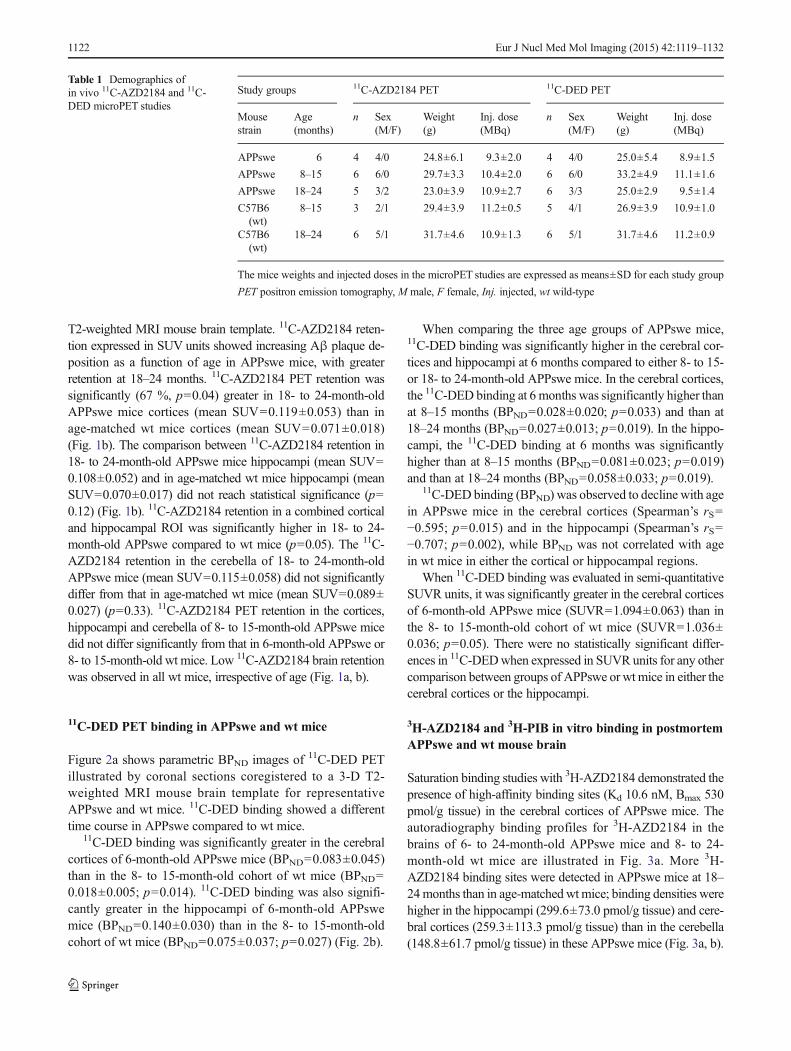
T2-weighted MRI mouse brain template. 11C-AZD2184 reten-tion expressed in SUV units showed increasing Aβ plaque de-position as a function of age in APPswe mice, with greaterretention at 18–24 months. 11C-AZD2184 PET retention wassignificantly (67 %, p=0.04) greater in 18- to 24-month-oldAPPswe mice cortices (mean SUV=0.119±0.053) than inage-matched wt mice cortices (mean SUV=0.071±0.018)(Fig. 1b). The comparison between 11C-AZD2184 retention in18- to 24-month-old APPswe mice hippocampi (mean SUV=0.108±0.052) and in age-matched wt mice hippocampi (meanSUV=0.070±0.017) did not reach statistical significance (p=0.12) (Fig. 1b). 11C-AZD2184 retention in a combined corticaland hippocampal ROI was significantly higher in 18- to 24-month-old APPswe compared to wt mice (p=0.05). The 11C-AZD2184 retention in the cerebella of 18- to 24-month-oldAPPswe mice (mean SUV=0.115±0.058) did not significantlydiffer from that in age-matched wt mice (mean SUV=0.089±0.027) (p=0.33). 11C-AZD2184 PET retention in the cortices,hippocampi and cerebella of 8- to 15-month-old APPswe micedid not differ significantly from that in 6-month-old APPswe or8- to 15-month-old wt mice. Low 11C-AZD2184 brain retentionwas observed in all wt mice, irrespective of age (Fig. 1a, b).
11C-DED PET binding in APPswe and wt mice
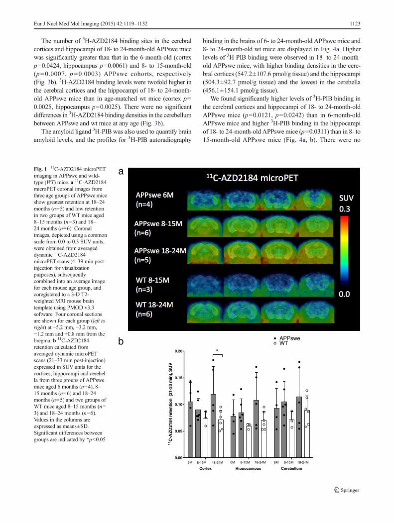
Figure 2a shows parametric BPND images of 11C-DED PETillustrated by coronal sections coregistered to a 3-D T2-weighted MRI mouse brain template for representativeAPPswe and wt mice. 11C-DED binding showed a differenttime course in APPswe compared to wt mice.
11C-DED binding was significantly greater in the cerebralcortices of 6-month-old APPswe mice (BPND=0.083±0.045)than in the 8- to 15-month-old cohort of wt mice (BPND=0.018±0.005; p=0.014). 11C-DED binding was also signifi-cantly greater in the hippocampi of 6-month-old APPswemice (BPND=0.140±0.030) than in the 8- to 15-month-oldcohort of wt mice (BPND=0.075±0.037; p=0.027) (Fig. 2b).
When comparing the three age groups of APPswe mice,11C-DED binding was significantly higher in the cerebral cor-tices and hippocampi at 6 months compared to either 8- to 15-or 18- to 24-month-old APPswe mice. In the cerebral cortices,the 11C-DED binding at 6 months was significantly higher thanat 8–15 months (BPND=0.028±0.020; p=0.033) and than at18–24 months (BPND=0.027±0.013; p=0.019). In the hippo-campi, the 11C-DED binding at 6 months was significantlyhigher than at 8–15 months (BPND=0.081±0.023; p=0.019)and than at 18–24 months (BPND=0.058±0.033; p=0.019).
11C-DED binding (BPND) was observed to decline with agein APPswe mice in the cerebral cortices (Spearman’s rS=−0.595; p=0.015) and in the hippocampi (Spearman’s rS=−0.707; p=0.002), while BPND was not correlated with agein wt mice in either the cortical or hippocampal regions.
When 11C-DED binding was evaluated in semi-quantitativeSUVR units, it was significantly greater in the cerebral corticesof 6-month-old APPswe mice (SUVR=1.094±0.063) than inthe 8- to 15-month-old cohort of wt mice (SUVR=1.036±0.036; p=0.05). There were no statistically significant differ-ences in 11C-DEDwhen expressed in SUVR units for any othercomparison between groups of APPswe or wt mice in either thecerebral cortices or the hippocampi.
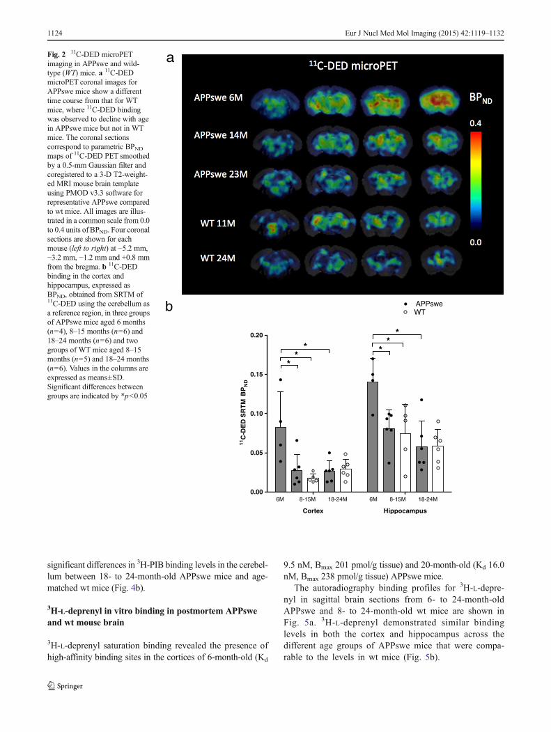
3H-AZD2184 and 3H-PIB in vitro binding in postmortemAPPswe and wt mouse brain
Saturation binding studies with 3H-AZD2184 demonstrated thepresence of high-affinity binding sites (Kd 10.6 nM, Bmax 530pmol/g tissue) in the cerebral cortices of APPswe mice. Theautoradiography binding profiles for 3H-AZD2184 in thebrains of 6- to 24-month-old APPswe mice and 8- to 24-month-old wt mice are illustrated in Fig. 3a. More 3H-AZD2184 binding sites were detected in APPswe mice at 18–24months than in age-matchedwtmice; binding densities werehigher in the hippocampi (299.6±73.0 pmol/g tissue) and cere-bral cortices (259.3±113.3 pmol/g tissue) than in the cerebella(148.8±61.7 pmol/g tissue) in these APPswe mice (Fig. 3a, b).
Table 1 Demographics ofin vivo 11C-AZD2184 and 11C-DED microPET studies
Study groups 11C-AZD2184 PET 11C-DED PET
Mousestrain
Age(months)
n Sex(M/F)
Weight(g)
Inj. dose(MBq)
n Sex(M/F)
Weight(g)
Inj. dose(MBq)
APPswe 6 4 4/0 24.8±6.1 9.3±2.0 4 4/0 25.0±5.4 8.9±1.5
APPswe 8–15 6 6/0 29.7±3.3 10.4±2.0 6 6/0 33.2±4.9 11.1±1.6
APPswe 18–24 5 3/2 23.0±3.9 10.9±2.7 6 3/3 25.0±2.9 9.5±1.4
C57B6(wt)
8–15 3 2/1 29.4±3.9 11.2±0.5 5 4/1 26.9±3.9 10.9±1.0
C57B6(wt)
18–24 6 5/1 31.7±4.6 10.9±1.3 6 5/1 31.7±4.6 11.2±0.9
The mice weights and injected doses in the microPET studies are expressed as means±SD for each study group
PET positron emission tomography, M male, F female, Inj. injected, wt wild-type
1122 Eur J Nucl Med Mol Imaging (2015) 42:1119–1132
The number of 3H-AZD2184 binding sites in the cerebralcortices and hippocampi of 18- to 24-month-old APPswe micewas significantly greater than that in the 6-month-old (cortexp=0.0424, hippocampus p=0.0061) and 8- to 15-month-old(p=0.0007, p=0.0003) APPswe cohorts, respectively(Fig. 3b). 3H-AZD2184 binding levels were twofold higher inthe cerebral cortices and the hippocampi of 18- to 24-month-old APPswe mice than in age-matched wt mice (cortex p=0.0025, hippocampus p=0.0025). There were no significantdifferences in 3H-AZD2184 binding densities in the cerebellumbetween APPswe and wt mice at any age (Fig. 3b).
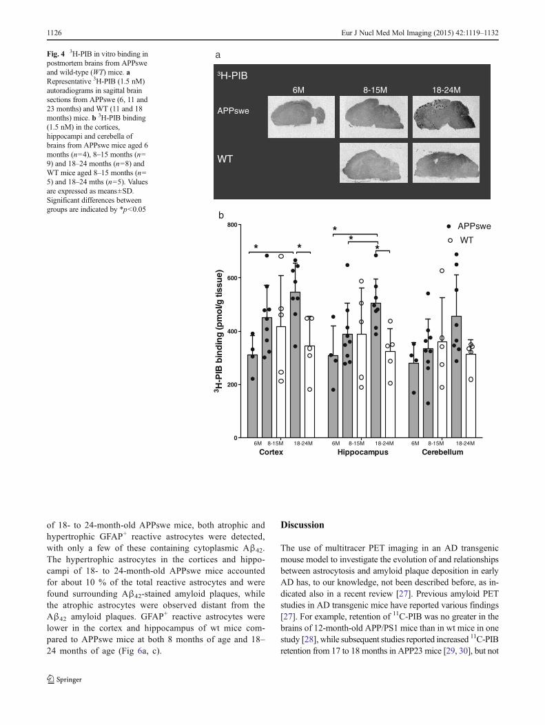
The amyloid ligand 3H-PIB was also used to quantify brainamyloid levels, and the profiles for 3H-PIB autoradiography
binding in the brains of 6- to 24-month-old APPswe mice and8- to 24-month-old wt mice are displayed in Fig. 4a. Higherlevels of 3H-PIB binding were observed in 18- to 24-month-old APPswe mice, with higher binding densities in the cere-bral cortices (547.2±107.6 pmol/g tissue) and the hippocampi(504.3±92.7 pmol/g tissue) and the lowest in the cerebella(456.1±154.1 pmol/g tissue).
We found significantly higher levels of 3H-PIB binding inthe cerebral cortices and hippocampi of 18- to 24-month-oldAPPswe mice (p=0.0121, p=0.0242) than in 6-month-oldAPPswe mice and higher 3H-PIB binding in the hippocampiof 18- to 24-month-old APPswe mice (p=0.0311) than in 8- to15-month-old APPswe mice (Fig. 4a, b). There were no
a
b
Cortex Hippocampus Cerebellum
0.00
0.05
0.10
0.15
0.20
11C
-AZ
D21
84re
ten
tion
(21-
33m
in),
SU
V
APPswe
6M 8-15M 18-24M 6M 8-15M 18-24M 6M 8-15M 18-24M
WT
*
Fig. 1 11C-AZD2184 microPETimaging in APPswe and wild-type (WT) mice. a 11C-AZD2184microPET coronal images fromthree age groups of APPswe miceshow greatest retention at 18–24months (n=5) and low retentionin two groups of WT mice aged8–15 months (n=3) and 18–24 months (n=6). Coronalimages, depicted using a commonscale from 0.0 to 0.3 SUV units,were obtained from averageddynamic 11C-AZD2184microPET scans (4–39 min post-injection for visualizationpurposes), subsequentlycombined into an average imagefor each mouse age group, andcoregistered to a 3-D T2-weighted MRI mouse braintemplate using PMOD v3.3software. Four coronal sectionsare shown for each group (left toright) at −5.2 mm, −3.2 mm,−1.2 mm and +0.8 mm from thebregma. b 11C-AZD2184retention calculated fromaveraged dynamic microPETscans (21–33 min post-injection)expressed in SUV units for thecortices, hippocampi and cerebel-la from three groups of APPswemice aged 6 months (n=4), 8–15 months (n=6) and 18–24months (n=5) and two groups ofWT mice aged 8–15 months (n=3) and 18–24 months (n=6).Values in the columns areexpressed as means±SD.Significant differences betweengroups are indicated by *p<0.05
Eur J Nucl Med Mol Imaging (2015) 42:1119–1132 1123
significant differences in 3H-PIB binding levels in the cerebel-lum between 18- to 24-month-old APPswe mice and age-matched wt mice (Fig. 4b).
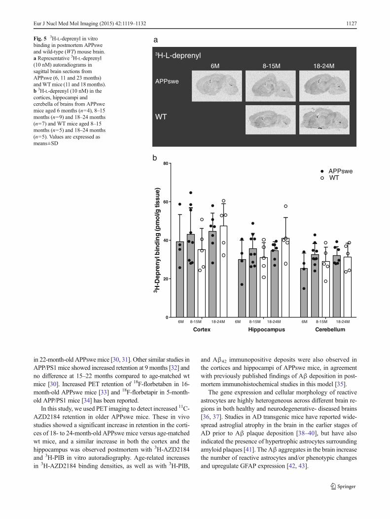
3H-L-deprenyl in vitro binding in postmortem APPsweand wt mouse brain
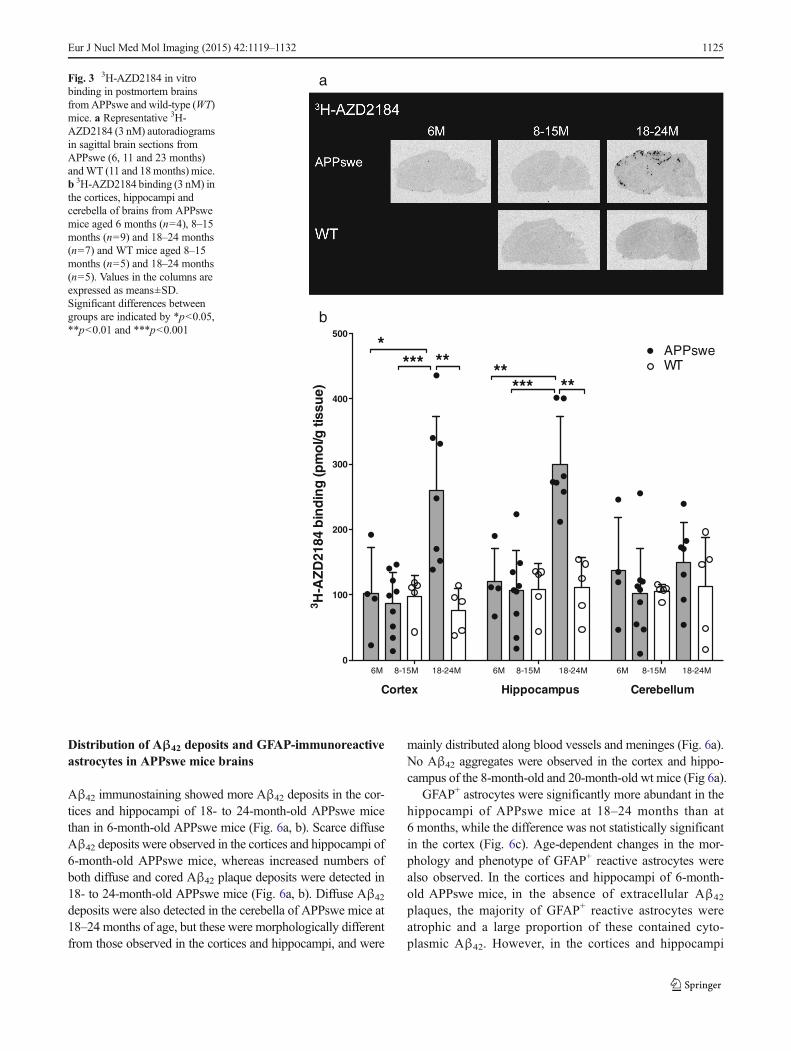
3H-L-deprenyl saturation binding revealed the presence ofhigh-affinity binding sites in the cortices of 6-month-old (Kd
9.5 nM, Bmax 201 pmol/g tissue) and 20-month-old (Kd 16.0nM, Bmax 238 pmol/g tissue) APPswe mice.
The autoradiography binding profiles for 3H-L-depre-nyl in sagittal brain sections from 6- to 24-month-oldAPPswe and 8- to 24-month-old wt mice are shown inFig. 5a. 3H-L-deprenyl demonstrated similar bindinglevels in both the cortex and hippocampus across thedifferent age groups of APPswe mice that were compa-rable to the levels in wt mice (Fig. 5b).
a
b
Cortex Hippocampus
0.00
0.05
0.10
0.15
0.20
11C
-DE
DS
RT
MB
PN
D
*
APPsweWT
6M 8-15M 18-24M 6M 8-15M 18-24M
*
* **
*
Fig. 2 11C-DED microPETimaging in APPswe and wild-type (WT) mice. a 11C-DEDmicroPET coronal images forAPPswe mice show a differenttime course from that for WTmice, where 11C-DED bindingwas observed to decline with agein APPswe mice but not in WTmice. The coronal sectionscorrespond to parametric BPNDmaps of 11C-DED PET smoothedby a 0.5-mm Gaussian filter andcoregistered to a 3-D T2-weight-ed MRI mouse brain templateusing PMOD v3.3 software forrepresentative APPswe comparedto wt mice. All images are illus-trated in a common scale from 0.0to 0.4 units of BPND. Four coronalsections are shown for eachmouse (left to right) at −5.2 mm,−3.2 mm, −1.2 mm and +0.8 mmfrom the bregma. b 11C-DEDbinding in the cortex andhippocampus, expressed asBPND, obtained from SRTM of11C-DED using the cerebellum asa reference region, in three groupsof APPswe mice aged 6 months(n=4), 8–15 months (n=6) and18–24 months (n=6) and twogroups of WT mice aged 8–15months (n=5) and 18–24 months(n=6). Values in the columns areexpressed as means±SD.Significant differences betweengroups are indicated by *p<0.05
1124 Eur J Nucl Med Mol Imaging (2015) 42:1119–1132
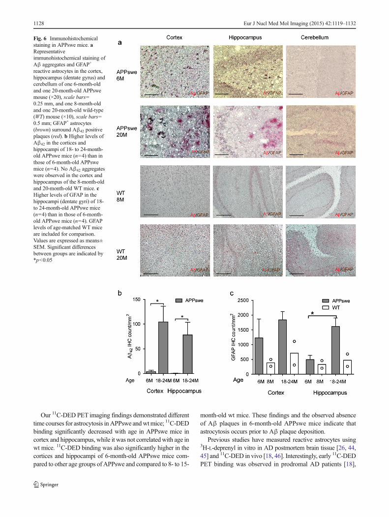
Distribution of Aβ42 deposits and GFAP-immunoreactiveastrocytes in APPswe mice brains
Aβ42 immunostaining showed more Aβ42 deposits in the cor-tices and hippocampi of 18- to 24-month-old APPswe micethan in 6-month-old APPswe mice (Fig. 6a, b). Scarce diffuseAβ42 deposits were observed in the cortices and hippocampi of6-month-old APPswe mice, whereas increased numbers ofboth diffuse and cored Aβ42 plaque deposits were detected in18- to 24-month-old APPswe mice (Fig. 6a, b). Diffuse Aβ42
deposits were also detected in the cerebella of APPswe mice at18–24 months of age, but these were morphologically differentfrom those observed in the cortices and hippocampi, and were
mainly distributed along blood vessels and meninges (Fig. 6a).No Aβ42 aggregates were observed in the cortex and hippo-campus of the 8-month-old and 20-month-old wt mice (Fig 6a).
GFAP+ astrocytes were significantly more abundant in thehippocampi of APPswe mice at 18–24 months than at6 months, while the difference was not statistically significantin the cortex (Fig. 6c). Age-dependent changes in the mor-phology and phenotype of GFAP+ reactive astrocytes werealso observed. In the cortices and hippocampi of 6-month-old APPswe mice, in the absence of extracellular Aβ42
plaques, the majority of GFAP+ reactive astrocytes wereatrophic and a large proportion of these contained cyto-plasmic Aβ42. However, in the cortices and hippocampi
0
100
200
300
400
500
6M 8-15M 18-24M
Cortex Hippocampus Cerebellum
6M 8-15M 18-24M 6M 8-15M 18-24M
**** ** **
*** **
3 H-A
ZD
2184
bin
din
g(p
mo
l/gtis
sue)
APPsweWT
b
aFig. 3 3H-AZD2184 in vitrobinding in postmortem brainsfromAPPswe and wild-type (WT)mice. a Representative 3H-AZD2184 (3 nM) autoradiogramsin sagittal brain sections fromAPPswe (6, 11 and 23 months)andWT (11 and 18months) mice.b 3H-AZD2184 binding (3 nM) inthe cortices, hippocampi andcerebella of brains from APPswemice aged 6 months (n=4), 8–15months (n=9) and 18–24 months(n=7) and WT mice aged 8–15months (n=5) and 18–24 months(n=5). Values in the columns areexpressed as means±SD.Significant differences betweengroups are indicated by *p<0.05,**p<0.01 and ***p<0.001
Eur J Nucl Med Mol Imaging (2015) 42:1119–1132 1125
of 18- to 24-month-old APPswe mice, both atrophic andhypertrophic GFAP+ reactive astrocytes were detected,with only a few of these containing cytoplasmic Aβ42.The hypertrophic astrocytes in the cortices and hippo-campi of 18- to 24-month-old APPswe mice accountedfor about 10 % of the total reactive astrocytes and werefound surrounding Aβ42-stained amyloid plaques, whilethe atrophic astrocytes were observed distant from theAβ42 amyloid plaques. GFAP+ reactive astrocytes werelower in the cortex and hippocampus of wt mice com-pared to APPswe mice at both 8 months of age and 18–24 months of age (Fig 6a, c).
Discussion
The use of multitracer PET imaging in an AD transgenicmouse model to investigate the evolution of and relationshipsbetween astrocytosis and amyloid plaque deposition in earlyAD has, to our knowledge, not been described before, as in-dicated also in a recent review [27]. Previous amyloid PETstudies in AD transgenic mice have reported various findings[27]. For example, retention of 11C-PIB was no greater in thebrains of 12-month-old APP/PS1 mice than in wt mice in onestudy [28], while subsequent studies reported increased 11C-PIBretention from 17 to 18 months in APP23 mice [29, 30], but not
0
200
400
600
800
6M 8-15M 18-24M Cortex Hippocampus Cerebellum
6M 8-15M 18-24M
6M 8-15M 18-24M
* **
**
3 H-P
IB b
ind
ing
(pm
ol/g
tiss
ue)
APPswe
WT
6M 8-15M 18-24M
3H-PIB
APPswe
WT
a
b
Fig. 4 3H-PIB in vitro binding inpostmortem brains from APPsweand wild-type (WT) mice. aRepresentative 3H-PIB (1.5 nM)autoradiograms in sagittal brainsections from APPswe (6, 11 and23 months) and WT (11 and 18months) mice. b 3H-PIB binding(1.5 nM) in the cortices,hippocampi and cerebella ofbrains from APPswe mice aged 6months (n=4), 8–15 months (n=9) and 18–24 months (n=8) andWT mice aged 8–15 months (n=5) and 18–24 mths (n=5). Valuesare expressed as means±SD.Significant differences betweengroups are indicated by *p<0.05
1126 Eur J Nucl Med Mol Imaging (2015) 42:1119–1132
in 22-month-old APPswe mice [30, 31]. Other similar studies inAPP/PS1mice showed increased retention at 9 months [32] andno difference at 15–22 months compared to age-matched wtmice [30]. Increased PET retention of 18F-florbetaben in 16-month-old APPswe mice [33] and 18F-florbetapir in 5-month-old APP/PS1 mice [34] has been reported.
In this study, we used PET imaging to detect increased 11C-AZD2184 retention in older APPswe mice. These in vivostudies showed a significant increase in retention in the corti-ces of 18- to 24-month-old APPswe mice versus age-matchedwt mice, and a similar increase in both the cortex and thehippocampus was observed postmortem with 3H-AZD2184and 3H-PIB in vitro autoradiography. Age-related increasesin 3H-AZD2184 binding densities, as well as with 3H-PIB,
and Aβ42 immunopositive deposits were also observed inthe cortices and hippocampi of APPswe mice, in agreementwith previously published findings of Aβ deposition in post-mortem immunohistochemical studies in this model [35].
The gene expression and cellular morphology of reactiveastrocytes are highly heterogeneous across different brain re-gions in both healthy and neurodegenerative- diseased brains[36, 37]. Studies in AD transgenic mice have reported wide-spread astroglial atrophy in the brain in the earlier stages ofAD prior to Aβ plaque deposition [38–40], but have alsoindicated the presence of hypertrophic astrocytes surroundingamyloid plaques [41]. The Aβ aggregates in the brain increasethe number of reactive astrocytes and/or phenotypic changesand upregulate GFAP expression [42, 43].
0
20
40
60
80
6M 8-15M 18-24M
Cortex Hippocampus Cerebellum
6M 8-15M 18-24M 6M 8-15M 18-24M
3 H-D
epre
nyl
bin
din
g(p
mo
l/gtis
sue)
APPsweWT
b
a
6M 8-15M 18-24M
3H-L-deprenyl
APPswe
WT
Fig. 5 3H-L-deprenyl in vitrobinding in postmortem APPsweand wild-type (WT) mouse brain.a Representative 3H-L-deprenyl(10 nM) autoradiograms insagittal brain sections fromAPPswe (6, 11 and 23 months)andWTmice (11 and 18months).b 3H-L-deprenyl (10 nM) in thecortices, hippocampi andcerebella of brains from APPswemice aged 6 months (n=4), 8–15months (n=9) and 18–24 months(n=7) and WT mice aged 8–15months (n=5) and 18–24 months(n=5). Values are expressed asmeans±SD
Eur J Nucl Med Mol Imaging (2015) 42:1119–1132 1127
Our 11C-DED PET imaging findings demonstrated differenttime courses for astrocytosis in APPswe andwtmice; 11C-DEDbinding significantly decreased with age in APPswe mice incortex and hippocampus, while it was not correlatedwith age inwt mice. 11C-DED binding was also significantly higher in thecortices and hippocampi of 6-month-old APPswe mice com-pared to other age groups of APPswe and compared to 8- to 15-
month-old wt mice. These findings and the observed absenceof Aβ plaques in 6-month-old APPswe mice indicate thatastrocytosis occurs prior to Aβ plaque deposition.
Previous studies have measured reactive astrocytes using3H-L-deprenyl in vitro in AD postmortem brain tissue [26, 44,45] and 11C-DED in vivo [18, 46]. Interestingly, early 11C-DEDPET binding was observed in prodromal AD patients [18],
Fig. 6 Immunohistochemicalstaining in APPswe mice. aRepresentativeimmunohistochemical staining ofAβ aggregates and GFAP+
reactive astrocytes in the cortex,hippocampus (dentate gyrus) andcerebellum of one 6-month-oldand one 20-month-old APPswemouse (×20), scale bars=0.25 mm, and one 8-month-oldand one 20-month-old wild-type(WT) mouse (×10), scale bars=0.5 mm; GFAP+ astrocytes(brown) surround Aβ42 positiveplaques (red). b Higher levels ofAβ42 in the cortices andhippocampi of 18- to 24-month-old APPswe mice (n=4) than inthose of 6-month-old APPswemice (n=4). No Aβ42 aggregateswere observed in the cortex andhippocampus of the 8-month-oldand 20-month-old WT mice. cHigher levels of GFAP in thehippocampi (dentate gyri) of 18-to 24-month-old APPswe mice(n=4) than in those of 6-month-old APPswe mice (n=4). GFAPlevels of age-matched WT miceare included for comparison.Values are expressed as means±SEM. Significant differencesbetween groups are indicated by*p<0.05
1128 Eur J Nucl Med Mol Imaging (2015) 42:1119–1132
consistent with our results in APPswe mice. Astrocytosis asmeasured by 11C-DED PETwas also observed to be negativelycorrelated with grey matter density in the parahippocampus atearly prodromal AD stages [47]. Early 11C-DED PET bindinghas also been reported in presymptomatic carriers of autosomaldominant AD mutations several decades before onset of symp-toms and earlier than Aβ deposition [48]. Therefore, our find-ings of early 11C-DED PET binding in APPswe mice beforeAβ deposition are consistent with reported in vivo PET find-ings in humans, highlighting the translational aspects of thisstudy.
In contrast to the in vivo 11C-DED PET binding results,there were no significant differences between APPswe and wtmice in the in vitro 3H-L-deprenyl autoradiography results.Possible explanations for this include either a reduction inmonoamine oxidase B (MAOB) enzyme activity when mea-sured in vitro compared to in vivo [46] or technical limitationsdue to a reduced in vitro enzyme activity in transgenicAPPswe mice compared to humans.
The level of GFAP upregulation in astrocytes has beenreported to be dependent on both the brain region and thecontext, which could in part underlie the heterogeneity offunctions mediated by astrocytes in the central nervoussystem, evidenced by their diverse neuroprotective orneurotoxic functions across different stages in AD [49].Our immunostaining results in APPswe mice revealedmore GFAP reactive astrocytes in the cortices and hippo-campi of 18- to 24-month-old than of 6-month-oldAPPswe mice, although the age-dependent increase wassignificant only in the hippocampus. There is increasingevidence that GFAP is not expressed uniformly by allastrocytes [50] and that different GFAP isoforms developin response to plaque-related gliosis, as shown in APP/PS1 and 3xTg AD transgenic mice [51]. It has been re-ported [52] that about 80 % of the astrocytes in the hip-pocampus express GFAP, compared to only 15–20 % ofthose in the cortex, which could at least partially explainour immunohistochemical results showing a significantincrease in GFAP with age in the hippocampus. Otherstudies in AD transgenic mice have reported GFAP ex-pression prior to Aβ plaque deposition [53] and also as anage-dependent process that is correlated with oligomericAβ but not with plaque burden [54]. GFAP expression inastrocytes has also been reported as a late event related toplaque formation and maturation, and as a neuroprotectiveevent that limits Aβ plaque growth [55].
In our present study, in contrast to the early increase in 11C-DED binding in the APPswe mouse brain cortex, GFAP im-munoreactivity was predominantly a late event. There was asignificant age-dependent increase in the number of GFAP+
reactive astrocytes in the hippocampus, possibly indicating thepresence of distinct subpopulations of astrocytes and/or dif-ferent stages of reactive astrocytosis. The different time course
of increases in 11C-DED binding and GFAP upregulation ob-served in APPswe mice is consistent with findings in humanAD. Quantitative autoradiography studies in postmortem ADbrains have shown a strong regional correlation between thenumber of GFAP+ reactive astrocytes and the extent of in vivo11C-PIB and in vitro 3H-PIB binding, but there appears to beno regional correlation between postmortem 3H-L-deprenyland in vivo 11C-PIB binding [45]. The regional and laminardistribution patterns of 3H-L-deprenyl reactive astrocytes alsodiffered from those of fibrillar Aβ in AD autopsy brains [26].Thus, our observation of early 11C-DED binding in the corti-ces of APPswe mice might reflect the presence of a subset ofactivated astrocytes that is functionally different from thosemeasured by GFAP at later stages.
The small, soluble Aβ forms that have been observed inAPPswe mice from birth [56, 20] could influence astrocytefunction. For example, Aβ25–35 peptides caused overexpres-sion of MAOB in cultured rat astrocytes [57]. Further, MAOBoverexpression in astrocytes led to production of proinflamma-tory molecules contributing to exacerbated neuroinflammationand Aβ plaque formation at later stages [58]. Our observationof early elevated 11C-DED binding might thus reflect a reactionof astrocytes to these small Aβ forms, with potential beneficialand/or neurotoxic consequences. Activated astrocytes appear toplay a role in the clearance of Aβ [59, 60].Whether the reactiveastrocytes that were observed early in the development of AD,as measured by elevated 11C-DED binding, have a phagocyticfunction requires further investigation.
One limitation of this study was the limited spatial resolu-tion of small animal PET imaging relative to the mouse brainregions selected for quantification, which contained some thinshapes being susceptible to partial volume effects. This mightaccount for the lower sensitivity of our in vivo versus in vitroimages. Inevitably, transgenic AD mice are an inherently lim-ited model for human disease and APPswe mice might phe-notypically reflect only some aspects of AD [19]. Astrocytosiswas elevated at 6 months and it subsequently declined withage in APPswe mice, consistent with findings of increasedastrocytosis in the prodromal phase of AD followed by de-creases in later stages in humans [18]. The cross-sectionaldesign of this study allowed a parallel in vivo/in vitro com-parison of the regional and temporal distributions of Aβ de-position and astrocytosis in the sameAPPswe and wt mice at agiven age interval. Further multitracer longitudinal PET imag-ing studies in AD transgenic animal models could provideadditional insights into the temporal evolution of neuropatho-logical changes during AD progression and could be usefulfor testing new therapies targeting astrocytes, especially at theearliest stages.
In conclusion, we provide in vivo evidence thatastrocytosis occurs early in AD and precedes Aβ plaque de-position. The increasing recognition of heterogeneous andcontext-dependent astrocytosis in the progression of AD
Eur J Nucl Med Mol Imaging (2015) 42:1119–1132 1129
indicates that more research is needed to elucidate the func-tions of the different astrocyte populations in the brain at dif-ferent stages of the disease.
Acknowledgments This work was supported by the Swedish ResearchCouncil (project 05817), Karolinska Institutet Strategic Neuroscienceprogram, Stockholm County Council-Karolinska Institutet regionalagreement onmedical training and clinical research (ALF grant), SwedishBrain Power, Swedish Brain Foundation, Alzheimer Foundation in Swe-den, Dementia Association (Demensfonden), Knut and Alice WallenbergFoundation, EU FP7 large-scale integrating project INMiND (http://www.uni-muenster. de/INMiND), Foundation for Old Servants,Karolinska Institutet’s Foundation for Aging Research, Gun and BertilStohne’s Foundation, Sigurd and Elsa Golje’s Foundation, Loo and HansOsterman’s Foundation, Lars Hierta Memorial Foundation, Ragnhild andEinar Lundström’s Memorial Foundation, Olle Engkvist ByggmästareFoundation, Åhlén Foundation, Magnus Bergvall’s Foundation, andWenner-Gren Foundation.
Conflicts of interest None.
Open Access This article is distributed under the terms of theCreative Commons Attribution 4.0 International License (http://creativecommons.org/licenses/by/4.0/), which permits unrestricted use,distribution, and reproduction in any medium, provided you giveappropriate credit to the original author(s) and the source, provide a linkto the Creative Commons license, and indicate if changes were made.
References
1. Hardy J, Selkoe DJ. The amyloid hypothesis of Alzheimer’s dis-ease: progress and problems on the road to therapeutics. Science2002;297(5580):353–6. doi:10.1126/science.1072994.
2. Mattson MP. Pathways towards and away from Alzheimer’s dis-ease. Nature 2004;430(7000):631–9. doi:10.1038/nature02621.
3. Nordberg A. Amyloid imaging in early detection of Alzheimer’sdisease. Neurodegener Dis 2010;7(1–3):136–8. doi:10.1159/000289223.
4. KlunkWE, Engler H, Nordberg A, Wang Y, Blomqvist G, Holt DP,et al. Imaging brain amyloid in Alzheimer’s disease with PittsburghCompound-B. Ann Neurol 2004;55(3):306–19.
5. Kudo Y, Okamura N, Furumoto S, Tashiro M, Furukawa K,Maruyama M, et al. 2-(2-[2-Dimethylaminothiazol-5-yl]ethenyl)-6- (2-[fluoro]ethoxy)benzoxazole: a novel PET agent for in vivodetection of dense amyloid plaques in Alzheimer’s disease patients.J Nucl Med 2007;48(4):553–61.
6. Nyberg S, Jönhagen ME, Cselényi Z, Halldin C, Julin P, Olsson H,et al. Detection of amyloid in Alzheimer’s disease with positronemission tomography using [11C]AZD2184. Eur J Nucl MedMol Imaging 2009;36(11):1859–63. doi:10.1007/s00259-009-1182-1.
7. Shoghi-Jadid K, Small GW, Agdeppa ED, Kepe V, Ercoli LM,Siddarth P, et al. Localization of neurofibrillary tangles and beta-amyloid plaques in the brains of living patients with Alzheimerdisease. Am J Geriatr Psychiatry 2002;10(1):24–35.
8. Clark CM, Schneider JA, Bedell BJ, Beach TG, BilkerWB,MintunMA, et al. Use of florbetapir-PET for imaging beta-amyloid pathol-ogy. JAMA 2011;305(3):275–83. doi:10.1001/jama.2010.2008.
9. Nelissen N, Van Laere K, Thurfjell L, Owenius R, VandenbulckeM, Koole M, et al. Phase 1 study of the Pittsburgh compound B
derivative 18F-flutemetamol in healthy volunteers and patients withprobable Alzheimer disease. J Nucl Med 2009;50(8):1251–9. doi:10.2967/jnumed.109.063305.
10. Rowe CC, Ackerman U, Browne W, Mulligan R, Pike KL,O’Keefe G, et al. Imaging of amyloid beta in Alzheimer’s diseasewith 18F-BAY94-9172, a novel PET tracer: proof of mechanism.Lancet Neurol 2008;7(2):129–35. doi:10.1016/S1474-4422(08)70001-2.
11. Cselényi Z, Jönhagen ME, Forsberg A, Halldin C, Julin P, SchouM, et al. Clinical validation of 18F-AZD4694, an amyloid-β-specific PET radioligand. J Nucl Med 2012;53(3):415–24. doi:10.2967/jnumed.111.094029.
12. Ni R, Gillberg PG, Bergfors A, Marutle A, Nordberg A. Amyloidtracers detect multiple binding sites in Alzheimer’s disease braintissue. Brain 2013;136(Pt 7):2217–27. doi:10.1093/brain/awt142.
13. Villemagne VL, Burnham S, Bourgeat P, Brown B, Ellis KA,Salvado O, et al. Amyloid β deposition, neurodegeneration, andcognitive decline in sporadic Alzheimer’s disease: a prospectivecohort study. Lancet Neurol 2013;12(4):357–67. doi:10.1016/S1474-4422(13)70044-9.
14. McGeer PL, McGeer EG. The inflammatory response system ofbrain: implications for therapy of Alzheimer and other neurodegen-erative diseases. Brain Res Brain Res Rev 1995;21(2):195–218.
15. Akiyama H, Barger S, Barnum S, Bradt B, Bauer J, Cole GM, et al.Inflammation and Alzheimer’s disease. Neurobiol Aging2000;21(3):383–421.
16. Pekny M, Wilhelmsson U, Pekna M. The dual role of astrocyteactivation and reactive gliosis. Neurosci Lett 2014;565:30–8. doi:10.1016/j.neulet.2013.12.071.
17. Verkhratsky A, Marutle A, Rodríguez-Arellano JJ, Nordberg A.Glial asthenia and functional paralysis: a new perspective on neu-rodegeneration and Alzheimer’s disease. Neuroscientist 2014. doi:10.1177/1073858414547132.
18. Carter SF, Schöll M, Almkvist O, Wall A, Engler H, Långström B,et al. Evidence for astrocytosis in prodromal Alzheimer diseaseprovided by 11C-deuterium-L-deprenyl: a multitracer PET para-digm combining 11C-Pittsburgh compound B and 18F-FDG. JNucl Med 2012;53(1):37–46. doi:10.2967/jnumed.110.087031.
19. Ashe KH, Zahs KR. Probing the biology of Alzheimer’s dis-ease in mice. Neuron 2010;66(5):631–45. doi:10.1016/j.neuron.2010.04.031.
20. Mustafiz T, Portelius E, Gustavsson MK, Hölttä M, Zetterberg H,Blennow K, et al. Characterization of the brain β-amyloid isoformpattern at different ages of Tg2576 mice. Neurodegener Dis2011;8(5):352–63.
21. Nagy K, Tóth M, Major P, Patay G, Egri G, Häggkvist J, et al.Performance evaluation of the small-animal nanoScan PET/MRIsystem. J Nucl Med 2013;54(10):1825–32. doi:10.2967/jnumed.112.119065.
22. Szanda I, Mackewn J, Patay G, Major P, Sunassee K, Mullen GE,et al. National Electrical Manufacturers Association NU-4 perfor-mance evaluation of the PET component of the NanoPET/CT pre-clinical PET/CT scanner. J Nucl Med 2011;52(11):1741–7. doi:10.2967/jnumed.111.088260.
23. Andersson JD, Varnäs K, Cselényi Z, Gulyás B, Wensbo D,Finnema SJ, et al. Radiosynthesis of the candidate beta-amyloidradioligand [(11)C]AZD2184: positron emission tomography ex-amination and metabolite analysis in cynomolgus monkeys.Synapse 2010;64(10):733–41. doi:10.1002/syn.20782.
24. Gulyás B, Pavlova E, Kása P, Gulya K, Bakota L, Várszegi S,et al. Activated MAO-B in the brain of Alzheimer patients,demonstrated by [11C]-L-deprenyl using whole hemisphereautoradiography. Neurochem Int 2011;58(1):60–8. doi:10.1016/j.neuint.2010.10.013.
25. Johnson AE, Jeppsson F, Sandell J, Wensbo D, Neelissen JA,Juréus A, et al. AZD2184: a radioligand for sensitive detection of
1130 Eur J Nucl Med Mol Imaging (2015) 42:1119–1132
beta-amyloid deposits. J Neurochem 2009;108(5):1177–86. doi:10.1111/j.1471-4159.2008.05861.x.
26. Marutle A, Gillberg PG, Bergfors A, Yu W, Ni R, Nennesmo I,et al. 3H-Deprenyl and 3H-PIB autoradiography show differentlaminar distributions of astroglia and fibrillar β-amyloid inAlzheimer brain. J Neuroinflammation 2013;10(1):90. doi:10.1186/1742-2094-10-90.
27. Zimmer ER, Parent MJ, Cuello AC, Gauthier S, Rosa-Neto P.MicroPET imaging and transgenic models: a blueprint forAlzheimer’s disease clinical research. Trends Neurosci2014;37(11):629–41. doi:10.1016/j.tins.2014.07.002.
28. Klunk WE, Lopresti BJ, Ikonomovic MD, Lefterov IM,Koldamova RP, Abrahamson EE, et al. Binding of the positronemission tomography tracer Pittsburgh compound-B reflects theamount of amyloid-beta in Alzheimer’s disease brain but not intransgenic mouse brain. J Neurosci 2005;25(46):10598–606. doi:10.1523/JNEUROSCI.2990-05.2005.
29. Maeda J, Zhang MR, Okauchi T, Ji B, Ono M, Hattori S, et al.In vivo positron emission tomographic imaging of glial responsesto amyloid-beta and tau pathologies in mouse models ofAlzheimer ’s disease and related disorders. J Neurosci2011;31(12):4720–30. doi:10.1523/JNEUROSCI.3076-10.2011.
30. Snellman A, López-Picón FR, Rokka J, Salmona M, Forloni G,Scheinin M, et al. Longitudinal amyloid imaging in mouse brainwith 11C-PIB: comparison of APP23, Tg2576, and APPswe-PS1dE9 mouse models of Alzheimer disease. J Nucl Med2013;54(8):1434–41. doi:10.2967/jnumed.112.110163.
31. Toyama H, Ye D, Ichise M, Liow JS, Cai L, Jacobowitz D, et al.PET imaging of brain with the beta-amyloid probe, [11C]6-OH-BTA-1, in a transgenic mouse model of Alzheimer’s disease. EurJ Nucl Med Mol Imaging 2005;32(5):593–600. doi:10.1007/s00259-005-1780-5.
32. Manook A, Yousefi BH, Willuweit A, Platzer S, Reder S, Voss A,et al. Small-animal PET imaging of amyloid-beta plaques with[11C]PiB and its multi-modal validation in an APP/PS1 mousemodel of Alzheimer’s disease. PLoS One 2012;7(3):e31310. doi:10.1371/journal.pone.0031310.
33. Rominger A, Brendel M, Burgold S, Keppler K, Baumann K,Xiong G, et al. Longitudinal assessment of cerebral β-amyloiddeposition in mice overexpressing Swedish mutant β-amyloid pre-cursor protein using 18F-florbetaben PET. J Nucl Med 2013;54(7):1127–34. doi:10.2967/jnumed.112.114660.
34. Poisnel G, Dhilly M, Moustié O, Delamare J, Abbas A, GuilloteauD, et al. PET imaging with [18F]AV-45 in an APP/PS1-21 murinemodel of amyloid plaque deposition. Neurobiol Aging2012;33(11):2561–71. doi:10.1016/j.neurobiolaging.2011.12.024.
35. Kawarabayashi T, Younkin LH, Saido TC, Shoji M, Ashe KH,Younkin SG. Age-dependent changes in brain, CSF, and plasmaamyloid (beta) protein in the Tg2576 transgenic mouse model ofAlzheimer’s disease. J Neurosci 2001;21(2):372–81.
36. Zhang Y, Barres BA. Astrocyte heterogeneity: an underappreciatedtopic in neurobiology. Curr Opin Neurobiol 2010;20(5):588–94.doi:10.1016/j.conb.2010.06.005.
37. Burda JE, Sofroniew MV. Reactive gliosis and the multicellularresponse to CNS damage and disease. Neuron 2014;81(2):229–48. doi:10.1016/j.neuron.2013.12.034.
38. Yeh CY, Vadhwana B, Verkhratsky A, Rodríguez JJ. Early astro-cytic atrophy in the entorhinal cortex of a triple transgenic animalmodel of Alzheimer’s disease. ASN Neuro 2011;3(5):271–9. doi:10.1042/AN20110025.
39. Kulijewicz-Nawrot M, Verkhratsky A, Chvátal A, Syková E,Rodríguez JJ. Astrocytic cytoskeletal atrophy in the medial prefron-tal cortex of a triple transgenic mouse model of Alzheimer’s dis-ease. J Anat 2012;221(3):252–62. doi:10.1111/j.1469-7580.2012.01536.x.
40. Beauquis J, Vinuesa A, Pomilio C, Pavía P, Galván V, Saravia F.Neuronal and glial alterations, increased anxiety, and cognitive im-pairment before hippocampal amyloid deposition in PDAPP mice,model of Alzheimer’s disease. Hippocampus 2014;24(3):257–69.doi:10.1002/hipo.22219.
41. Olabarria M, Noristani HN, Verkhratsky A, Rodríguez JJ.Concomitant astroglial atrophy and astrogliosis in a triple transgen-ic animal model of Alzheimer’s disease. Glia 2010;58(7):831–8.doi:10.1002/glia.20967.
42. Allaman I, Gavillet M, Bélanger M, Laroche T, Viertl D, LashuelHA, et al. Amyloid-beta aggregates cause alterations of astrocyticmetabolic phenotype: impact on neuronal viability. J Neurosci2010;30(9):3326–38. doi:10.1523/JNEUROSCI.5098-09.2010.
43. Serrano-Pozo A, Gómez-Isla T, Growdon JH, Frosch MP, HymanBT. A phenotypic change but not proliferation underlies glial re-sponses in Alzheimer disease. Am J Pathol 2013;182(6):2332–44.doi:10.1016/j.ajpath.2013.02.031.
44. Jossan SS, Gillberg PG, d’Argy R, Aquilonius SM, Långström B,Halldin C, et al. Quantitative localization of human brain mono-amine oxidase B by large section autoradiography using L-[3H]deprenyl. Brain Res 1991;547(1):69–76.
45. Kadir A, Marutle A, Gonzalez D, Schöll M, Almkvist O, MousaviM, et al. Positron emission tomography imaging and clinical pro-gression in relation to molecular pathology in the first PittsburghCompound B positron emission tomography patient withAlzheimer’s disease. Brain 2011;134(Pt 1):301–17. doi:10.1093/brain/awq349.
46. Fowler JS, MacGregor RR, Wolf AP, Arnett CD, Dewey SL,Schlyer D, et al. Mapping human brain monoamine oxidase Aand B with 11C-labeled suicide inactivators and PET. Science1987;235(4787):481–5.
47. Choo IH, Carter SF, Schöll ML, Nordberg A. Astrocytosis mea-sured by (11)C-deprenyl PET correlates with decrease in gray mat-ter density in the parahippocampus of prodromal Alzheimer’s pa-tients. Eur J Nucl Med Mol Imaging 2014;41(11):2120–6. doi:10.1007/s00259-014-2859-7.
48. Nordberg A. Molecular imaging in sporadic Alzheimer’s diseasepopulations and those genetically at risk. Neurodegener Dis2014;13(2–3):160–2. doi:10.1159/000356333.
49. Anderson MA, Ao Y, Sofroniew MV. Heterogeneity of reactiveastrocytes. Neurosci Lett 2014;565:23–9. doi:10.1016/j.neulet.2013.12.030.
50. Kamphuis W, Middeldorp J, Kooijman L, Sluijs JA, Kooi EJ,Moeton M, et al. Glial fibrillary acidic protein isoform expressionin plaque related astrogliosis in Alzheimer’s disease. NeurobiolAging 2014;35(3):492–510. doi:10.1016/j.neurobiolaging.2013.09.035.
51. Kamphuis W, Mamber C, Moeton M, Kooijman L, Sluijs JA,Jansen AH, et al. GFAP isoforms in adult mouse brain with a focuson neurogenic astrocytes and reactive astrogliosis in mouse modelsof Alzheimer disease. PLoS One 2012;7(8):e42823. doi:10.1371/journal.pone.0042823.
52. Rodríguez JJ, Yeh CY, Terzieva S, OlabarriaM, Kulijewicz-NawrotM, Verkhratsky A. Complex and region-specific changes inastroglial markers in the aging brain. Neurobiol Aging2014;35(1):15–23. doi:10.1016/j.neurobiolaging.2013.07.002.
53. Heneka MT, Sastre M, Dumitrescu-Ozimek L, Dewachter I, WalterJ, Klockgether T, et al. Focal glial activation coincides with in-creased BACE1 activation and precedes amyloid plaque depositionin APP[V717I] transgenic mice. J Neuroinflammation 2005;2:22.doi:10.1186/1742-2094-2-22.
54. DaRocha-Souto B, Scotton TC, Coma M, Serrano-Pozo A,Hashimoto T, Serenó L, et al. Brain oligomeric β-amyloid but nottotal amyloid plaque burden correlates with neuronal loss and as-trocyte inflammatory response in amyloid precursor protein/tau
Eur J Nucl Med Mol Imaging (2015) 42:1119–1132 1131
transgenic mice. J Neuropathol Exp Neurol 2011;70(5):360–76.doi:10.1097/Nen.0b013e318217a118.
55. Kraft AW, HuX,YoonH, Yan P, XiaoQ,WangY, et al. Attenuatingastrocyte activation accelerates plaque pathogenesis in APP/PS1mice. FASEB J 2013;27(1):187–98. doi:10.1096/fj.12-208660.
56. Unger C, Hedberg MM, Mustafiz T, Svedberg MM, Nordberg A.Early changes in Abeta levels in the brain of APPswe transgenicmice–implication on synaptic density, alpha7 neuronal nicotinicacetylcholine- and N-methyl-D-aspartate receptor levels. Mol CellNeurosci 2005;30(2):218–27. doi:10.1016/j.mcn.2005.07.012.
57. SongW, Zhou LJ, Zheng SX, ZhuXZ. Amyloid-beta 25-35 peptideinduces expression of monoamine oxidase B in cultured rat astro-cytes. Acta Pharmacol Sin 2000;21(6):557–63.
58. Miller G. Neuroscience. The dark side of glia. Science2005;308(5723):778–81. doi:10.1126/science.308.5723.778.
59. Wyss-Coray T, Loike JD, Brionne TC, Lu E, Anankov R, Yan F,et al. Adult mouse astrocytes degrade amyloid-beta in vitro and insitu. Nat Med 2003;9(4):453–7. doi:10.1038/nm838.
60. Thal DR. The role of astrocytes in amyloid β-protein toxicity andclearance. Exp Neurol 2012;236(1):1–5. doi:10.1016/j.expneurol.2012.04.021.
1132 Eur J Nucl Med Mol Imaging (2015) 42:1119–1132