Association between salivary sialic acid and periodontal … · Association between salivary sialic...
12
ORIGINAL ARTICLE Association between salivary sialic acid and periodontal health status among smokers Jwan Ibrahim Jawzali Basic science Department, College of Nursing, Erbil, Iraq Received 7 August 2015; revised 2 March 2016; accepted 30 May 2016 Available online 9 July 2016 KEYWORDS Cigarette smoking; Sialic acid; Periodontal diseases Abstract Background: Smoking is an environmental risk factor causing poor dental health. Sialic acid is a salivary marker of oxidative stress for research of periodontal diseases. Aims: To identify diagnostic sialic acid fraction and its scavenger effect for periodontal diseases among smokers and periodental health status. Subject and method: This study carried out in the Khanzad specialized dental center – Erbil city. The study population is composed of 62 convenient samples. A structured interview questionnaire form was used to collect data about socio-demographic properties and smoking history. Clinical measurements were carried out to measure periodontal health status. Un-stimulated whole saliva samples were collected for measuring sialic acid fractions. Statistical package for social science (SPSS, version 18), was used for analysis and odds ratio. Results: Risk of smoking increased significantly in young to mid ages, which included most of the current smokers, with periodontal diseases, and high total free sialic acid. Risk of periodontitis and teeth missing increased significantly by long duration of smoking, bad tooth brushing, and poor eating habits. Risk of teeth mobility and loss decreased significantly by early smoking cessation and low income. High levels of free sialic acid correlated significantly in current smokers with medium and deep pocket depth. Conclusion: Salivary free sialic acid may be used as a diagnostic oxidative stress biomarker for periodontal diseases among young current smokers. Cumulative destructive effect of long duration of smoking on the periodontum can be controlled by smoking cessation, good oral hygiene and diet habit in early old ages. Ó 2016 The Author. Production and hosting by Elsevier B.V. on behalf of King Saud University. This is an open access article under the CC BY-NC-ND license (http://creativecommons.org/licenses/by-nc-nd/4.0/). 1. Introduction Periodontal diseases (gingivitis and periodontitis) are the most prevalent chronic diseases affecting population worldwide. Gingivitis is inflammation of the gum due to the accumulation of plaque, and affects 50% of the adult population (Sculley and Langley-Evans, 2003). Periodontitis affects the supporting structures of the teeth and if not promptly recognized and E-mail address: [email protected] Peer review under responsibility of King Saud University. Production and hosting by Elsevier The Saudi Dental Journal (2016) 28, 124–135 King Saud University The Saudi Dental Journal www.ksu.edu.sa www.sciencedirect.com http://dx.doi.org/10.1016/j.sdentj.2016.05.002 1013-9052 Ó 2016 The Author. Production and hosting by Elsevier B.V. on behalf of King Saud University. This is an open access article under the CC BY-NC-ND license (http://creativecommons.org/licenses/by-nc-nd/4.0/).
Transcript of Association between salivary sialic acid and periodontal … · Association between salivary sialic...

The Saudi Dental Journal (2016) 28, 124–135
King Saud University
The Saudi Dental Journal
www.ksu.edu.sawww.sciencedirect.com
ORIGINAL ARTICLE
Association between salivary sialic acid
and periodontal health status among smokers
E-mail address: [email protected]
Peer review under responsibility of King Saud University.
Production and hosting by Elsevier
http://dx.doi.org/10.1016/j.sdentj.2016.05.0021013-9052 � 2016 The Author. Production and hosting by Elsevier B.V. on behalf of King Saud University.This is an open access article under the CC BY-NC-ND license (http://creativecommons.org/licenses/by-nc-nd/4.0/).
Jwan Ibrahim Jawzali
Basic science Department, College of Nursing, Erbil, Iraq
Received 7 August 2015; revised 2 March 2016; accepted 30 May 2016
Available online 9 July 2016
KEYWORDS
Cigarette smoking;
Sialic acid;
Periodontal diseases
Abstract Background: Smoking is an environmental risk factor causing poor dental health. Sialic
acid is a salivary marker of oxidative stress for research of periodontal diseases.
Aims: To identify diagnostic sialic acid fraction and its scavenger effect for periodontal diseases
among smokers and periodental health status.
Subject and method: This study carried out in the Khanzad specialized dental center – Erbil city.
The study population is composed of 62 convenient samples. A structured interview questionnaire
form was used to collect data about socio-demographic properties and smoking history. Clinical
measurements were carried out to measure periodontal health status. Un-stimulated whole saliva
samples were collected for measuring sialic acid fractions. Statistical package for social science
(SPSS, version 18), was used for analysis and odds ratio.
Results: Risk of smoking increased significantly in young to mid ages, which included most of
the current smokers, with periodontal diseases, and high total free sialic acid. Risk of periodontitis
and teeth missing increased significantly by long duration of smoking, bad tooth brushing, and poor
eating habits. Risk of teeth mobility and loss decreased significantly by early smoking cessation and
low income. High levels of free sialic acid correlated significantly in current smokers with medium
and deep pocket depth.
Conclusion: Salivary free sialic acid may be used as a diagnostic oxidative stress biomarker for
periodontal diseases among young current smokers. Cumulative destructive effect of long duration
of smoking on the periodontum can be controlled by smoking cessation, good oral hygiene and diet
habit in early old ages.� 2016 The Author. Production and hosting by Elsevier B.V. on behalf of King Saud University. This is an
open access article under the CC BY-NC-ND license (http://creativecommons.org/licenses/by-nc-nd/4.0/).
1. Introduction
Periodontal diseases (gingivitis and periodontitis) are the most
prevalent chronic diseases affecting population worldwide.Gingivitis is inflammation of the gum due to the accumulationof plaque, and affects 50% of the adult population (Sculley
and Langley-Evans, 2003). Periodontitis affects the supportingstructures of the teeth and if not promptly recognized and
Association between salivary sialic acid and periodontal health status 125
correctly managed can ultimately lead to gum recession, loss ofgingival tissue, underlying alveolar bone and tooth, resulting inreduced masticatory function and subsequent alterations in
dietary intake and nutritional status (Milward and Chapple,2013).
Periodontal disease is initiated by the colonization of the
gum by specific bacteria and their products which causesabnormal host response, involving the release of excess prote-olytic enzymes and reactive oxygen species (ROS), that cause
increased levels of biomarkers for host tissue damage(Chapple et al., 2007). Tissue injury from free radical produc-tion in periodontitis is related to low antioxidant (AO) capac-ity and may be caused by a number of factors including
smoking and poor nutritional status (Sculley and Langley-Evans, 2003).
Smoking is a single, modifiable environmental risk factor
responsible for excess prevalence of periodontal disease inthe population and has a direct influence on periodontal vari-ables (Sceedev et al., 2012). Smoking effects include chronic
reduction of blood flow, altered neutrophil function, cytokineand growth factor production, inhibition of fibroblast growthand attachment, and decreased collagen production and vascu-
larity (Naresh Kumar, 2012). It was demonstrated that smok-ing increases the levels of free radicals and lipid peroxidation inperiodontal tissues. In addition to decreased antioxidant levelsin blood, gingival tissue, saliva, and gingival crevicular fluid
(GCF) of periodontitis and gingivitis smokers (Kurtul andGokpinar, 2012). Smejkalova et al. (2012) reported thatsocioeconomic disadvantages, poor oral hygiene habit, and
bad eating behavior associated with smoking and smokingrelated diseases.
Laboratory tests of samples from plaque, saliva or gingival
crevicular fluid are more accurate than clinical measurementsand are developed to measure biomarkers (derived from bacte-rial structure or the host inflammatory system) of periodontal
diseases to detect of ‘high-risk’ individuals and an increasedprobability of disease (Beltran-Aguilar et al., 2012).
Saliva is the first defense fluid and an important salivarybiomarker is sialic acids, they are family of nine carbon acidic
monosaccharide, systemic inflammatory marker, and compo-nent of salivary glycolipids, glycoproteins including IgA andother immunological and acute phase proteins (Sculley and
Langley-Evans, 2003). Sialic acid levels increased in periodon-titis, because it is protective constituent of human salivarymucin, and lipid bound sialic acid fraction can be used as diag-
nostic parameter for periodontitis (Jawzaly, 2010). Ogasawaraet al. (2007) concluded that sialic acids of mucin acts as scav-engers for hydroxyl (OH) free radical and react directly with it.Therefore this study was conducted to identify sialic acid frac-
tions levels among smokers as biomarkers for periodontal dis-eases and its prognoses.
2. Material and methods
2.1. The study population
Sixty-two (62) convenient samples attending Khanzad special-ized dental center/ Erbil city were recruited to this descriptive
study after their consent had been taken.Inclusion criteria: Patients should have been smoking
at least one year and more, should not have any systemic
condition and should not have be subjected to periodontaltherapy or any antibiotic medication during the last 3 months.
Exclusion criteria: Patients just received dental treatment
were included to avoid contamination.The studied population ages were between 21 and 75 years
with a mean of 48.3 ± 13.5 years, composed from 52 males
and 10 females and were divided to two groups after clinicalmeasurements of dental condition;
Group I: 41 smoker patients with periodontal diseases, con-
sist of 35 periodontits patients and 6 gingivitis patients.Group II: 21 non periodontal smokers consist of 6 patients
with simple caries, 7 with partial edentulous patients (loss ofteeth as a result of previous caries and periodontities) and 8
individuals with healthy dental condition, regarded as the con-trol group.
2.2. Data collection and measures
A structured interview questionnaire form was used to collectdata, which are composed of three parts:
2.2.1. First part
Clinical measurements were carried out by the trained dentist.Periodontal examinations were repeated in 10% of the sample
for calibration by the heads of the periodontal department inthe center to measure the number of teeth remaining, andthe indices of; gingivitis, periodontitis and caries.
Oral examination:Bleeding on probing for gingivitis (Saxer and Muhlemann,
2004), Probing Pocket depth (PPD): Ramjford teeth were
selected for pocket depth measurement as there was highagreement between these index teeth and full mouth situationconcluded by Mumghamba et al. (2004). These teeth are (16,21, 24, 36, 41, and 44). If one of these teeth is missing its distal
neighbor (17, 11, 25, 37, 42, or 45, respectively) may be substi-tuted. Probing pocket depth was done by measuring the dis-tance with Williams periodontal probe from the gingival
margin to the bottom of the pocket for each tooth and atthe six sites (mesial, middle, and distal area of the facial andlingual surfaces). The greatest single measurement determines
the pocket score for the tooth. Navy Periodontal DiseasesIndex (NPDI) second component (pocket scores) criteria wereused for periodontal destruction diagnosis (Grossman, 1974)and classification of (Silvestre et al., 2009) was used for identi-
fying severity of disease:
0 probing depth not over 3 mm, (1–3 mm mild pockets).
5 probing pocket depth greater than 3 mm but not over5 mm, (4–5 mm medium pockets).8 probing pocket depth greater than 5 mm (P6 mm deep
pockets).
Mobility degree of teeth (Fermin and Henry, 2005), missing
of teeth and caries by World Health Organization diagnosticcriteria was used for determining the decayed, missing, filledteeth (DMFT) index.
2.2.2. Second part
Socio-demographic data and smoking history, collected byasking the studied population about social behavior factors,
included; ages, sex, familial history of oral diseases, eating
126 J.I. Jawzali
habit of sweet foods, and oral hygiene habit (frequency oftooth brushing and use of dental floss). Smoking historyincluded: Smoking status, former (had used cigarettes in the
past) and current smokers (who were continuous in cigarettessmoking). Dose of cigarette was determined on the bases ofsticks of cigarette per day and they were classified into two cat-
egories; (1–10 cigarettes/day) smokers, and (>10 cigarette/-day) high smokers. Duration of smoking (the number ofyears of smoking) was computed by subtracting the partici-
pant’s age at smoking initiation from their age at smoking ces-sation for former smokers, or subtracting the participant’s ageat smoking initiation from their current age for current smok-ers and categorized to; 1–5, 6–10, 11–15, and >15 years. Dura-
tion of smoking cessation for former smokers is computed bysubtracting participant’s age at smoking cessation from theircurrent ages and categorized as; <5 years, 5–10 years, 11–
15 years and >15 years of cessation.
2.2.3. Third part
Biochemical measurements were done for saliva for all subjects
in the laboratory of basic science – College of Nursing/HawlerMedical University. Un-stimulated whole saliva samples (priorto the clinical measurements) were collected from all subjects
between 9 and 11 h AM., by the spitting method. The sampleswere stored at �20 �C for one hour, then centrifuged immedi-ately at (10,000)g and at 4 �C for 20 min to obtain a clear
supernatant. The remaining sediment (precipitate) was washedand then re-suspended to the original volume with saline solu-tion (7% pH 7.0) and stored at �20 �C for analysis of total sia-
lic acid (TSA) and free sialic acid (FSA) and measured with themodified thio-barbituric acid method of Skoza and Mohos,1976). Extraction and determination of lipid -bound sialic acid(LSA) (Masami, 1989). Determination of sialic acid bound to
proteins (PSA) (Shetty and Pattabiraman, 2004).
2.3. Statistical analysis
The collected data were analyzed statistically by using statisticalpackage for social science (SPSS, version 18) that includeddescriptive tests for frequency, and means with standard devia-
tion. Independent T-Test was used to compare between twomeans and a one way ANOVA, to compare three or moremeans. Chi – square, Eta-test and Pearson’s R to test relations;between nominal with nominal, nominal with interval and inter-
val with interval (data scale) respectively. A P-value less than0.05 was considered as statistically significant, and less than0.01 highly significant. Logistic regression was used for vari-
ables to calculate odds ratio and adjust of cofounders effects.
3. Results
3.1. Dental health status of the studied smokers
Fig. 1 represents distribution of the studied smokers in different status
of; dental health (cases) and smoking. They are composed of 35
(56.5%), 33 male and 2 female periodontitis patients, 6 (9.7%) gingivi-
tis (5 male and 1 female), 6 (9.7%) caries (4 male and 2 female), 7
(11.3%) of partial edentulous (4 male and 3 female), and 8 (12.3%)
of the control group (6 male and 2 female) that were free from oral dis-
eases. Majority of periodontitis 62.86%, and 83.3% of gingivitis were
current smokers, while 50%, 58.1%, 62.5.5% of caries, partial edentu-
lous, and controls respectively were former smokers.
3.2. Characteristic of periodontal health status among smokers
Figs. 2 and 3, show significant differences of dental parameters
between cases. Periodontitis smokers had significantly highest score
5.43 ± 1.6 mm of probe pocket depth, it is in the third criteria of
(NPDI) and regarded approximately as deep pocket according to
(Silvestre et al., 2009) classification, while smokers in other dental
health status had medium means of pocket depth (3.44) mm. There
was also significant difference in the degree of teeth mobility between
periodontitis (1.3) score and cases except partial edentulous smokers
and did not differ in teeth mobility (0.86) score with periodontitis.
3.3. Relation between dental oral health and smoking parameters
Duration of smoking and its cessation with cases (dental oral health):
Table 1 shows high relation between durations of smoking and cases,
because majority of studied smokers (36) 58.1% had longest duration
of smoking and 21, 58.3% of these smokers had periodontitis diseases.
Table 1 also shows low relation between cases and duration of smoking
cessation. The highest percentage (42.3%) of smokers were in P10
years of smoking cessation and 9 (81.8%) of these smokers had peri-
odontitis and were partially edentulous, while 62.5% of caries and con-
trol groups were in short duration of smoking cessation <5 years.
3.3.1. Smoking duration and cessation with teeth missing
Table 2 shows significant relations between duration of smoking and
duration of smoking cessation with teeth missing. There was also sig-
nificant difference in the means of teeth missing among smokers with
different smoking duration and duration of smoking cessation. The
highest number (36) 58.1% of smokers had teeth missing in longest
(>15) year duration of smoking and (15) smokers with high number
(>6) of teeth missing occupies 41.7%. Also high number (11),
42.3% of smokers had teeth missing in >10 years of duration of smok-
ing cessation and smokers with high number (>6) of teeth missing
occupy 72.7%. Multinomial regression analysis showed increase dura-
tion of smoking caused significant increased risk of teeth missing (3- to
>6) teeth. Smoking cessation showed a significantly increased risk of
low number of teeth missing (3–6) teeth in groups <5 and 5–10 years
of smoking cessation comparing to the high number of teeth missing
(>6 teeth) as reference in >10 years duration of cessation.
3.3.2. Smoking duration and teeth mobility
Table 3 shows significant correlations between duration of smoking
and degree of teeth mobility. There was a significant difference
between duration of smoking with different scores of teeth mobility.
The highest number (36) 58.1% of smokers with teeth mobility were
in the group >15 years of smoking. Logistic regression analysis
showed that low duration of smoking (1–5) years caused a significant
decrease in teeth mobility among the studied smokers.
3.4. Relation between socio-demographic properties with smoking
parameters and dental health status
3.4.1. Smoking status, cases and ages
The studied population ages ranged between 21 and 75 years with
mean 48.3 ± 13.5 years and were divided into five age groups. Table 4
shows a high and significant relation of age groups with smoking status
and cases respectively. There was a high significant difference in ages of
smokers among cases. Also between means of former smoker’s ages
(53.9 ± 12.4) years and current smokers (44.3 ± 12.9) years. The high-
est number (18) and percentage 69.2% of former smokers were in old
age groups (51 to P61) years, and included all partial edentulous (7
smokers). While (17), 47% of current smokers were in young age
(21–40) years. Logistic regression analysis showed significant increase

2
32
42
52
62
72
82
92
periodon��sPar�al edentulous
Gingivi�sCariesControlsTotal
13
4 1 3 5
22
3 5 3 3
36 35
7 6 6 8
62 Former Smokers
Current Smokers
Total
Figure 1 Distribution study sample in different cases with different smoking status.
Figure 2 Significant difference between smokers in the means of teeth probe pocket depth.
Figure 3 Significant difference between smokers in the means of teeth mobility degree.
Association between salivary sialic acid and periodontal health status 127
in the risk of smoking by (10.7) times at age group (31–40) years, 80%
(12) smokers of it were current smokers, and 66.7% of this age had
periodontitis.
3.4.2. Smoking duration, smoking cessation and ages
Table 5 shows significant relation between age groups with duration of
smoking, and duration of smoking cessation. The highest percentage
32.2.1% of smokers were in age group (51–60) years and longest dura-
tion of smoking (>15) years occupy 85% of it. Also the highest per-
centage 38.5% of smokers who ceased smoking was in the age group
51–60 years. While the age group P61 years included large number
(6) smokers with longest duration of smoking cessation (>10) years.
There was also a significant difference between groups of ages in the
duration of smoking and duration of smoking cessation.
3.4.3. Education and dose of smoking:
Table 6 shows a significant relation between dose of cigarette intake
and education levels. There was a significant difference in dose of
cigarette intake among smokers with different education levels. Major-
ity 38.7% were illiterate and 22 smokers, 91.7% of illiterate had a high
dose of cigarette intake. Lowest intake 1.6% was among smokers with
institute education levels.
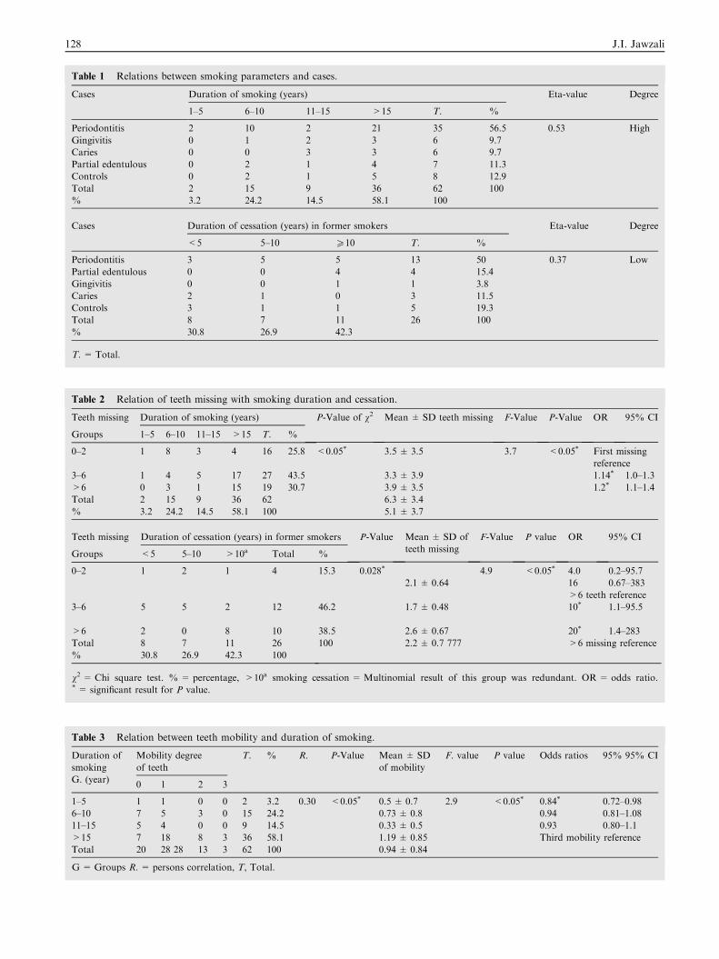
Table 2 Relation of teeth missing with smoking duration and cessation.
Teeth missing Duration of smoking (years) P-Value of v2 Mean ± SD teeth missing F-Value P-Value OR 95% CI
Groups 1–5 6–10 11–15 >15 T. %
0–2 1 8 3 4 16 25.8 <0.05* 3.5 ± 3.5 3.7 <0.05* First missing
reference
3–6 1 4 5 17 27 43.5 3.3 ± 3.9 1.14* 1.0–1.3
>6 0 3 1 15 19 30.7 3.9 ± 3.5 1.2* 1.1–1.4
Total 2 15 9 36 62 6.3 ± 3.4
% 3.2 24.2 14.5 58.1 100 5.1 ± 3.7
Teeth missing Duration of cessation (years) in former smokers P-Value Mean ± SD of
teeth missing
F-Value P value OR 95% CI
Groups <5 5–10 >10a Total %
0–2 1 2 1 4 15.3 0.028* 4.9 <0.05* 4.0 0.2–95.7
2.1 ± 0.64 16 0.67–383
>6 teeth reference
3–6 5 5 2 12 46.2 1.7 ± 0.48 10* 1.1–95.5
>6 2 0 8 10 38.5 2.6 ± 0.67 20* 1.4–283
Total 8 7 11 26 100 2.2 ± 0.7 777 >6 missing reference
% 30.8 26.9 42.3 100
v2 = Chi square test. %= percentage, >10a smoking cessation =Multinomial result of this group was redundant. OR= odds ratio.* = significant result for P value.
Table 1 Relations between smoking parameters and cases.
Cases Duration of smoking (years) Eta-value Degree
1–5 6–10 11–15 >15 T. %
Periodontitis 2 10 2 21 35 56.5 0.53 High
Gingivitis 0 1 2 3 6 9.7
Caries 0 0 3 3 6 9.7
Partial edentulous 0 2 1 4 7 11.3
Controls 0 2 1 5 8 12.9
Total 2 15 9 36 62 100
% 3.2 24.2 14.5 58.1 100
Cases Duration of cessation (years) in former smokers Eta-value Degree
<5 5–10 P10 T. %
Periodontitis 3 5 5 13 50 0.37 Low
Partial edentulous 0 0 4 4 15.4
Gingivitis 0 0 1 1 3.8
Caries 2 1 0 3 11.5
Controls 3 1 1 5 19.3
Total 8 7 11 26 100
% 30.8 26.9 42.3
T. = Total.
Table 3 Relation between teeth mobility and duration of smoking.
Duration of
smoking
G. (year)
Mobility degree
of teeth
T. % R. P-Value Mean ± SD
of mobility
F. value P value Odds ratios 95% 95% CI
0 1 2 3
1–5 1 1 0 0 2 3.2 0.30 <0.05* 0.5 ± 0.7 2.9 <0.05* 0.84* 0.72–0.98
6–10 7 5 3 0 15 24.2 0.73 ± 0.8 0.94 0.81–1.08
11–15 5 4 0 0 9 14.5 0.33 ± 0.5 0.93 0.80–1.1
>15 7 18 8 3 36 58.1 1.19 ± 0.85 Third mobility reference
Total 20 28 28 13 3 62 100 0.94 ± 0.84
G= Groups R. = persons correlation, T, Total.
128 J.I. Jawzali

Table 4 Relation of ages with smoking status and cases.
Age groups
(years)
Smoking status Relation Differences 95% CI
Former
smokers
Current
smokers
T. Eta-
test
Value T-
Value
P-Value Odds
ratio
Low Up
21–30 2 5 7 0.8 High Smoking
status
Means ± SD
of ages
8.7 <0.01** 6.7 0.8–
54.9
31–40* 3 12 15 Former
smokers
53.9 ± 12.4 10.7* 1.7–
66.7
41–50 3 5 8 Current
smoker
44.3 ± 12.9 4.4 0.6–
31.3
51–60 10 11 21 2.9 0.6–
14.2
P61 8 3 11 0.4
Total 26 36 62
Age G
(years)
Dental health status (cases) T. Relation Differences
Periodontitis 1 P .ed. 2 Gingivitis 3 Caries 4 Control 5 P-v2 Cases Means ± SD of ages (y) F. value P value
21–30 5 0 1 0 1 7 0.05* 1 45.7 ± 13.5 3.9 <0.01*
31–40* 10 0 2 1 2 15 2 63.4 ± 9.9
41–50 5 0 2 1 0 8 3 40.5 ± 11.5
51–60 11 2 1 2 5 21 4 53.8 ± 9.2
P61 4 5 0 2 0 11 5 48.6 ± 10.9
Total 35 7 5 6 8 62 Total 48.3 ± 13.5
v2 = Chi square value, P .ed. = Partial edentulous, T. = Total.
Table 5 Relations between Smoking duration and cessation with age groups.
Age G. (years) Duration of smoking categories (years) v2 P-value Mean ± SD of duration F-Value Sig.
1–5 6–10 11–15 >15 T. (%)
21–30 1 6 0 0 7 11.3 32.6 <.01** 7.6 ± 2.6 7.9 <.01**
31–40 0 4 4 7 15 24.2 14.9 ± 4.1
41–50 0 3 1 5 9 14.5 17.4 ± 7.5
51–60 0 0 3 17 20 32.2 22.5 ± 6.9
P61 1 2 1 7 11 17.8 17.8 ± 8.6
Total 2 15 9 36 62 100 17.5 ± 7
% 6.5 24.2 14.5 58.1
Smoking cessation (years) Age groups (years) Eta test D. Mean ± SD of ages F value Sig
21–30 31–40 41–50 51–60 >61 T. %
<5 1 1 0 4 2 8 30.8 0.9 High 51.9 ± 12.5 3.2 P.05*
5–10 1 2 1 3 0 7 26.9 46.6 ± 10.8
>10 0 0 2 3 6 11 42.3 60.1 ± 10.9
Total 2 3 3 10 8 26 100 53.9 ± 12.4
% 7.7 11.5 11.5 38.5 30.8 100
%= Percentage, T. = Total, v2 = Chi square test, D. = Degree. * = significan.
Association between salivary sialic acid and periodontal health status 129
3.4.4. Occupation and smoking status
Table 7 shows a significant relation between occupation and smoking
status. There was a significant difference in smoking status among
smokers with various occupations status. Most 33.9% were govern-
ment employed and 14, (66.7%) of them were current smokers, while
the highest 85.7% percentage of retired smokers were former smokers.
Logistic regression showed that risk of current smoking decreased by
income, significantly among retired smokers.
3.4.5. Oral hygiene habit with smoking duration and cases
Table 8 shows high relation of oral hygiene habit (tooth brushing fre-
quency) and duration of smoking as data scale variables. The highest
percentage 44.4%, 16 smokers among the longest duration >15 years
of smoking had no brushing habit of teeth. Logistic regression showed
increased frequency of tooth brushing significantly among smoking
duration 11–15 years, 55% of these smokers had once/day tooth
brushing compared to smokers which had >15 years duration of
smoking.
Relation of oral hygiene with cases: Table 9 shows low relation of
oral hygiene habit (tooth brushing frequency) with cases. High num-
ber of smokers 15, 62.5% with no brushing habit of teeth had peri-
odontitis disease. Logistic regression showed that tooth brushing
frequency decreased in all cases except caries smokers and increased
significantly compared to control, 50% of controls had once/day
teeth brushing.

Table 8 Relations between duration of smoking and tooth brushing.
Duration of smoking (years) Tooth brushing frequency/week Eta-test value Degree of relation Odds ratio 95% CI
Not 1–4 >7 Total %
1–5 0 1 1 2 3.2 0.6 High 1.2 0.9–1.7
6–10 6 1 8 15 24.2 1.1 0.92–1.3
11–15 2 2 5 9 14.5 1.8* 1.0–1.4
>15 16 10 10 36 58.1 >15 is reference
Total 24 14 24 62 100
% 38.7 22.6 38.7 100
Table 9 Relations between dental health status (cases) and tooth brushing.
Cases Tooth brushing frequency/week Eta-test value Degree of relation Odds ratio 95% CI
Not 1-4 >7 Total %
Periodontitis 15 8 12 35 56.5 0.31 Low .964 0.8–1.16
Gingivitis 3 1 3 7 11.3 .976 0.76–1.3
Caries 1 3 2 6 9.7 1.104* 0.87–1.4
Partial edentulous 2 1 3 6 9.6 .952 0.74–1.2
Controls 3 1 4 8 12.9 Control is reference
Total 24 14 24 62
Table 6 Relation between education levels and dose of cigarette/day.
Education status (1–10) cigarette/d >10 cigarette/d Total % Chi squire value P-Value F-Value P-Value
Illiterate 2 22 24 38.7 11.33 <.05* 2.45 <.05*
Primary 1 13 14 22.6
Intermediate 2 10 12 19.4
Secondary 0 8 8 12.9
Institute 1 0 1 1.6
University 0 3 3 4.8
Total 6 56 62 100
Table 7 Relation between occupations status and smoking status.
Occupation Former smoker Current smoker Total % v2 P-Value F-Value P-Value Odds ratio 95% CI
Self employed 10 10 20 3.2 10.2 <0.05* 2.8 <0.05*
Government employed 7 14 21 33.9 0.13 0.013–1.2
Students 2 3 5 8.1 0.25 0.03–2.4
Retired 6 1 7 11.3 0.19 0.012–2.9
House wife 1 8 9 14.5 0.02* 0.001–0.4
Total 26 36 62 100 Last as reference
130 J.I. Jawzali
3.4.6. Diet habit and dental oral health
Table 10 shows relation of sweet diet intake and dental health status
(cases). There was low relation and significant difference in sweet diet
intake among cases. Caries smokers had significantly low to medium
intake of sweet diet compare to periodontitis, while controls (healthy
dental smokers) ingested significantly larger amount of medium con-
tent of sweet diet compared to periodontitis.
3.5. Sialic acid, smoking and dental health
3.5.1. Levels of total sialic acid and their fractions among smokers
Figs. 4–6 show difference in the mean levels of total sialic acid (TSA),
total free sialic acid (TFSA), and total protein bound sialic acid
(TPSA) respectively in different dental health status. Statistical analy-
sis showed high significant difference between periodontal groups
(periodontitia and gingivitis) and groups; partial edentulous, caries
and controls in the level of total free sialic acid, while TSA and TPSA,
were significantly different between periodontitis and cases.
3.5.2. Relation between salivary sialic acid and smoking status
Table 11 shows significant correlation between TFSA and smoking sta-
tus, and distribution of current smokers among cases. There was a sig-
nificant difference of means of total free sialic acid (TFSA) between
current smokers and former smokers. Logistic regression showed
increase in risk of high levels of total free sialic acid significantly in cur-
rent smoking. Total protein bound sialic acid (TPSA) showed only a

Table 10 Relation between oral health status and sweet diet intake.
Parameters Degree of sweet diets F P value Intake OR 95% CI
Cases No Low Med High T. Eta value
Periodontitis 9 11 4 11 35 0.4 5.0 < 0.01**
Partial edentulous 2 2 1 2 7
Gingivitis 1 1 2 2 6
Caries 1 2 3 0 6 Not 5.7
Little 9.2* 1.1–7.4
Med 3.8
High
Controls 0 2 5 1 8 Not
Little
Med 13.8* 1.2–156.7
High
Total 13 18 15 16 62
Med =Medium.
Figure 4 Significant difference among smokers in the mean of total sialic acid.
Figure 5 Significant difference among smokers in the mean of total free sialic acid.
Association between salivary sialic acid and periodontal health status 131
significant positive relation with duration of smoking, while total sialic
acid and other fractions of sialic acid showed no significant relations.
3.5.3. Relation between salivary free sialic acid and periodontal pocket
depth
Total free sialic acid (TFSA) fraction and current smokers were used
for identifying relation between salivary sialic acid and dental parame-
ters as a result of significant increase (TFSA) level in current smoking
(Table 11). Levels of free sialic acid were divided to two groups (627
to >27) mg/l depending on the normal range of free sialic acid in
healthy dental status of non smokers, concluded by (Jawzaly, 2010).
Also probe pocket depths were divided to two groups (63 mm) and
(4 to >6 mm) depending on NPDI. 75% of current smokers had peri-
odontal diseases composed of 61.1% periodontitis and 13.9% gingivitis
(Table 11). Also 75% of current smokers had high levels (>27 mg/l) of
(TFSA) and 63.9% of them had (P4 mm) probe pocket depth
(Table 12). Statistical analysis showed significant relation between
TFSA and probe pocket depth, and significant difference (P
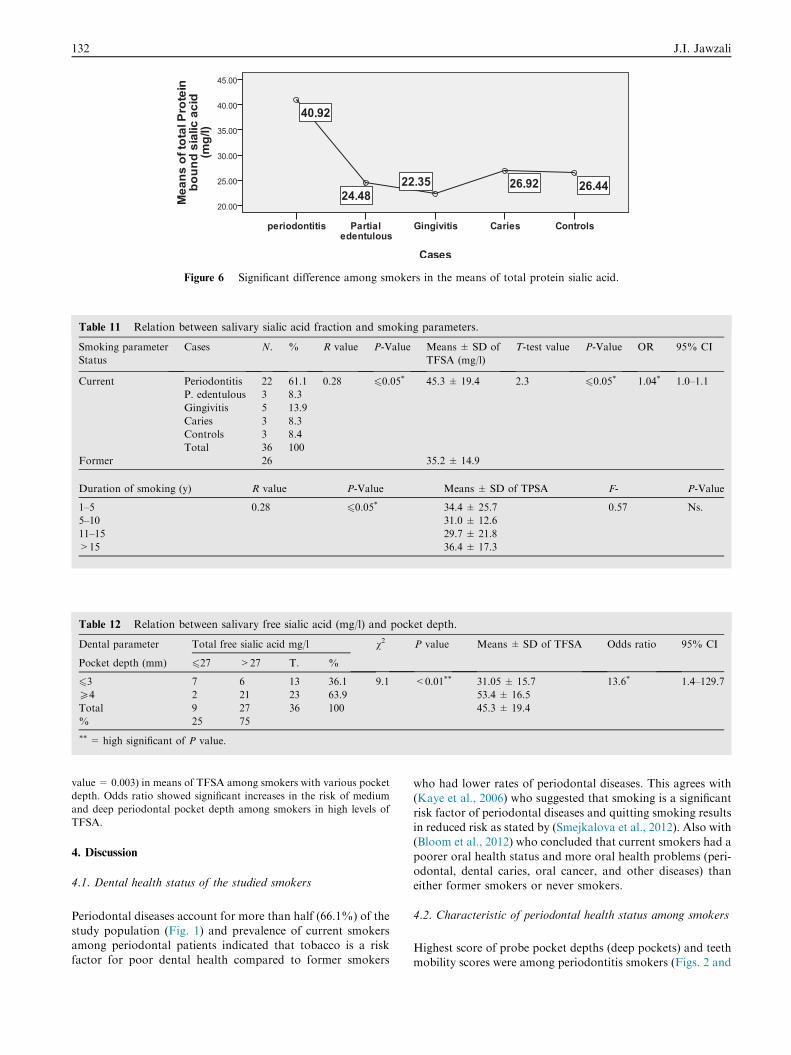
Figure 6 Significant difference among smokers in the means of total protein sialic acid.
Table 11 Relation between salivary sialic acid fraction and smoking parameters.
Smoking parameter Cases N. % R value P-Value Means ± SD of
TFSA (mg/l)
T-test value P-Value OR 95% CI
Status
Current Periodontitis 22 61.1 0.28 60.05* 45.3 ± 19.4 2.3 60.05* 1.04* 1.0–1.1
P. edentulous 3 8.3
Gingivitis 5 13.9
Caries 3 8.3
Controls 3 8.4
Total 36 100
Former 26 35.2 ± 14.9
Duration of smoking (y) R value P-Value Means ± SD of TPSA F- P-Value
1–5 0.28 60.05* 34.4 ± 25.7 0.57 Ns.
5–10 31.0 ± 12.6
11–15 29.7 ± 21.8
>15 36.4 ± 17.3
Table 12 Relation between salivary free sialic acid (mg/l) and pocket depth.
Dental parameter Total free sialic acid mg/l v2 P value Means ± SD of TFSA Odds ratio 95% CI
Pocket depth (mm) 627 >27 T. %
63 7 6 13 36.1 9.1 <0.01** 31.05 ± 15.7 13.6* 1.4–129.7
P4 2 21 23 63.9 53.4 ± 16.5
Total 9 27 36 100 45.3 ± 19.4
% 25 75
** = high significant of P value.
132 J.I. Jawzali
value = 0.003) in means of TFSA among smokers with various pocket
depth. Odds ratio showed significant increases in the risk of medium
and deep periodontal pocket depth among smokers in high levels of
TFSA.
4. Discussion
4.1. Dental health status of the studied smokers
Periodontal diseases account for more than half (66.1%) of the
study population (Fig. 1) and prevalence of current smokersamong periodontal patients indicated that tobacco is a riskfactor for poor dental health compared to former smokers
who had lower rates of periodontal diseases. This agrees with(Kaye et al., 2006) who suggested that smoking is a significant
risk factor of periodontal diseases and quitting smoking resultsin reduced risk as stated by (Smejkalova et al., 2012). Also with(Bloom et al., 2012) who concluded that current smokers had a
poorer oral health status and more oral health problems (peri-odontal, dental caries, oral cancer, and other diseases) thaneither former smokers or never smokers.
4.2. Characteristic of periodontal health status among smokers
Highest score of probe pocket depths (deep pockets) and teethmobility scores were among periodontitis smokers (Figs. 2 and

Association between salivary sialic acid and periodontal health status 133
3), this indicates ligament and alveolar bone destruction withpocket formation and it is in agreement with (Sceedev et al.,2012) who reported that smokers had significantly more sites
with probing depths 4–7 mm, and (Smejkalova et al., 2012)who found shallow or deep gingival pocket in all sextants ofsmokers. Bergstrom et al. (2000) found that smokers not only
have significantly increased probing depths and alveolar boneloss, but also increased tooth mobility. High teeth mobilityscore in partial edentulous smokers who lost teeth as a result
of peridontitis or caries may due to their old ages (P50 years)and extended tobacco use which cause permanent loss of thealveolar bone. This in line with (Jette et al., 1993) who con-firmed that exposure to tobacco is a risk factor for tooth loss
in older adults.
4.3. Dental health status and smoking parameters (Table 1–3)
Most studied smokers (current and former smokers) hadlongest duration of smoking (>15) years especially peri-odontitis smokers, who had highest teeth missing and teeth
mobility scores. This was apparent by a significant associa-tion of teeth missing and teeth mobility with duration ofsmoking and included a large number smokers with highest
teeth missing, (Tables 2 and 3). Therefore long duration ofsmoking may be a risk factor for poor oral health; periodon-titis and teeth loses. This result is consistent with (Jette et al.,1993) who found that duration of tobacco use among cur-
rent and past tobacco users is a significant risk factor fortooth loss.
Majority (81.1) of former smokers with the longest dura-
tion of smoking cessation >10 years were periodontitis andpartially edentulous (Table 1) and included approximately3/4 of smokers with high teeth missing (>6) and increased risk
of teeth missing in contrast to short duration of smoking ces-sation and decreased risk of low number of teeth missing(Table 2). These results indicate lifelong permanent cumulative
effect of smoking on periodontum destruction, which increasesby duration of smoking, and requires more years to reducetooth loss. This result is in consistence with (Kaye et al.,2006) who concluded that smoking cessation is beneficial for
tooth retention, but long term abstinence is required to reducethe risk to the level of people who have never smoked, becauseof irreversible alveolar bone loss.
4.4. Smoking parameter, dental health status and socio-
demographic properties
4.4.1. Relation smoking status and dental health status with ages(Table 4)
Age group 31–40 years, increased the risk of smoking because80% of this age group were current smokers, and more thanhalf had periodontitis diseases. While 3/4 of old ages groups51 to P61 years were former smokers, included all partially
edentulous, simple caries diseases, and healthy dental group(control). These results indicate that current smoking in youngages have a higher than average risk of periodontal disease and
declined with age as concluded by (Wayne, 2007).
4.4.2. Smoking duration and cessation with ages (Table 5)
Longest duration of smoking which increased teeth missing
and mobility was in age group 51–60 years. So at this age
group; year’s exposure to tobacco accumulated and increaserisk of missing and mobility as in periodontitis which occupies11 smokers 53.4% of this age (Table 4). Short cessation of
smoking which decreased the risk of low number of teeth miss-ing was also in this age group and included control and cariesgroups (Table 1) especially 5 smokers represent 62.5%, of the
control at age group 51–60 years (Table 4). This may be due toa protective effect of smoking cessation in early old ages not>60 years because older included more than half of smokers
that had the longest duration of smoking cessation with thehighest teeth missing, which were former periodontitis andpartial edentulous smokers. These results are in agreementwith (Mai et al., 2013) who concluded that smoking may be
a major factor in tooth loss especially in the older populationdue to persistent periodontitis, and may decrease in youngadults and it is less important factor in tooth loss due to caries
which occur in all age groups.
4.4.3. Smoking with education and occupation (Tables 6 and 7)
The result indicates that low levels of education, cause less
awareness to high dose of cigarette intake in contrast to highlevels of education. Smoking risk is associated positively withoccupation status and income, which increased in government
employers, and decreased in retired former smokers as con-firmed by logistic analysis and showed a significantly decreasedrisk of current smoking in retired old age smokers compared to
house wives, majority were current smokers. This result is inconsistence with (Smejkalova et al., 2012) that found a preva-lence of current smokers in the group of respondents with the
highest income and is in contrast with (Wayne, 2007) inincome but agrees with him in the levels of education andage who stated that the prevalence of current smoking declinedwith advancing age and was inversely associated with house-
hold income and level of education.
4.4.4. Duration of smoking and dental health status with oral
hygiene habit and diet habit (Tables 8–10)
The result indicates protective effect of tooth brushing habit(once/day), short duration (<15 years) of smoking and lowto medium content of sweet diets, especially among patients
who quit smoking which includes simple caries and controlbecause; most of them were former smokers (Fig. 1) and50% of caries and 32.7% of controls quit smoking after a short
duration of smoking 11–15 years (Table 1) and most of themwere at age groups <61 years (Table 4). These results indi-cated that, smoking cessation before age of 61 years, oral
hygiene and medium content of sweet diet may decrease oxida-tive stress and conserves oral health. These results confirm theresults of Arowojolu et al. (2013) who concluded that smokersshould be instructed to detailed oral hygiene and encouraged
to quit smoking. Ozturk et al. (2008) who reported that goodoral hygiene may contribute to saliva composition as an oxida-tive stress-decreasing factor in dental diseases. Smejkalova
et al. (2012) who found that current smokers consumed sweet-ened diet more frequently than non smokers and former smok-ers and reported that daily smoking was associated with
increased use of sugar in tea and coffee, and with (Milwardand Chapple, 2013) who suggested that oxidative stress thatwas generated from frequency intake of simple sugar, carbohy-
drate and fat is the mechanism by which smoking increases therisk of periodontitis.

134 J.I. Jawzali
4.4.5. Levels of sialic acid and its fraction among the studied
sample (Figs. 4–6)
High significant levels of total sialic acid (TSA), protein boundsialic acid (PSA), among periodontitis and free sialic acid(FSA) among periodontitis and gingivitis smokers (most were
current smokers) compared to partial edentulous, caries andcontrols,(majority were former smokers Fig. 1), may indicatedifference in inflammation, and oxidative stress status, free
radical production and destructive process .These resultsmay be due to differences; in ages, smoking habit, adequateantioxidant defense and social behaviors and are in consistencewith (Dhotre et al., 2012) who revealed that smoking increases
the level of free radicals in periodontal tissues, which may beresponsible for the destruction seen in periodontal diseasesand with (Kurtul and Gokpinar, 2012) who concluded
increased salivary total sialic acid (TSA) and malonyl aldehyde(MDA) levels in smokers and contribute it to lipidperoxidation.
No significant difference between caries and control groupin; TSA, and its fraction (FSA, TPA) was confirmed by nosignificant difference of periodontal indexes (probe depth
and mobility) and indicated to no periodontal damage andoxidative stress in these groups. This result may be due tosmoking cessation and similarity of control with caries iningestion of sweet diet and tooth brushing. It is online with
(Ozturk et al., 2008) who found similar LPO levels for boththe caries and caries-free groups and was attributed to toothbrushing. Mai et al. (2013) concluded that smoking appears
to be a less important factor in tooth loss due to caries.Smejkalova et al. (2012) reported that association betweencaries and smoking due to other contributing factors; ages,
oral hygiene and eating habit and caries is well documentedin old age groups.
4.4.6. Relation between salivary free sialic acid with smokingstatus and periodontal pocket depth: (Tables 11 and 12)
Significant relation of high levels of free sialic acid (>27) withcurrent smoking (47% were in young ages Table 4) and probe
pocket depth P4 mm, indicate to risk of smoking and severperiodontitis disease. Free sialic acid may be released fromhydrolysis of glycosidic linkage of terminal sialic acid of mucinby reactive oxygen species (ROS) resulted from current smok-
ing, as indicated by (Eguchi et al., 2005), and cause injury, lipidperoxidation, and destruction of periodontal tissues. ROS maybe produced from defects of the inflammatory response, lack
of adequate antioxidant (AO) defence in young ages, smoking(which cause change in neutrophil function and cytokine) andpoor dietary habit as reported by (Sculley and Langley-Evans,
2003). These results confirm that sialic acid besides being aninflammation marker may be an alternative oxidative stressmarker in tobacco exposure, and agree with (Bloomer, 2007)
who indicated that young, novice cigarette smokers have lowerblood antioxidant capacity and higher lipid peroxidation levelscompared to nonsmokers, and revealed that smoking increasesthe level of free radicals and periodontal diseases. Also with
(Cavas et al., 2005) who concluded that over-excreted sialicacids to saliva might have an important role in the removalof hydrogen peroxide and increase in FSA levels in saliva,
and has been found to be in good accordance with antioxidantenzymes and concluded that FSA may be an alternative oxida-tive stress marker.
5. Conclusion
Salivary free sialic acid can be used as diagnostic oxidativestress bio-market for periodontal disease among young to
mid ages of current smokers. Cumulative destruction effectof long duration of smoking on periodontal tissue can bedecreased by early smoking cessation, accompanied with good
oral hygiene, eating habits and low income, especially in agesnot more than 60 years.
Conflicts of interest
There was no conflict of interest to declare.
Acknowledgments
Thanks first given to all study participants for their contribu-tions and periodontic clinic staff for their support during datacollection. Thanks to College of Nursing/Hawler Medical
University and Khanzad specialized dental center hospitalfor their permission to collect data.
References
Arowojolu, Modupe O., Fawole, Olufunmilayo I., Dosumu, Elizabeth
B., Opeodu, O.I., 2013. A comparative study of the oral hygiene
status of smokers and non-smokers in Ibadan, Oyo state. Niger.
Med. J. 54 (4), 240–243. http://dx.doi.org/10.4103/0300-
1652.119627.
Bergstrom, J., Eliasson, S., Dock, J., 2000. Exposure to tobacco
smoking and periodontal health. J. Clin. Periodontol. 27, 61–68.
Bloom, Barbara, Adams, Patricia F., Cohen, Robin A., Simile,
Catherine M., 2012. Centers for Disease Control and Prevention’s
(CDC) National Center for Health Statistics, Division of Health
Interview Statistics. Smoking and Oral Health in Dentate Adults
aged 18-64. NCHS Data Brief, Number 85.
Bloomer, Richard J., 2007. Decreased blood antioxidant capacity and
increased lipid peroxidation in young cigarette smokers compared
to nonsmokers: impact of dietary intake. Nutr. J. 6, 39. http://dx.
doi.org/10.1186/1475-2891-6-39.
Cavas, L., Arpinar, P., Yurdakoc, K., 2005. Possible interactions
between antioxidant enzymes and free sialic acids in saliva: a
preliminary study on elite judoists. Int. J. Sports Med. 26 (10), 832–
835.
Chapple, Iain L.C., Milward, Mike R., Dietrich, Thomas, 2007. The
prevalence of inflammatory periodontitis is negatively associated
with serum antioxidant concentrations. Am. Soc. Nutr. 137, 657–
664.
Dhotre, Pradnya Shree, Suryakar, Adinath N., Bhogade, Rajashree B.,
2012. Oxidative Stress in Periodontitis. Eur. J. Gen. Med. 9 (2), 81–
84.
Eguchi, H., Ikeda, Y., Ookawara, T., Koyota, S., Fujiwara, N.,
Honke, K., Wang, P.G., Taniguchi, N., Suzuki, K., 2005. Mod-
ification of oligosaccharides by reactive oxygen species decreases
sialyl lewis x-mediated cell adhesion. Glycobiology 15.
Beltran-Aguilar, Eugenio D., Eke, Paul I., Thornton-Evans, Gina,
Petersen, Poul E., 2012. Recording and surveillance systems for
periodontal diseases. Periodontol 2000 60 (1), 40–53. http://dx.doi.
org/10.1111/j.1600-0757.2012.00446.x.
Fermin, A.C., Henry, H.T., 2005. Clinical diagnosis. In: Fermin, A.
Carranza (Ed.), Carrnzas Clinical Periodontology, 10th ed. Middle
East Sanders Elsevier.
Grossman, F.D., 1974. Navy periodontal screening exam. J. Am. Soc.
Prev. Den. 3, 41–45.

Association between salivary sialic acid and periodontal health status 135
Jawzaly, J., 2010. A PhD thesis, College of Dentistry, Hawler
Medicinal University, Erbil, Iraq.
Jette, Alan M., Feldman, Henry A., Tennstedt, Sharon L., 1993.
Tobacco use: a modifiable risk factor for dental disease among the
elderly. Am. J. Public Health 83 (9).
Kaye, Elizabeth Krall, Dietrich, Thomas, Nunn, Martha E., Garcia,
Raul I., 2006. Risk of tooth loss after cigarette smoking cessation.
Prev. Chron. Dis. 3 (4), A115.
Kurtul, Naciye, Gokpinar, Engin, 2012. Salivary lipid peroxidation
and total sialic acid levels in smokers and smokeless tobacco users
as maras, powder. Mediat. Inflamm., 8 http://dx.doi.org/10.1155/
2012/619293, Hindawi Publishing Corporation.
Mai, Xiaodan, Wactawski-Wende, Jean, Hovey, Kathleen M.,
LaMonte, Michael J., Chen, Chaoru, Tezal, Mine, Genco, Robert
J., 2013. Associations between smoking and tooth loss according to
the reason for tooth loss. JADA 144 (3), http://jada.ada.org.
Masami, S., 1989. Method of measuring lipid bound sialic acid. United
States Patent 4837144; 6/6/1989.
Milward, Chapple, 2013. The role of diet in periodontal diseases.
Clinical 52.
Mumghamba, E.G.S., Matee, M.I.N., Pitiphat, W., Simon, E., 2004.
The usefulness of using Ramfjord teeth in predicting periodontal
status of a Tanzanian adult population. J. Clin. Periodontol. 31 (1).
Naresh Kumar, C., 2012. A comparative study to assess and correlate
the levels of superoxide dismutase, glutathione peroxidase, malon-
dialdehyde and sialic acid in smokers and nonsmokers with chronic
periodontitis. Master thesis of dental surgery, Periodontology,
College of Dental surgery and Research Center. Bangalore. No 3 of
6).
Ogasawara, Y., Namai, T., Yoshino, F., Lee, M.C., Ishii, K., 2007.
Sialic acid is an essential moiety of mucin as a hydroxyl radical
scavenger. FEBS Lett. 581, 2473–2477.
Ozturk, L.K., Furuncuoglu, H., Atala, M.H., Ulukoylu, O., Akyuz, S.,
Yarat, A., 2008. Association between dental-oral health in young
adults and salivary glutathione, lipid peroxidation and sialic acid
levels and carbonic an hyhydase acrtivity. Braz J Med Bio Res 41
(11), 956–959.
Saxer, Muhlemann, 2004. Epidemiology of periodontal diseases. In:
Muelle (Ed.), Periodontology. Thiem, Stuttgart: New York, pp.
38–46.
Sceedev, Maddipati, Ramesh, Alampall, Dwarakenath, Chin, 2012.
Periodontal status in smokers and nonsmokers: a clinical, micro-
biological, and histopathological study. Int. J. Dentistry 571590.
Sculley, Langley-Evans, 2003. Periodontal disease is associated with
lower antioxidant capacity in whole saliva and evidence of
increased protein oxidation. Clin. Sci. 105, 167–172.
Shetty, P.K., Pattabiraman, T.N., 2004. Salivary glycoproteins as
indicators of oral diseases. Indian J. Clin. Biochem. 19 (1), 97–101.
Silvestre, Francisco-J., Miralles, Lucıa, Llambes, Fernando, Bautista,
Daniel, Sola-Izquierdo, Eva, Hernandez-Mijares, Antonio, 2009.
Type 1 diabetes mellitus and periodontal disease relationship to
different clinical variables. Med. Oral Patol. Oral Circ. Bucal. 14
(4), E175–E179.
Skoza, L., Mohos, S., 1976. Stable thiobarbituric acid chromophor
with dimethyl sulphoxide. Biochem. J. 159, 457–462.
Smejkalova, Jindra, Jacob, Vimal, Hodacova, Lenka, Fiala, Zdenek,
Slezak, Radovan, Vellappally, Sajith, 2012. The influence of
smoking on dental and periodontal status. In: Virdi, Mandeep
(Ed.), Oral Health Care – Pediatric, Research, Epidemiology and
Clinical Practices. InTech, ISBN: 978-953-51-0133-8, <http://
www.intechopen.com>.
Wayne, J., 2007. Millar and David locker. Smoking and oral health
status. JCDA 73 (2).