Associate Member, Bloodworks Associate Professor ...
28
Transfusion Medicine Jill Johnsen, MD Associate Member, Bloodworks Associate Professor, University of Washington
Transcript of Associate Member, Bloodworks Associate Professor ...

Transfusion Medicine
Jill Johnsen, MDAssociate Member, Bloodworks Associate Professor, University of Washington

Source: ASH Image Bank.
Transfusion is one of the most common inpatient procedures1
Transfused daily in the U.S.2 :– 36,000 U red blood cells (RBCs)– 7000 U platelets– 10,000 U plasma
1. Delaney M, Wendel S, Bercovitz RS, et al; Biomedical Excellence for Safer Transfusion (BEST) Collaborative. Transfusion reactions: prevention, diagnosis, and treatment. Lancet. 2016;388(10061):2825-2836.2. American Red Cross. Blood needs & blood supply. Available at: https://www.redcrossblood.org/donate-blood/how-to-donate/how-blood-donationshelp/blood-needs-blood-supply.
Transfusion

Transfusion: Progress from a potentially lethal procedure to a now largely safe and common treatment
A patient having blood let from his right arm, while the blood of a dog is transfused into his left arm. Engraving, ca.1692.Wellcome Library, London
Blood transfusion used during childbirth, including instruments. From Gustave-Joseph Alphonse Witkowski’s Histoire des accouchements, ca. 1887. Wellcome Library, London

Discovery of ABO made safe transfusion possible
The four blood groups. From Laurence H Snyder’s Blood Grouping in Relation to Clinical and Legal Medicine, 1929. WellcomeLibrary, London

Now over 300 known red cell blood group antigens1
1. Johnsen J. Hematology (ASH Education Program). Dec 5;2015(1):168-76

Patient may need a transfusion? - order a “type and screen”
“TYPE” is a test to determine blood typeABO and RhD (D) are considered in all transfusionsExtended typing tests for other blood groups (next most common: C,E,K)
Forward type: detects antigens on the patient’s RBCs using reagent antibodyReverse type: detects antibodies in plasma/serum using reagent RBCs
“SCREEN” is a test to identify presence of anti-RBC antibodies
(Ordering a type and screen is a good time to consider IV access)

TYPE: Routine blood group testing is based on interactionof RBCs with anti-RBC antibodies
Tube testing image courtesy of Kerry Lannert
All transfusions: ABO, D. Higher risk transfusions: C, E, K, potentially others
Genotyping can also be done, can test more blood groups at once

Incubation of patient plasma/serum with red cells from 2-3 very well characterized “reagent” blood cell donors
– Collectively, these donors present the non-ABO blood group antigens likely to provoke allosensitization and transfusion reactions
SCREEN: test for anti-RBC antibodies

Antibody screen positive? Next step: Antibody identification
Antibody identificationA logic puzzle Tests agglutination against a panel of 10-16 human red cells that express blood group antigens in different combinations• Can take hours to days to solve• Can be confounded by interfering agents
(e.g. warm antibodies, some drugs)

Patient needs a transfusion: order a “Type and Cross”TYPE, SCREEN, and Crossmatch
CROSSMATCH: identifies blood components for transfusion in this patientIf negative antibody screen:• Electronic Crossmatch (most common!)• Immediate Spin Crossmatch
– Rapid mixing of patient serum with donor RBCs for ABO compatibility
If positive antibody screen:• Full Crossmatch
– Requires incubations and Coombs reagent to test that the patient’s serum does not react with donor RBCs
– Takes >45 minutes– Takes a lot longer if there is an antibody to a high incidence (very common) antigen

Blood components are from blood donors
• Volunteer blood donors• Complete a health assessment and questionnaire• Meet minimum physiologic criteria• Blood is sampled for testing
– Blood groups: minimum ABO and D (including testing for weak D)• Other blood groups in recurring donors and/or for special situations
– Blood borne pathogen testing:• Serology: HIV-1/2, HCV, HTLV-I/II, HBc, HBsAg, syphilis• Nucleic acid testing: HCV RNA, HIV-1 RNA, WNV RNA, HBV DNA• At least once: serology negative for Trypanosoma cruzi• More recent additions: Zika, Babesia microti

• Red cells (packed red blood cells, or PRBCs): increase Hgb ~1 g/dL*– Hct 65-80% in 225-350mL, stored at 4C, shelf life 42 days
• Platelets (160-400 mL in plasma): increase platelets ~40-50 K/uL*– single donor (pharesis) or pooled (4-6 donors from whole blood centrifugation)– stored at RT, shelf life 5 days
• Plasma (albumin, coag factors, fibrinolytic proteins, Igs, others): 200-250mL– fresh plasma or fresh frozen plasma (FFP)– stored frozen, shelf life one year; thawed shelf life 24 hours
Further manufacturing:• Cryoprecipitate (insoluble cold precipitate of plasma):
– fibrinogen, VWF, factor VIII, factor XIII
• Prothrombin complex concentrates (thrombin, FIX, FX, FVII), IVIg, Albumin, etc.*in average-sized adults
Blood components for transfusion

Infectious risks of transfusion
Weinstein R. Red Blood Cell Transfusion: A Pocket Guide for the Clinician. American Society of Hematology. November, 2016.

Immediate immunologic complications of transfusion
• Hemolytic transfusion reaction (HTR) – Destruction of RBCs by anti-blood group antibodies, life-threatening
• Immune-mediated platelet destruction (alloantibodies: HLA or platelet)• Febrile non-hemolytic reaction (anti-WBC antibodies, cytokines)
– Anti-pyretics can offer symptom relief; if recurrent consider leukocyte reduction• Incidence <1% leukocyte reduced RBCs, 5% leukocyte reduced platelets
• Transfusion-related lung injury (TRALI)– Acute hypoxemia, non-cardiogenic pulmonary edema within 6 hours– Due to donor anti-WBC antibodies, pro-inflammatory molecules in stored components
• Allergic reactions (1-3% of plasma-containing components)– Common, mild, self-limited urticarial reaction, usually responsive to antihistamines
• Anaphylactoid/anaphylactic reactions (rare, IgA-deficient patients high risk)– If refractory to meds, consider washed cellular components to reduce plasma exposure

Delayed immunologic complications of transfusion
• Delayed hemolytic transfusion reaction (destruction of RBCs)– Similar to HTR: hemolysis due to either anamnestic or new alloimmune response
• Alloimmunization to (donor) antigens (any blood cell antigens or plasma proteins)– Blood components contain things not on the label (e.g. in platelets: some RBCs, WBCs)
• Post-transfusion purpura (PTP)– Rare, dramatic, self-limited purpura 7-10 days later – Platelet specific antibody destroys autologous and allogeneic platelets, IVIg can treat
• Transfusion-associated graft-vs-host disease (TA-GVHD) (rare)– Transfused allogeneic T-cells (from any component with viable T-cells)– Risks for TA-GVHD: severe cellular immunodeficiency, purine analagues (e.g.
fludarabine), haploidentical HLA to a homozygous donor– Irradiated components are indicated for patients at risk for TA-GVHD

Model of events leading todelayed hemolytic transfusion reaction (DHTR)1
1. Tormey C. and Hendrickson J. Transfusion-related red blood cell alloantibodies:induction and consequences. Blood 2019;133(17);1821-1830. (2019)
Primary allosensitization Antibody evanescence Anamnestic
response

Other (non-immune) complications of transfusion
• Transfusion-associated circulatory overload (TACO)– From excessive volumes or excessively rapid rates: treat pulmonary edema, reduce fluids
• Hypothermia: from infusing large volumes of cold components– Risks arrhythmia/arrest and coagulopathy: mitigated by blood warmers
• Metabolic complications: usually with large volume / rapid transfusions– Citrate “toxicity” : chelation of ionized calcium by the citrate anticoagulant in blood components
• Iron overload• Donor-transmitted infectious agents: Viruses, bacteria, parasites, variant Creutzfeldt-Jakob
• Bacterial sepsis or endotoxin rxns from contamination (infrequent, life-threatening): – Most common culprit component is platelets– Treat aggressively with antibiotics and supportive care
• Cytomegalovirus (CMV): can reside in donor WBCs– Risks for immunocompromised patients and premature infants of seronegative mothers– Risks reduced by transfusing CMV-seronegative or leukocyte-reduced components
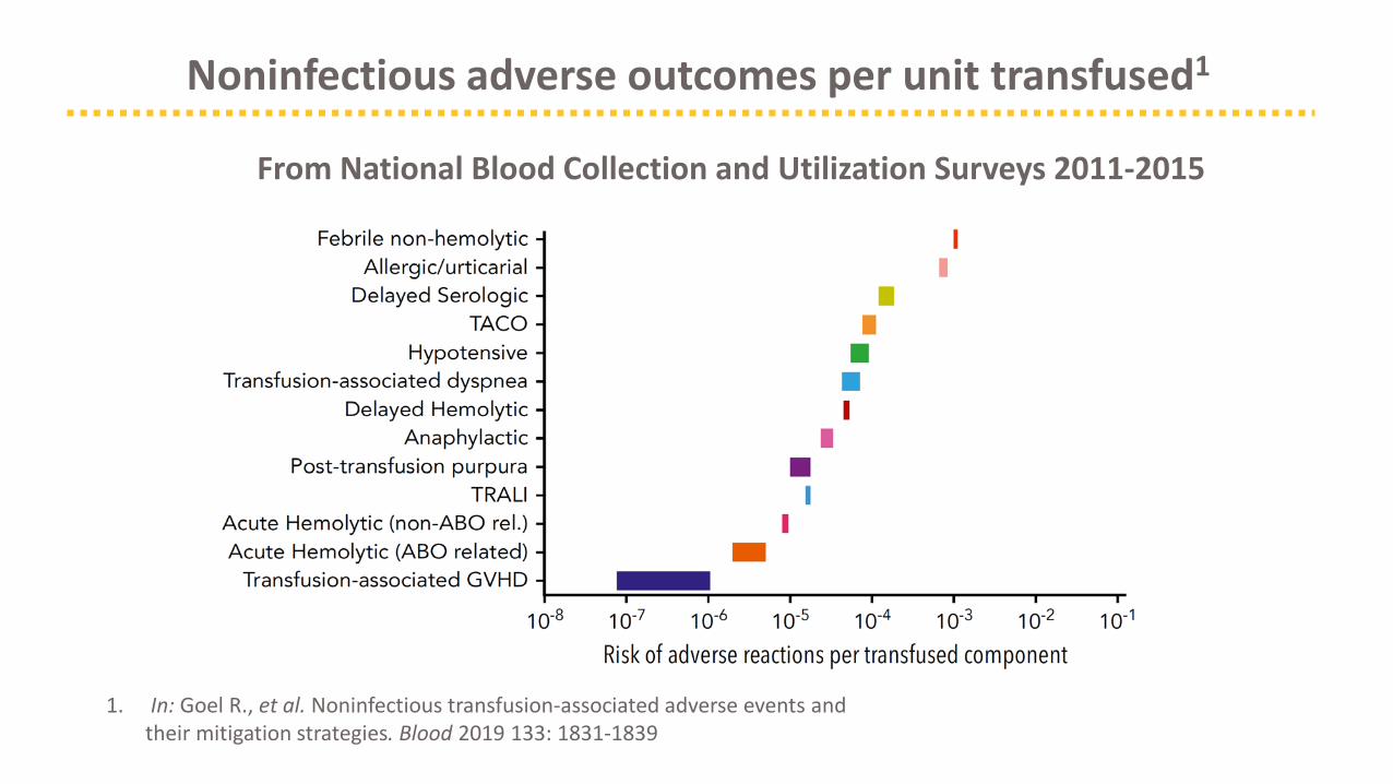
Noninfectious adverse outcomes per unit transfused1
1. In: Goel R., et al. Noninfectious transfusion-associated adverse events and their mitigation strategies. Blood 2019 133: 1831-1839
From National Blood Collection and Utilization Surveys 2011-2015

Serious Hazards Of Transfusion (SHOT) 20191
Summary data for 2019 (n=3397)
1. www.shotuk.org
Mistakes are the most common of the
serious hazards

When to transfuse PRBCs?
AABB Choosing Wisely (#1): Don’t transfuse more units of blood than absolutely necessary.
ASH Choosing Wisely (#1): Don’t transfuse more than the minimum number of red blood cell (RBC) units necessary to relieve symptoms of anemia or to return a patient to a safe hemoglobin range (7 to 8 g/dL in stable, non-cardiac in-patients).
• Transfuse for symptoms and/or hemoglobin – Threshold 7.0-8.0g/dL for most hospitalized, stable patients– Threshold 8.0g/dL for pre-existing cardiovascular disease
• Order one PRBC unit unless actively bleeding (use weight-based dosing in children)– Order more units only after re-assessment– Remember that each unit of blood carries risks
• Liberal transfusion strategies do not improve outcomes compared to restrictive strategies • Unnecessary transfusion generates costs and exposes patients to risks without likely benefit
Adapted from www.choosingwisely.org

AABB Choosing Wisely (#2): Don’t transfuse red blood cells for iron deficiency without hemodynamic instability.• Cheaper and safer alternatives to treat iron deficiency (e.g. iron treatment)• Unless otherwise meet criteria for transfusion, don’t transfuse
When not to transfuse: Asymptomatic iron deficiency anemia
Adapted from www.choosingwisely.org

ASH Choosing Wisely (#7): Don’t routinely transfuse patients with sickle cell disease (SCD) for chronic anemia or uncomplicated pain crisis without an appropriate clinical indication.
• SCD patients are at higher risk for harm from unnecessary PRBC transfusion – alloimmunization to minor blood group antigens– iron overload
• Even most severe types of SCD (baseline hemoglobin 7-10 g/dl) usually tolerate further temporary hemoglobin reductions without symptoms. – IV fluids may contribute to a decrease in hemoglobin by 1-2 g/dL– routine transfusion in this setting should be avoided
• No evidence transfusion reduces SCD vaso-occlusive crisis pain! • Guidance for transfusion in SCD is in the NHLBI 2014 guidelines
High risk for transfusion AEs:Sickle cell disease patients
Adapted from www.choosingwisely.org

“Universal units”: O-negative RBCs, AB-positive (male) plasma– Mitigate risks of ABO incompatibility– Reduce risks of allosensitization to D
AABB Choosing Wisely (#5): Don’t transfuse O negative blood EXCEPT:
– to O negative patients– in emergencies for women of child bearing potential with unknown blood group.
• O-negative PRBC units are in chronic short supply– Shortages are exacerbated by overutilization for patients who are not O-negative– Common practice during shortages to transfuse O-positive in males, use low titer anti-A plasma
“Universal” blood and emergencies
Adapted from www.choosingwisely.org

Blood testing for transfusion:Monitoring recommendations
Adapted from www.choosingwisely.org
AABB Choosing Wisely (#4):Don’t perform serial blood counts on clinically stable patients.
• Unless bleeding or otherwise unstable, transfusion (PRBCs or platelets) should use the results from the first labs of the day
• Multiple blood draws to recheck the transfusion threshold can lead to: – excessive phlebotomy– iatrogenic anemia– unnecessary transfusions
Limit blood draws!!

AABB Choosing Wisely (#3): Don’t routinely use blood products to reverse warfarin.ASH Choosing Wisely (#4): Don’t administer plasma or prothrombin complex concentrates for non-emergent reversal of vitamin K antagonists
(i.e. outside of the setting of major bleeding, ICH, or emergency surgery)
• Rationale: blood products have risks, are costly, and are rarely indicated• Most patients can be reversed with holding warfarin and/or vitamin K• For serious bleeding or emergency surgery / invasive procedures only:
– prothrombin complex concentrates (PCCs)– (plasma)
When not to use plasma and PCCs: Warfarin reversal
Adapted from www.choosingwisely.org

• Preoperative or bleeding patients who require replacement of multiple coagulation factors (e.g., liver disease, DIC)
• Patients undergoing massive transfusion • Patients on warfarin who are actively bleeding or in need of an immediate invasive
procedure• Patients with coagulation factor or plasma protein deficiencies, congenital or
acquired, for which no specific products are available (e.g. FXI, C1 inhibitor)• Thrombotic thrombocytopenic purpura (TTP)
See lectures on coagulation for underlying disorders and management
When to transfuse plasma

• Thresholds for platelet transfusion are evolving: the general trend is towards more conservative use of platelet transfusion– PLADO trial– Pragmatic use of a scarce resource
• Thrombocytopenia: correction of quantitative defects– Prophylactic transfusion: historically PLT <10k, but PLADO trial used threshold < 5K– For invasive procedures, trauma, and active bleeding in patients with moderate to severe
thrombocytopenia – Rapidly falling platelet count with active bleeding or significant consumption
• Platelet dysfunction: correction of qualitative defects– Consider functional platelet count to be the predicted post-transfusion platelet count
See lectures on thrombocytopenia for causes and management
When to transfuse platelets?

Transition slides should be kept clean and simple; two slide masters are provided for transitions between topics.
THANK YOU!