Associate editor: H. Bo¨nisch Heat shock proteins in the ... · Heat shock proteins in the...
31
Associate editor: H. Bo ¨nisch Heat shock proteins in the regulation of apoptosis: new strategies in tumor therapy A comprehensive review Amere Subbarao Sreedhar 1 , Peter Csermely * Department of Medical Chemistry, Semmelweis University, P.O. Box 260, H-1444 Budapest, Hungary Abstract Heat shock proteins (Hsp) form the most ancient defense system in all living organisms on earth. These proteins act as molecular chaperones by helping in the refolding of misfolded proteins and assisting in their elimination if they become irreversibly damaged. Hsp interact with a number of cellular systems and form efficient cytoprotective mechanisms. However, in some cases, wherein it is better if the cell dies, there is no reason for any further defense. Programmed cell death is a widely conserved general phenomenon helping in many processes involving the reconstruction of multicellular organisms, as well as in the elimination of old or damaged cells. Here, we review some novel elements of the apoptotic process, such as its interrelationship with cellular senescence and necrosis, as well as bacterial apoptosis. We also give a survey of the most important elements of the apoptotic machinery and show the various modes of how Hsp interact with the apoptotic events in detail. We review caspase-independent apoptotic pathways and anoikis as well. Finally, we show the emerging variety of pharmacological interventions inhibiting or, just conversely, inducing Hsp and review the emergence of Hsp as novel therapeutic targets in anticancer protocols. D 2004 Elsevier Inc. All rights reserved. Keywords: Apoptosis; Cancer; Heat shock proteins; Molecular chaperones; Necrosis; Tumor suppression Abbreviations: 17-AAG, 17-allylamino-17-demethoxy-geldanamycin; AIF, apoptosis-inducing factor; Ask1, apoptosis signal-regulating kinase-1; BAD, Bcl-2- associated death protein; BAG, Bcl-2-associated athanogene protein; Bax, B-cell lymphoma-2 protein (Bcl-2)-associated X protein; Bcl-2, B-cell lymphoma-2 protein; CAD, caspase-activated deoxyribonuclease; DNase, deoxyribonuclease; ER, endoplasmic reticulum; FADD, Fas-associated death domain protein; Grp, glucose-regulated protein; Hip, Hsc70-interacting protein; Hop, Hsp70-Hsp90 organizing protein; HSF, heat shock factor; Hsp, heat shock protein; IAP, inhibitors of apoptosis proteins; JNK, c-Jun N-terminal kinase; MMPT, matrix metalloprotease; Mn-SOD, manganese superoxide dismutase; NF-nB, nuclear factor-nB; NOS, nitric oxide synthase; p23, 23-kDa co-chaperone of Hsp90; PARP, poly-ADP ribose polymerase; PCD, programmed cell death; PI-3-kinase, phosphatidyl-inositol-3-kinase; PS, phosphatidylserine; PTP, permeability transition pore; ROS, reactive oxygen species; SAPK, stress-activated protein kinase; Smac/DIABLO, second mitochondria-derived activator of caspases; TNF, tumor necrosis factor. Contents 1. Introduction ............................................ 228 1.1. Heat shock proteins: molecular chaperones ........................ 228 1.2. Cellular senescence, apoptosis, and necrosis: chaperone overload as a potential regulator 229 1.3. Evolution of apoptosis: bacteria and eukaryotes ...................... 229 1.4. Major elements of the mechanism of apoptosis ...................... 230 2. Heat shock proteins and caspase-dependent apoptosis ...................... 230 2.1. Sites of initial signaling events .............................. 230 2.1.1. Plasma membrane ................................ 230 2.1.2. Cytosol ...................................... 232 2.1.3. Nucleus ...................................... 232 0163-7258/$ – see front matter D 2004 Elsevier Inc. All rights reserved. doi:10.1016/j.pharmthera.2003.11.004 * Corresponding author. Tel.: +36-1-266-2755x4102; fax: +36-1-266-7480. E-mail address: [email protected] (P. Csermely). 1 Currently on leave from the Centre for Cellular and Molecular Biology, Hyderabad 500 007, India. www.elsevier.com/locate/pharmthera Pharmacology & Therapeutics 101 (2004) 227 – 257
Transcript of Associate editor: H. Bo¨nisch Heat shock proteins in the ... · Heat shock proteins in the...

www.elsevier.com/locate/pharmthera
Pharmacology & Therapeutics 101 (2004) 227–257
Associate editor: H. Bonisch
Heat shock proteins in the regulation of apoptosis:
new strategies in tumor therapy
A comprehensive review
Amere Subbarao Sreedhar1, Peter Csermely*
Department of Medical Chemistry, Semmelweis University, P.O. Box 260, H-1444 Budapest, Hungary
Abstract
Heat shock proteins (Hsp) form the most ancient defense system in all living organisms on earth. These proteins act as molecular
chaperones by helping in the refolding of misfolded proteins and assisting in their elimination if they become irreversibly damaged. Hsp
interact with a number of cellular systems and form efficient cytoprotective mechanisms. However, in some cases, wherein it is better if the
cell dies, there is no reason for any further defense. Programmed cell death is a widely conserved general phenomenon helping in many
processes involving the reconstruction of multicellular organisms, as well as in the elimination of old or damaged cells. Here, we review
some novel elements of the apoptotic process, such as its interrelationship with cellular senescence and necrosis, as well as bacterial
apoptosis. We also give a survey of the most important elements of the apoptotic machinery and show the various modes of how Hsp interact
with the apoptotic events in detail. We review caspase-independent apoptotic pathways and anoikis as well. Finally, we show the emerging
variety of pharmacological interventions inhibiting or, just conversely, inducing Hsp and review the emergence of Hsp as novel therapeutic
targets in anticancer protocols.
D 2004 Elsevier Inc. All rights reserved.
Keywords: Apoptosis; Cancer; Heat shock proteins; Molecular chaperones; Necrosis; Tumor suppression
Abbreviations: 17-AAG, 17-allylamino-17-demethoxy-geldanamycin; AIF, apoptosis-inducing factor; Ask1, apoptosis signal-regulating kinase-1; BAD, Bcl-2-
associated death protein; BAG, Bcl-2-associated athanogene protein; Bax, B-cell lymphoma-2 protein (Bcl-2)-associated X protein; Bcl-2, B-cell lymphoma-2
protein; CAD, caspase-activated deoxyribonuclease; DNase, deoxyribonuclease; ER, endoplasmic reticulum; FADD, Fas-associated death domain protein;
Grp, glucose-regulated protein; Hip, Hsc70-interacting protein; Hop, Hsp70-Hsp90 organizing protein; HSF, heat shock factor; Hsp, heat shock protein; IAP,
inhibitors of apoptosis proteins; JNK, c-Jun N-terminal kinase; MMPT, matrix metalloprotease; Mn-SOD, manganese superoxide dismutase; NF-nB, nuclearfactor-nB; NOS, nitric oxide synthase; p23, 23-kDa co-chaperone of Hsp90; PARP, poly-ADP ribose polymerase; PCD, programmed cell death; PI-3-kinase,
phosphatidyl-inositol-3-kinase; PS, phosphatidylserine; PTP, permeability transition pore; ROS, reactive oxygen species; SAPK, stress-activated protein
kinase; Smac/DIABLO, second mitochondria-derived activator of caspases; TNF, tumor necrosis factor.
Contents
1. Introduction . . . . . . . . . . . . . . . . . . . . . . . . . . . . . . . . . . . . . . . . . . . . 228
1.1. Heat shock proteins: molecular chaperones . . . . . . . . . . . . . . . . . . . . . . . . 228
1.2. Cellular senescence, apoptosis, and necrosis: chaperone overload as a potential regulator 229
1.3. Evolution of apoptosis: bacteria and eukaryotes . . . . . . . . . . . . . . . . . . . . . . 229
1.4. Major elements of the mechanism of apoptosis . . . . . . . . . . . . . . . . . . . . . . 230
2. Heat shock proteins and caspase-dependent apoptosis . . . . . . . . . . . . . . . . . . . . . . 230
2.1. Sites of initial signaling events . . . . . . . . . . . . . . . . . . . . . . . . . . . . . . 230
2.1.1. Plasma membrane . . . . . . . . . . . . . . . . . . . . . . . . . . . . . . . . 230
2.1.2. Cytosol . . . . . . . . . . . . . . . . . . . . . . . . . . . . . . . . . . . . . . 232
2.1.3. Nucleus . . . . . . . . . . . . . . . . . . . . . . . . . . . . . . . . . . . . . . 232
0163-7258/$ – see front matter D 2004 Elsevier Inc. All rights reserved.
doi:10.1016/j.pharmthera.2003.11.004
* Corresponding author. Tel.: +36-1-266-2755x4102; fax: +36-1-266-7480.
E-mail address: [email protected] (P. Csermely).1 Currently on leave from the Centre for Cellular and Molecular Biology, Hyderabad 500 007, India.
A.S. Sreedhar, P. Csermely / Pharmacology & Therapeutics 101 (2004) 227–257228
2.1.4. Mitochondria and reactive oxygen species . . . . . . . . . . . . . . . . . . . . 233
2.1.5. Endoplasmic reticulum . . . . . . . . . . . . . . . . . . . . . . . . . . . . . . 235
2.2. Effector molecules . . . . . . . . . . . . . . . . . . . . . . . . . . . . . . . . . . . . . 236
2.2.1. Caspases . . . . . . . . . . . . . . . . . . . . . . . . . . . . . . . . . . . . . 236
2.2.2. Nucleases . . . . . . . . . . . . . . . . . . . . . . . . . . . . . . . . . . . . . 236
2.2.3. Transglutaminases . . . . . . . . . . . . . . . . . . . . . . . . . . . . . . . . 236
3. Heat shock proteins and caspase-independent apoptosis . . . . . . . . . . . . . . . . . . . . . 237
3.1. Serine proteases . . . . . . . . . . . . . . . . . . . . . . . . . . . . . . . . . . . . . . 237
3.2. Cathepsins . . . . . . . . . . . . . . . . . . . . . . . . . . . . . . . . . . . . . . . . . 237
3.3. Calpains . . . . . . . . . . . . . . . . . . . . . . . . . . . . . . . . . . . . . . . . . . 237
3.4. Ceramide-induced apoptosis . . . . . . . . . . . . . . . . . . . . . . . . . . . . . . . . 238
3.5. Apoptosis-inducing factor . . . . . . . . . . . . . . . . . . . . . . . . . . . . . . . . . 238
3.6. Anoikis . . . . . . . . . . . . . . . . . . . . . . . . . . . . . . . . . . . . . . . . . . 238
4. Heat shock proteins and antiapoptotic mediators . . . . . . . . . . . . . . . . . . . . . . . . . 239
4.1. Heat shock proteins and B-cell lymphoma-2 protein pathway. . . . . . . . . . . . . . . 239
4.2. Heat shock proteins and phosphatidyl-inositol-3-kinase/Akt pathway . . . . . . . . . . . 239
5. Modulation of modulators: effect of apoptosis on heat shock protein induction . . . . . . . . . 240
6. Molecular mechanism of heat shock protein action . . . . . . . . . . . . . . . . . . . . . . . . 240
6.1. Heat shock proteins as molecular chaperones of apoptosis . . . . . . . . . . . . . . . . 240
6.2. Role of co-chaperones . . . . . . . . . . . . . . . . . . . . . . . . . . . . . . . . . . . 240
6.3. Heat shock proteins and cellular homeostasis . . . . . . . . . . . . . . . . . . . . . . . 241
6.3.1. Redox homeostasis . . . . . . . . . . . . . . . . . . . . . . . . . . . . . . . . 241
6.3.2. Cell organization . . . . . . . . . . . . . . . . . . . . . . . . . . . . . . . . . 241
7. Heat shock proteins as pharmacological targets in apoptosis modulation . . . . . . . . . . . . . 242
7.1. Heat shock protein inhibition as an efficient way to induce the apoptosis of tumor cells 242
7.1.1. Apoptosis in tumor cells . . . . . . . . . . . . . . . . . . . . . . . . . . . . . 242
7.1.2. Heat shock proteins in tumor cells . . . . . . . . . . . . . . . . . . . . . . . . 242
7.1.3. Enforced apoptosis of tumor cells . . . . . . . . . . . . . . . . . . . . . . . . 243
7.1.4. Heat shock protein inhibitors as heat shock protein inducers. . . . . . . . . . . 244
7.1.5. Ongoing clinical trials with heat shock protein inhibitors . . . . . . . . . . . . 244
7.2. Therapeutic use of heat shock protein up-regulation . . . . . . . . . . . . . . . . . . . 244
7.2.1. Sensitization of cancer cells for immune attack . . . . . . . . . . . . . . . . . 244
7.2.2. Protection of cells from apoptosis . . . . . . . . . . . . . . . . . . . . . . . . 245
7.2.3. Methods of heat shock protein activation. . . . . . . . . . . . . . . . . . . . . 245
8. Conclusions and perspectives . . . . . . . . . . . . . . . . . . . . . . . . . . . . . . . . . . . 246
8.1. Pleiotropic role of heat shock proteins in apoptosis . . . . . . . . . . . . . . . . . . . . 246
8.2. Exciting areas of further research . . . . . . . . . . . . . . . . . . . . . . . . . . . . . 246
Acknowledgments . . . . . . . . . . . . . . . . . . . . . . . . . . . . . . . . . . . . . . . . . . . 247
References . . . . . . . . . . . . . . . . . . . . . . . . . . . . . . . . . . . . . . . . . . . . . . . 247
1. Introduction
Programmed cell death (PCD) is a common phenome-
non in developmental processes or in normal physiological
conditions, where the old or damaged cells have to be
eliminated. Two distinct forms of cell death, apoptosis and
necrosis, have been characterized. Apoptosis is induced by
an array of extra- or intracellular stimuli. Organisms are
equipped with their own physiological defense to cope
with environmental stress and are under the control of
genetic machinery in order to prevent or induce cell death
depending upon the severity of the stress. Heat shock
proteins (Hsp) are highly conserved and play a major role
in cytoprotection. Apoptosis resistance is associated with
the high expression of Hsp, hence, the present discussion
is focused on the functions of Hsp in apoptosis regulation.
We also review the pharmacological applications using
Hsp inhibitors to induce apoptosis in various tumor
models.
1.1. Heat shock proteins: molecular chaperones
The eukaryotic stress response is highly conserved and
involves the induction of Hsp. Hsp are detected in all living
cells. Both in vivo and in vitro studies have shown that
various stressors transiently increase the production of Hsp
as protection against harmful insults. Hsp induction was
first identified in the salivary glands of Drosophila mela-
nogaster upon application of heat shock (Ritossa, 1962).
The unique nature of Hsp synthesis was correlated with the
acquisition of thermotolerance and cytoprotection. Later, the
interest on Hsp tremendously expanded as more and more
functions of normal resting cells involving Hsp were un-
covered. The multifunctional roles of Hsp in cells show that
A.S. Sreedhar, P. Csermely / Pharmacology & Therapeutics 101 (2004) 227–257 229
Hsp is one of the major regulatory proteins in the cell, and
vital functions of cell, such as the maintenance of the cell
cycle, are associated with Hsp. The role of Hsp in cell
proliferation was reviewed (Pechan, 1991; Helmbrecht et
al., 2000).
In mammalian cells, the stress response involves the
induction of 5 major classes of Hsp families, namely, the
small Hsp exemplified by Hsp27, Hsp60, Hsp70, Hsp90,
and Hsp104 (Table 1; Lindquist & Craig, 1988; Craig et al.,
1994; Morimoto et al., 1994; Scharf et al., 1998). Hsp
synthesis is tightly regulated at the transcriptional level by
heat shock factors (HSF). Although various HSF were
reported, HSF-1 was shown to be the main regulator of
the short-term induction of Hsp. In resting cells, HSF-1 is a
monomer; however, active HSF-1 exists as a trimer and
binds to the heat shock elements (the consensus DNA
sequence for heat shock factor binding; Morimoto et al.,
1992). In addition, some members of the HSF family help
the long-term induction of Hsp or exhibit important roles in
the regulation of gene expression and developmental pro-
cesses (Morimoto, 1998).
Hsp function as molecular chaperones in regulating
cellular homeostasis and promoting cell survival (Hartl,
1996; Bukau & Horwich, 1998). Various studies demon-
strate that Hsp-induced cytoprotection can be attributed
partly to the suppression of apoptosis (Samali & Orrenius,
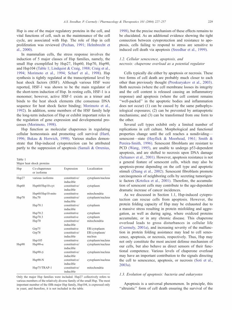
Table 1
Major heat shock proteins
Hsp Co-chaperones
or isoforms
Expression Localization
Hsp27 various isoforms constitutive/
inducible
cytoplasm/nucleus
Hsp60 Hsp60/Hsp10-cyt constitutive/
inducible
cytoplasm
Hsp60/Hsp10-mito constitutive mitochondria
Hsp70 Hsc70 constitutive/
inducible
cytoplasm/nucleus
Hsp70.1 constitutive/
inducible
cytoplasm
Hsp70.2 constitutive cytoplasm
Hsp70.3 constitutive cytoplasm
Hsp70 constitutive/
inducible
mitochondria
Grp75 constitutive ER/cytoplasm
Grp78 constitutive/
inducible
ER/cytoplasm/
nucleus
Hsp105 constitutive cytoplasm/nucleus
Hsp90 Hsp90-a constitutive/
inducible
cytoplasm/nucleus
Hsp90-h constitutive/
inducible
cytoplasm/nucleus
Hsp90-N constitutive/
inducible
cytoplasm/nucleus
Hsp75/TRAP-1 constitutive/
inducible
mitochondria
Only the major Hsp families were included. Hsp27 collectively refers to
various members of the relatively diverse family of the small Hsp. The most
important member of the fifth major Hsp family, Hsp104, is expressed only
in yeast, and therefore, it is not included in the table.
1998), but the precise mechanism of these effects remains to
be elucidated. As an additional evidence showing the tight
connection between cytoprotection and resistance to apo-
ptosis, cells failing to respond to stress are sensitive to
induced cell death via apoptosis (Sreedhar et al., 1999).
1.2. Cellular senescence, apoptosis, and
necrosis: chaperone overload as a potential regulator
Cells typically die either by apoptosis or necrosis. These
two forms of cell death are probably much closer to each
other than previously thought (Proskuryakov et al., 2003).
Both necrosis (where the cell membrane looses its integrity
and the cell content is released causing an inflammatory
response) and apoptosis (where the cell content remains
‘‘well-packed’’ in the apoptotic bodies and inflammation
does not occur) (1) can be caused by the same pathophys-
iological exposures; (2) can be prevented by antiapoptotic
mechanisms; and (3) can be transformed from one form to
the other.
Several cell types exhibit only a limited number of
replications in cell culture. Morphological and functional
properties change until the cell reaches a nondividing—
senescent—state (Hayflick & Moorhead, 1961; Smith &
Pereira-Smith, 1996). Senescent fibroblasts are resistant to
PCD (Wang, 1995), are unable to undergo p53-dependent
apoptosis, and are shifted to necrosis upon DNA damage
(Seluanov et al., 2001). However, apoptosis resistance is not
a general feature of senescent cells, which may also be
apoptosis-prone depending on the cell type and apoptotic
stimuli (Zhang et al., 2002). Senescent fibroblasts promote
carcinogenesis of neighboring cells by secreting tumorigen-
ic factors (Krtolica et al., 2001). Therefore, the accumula-
tion of senescent cells may contribute to the age-dependent
dramatic increase of cancer incidences.
As we discussed in Section 1.1, Hsp-induced cytopro-
tection can rescue cells from apoptosis. However, the
protein folding capacity of Hsp may be exhausted due to
a massive stress resulting in protein misfolding and aggre-
gation, as well as during aging, where oxidized proteins
accumulate, or in any chronic disease. This chaperone
overload leads to gross disturbances in cellular life
(Csermely, 2001a), and increasing severity of the malfunc-
tion in protein folding assistance may lead to cell senes-
cence, apoptosis, or necrosis, respectively. Thus, Hsp may
not only constitute the most ancient defense mechanism of
our cells, but also behave as direct sensors of their func-
tional competence. Various levels of chaperone overload
may have an important contribution to the signals directing
the cell to senescence, apoptosis, or necrosis (Soti et al.,
2003a).
1.3. Evolution of apoptosis: bacteria and eukaryotes
Apoptosis is a universal phenomenon. In principle, this
‘‘altruistic’’ form of cell death ensuring the survival of the
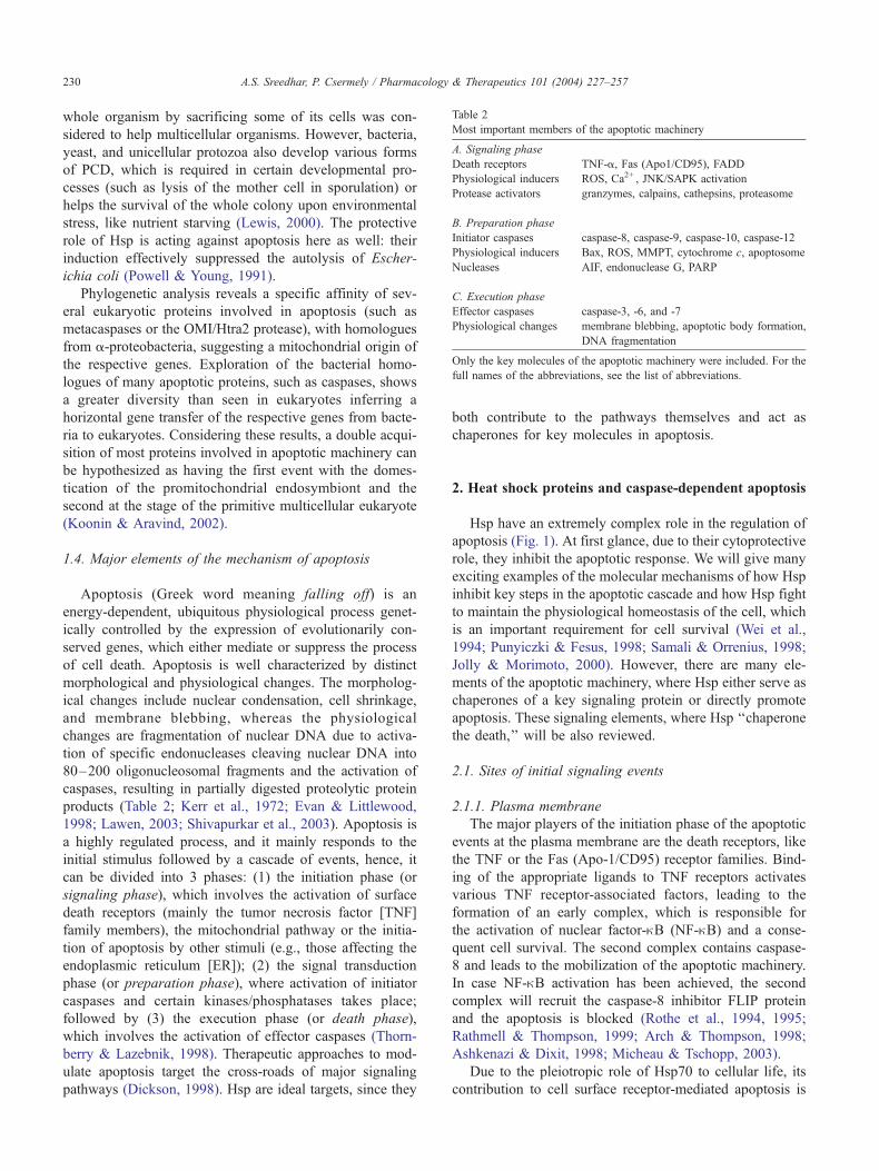
Table 2
Most important members of the apoptotic machinery
A. Signaling phase
Death receptors TNF-a, Fas (Apo1/CD95), FADD
Physiological inducers ROS, Ca2+ , JNK/SAPK activation
Protease activators granzymes, calpains, cathepsins, proteasome
B. Preparation phase
Initiator caspases caspase-8, caspase-9, caspase-10, caspase-12
Physiological inducers Bax, ROS, MMPT, cytochrome c, apoptosome
Nucleases AIF, endonuclease G, PARP
C. Execution phase
Effector caspases caspase-3, -6, and -7
Physiological changes membrane blebbing, apoptotic body formation,
DNA fragmentation
Only the key molecules of the apoptotic machinery were included. For the
full names of the abbreviations, see the list of abbreviations.
A.S. Sreedhar, P. Csermely / Pharmacology & Therapeutics 101 (2004) 227–257230
whole organism by sacrificing some of its cells was con-
sidered to help multicellular organisms. However, bacteria,
yeast, and unicellular protozoa also develop various forms
of PCD, which is required in certain developmental pro-
cesses (such as lysis of the mother cell in sporulation) or
helps the survival of the whole colony upon environmental
stress, like nutrient starving (Lewis, 2000). The protective
role of Hsp is acting against apoptosis here as well: their
induction effectively suppressed the autolysis of Escher-
ichia coli (Powell & Young, 1991).
Phylogenetic analysis reveals a specific affinity of sev-
eral eukaryotic proteins involved in apoptosis (such as
metacaspases or the OMI/Htra2 protease), with homologues
from a-proteobacteria, suggesting a mitochondrial origin of
the respective genes. Exploration of the bacterial homo-
logues of many apoptotic proteins, such as caspases, shows
a greater diversity than seen in eukaryotes inferring a
horizontal gene transfer of the respective genes from bacte-
ria to eukaryotes. Considering these results, a double acqui-
sition of most proteins involved in apoptotic machinery can
be hypothesized as having the first event with the domes-
tication of the promitochondrial endosymbiont and the
second at the stage of the primitive multicellular eukaryote
(Koonin & Aravind, 2002).
1.4. Major elements of the mechanism of apoptosis
Apoptosis (Greek word meaning falling off) is an
energy-dependent, ubiquitous physiological process genet-
ically controlled by the expression of evolutionarily con-
served genes, which either mediate or suppress the process
of cell death. Apoptosis is well characterized by distinct
morphological and physiological changes. The morpholog-
ical changes include nuclear condensation, cell shrinkage,
and membrane blebbing, whereas the physiological
changes are fragmentation of nuclear DNA due to activa-
tion of specific endonucleases cleaving nuclear DNA into
80–200 oligonucleosomal fragments and the activation of
caspases, resulting in partially digested proteolytic protein
products (Table 2; Kerr et al., 1972; Evan & Littlewood,
1998; Lawen, 2003; Shivapurkar et al., 2003). Apoptosis is
a highly regulated process, and it mainly responds to the
initial stimulus followed by a cascade of events, hence, it
can be divided into 3 phases: (1) the initiation phase (or
signaling phase), which involves the activation of surface
death receptors (mainly the tumor necrosis factor [TNF]
family members), the mitochondrial pathway or the initia-
tion of apoptosis by other stimuli (e.g., those affecting the
endoplasmic reticulum [ER]); (2) the signal transduction
phase (or preparation phase), where activation of initiator
caspases and certain kinases/phosphatases takes place;
followed by (3) the execution phase (or death phase),
which involves the activation of effector caspases (Thorn-
berry & Lazebnik, 1998). Therapeutic approaches to mod-
ulate apoptosis target the cross-roads of major signaling
pathways (Dickson, 1998). Hsp are ideal targets, since they
both contribute to the pathways themselves and act as
chaperones for key molecules in apoptosis.
2. Heat shock proteins and caspase-dependent apoptosis
Hsp have an extremely complex role in the regulation of
apoptosis (Fig. 1). At first glance, due to their cytoprotective
role, they inhibit the apoptotic response. We will give many
exciting examples of the molecular mechanisms of how Hsp
inhibit key steps in the apoptotic cascade and how Hsp fight
to maintain the physiological homeostasis of the cell, which
is an important requirement for cell survival (Wei et al.,
1994; Punyiczki & Fesus, 1998; Samali & Orrenius, 1998;
Jolly & Morimoto, 2000). However, there are many ele-
ments of the apoptotic machinery, where Hsp either serve as
chaperones of a key signaling protein or directly promote
apoptosis. These signaling elements, where Hsp ‘‘chaperone
the death,’’ will be also reviewed.
2.1. Sites of initial signaling events
2.1.1. Plasma membrane
The major players of the initiation phase of the apoptotic
events at the plasma membrane are the death receptors, like
the TNF or the Fas (Apo-1/CD95) receptor families. Bind-
ing of the appropriate ligands to TNF receptors activates
various TNF receptor-associated factors, leading to the
formation of an early complex, which is responsible for
the activation of nuclear factor-nB (NF-nB) and a conse-
quent cell survival. The second complex contains caspase-
8 and leads to the mobilization of the apoptotic machinery.
In case NF-nB activation has been achieved, the second
complex will recruit the caspase-8 inhibitor FLIP protein
and the apoptosis is blocked (Rothe et al., 1994, 1995;
Rathmell & Thompson, 1999; Arch & Thompson, 1998;
Ashkenazi & Dixit, 1998; Micheau & Tschopp, 2003).
Due to the pleiotropic role of Hsp70 to cellular life, its
contribution to cell surface receptor-mediated apoptosis is
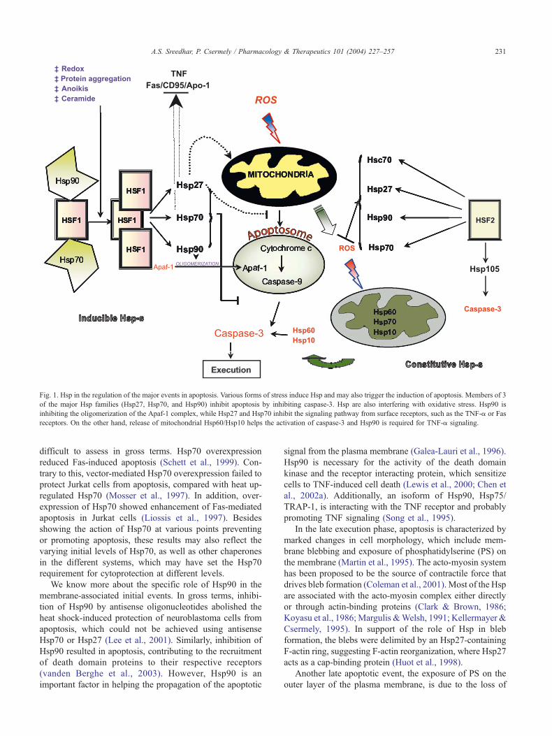
Fig. 1. Hsp in the regulation of the major events in apoptosis. Various forms of stress induce Hsp and may also trigger the induction of apoptosis. Members of 3
of the major Hsp families (Hsp27, Hsp70, and Hsp90) inhibit apoptosis by inhibiting caspase-3. Hsp are also interfering with oxidative stress. Hsp90 is
inhibiting the oligomerization of the Apaf-1 complex, while Hsp27 and Hsp70 inhibit the signaling pathway from surface receptors, such as the TNF-a or Fas
receptors. On the other hand, release of mitochondrial Hsp60/Hsp10 helps the activation of caspase-3 and Hsp90 is required for TNF-a signaling.
A.S. Sreedhar, P. Csermely / Pharmacology & Therapeutics 101 (2004) 227–257 231
difficult to assess in gross terms. Hsp70 overexpression
reduced Fas-induced apoptosis (Schett et al., 1999). Con-
trary to this, vector-mediated Hsp70 overexpression failed to
protect Jurkat cells from apoptosis, compared with heat up-
regulated Hsp70 (Mosser et al., 1997). In addition, over-
expression of Hsp70 showed enhancement of Fas-mediated
apoptosis in Jurkat cells (Liossis et al., 1997). Besides
showing the action of Hsp70 at various points preventing
or promoting apoptosis, these results may also reflect the
varying initial levels of Hsp70, as well as other chaperones
in the different systems, which may have set the Hsp70
requirement for cytoprotection at different levels.
We know more about the specific role of Hsp90 in the
membrane-associated initial events. In gross terms, inhibi-
tion of Hsp90 by antisense oligonucleotides abolished the
heat shock-induced protection of neuroblastoma cells from
apoptosis, which could not be achieved using antisense
Hsp70 or Hsp27 (Lee et al., 2001). Similarly, inhibition of
Hsp90 resulted in apoptosis, contributing to the recruitment
of death domain proteins to their respective receptors
(vanden Berghe et al., 2003). However, Hsp90 is an
important factor in helping the propagation of the apoptotic
signal from the plasma membrane (Galea-Lauri et al., 1996).
Hsp90 is necessary for the activity of the death domain
kinase and the receptor interacting protein, which sensitize
cells to TNF-induced cell death (Lewis et al., 2000; Chen et
al., 2002a). Additionally, an isoform of Hsp90, Hsp75/
TRAP-1, is interacting with the TNF receptor and probably
promoting TNF signaling (Song et al., 1995).
In the late execution phase, apoptosis is characterized by
marked changes in cell morphology, which include mem-
brane blebbing and exposure of phosphatidylserine (PS) on
the membrane (Martin et al., 1995). The acto-myosin system
has been proposed to be the source of contractile force that
drives bleb formation (Coleman et al., 2001). Most of the Hsp
are associated with the acto-myosin complex either directly
or through actin-binding proteins (Clark & Brown, 1986;
Koyasu et al., 1986; Margulis &Welsh, 1991; Kellermayer &
Csermely, 1995). In support of the role of Hsp in bleb
formation, the blebs were delimited by an Hsp27-containing
F-actin ring, suggesting F-actin reorganization, where Hsp27
acts as a cap-binding protein (Huot et al., 1998).
Another late apoptotic event, the exposure of PS on the
outer layer of the plasma membrane, is due to the loss of

A.S. Sreedhar, P. Csermely / Pharmacology & Therapeutics 101 (2004) 227–257232
plasma membrane phospholipid asymmetry (Martin et al.,
1995). The role of Hsp in PS externalization is not known
but external PS can be recognized by the immune system
where Grp94, a homologue of Hsp90, helps the recognition
(Schild & Rammensee, 2000).
In several cases, Hsp translocate to the cell surface after
the initiation of apoptosis, making the cells more vulnerable
to immune lysis (Feng et al., 2002; Sapozhnikov et al.,
2002). There is a correlation between surface expression of
Fas and Hsp70 protein during heat-induced apoptosis in rat
histiocytoma cells (Sreedhar et al., 2000).
2.1.2. Cytosol
In the cytosol, stress kinases are important elements of
the signal transduction pathway in inducing and or modu-
lating the apoptotic response. Among the mitogen-activated
protein kinases, the activation of the signal-regulated protein
kinase (ERK1/2) is associated with a mitogenic stimulus,
whereas the c-Jun N-terminal kinase (JNK) and the p38
kinases are stress responsive.
The small Hsp, Hsp27, is phosphorylated in vivo by a
protein kinase termed MAPKAP kinase-2, which is activat-
ed by p38 kinase-induced phosphorylation. Charette et al.
(2000) demonstrated that the phosphorylated dimers of
Hsp27 interact with Daxx, a protein that contains a death
domain, specifically binds to Fas, and through the activation
of the JNK kinase, enhances Fas-mediated apoptosis. Bind-
ing of the phosphorylated form of Hsp27 to Daxx prevents
Daxx interaction with the apoptosis signal-regulating ki-
nase-1 (Ask1), a serine/threonine kinase, thereby inhibiting
the Fas-mediated apoptotic pathway.
Hsp70 has a rather general inhibitory role in stress kinase
pathways. Hsp72 acts as a direct inhibitor of Ask1: its
physical interaction with the kinase was demonstrated in in
vitro binding assays using Ni+-NTA-agarose beads. Hsp72
antisense oligonucleotides prevent the inhibitory effects of
Hsp72 in H2O2-induced Ask1 activation and consequent
apoptosis of NIH3T3 cells (Park et al., 2002). Hsp72 also
interacts directly with the peptide-binding domain of JNK
(Park et al., 2001; Gabai et al., 2002). Using the antisense
RNA approach, it was found that accumulation of Hsp72 is
necessary for JNK down-regulation. However, mutant
Hsp70 was able to inhibit JNK activation but not apoptosis,
suggesting that the chaperone activity of Hsp70 is required
for the inhibition of apoptosis but not for JNK inhibition
(Mosser et al., 2000). Hsp70-induced JNK inhibition regu-
lates the Bid-dependent apoptotic pathway (Gabai et al.,
2002). Constitutive expression of another member of the
Hsp70 family, Hsp105a, also protected cells from stress-
induced apoptosis through JNK inhibition in neuronal PC12
cells (Hatayama et al., 2001), although the molecular
mechanism behind this phenomenon is not known, and
overexpression of the same protein enhanced oxidative
stress-mediated apoptosis in mouse embryonal F9 cells
(Yamagishi et al., 2002). Similar to the inhibitory effect of
Hsp72 on the JNK kinase, addition of purified recombinant
Hsp72 to a crude cell lysate reduced p38 kinase activation,
while depletion of the whole family of Hsp70 proteins with
a monoclonal antibody enhanced p38 activation (Gabai et
al., 1997, 2000).
The role of Hsp90 in stress kinase regulation is not
known in such details like that of Hsp27 or Hsp70.
However, Hsp90 is necessary for the folding and activation
competence of a large number of kinases (Pratt & Toft,
2003). Dissociation of the Hsp90-Raf1 complex results in
apoptosis in mast cells (Cissel & Beaven, 2000) and in B-
lymphocytes (Piatelli et al., 2002), where the disruption of
the mitogen-activated protein kinase cascade is accompa-
nied with the activation of JNK in the dexamethasone-
induced mast cell apoptosis model (Cissel & Beaven,
2000). The down-regulation of Raf can also be induced
by the sequestration of its activator, B-cell lymphoma-2
protein (Bcl-2)-associated athanogene protein-1 (BAG-1),
by Hsp70 after various forms of stress (Song et al., 2001).
2.1.3. Nucleus
Nucleosomal fragmentation of DNA is a biochemical
signature of apoptosis resulting from the activation of
specific endonuclease activation resulting in the cleavage
of the chromatin to shorter DNA fragments called oligo-
nucleosomal DNA fragments (Compton, 1992). However,
DNA damage may also be an early signaling event of the
apoptotic cascade as well. In a few cell types, the early
phase of apoptosis is associated with the appearance of
high molecular weight DNA fragments suggesting a large-
scale reorganization of chromatin. Formation of high
molecular weight DNA fragments in cerebellar granule
neurons accompanies both caspase-dependent and -inde-
pendent types of cell death, indicative of multiple mecha-
nisms in the regulation of excision of DNA loop domains
during neuronal cell death (Bezvenyuk et al., 2000). As
another early nucleus-dependent event, histone H1.2, has
been recently shown to translocate to the cytoplasm in
response to DNA damage and promote the release of
cytochrome c by activating the Bcl-2 protein, Bak (Konishi
et al., 2003).
Hsp play a major role in protecting the cells from DNA
damage induced by various damaging agents (Samali &
Orrenius, 1998). Most Hsp translocate to the nucleus after
stress, and members of Hsp27 and Hsp70 families have a
protective role against oxidative stress as we will describe in
Sections 2.1.4 and 6.3.1. These features pose Hsp as
elements of an efficient mechanism to protect the integrity
of DNA. Indeed, overexpression of Hsp25 was shown to
reduce oxidative DNA damage after TNF-a treatment (Park
et al., 1998). One of the major hydrophilic by-products of
lipid peroxidation, trans-4-hydroxy-2-nonenal, binds to
DNA, resulting in the formation of exocyclic guanosine
adducts and acts as a mutagen (Hu et al., 2002). Appearance
of Hsp72 in cell nucleus was seen in dimethylarsinic acid-
treated human alveolar L-132 cells, where nuclear Hsp72
suppresses the appearance of apoptosis after DNA damage

A.S. Sreedhar, P. Csermely / Pharmacology
(Kato et al., 1999). A smaller fraction of Hsp90 also trans-
locates to the nucleus upon stress (Csermely et al., 1995),
and Hsp90 tightly interacts with histones (Schnaider et al.,
1999), inducing a condensed state of the chromatin
(Csermely et al., 1994). However, a role of Hsp90 in the
protection of DNA against oxidative or other forms of
damage has not been shown yet.
A rather specific form of DNA damage occurs with
telomere shortening. At a critical length of the telomere
regions at the end of the chromosomes around 7 kb, cells go
to the state of cellular senescence, which may further
proceed to apoptosis. Telomere regions are synthesized/
maintained by the telomerase enzyme. Hsp90 directly inter-
acts with telomerase and is necessary to achieve its enzyme
activity (Holt et al., 1999). Consequently, Hsp90 was
reported to increase telomerase activity in prostate carcino-
mas (Akalin et al., 2001). Moreover, Hsp90 is an essential
component for the activation of the Akt kinase, which is
activates the telomerase and thus acts against apoptosis
(Haendeler et al., 2003).
Fig. 2. Major pathways of mitochondrial apoptosis. Many proapoptotic and signa
membrane permeabilization. The PTP induces the mitochondrial translocation an
permeabilization of the inner mitochondrial membrane resulting in the leakage of
‘‘go’’ signal for the execution phase of apoptosis. The second mitochondria-deri
inhibits the IAP (such as XIAP, c-IAP1, and c-IAP2) after its release to the cytoso
and -9. Apart from Smac/DIABLO, there is another mitochondrial protein, HtrA2/
drives the assembly of the high molecular weight caspase-activating complex cal
presence of dATP and caspase-9, recruits and helps the autoactivating cleavage o
ROS-induced apoptosis are also included, such as the formation of the highly t
apoptosis, endonucleases, such as CAD, are activated.
2.1.4. Mitochondria and reactive oxygen species
Although initial studies suggested that organelles like
mitochondria, ER, and lysosomes do not play a major role
during apoptosis (Kerr et al., 1972), later, it was found
that mitochondria are the central coordinators of apoptotic
events (Ferri & Kroemer, 2001). Many proapoptotic and
signal transduction pathways converge on the mitochon-
dria to induce mitochondrial membrane permeabilization
(Fig. 2). There are several competing models to explain
the rupture of the outer mitochondrial membrane as a
result of the opening of the mega-channel, called the
permeability transition pore (PTP). The adenine nucleotide
translocator present in the inner mitochondrial membrane
and the voltage-dependent anion channel in the outer
membrane are the major components of the PTP and are
responsible for the lethal change in mitochondrial mem-
brane potential (Martinou et al., 2000). The PTP induces
the mitochondrial translocation and multimerization of the
proapoptotic protein, B-cell lymphoma-2 protein (Bcl-2)-
associated X protein (Bax). Bax, in turn, helps the
& Therapeutics 101 (2004) 227–257 233
l transduction pathways converge on mitochondria to induce mitochondrial
d multimerization of the proapoptotic protein, Bax. Bax, in turn, helps the
cytochrome c and other mitochondrial inter-membrane proteins that give a
ved activator of caspases (Smac/DIABLO) is a mitochondrial protein that
l. IAP are known to block the processing of the effector caspases, caspase-3
Omi, that also inhibits IAP. The release of cytochrome c from mitochondria
led apoptosome. The apoptosome contains oligomerized Apaf-1, which in
f caspase-3, an executioner of apoptosis. On the figure, several elements of
oxic nitrogen reactive species (NRS). Finally, in the executioner phase of

A.S. Sreedhar, P. Csermely / Pharmacology & Therapeutics 101 (2004) 227–257234
permeabilization of the inner mitochondrial membrane
resulting in the leakage of cytochrome c and other
mitochondrial intermembrane proteins that give a ‘‘go’’
signal for the execution phase of apoptosis (De Giorgi et
al., 2002).
The second mitochondria-derived activator of caspases
(Smac/DIABLO) is a mitochondrial protein that inhibits the
‘‘inhibitors of apoptosis proteins’’ (IAP, such as XIAP, c-
IAP1, and c-IAP2) after its release to the cytosol. IAP are
known to block the processing of the effector caspases,
caspase-3 and -9 (Shibata et al., 2002). Apart from Smac/
DIABLO, there is another mitochondrial protein, HtrA2/
Omi, that also inhibits IAP (van Loo et al., 2002).
The release of cytochrome c from the mitochondria
drives the assembly of the high molecular weight caspase
activating complex called apoptosome. The apoptosome
contains oligomerized Apaf-1, which, in the presence of
dATP and caspase-9, recruits and helps the autoactivating
cleavage of caspase-3, an executioner of apoptosis (Acehan
et al., 2002).
Immunodepletion of Hsp27 from cytochrome c-activated
cytosol resulted in a decreased caspase activity. In parallel
experiments, Hsp27 was co-precipitated with both cyto-
chrome c and procaspase-3. These data suggest that
Hsp27 sequesters both cytochrome c and procaspase-3,
and thus prevents the correct formation/function of the
apoptosome complex (Concannon et al., 2001). Indeed,
Hsp27 binds to cytochrome c released from the mitochon-
dria to the cytosol and prevents cytochrome c-mediated
interaction of Apaf-1 with procaspase-9, thus interfering
specifically with the mitochondrial pathway of caspase-
dependent cell death (Garrido et al., 1999; Bruey et al.,
2000). Later studies showed that Hsp27 is also localized in
the mitochondria and promotes the retention of cytochrome
c (Paul et al., 2002). Similarly, Hsp27 inhibits the release of
another proapoptotic molecule, Smac/DIABLO, as well
(Chauhan et al., 2003). Interestingly, Hsp27 was shown to
form a complex with cell death-inhibiting RNA, which is a
noncoding RNA competing with the mRNA of several
antiapoptotic proteins, such as that of Bcl-2 for ribonucleo-
protein-mediated degradation (Shchors et al., 2002). Alto-
gether, these observations show that Hsp27 plays a major
role in protecting mitochondria during activation of apopto-
sis (Samali et al., 2001).
Mitochondrial Hsp60 and its co-chaperone, Hsp10, are
associated with procaspase-3 in Jurkat cells. In the staur-
osporin-induced apoptosis model, Hsp60 and Hsp10 release
the active caspase-3. Similar effects were demonstrated in
cell free systems, suggesting a role for Hsp60 and Hsp10 in
regulating apoptosis in presence of cytochrome c and dATP
(Samali et al., 1999; Xanthoudakis et al., 1999). In agree-
ment with the previous findings, antisense oligonucleotide-
induced decrease in mitochondrial Hsp60 induced the re-
lease of cytochrome c. On the contrary, cytoplasmic Hsp60
was found to sequester several antiapoptotic molecules,
such as Bax or Bak (Kirchhoff et al., 2002).
In vitro experiments with purified recombinant Hsp70
showed its inhibitory role for cytochrome c/dATP-mediated
caspase activation but not for the oligomerization of Apaf-1.
Hsp70 suppresses apoptosis by directly associating with
Apaf-1 and blocking the assembly of a functional apopto-
some (Beere et al., 2000). In contrary, overexpression of
Hsp105a, a Hsp70 homologue, is associated with the induc-
tion of apoptosis involving cytochrome c release, caspase-3
activation, and poly-ADP ribose polymerase (PARP) cleav-
age in mouse embryonal F9 cells (Yamagishi et al., 2002).
Hsp90 directly binds to Apaf-1 to prevent the formation
of apoptosome complex (Pandey et al., 2000). In addition,
the reactive cysteines present on Hsp90 were able to reduce
cytochrome c, suggesting a role for Hsp90 in modulating the
redox status in resting and apoptotic cells (Nardai et al.,
2000). Hsp90 is known to help the vascular endothelial
growth factor (VEGF)-induced expression of the antiapop-
totic Bcl-2 (Dias et al., 2002); however, the exact mecha-
nism of this action is unknown.
It seems that we are just starting to uncover the role of
Hsp in mitochondrial apoptotic events. Data on their in-
volvement in the formation of the PTP, as well as in the
action of several pro- or antiapoptotic proteins, such as
HtrA2/Omi or the IAP, are spuriously lacking. Discovery of
the plenitude of the possible regulatory functions of Hsp in
these processes is an exciting task of future research efforts.
Mitochondria are primary sites of reactive oxygen species
(ROS) formation. It has been shown in many model systems
that the cellular redox homeostasis plays an essential role in
cell survival and cellular signaling. ROS or free radicals
were reported to have a major role in the mediation of
cellular damage (Thannickal & Fanburg, 2000). Although
the origin of ROS remains to be determined, mitochondria
are thought to be the major source of ROS in vivo (Boveris
& Chance, 1973). However, ROS can be generated by a
variety of mechanisms, like the mitochondrial and micro-
somal electron transport chain, xanthine oxidase and other
flavoprotein oxidases, auto-oxidation of hydroquinones,
catecholamines, and thiols, intracellular xenobiotic mecha-
nisms, NADP(H)-oxidase, as well as by the auto-oxidation
of hemoglobin. In a normal cell, there is always a balance
between pro- and antioxidant pathways. Upon stress stimuli,
an imbalance of the redox milieu develops and leads to the
accumulation of ROS. ROS may induce some specific
effects, serving as messengers of cellular damage and can
also cause a general damage in the cell by oxidizing the
membrane lipids, proteins, and DNA. The overproduction of
ROS is associated with many forms of apoptosis and
necrosis (Buttke & Sandstrom, 1995; Zamzami et al.,
1995; Suzuki et al., 1997).
ROS-induced apoptosis was shown to be associated with
the up-regulation of the Fas death receptor (Bauer et al.,
1998). As an interesting cross-talk between different path-
ways, the antiapoptotic protein Bcl-2 also prevents the
mitochondrial generation of ROS after the opening of the
PTP (Gottlieb et al., 2000).

A.S. Sreedhar, P. Csermely / Pharmacology & Therapeutics 101 (2004) 227–257 235
There is a correlation between ROS generation and the
induction of Hsp (Schoeniger et al., 1994; Gorman et al.,
1999). Showing a general protective role of Hsp against
ROS, heat shock transcription factor-1 deficiency increases
mitochondrial oxidative damage in mouse hearts (Yuan
et al., 2002a).
Small Hsp, such as Hsp27, emerged as novel and
important factors to protect against oxidative stimuli, block-
ing an important initiation factor of apoptotic processes
(Arrigo, 2001). Indeed, small Hsp elevate reduced glutathi-
one levels by promoting an increase in glucose-6-phosphate
dehydrogenase activity and by a somewhat smaller activa-
tion of glutathione reductase and glutathione transferase
(Preville et al., 1999; Arrigo, 2001). As another example
of their action, the cytotoxicity induced by TNF-a and
inflammatory cytokines via ROS production is inhibited
by overexpression of Hsp27 or other small Hsp, such as a-
crystalline, in L929 fibroblasts through the overexpression
of Bcl-2, an antiapoptotic protein, which helps in maintain-
ing the mitochondrial integrity (Park et al., 1998).
Members of the Hsp70 family play a similar role in
oxidative protection like the small Hsp. Hyperoxia-mediated
lipid peroxidation was attenuated in A549 human lung
adenocarcinoma cells by overexpressing Hsp70. Increased
expression of Hsp70 did not detectably alter a number of
major antioxidant enzymes, such as manganese superoxide
dismutase (Mn-SOD), catalase, and glutathione peroxidase,
suggesting a specific protective role for Hsp70 against
hyperoxia (Wong et al., 1998). Hsp70 was shown to be
cardioprotective by conferring oxidative protection after
heat shock preconditioning (Su et al., 1999). There is also
a correlation between Mn-SOD activity and Hsp72-mediat-
ed cardioprotection, although Hsp70 does not bind to Mn-
SOD directly (Suzuki et al., 2002). Rat histiocytic cells
expressing Hsp70 were shown to be resistant to heat-
induced apoptosis partially through the inhibition of ROS
induction. In this system, hydrogen peroxide-induced Hsc70
expression along with Hsp70 suggested a role for Hsc70 in
protection against ROS (Sreedhar et al., 2002a). Hsp72
reduces the formation of 8-hydroxy-2V-deoxyguanosine-and trans-4-hydroxy-2-nonenal-modified proteins in ische-
mia-reperfused liver of rats (Yamagami et al., 2002). The
role for Hsc70 as an antioxidant has also been shown in
reoxygenation injury in the intestinal cell line Caco-2
(Gebhardt et al., 1999) and in monocytes (Chong et al.,
1998).
Nitric oxide (NO) is an important signaling molecule
regulating a number of diverse physiological processes and
is produced by a group of enzymes called nitric oxide
synthases (NOS), namely, neuronal (nNOS), endothelial
(eNOS), and inducible (iNOS) (Christopherson & Bredt,
1997; Mayer & Hemmens, 1997). NO inhibits apoptosis
acting through up-regulation of survival kinases, like Akt
(Dimmeler et al., 1998), and in an in vitro model by a direct
inhibition of caspase-3 via S-nitrosylation (Rossig et al.,
1999). Indeed, NOS is interacting with caspase-3 in an NO-
dependent manner (Matsumoto et al., 2003). On the other
hand, NO is combined with superoxides forming nitrogen
peroxides, which are efficient initiators of the apoptotic
response. Hsp70 attenuates the NO-induced apoptosis in
RAW264.7 macrophages by maintaining the mitochondrial
integrity (Klein & Brune, 2002), but protection by Hsp70
also involves the up-regulation of intracellular glutathione
(GSH) (Calabrese et al., 2002; Sreedhar et al., 2002a).
Hsp90 has been shown to have a regulatory role in
eNOS- and nNOS-mediated NO production; hence, inhibi-
tion of Hsp90 helps the induction of apoptosis by dimin-
ished NO production and by increased NOS-dependent
superoxide production in certain cellular systems (Garcia-
Cardena et al., 1998; Bender et al., 1999; Pritchard et al.,
2001; Billecke et al., 2002). Hsp90 was also shown to
inhibit superoxides generated by nNOS but not from xan-
thine oxidase (Song et al., 2002). As a more direct role in
oxidative stress, Hsp90 protects cells from iron overload-
induced oxidative stress (Fukuda et al., 1996).
2.1.5. Endoplasmic reticulum
The ER plays a critical role in protein biosynthesis and
maintenance of intracellular calcium homeostasis. A variety
of conditions, including disruption of intracellular homeo-
stasis and alteration of ER intraluminal oxidative environ-
ment, can induce ER stress and lead to apoptosis (Chen &
Gao, 2002). The participation of the ER in apoptosis
initiation and progression is assumed to involve at least
two mechanisms, the unfolded protein response and dis-
turbed Ca2+ signaling (Kaufman, 1999; Patil & Walter,
2001). Although the complete understanding of the contri-
bution of ER stress to the development of apoptosis is
missing, recent studies suggest the involvement of the ER
proteases, caspase-7 and -12, in this process (Nakagawa et
al., 2000). The unfolded protein response also induces the
CHOP/GADD153 transcription factor, which also promotes
ER-induced apoptosis (Kaufman, 1999).
Glucose-regulated proteins (Grp) are a class of ER
proteins composed of several members, which are induced
by ER stress, like depletion of ER intraluminal Ca2+ or
disturbances of protein glycosylation. Grp78 is involved in
polypeptide translocation across the ER membranes and
acts as an apoptotic regulator by protecting the host cell
against ER stress-induced cell death. However, the mech-
anism of this protection is obscure. From a cell free system,
it was found that Grp78 inhibits ER-induced apoptosis
through direct binding and inhibition of the proapoptotic
caspase-7 and -12 (Rao et al., 2002; Xie et al., 2002;
Reddy et al., 2003). Recently, Grp78 induction was found
to be associated with NF-nB activation (Chen & Gao,
2002); further, the Grp78-induced inhibition of Ca2+ dis-
turbances and oxidative ER stress-induced apoptosis was
extensively studied. Other important chaperone of the ER,
calreticulin, and protein disulfide isomerase exert similar
protective role like Grp78 (Liu et al., 1997a; Ko et al.,
2002).

A.S. Sreedhar, P. Csermely / Pharmacology & Therapeutics 101 (2004) 227–257236
Recently, a rather exciting interplay between the ER
and mitochondria has been uncovered during the apoptotic
process. This interaction involves the junctions between
the two organelle and opens the possibility that pro- and
antiapoptotic molecules, like Bcl-2, regulate calcium
fluxes through this junction (Berridge, 2002). As an
additional hint for the specific interaction of these two
organelles, two proapoptotic mitochondrial molecules, Bax
and Bak, were shown to be localized to the ER and
promote caspase-12-dependent apoptosis (Zong et al.,
2003). It is a completely unexplored question if ER chap-
erones are involved in the regulation of the ER/mitochon-
drial junctions.
2.2. Effector molecules
2.2.1. Caspases
The family of proteases known as caspases specifically
cleave proteins at aspartate residues. Caspases exists as
inactive zymogens (procaspases). There are about 14 types
of caspases reported in the literature (Ahmad et al., 1998;
Thornberry & Lazebnik, 1998) and are classified into 3
major groups, which are initiator, inflammatory, and effector
caspases (Nicholson & Thornberry, 1997). The activation of
caspases is organized as a cascade in various apoptotic
pathways. Thus, TNF-induced apoptosis involves the acti-
vation of the initiator caspases-8 (FLICE) and -10, which
further activate the effector caspases-3, -6, and -7. The
mitochondrial apoptosome-mediated pathways involve the
activation of the initiator caspase-9, which further activates
the same set of effector caspases, caspase-3, -6, and -7.
Hsp27 helps in retaining the mitochondrial integrity and
inhibits mitochondrion-dependent caspase activation, but its
direct involvement in caspase inhibition was not shown
(Samali et al., 2001). However, immunoprecipitation experi-
ments suggest that Hsp27 binds to procaspase-3 and inhibits
its processing (Concannon et al., 2001). Interestingly, meth-
ylglyoxal modification of Hsp27 inhibits its interaction with
procaspase-3 (Sakamoto et al., 2002). The small Hsp, a-
and h-crystallines, inhibit both mitochondrial and death
receptor pathways and are thought to be involved in the
inhibition of autocatalytic maturation of caspase-3 (Kamradt
et al., 2002).
Hsp70 binds to caspase-3 through its pseudosubstrate,
the carboxy terminal EEVD sequence, and inhibits caspase-
3 activity. However, the availability of the EEVD sequence
depends on Hsp70 conformation and is dependent on ATP
binding to Hsp70. Later, it was shown that Hsp70-mediated
caspase inhibition is a result of reduced processing of
procaspase-3 but not due to inhibition of the activity of
the processed enzyme (Mosser et al., 1997).
2.2.2. Nucleases
From the functional point of view, the deoxyribonuclease
(DNase) implicated in apoptosis may be classified in the
groups of Ca2+ /Mg2+ endonucleases, Mg2+ endonucleases,
acid endonucleases, cation-independent endonucleases, and
Zn2+ -sensitive endonucleases (Ribeiro & Carson, 1993;
Counis & Torriglia, 2000). However, Zn2+ -sensitive endo-
nucleases were reported only in in vitro reconstitution of
intact HeLa S3 nuclei and in apoptotic U937 cytosolic
extracts (Kimura et al., 1998). Recently, a new Ca2+ /Mg2+
endonuclease was identified and called as the NUC70 cyto-
plasmic endonuclease. The activity of NUC70 is inhibited by
caspase inhibitors suggesting that this nuclease is a cytoplas-
mic target for effector caspases (Urbano et al., 1998). An
additional set of reports showed a Ca2+ /Mg2+ endonuclease
activity associated with recombinant cyclophilins A, B, and C
(Montague et al., 1994, 1997).
The major nuclease responsible for apoptosis-induced
DNA fragmentation is a Mg2+ endonuclease called as
caspase-activated DNase (CAD) or DNA fragmentation
factor (DEF40). In proliferating cells, CAD exists in a
complex of its inhibitor ICAD/DEF45, and the caspase-3-
induced cleavage of ICAD leads to the release of the CAD
endonuclease activity (Liu et al., 1997b; Enari et al.,
1998). Hsc70, with its cofactor Hsp40, is involved in the
folding of CAD. CAD is released from ribosomes as a
heterocomplex with ICAD, which also assists in the
folding of CAD during its synthesis (Sakahira & Nagata,
2002).
2.2.3. Transglutaminases
Tissue transglutaminase (TGase) is a member of the
transglutaminase family catalyzing protein cross-linking
by transamidation. There are several classes of enzymati-
cally active TGases identified as having an important role in
packing the cell at the late phase of apoptosis to prevent a
massive inflammatory process (Lorand & Graham, 2003).
Apart from transamidation, TGases also bind and hydrolyze
ATP and guanosine triphosphate (GTP) (Tucholski & John-
son, 2002). The intracellular TGase activity is inhibited by
GTP and NO and is enhanced by increases in intracellular
Ca2+ level (Fesus, 1998).
TGase expression is inversely correlated with the expres-
sion of the antiapoptotic protein Bcl-2, and inhibition of the
enzyme confers protection against apoptosis (Oliverio et al.,
1999). Indeed, initially it was thought that TGase is only
proapoptotic, as it sensitized SK-N-BE and 3T3 cells for
apoptosis-inducing hyperpolarization of mitochondria fol-
lowed by increased production of ROS. Furthermore, TGase
overexpressing cells were sensitive to staurosporin-induced
apoptosis (Piacentini et al., 2002). However, recent evidence
suggests that TGase may also act as an apoptotic inhibitor
through the cell cycle-regulating retinoblastoma protein
(Antonyak et al., 2001; Boehm et al., 2002; Tucholski &
Johnson, 2002).
Although the role of Hsp in TGase action has not been
explored yet, it is of considerable interest that the ER
chaperone protein disulfide isomerases, like Erp57, were
shown to possess TGase activity (Chandrashekar et al.,
1998; Natsuka et al., 2001).
A.S. Sreedhar, P. Csermely / Pharmacology & Therapeutics 101 (2004) 227–257 237
3. Heat shock proteins and
caspase-independent apoptosis
In this section, we will review the emerging alternative
pathways of apoptosis, which are not centered around caspase
activation. We would like to apologize for this dissection,
which will turn to be rather artificial in some cases. Obvi-
ously, the signaling pathways are interrelated and, therefore,
‘‘caspase-independent’’ pathways may sometimes converge
with caspase-dependent ones. In Section 3.6, we will briefly
summarize anoikis, where currently no caspase-independent
pathways are known. However, the therapeutic use of Hsp
modulation in anticancer protocols makes it especially im-
portant to review their effects on caspase-independent apo-
ptotic pathways, which are many times the major pathways of
apoptosis in tumor cells (Nylandsted et al., 2000).
3.1. Serine proteases
Granzymes are a family of serine proteases often associ-
ated with perforin in activated T-lymphocytes and natural
killer cells. A number of granzymes, granzyme A–G, have
been isolated and cloned from mouse cytotoxic lymphocytes
and natural killer cells; however, in humans, only a few of
them were identified. Granzyme B has been found in the
nucleus, as well as in cytoplasmic granules of killer thymo-
cytes, and is involved in target cell apoptosis during lym-
phocyte-mediated cytotoxicity (Trapani, 2001; Pardo et al.,
2002; Raja et al., 2003).
Granzyme cascade cleaves caspases in vivo, resulting in
a massive amplification of caspase-dependent apoptotic
pathways (van de Craen et al., 1997; Barry et al., 2000).
Although these proteases can directly activate caspases,
predominantly they induce the activation of the Bid protein
and the consequent mitochondrial membrane changes and
cytochrome c release (Wang et al., 2001). Furthermore,
granzyme-mediated apoptosis requires Bid cleavage not
involving caspase activation (Sutton et al., 2000), suggest-
ing that granzyme-mediated apoptosis can be caspase-inde-
pendent. Granzyme A is a typical mediator of caspase-
independent apoptotic pathways, where it directly activates
the ER DNase, GAAD, and promotes single strand DNA
nicks and consequent apoptosis (Fan et al., 2003). Similarly,
the involvement of granzyme C during caspase- and mito-
chondrial depolarization-independent cell death was
reported (Johnson et al., 2003). In contrast, a recent report
shows that granzyme B requires procaspase-3 for apoptosis
initiation, where caspase-3 mutant cells failed to suffer
granzyme B-dependent apoptosis (Metkar et al., 2003).
Very little is known about the role of Hsp in granzyme-
mediated cell death pathways. It has been shown that Hsp27
does not interfere with granzyme B-induced activation of
caspases (Bruey et al., 2000). However, Hsp27 co-precip-
itates with granzyme A from cytoplasmic lysates, although
it is not a substrate for granzyme A (Beresford et al., 1998).
Surface-expressed Hsp70 mediates the apoptosis of tumor
cells by binding and uptake of granzyme B (Gross et al.,
2003).
3.2. Cathepsins
Cathepsins are a class of proteolytic enzymes containing
several types of proteases. The 3 major classes of cathepsins
are the cysteine proteases, comprising cathepsins B, C, L, H,
K, S, and O, followed by the aspartyl proteases, comprising
cathepsins D, E, and F, and finally, a serine protease,
cathepsin G. Being of lysosomal origin, these enzymes play
a major role in peptide formation and protein degradation
(Buhling et al., 2002; Takuma et al., 2003). Cathepsins are
often involved in various forms of autophagy-associated
apoptosis (Uchiyama, 2001). Oxidative stress-induced, cas-
pase-independent apoptosis often involves the activation of
cathepsin D (Kagedal et al., 2001; Takuma et al., 2003).
Cathepsin B is released from lysosomes in liver cells after
TNF-a treatment or in p53-induced apoptosis and contrib-
utes to cell death (Werneburg et al., 2002; Yuan et al.,
2002b). In several tumor cells, cathepsin B is the most
important mediator of cell death (Foghsgaard et al., 2002).
Its activation can be prevented by the activation of the NF-
nB pathway (Liu et al., 2003). Cathepsins emerge as
important players of caspase-independent apoptosis, but in
several cases, their activation also converges to caspase-
dependent pathways (Mathiasen & Jaattela, 2002; Turk
et al., 2002).
Although little is known about the role of Hsp in the
regulation of these proteases compared with caspases, Hsp73
andHsp90were found accumulated in lysosomes of proximal
tubular epithelial cells in rat kidneys in acute gentamicin
nephropathy, suggesting a role of these proteins in protein
degradation (Komatsuda et al., 1999; Agarraberes & Dice,
2001). The whole Hsp70/Hsp90 chaperone complex was
found in lysosomal membranes, suggesting a major role for
these chaperones and additional co-chaperones in proteolytic
pathways (Agarraberes & Dice, 2001). Hsc70 was also found
within the lumen of lysosome and the effective uptake of
cytosolic proteins by these organelles was shown to depend
on the availability of Hsc70 (Terlecky et al., 1992; Cuervo
et al., 1997).
3.3. Calpains
Calpains are calcium-dependent proteases thought to play
an important role in cytoskeletal reorganization and muscle
protein degradation. Calpains exist as heterodimers com-
prised of a small regulatory subunit and one of the 3 large
catalytic subunits: calpain-1, -2, or -3 (Goll et al., 2003).
Calpains and caspases often synergize in the apoptotic
process, especially in neuronal cells. However, in various
cancer cells, such as in ovarian and breast cancer (Bao et al.,
2002; Mathiasen et al., 2002), or in cisplatin-mediated
apoptosis of melanoma cells (Mandic et al., 2002), calpain-
mediated, but caspase-independent, apoptosis is the major

A.S. Sreedhar, P. Csermely / Pharmacology & Therapeutics 101 (2004) 227–257238
pathway of cell death. In the latter model, calpain cleaves Bid
independently of caspase activation and thus triggers a
consecutive apoptosis (Mandic et al., 2002).
Grp94 was shown to protect human neuroblastoma cells
from hypoxia/reoxygenation-induced apoptosis involving
calpains (Bando et al., 2003). Grp94 was also shown to be
cleaved by calpain in etoposide-induced apoptosis (Reddy et
al., 1999). It is interesting to note that cisplatin, which may
interact with Grp94 (Itoh et al., 1999b; Soti et al., 2002),
induces the activation of calpain in the apoptotic process
(Mandic et al., 2003).
3.4. Ceramide-induced apoptosis
Ceramide has been recognized as a lipid mediator in the
induction of apoptosis. Multiple, diverse apoptotic inducers
are known to increase ceramide concentration in apoptotic
cells (van Blitterswijk et al., 2003). Ceramide-induced apo-
ptosis can be inhibited by Bcl-2 (Ruvolo et al., 2002). In
agreement with this, mitochondria were shown to sense the
sphingolipid signals (Tomassini & Testi, 2002). Fas induces
ceramide generation during the initiation phase of apoptosis,
which, in turn, may contribute to death receptor clustering
(Rufini & Testi, 2000; van Blitterswijk et al., 2003). Ceram-
ide was also shown to activate apoptosis by activating
multiple pathways, which are both caspase-9 and -3 depen-
dent (Movsesyan et al., 2002). Ceramide has multiple targets
in the cell, including various kinases and phosphatases,
leading to the activation of certain transcriptional factors
(Alesse et al., 1998; Pettus et al., 2002). Its role and regulation
was suggested to be dependent on intracellular glutathione
(GSH) levels (Lavrentiadou et al., 2001). Besides caspase-
dependent routes of ceramide-induced apoptosis (Movsesyan
et al., 2002; Ruvolo et al., 2002), cathepsin-(De Stefanis et al.,
2002) and calpain-dependent pathways (Poppe et al., 2002)
were also shown to participate in ceramide-induced cell
death. Indeed, ceramide has been shown to interact with
cathepsin D directly and promote the autocatalytic activation
of the enzyme (Pettus et al., 2002).
Although ceramide inhibits the heat shock response in
some cells, especially suppressing the induction of Hsp70
(Kondo et al., 2000), its major action was proved to be the
induction Hsp, such as small Hsp (Chang et al., 1995).
Ceramide also induces stress-activated protein kinase
(SAPK) signaling (Westwick et al., 1995). It was hypothe-
sized that Hsp70 protect cells from ceramide-induced apo-
ptosis, where increased ceramide levels are associated with
the activation of SAPK/JNK kinases and induced Hsp70
synthesis (Verheij et al., 1996). In agreement with this
assumption, a later study showed the suppressive effect of
Hsp70 on ceramide-induced cell death (Ahn et al., 1999).
3.5. Apoptosis-inducing factor
The apoptosis-inducing factor (AIF) is a recently iden-
tified mediator of caspase-independent apoptosis. AIF
translocates from the mitochondria to both the cytosol
and the nucleus. Nuclear AIF induces peripheral chromatin
condensation and high molecular weight DNA fragmenta-
tion to about 50-kb-long DNA fragments. Overexpression
of one of the major antiapoptotic proteins, Bcl-2, inhibited
AIF translocation (Cande et al., 2002a, 2002b). Hsp70 and
Hsc70 (but not Hsp27) directly inhibited AIF translocation
both in vitro and in intact cells. In agreement with this, the
depletion of Hsp70 using antisense oligonucleotides sensi-
tized cells to AIF-mediated apoptosis (Ravagnan et al.,
2001).
3.6. Anoikis
Anoikis (Greek word meaning state of homelessness) is
defined as a type of cell death where cells fail to find their
substratum and the lack of the integrin-mediated extracellu-
lar matrix interaction induces apoptosis (Frisch & Screaton,
2001; Grossmann, 2002). Anoikis mainly occurs in epithe-
lial cells, functioning to prevent the shedding and relocali-
zation of these cells. It also assures proper developmental
positioning of epithelial cells in specialized structures, such
as the luminal structures of the mammary gland. Failure of
anoikis contributes substantially to human tumor progres-
sion and facilitates metastasis (Frisch & Screaton, 2001).
Anoikis utilizes mostly the Fas pathway, leading to caspase-
dependent apoptosis. However, besides the general mecha-
nism, there are some anoikis-specific elements, like the
direct recruitment of caspase-8 to unligated integrin recep-
tors (Stupack et al., 2001). An additional specific pathway
involves FADD (Fas-associated death domain protein),
which is primarily a nuclear protein, but its localization
may be regulated by cell adhesion. Signaling pathways from
integrins to FADD are currently being studied (Frisch,
1999). It is possible that cytoskeletal alterations, which
accompany cell-matrix detachment, could release death
receptors from a sequestered state, leading to death do-
main-induced apoptosis.
We have rather indirect evidence for the participation of
Hsp in anoikis. The phosphorylated form of Hsp27 was
shown to help the stability of integrin in platelets together
with Hsp70 and Hsp90 (Polanowska-Grabowska & Gear,
2000). As a possible consequence of its role in anoikis
regulation, overexpression of Hsp27 was shown to inhibit
the invasive and metastatic potential of melanoma cells
(Aldrian et al., 2002), where anchorage-dependent cell
growth is related not only to the Hsp27 protein content, but
also to its phosphorylation status by p38 kinases and its F-
actin association. Hsp60 activates a3h1-integrin, which is
involved in the adhesion of metastatic breast cancer cells to
lymph nodes and osteoblasts (Barazi et al., 2002).
BAG, a co-chaperone of Hsp70/Hsc70, binds and
sequesters the death domain protein, FADD, leading to
the inhibition of anoikis. Conversely, the inhibition of the
chaperone activity of BAG leads to anoikis (Frisch, 1999).
In agreement with a key role of BAG in anoikis regulation,
A.S. Sreedhar, P. Csermely / Pharmacology & Therapeutics 101 (2004) 227–257 239
most of the metastatic tumors show a high expression of
BAG (Cutress et al., 2002).
Currently, we do not know caspase-independent path-
ways of anoikis. However, the down-regulation of caspase-
induced apoptosis in various tumors indicates that their
discovery, and the characterization of the involvement of
Hsp in caspase-independent anoikis regulation, is probably
only a question of time.
4. Heat shock proteins and antiapoptotic mediators
Hsp are involved not only in the regulation of the various
proapoptotic pathways, but also in the maintenance and
activation of antiapoptotic mediators (Fig. 3). To better
understand the pleiotropic role of Hsp, we chose to give a
separate summary of their role in the regulation of anti-
apoptotic pathways in the following section.
4.1. Heat shock proteins and
B-cell lymphoma-2 protein pathway
The Bcl-2 family of proteins consists of more than 20
members, which form various homo- and heterodimers with
each other, either promoting or inhibiting apoptosis (Gross
et al., 1999; Cory & Adams, 2002). The relative expression
of these pro- and antiapoptotic proteins is thought to decide
the fate of cell during apoptosis by regulating mitochondrial
membrane integrity (Cecconi, 1999; Gross et al., 1999).
As we will describe in detail in Section 6.2, BAG is a
nucleotide exchange factor for Hsp70 (Hohfeld, 1998). The
BAG proteins make a direct link between Hsp and Bcl-2
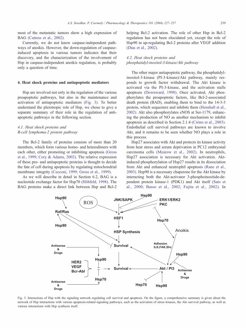
Fig. 3. Interactions of Hsp with the signaling network regulating cell survival an
network of Hsp interactions with various apoptosis-related signaling pathways, su
various interactions with Hsp synthesis itself.
helping Bcl-2 activation. The role of other Hsp in Bcl-2
regulation has not been elucidated yet, except the role of
Hsp90 in up-regulating Bcl-2 proteins after VEGF addition
(Dias et al., 2002).
4.2. Heat shock proteins and
phosphatidyl-inositol-3-kinase/Akt pathway
The other major antiapoptotic pathway, the phosphatidyl-
inositol-3-kinase (PI-3-kinase)/Akt pathway, mainly res-
ponds to growth factor withdrawal. The Akt kinase is
activated via the PI-3-kinase, and the activation stalls
apoptosis (Downward, 1998). Once activated, Akt phos-
phorylates the proapoptotic factors, like Bcl-2-associated
death protein (BAD), enabling them to bind to the 14-3-3
protein, which sequesters and inhibits them (Henshall et al.,
2002). Akt also phosphorylates eNOS at Ser-1179, enhanc-
ing the production of NO as another mechanism to inhibit
apoptosis as described in Section 2.1.4 (Cirino et al., 2003).
Endothelial cell survival pathways are known to involve
Akt, and it remains to be seen whether NO plays a role in
this process.
Hsp27 associates with Akt and protects its kinase activity
from heat stress and serum deprivation in PC12 embryonal
carcinoma cells (Mearow et al., 2002). In neutrophils,
Hsp27 association is necessary for Akt activation. Akt-
induced phosphorylation of Hsp27 results in its dissociation
from Akt and enhanced neutrophil apoptosis (Rane et al.,
2003). Hsp90 is a necessary chaperone for the Akt kinase by
interacting both the Akt-activator 3-phosphoinositide-de-
pendent protein kinase-1 (PDK1) and Akt itself (Sato et
al., 2000; Basso et al., 2002; Fujita et al., 2002). In
d apoptosis. On the figure, a comprehensive summary is given about the
ch as the activation of stress kinases, the Akt survival pathway, as well as

A.S. Sreedhar, P. Csermely / Pharmacology & Therapeutics 101 (2004) 227–257240
connection with this, Hsp90 serves as a molecular scaffold
to promote the Akt-induced phosphorylation and activation
of eNOS (Fontana et al., 2002). Interestingly, the ER
chaperone, calreticulin, suppressed the Akt kinase activity
during cardiac differentiation (Kageyama et al., 2002).
5. Modulation of modulators: effect of
apoptosis on heat shock protein induction
The preceding sections gave numerous examples of how
Hsp regulate apoptosis. However, key pro- and antiapop-
totic processes also regulate Hsp synthesis. In this section,
we will briefly summarize the (unfortunately rather limited)
knowledge on the regulation of Hsp synthesis by the
apoptotic process. In principle, apoptosis inhibits Hsp
synthesis by down-regulating the respective transcription
factor, HSF-1. Thus, activation of Fas inhibited heat-in-
duced activation of HSF-1 and the up-regulation of Hsp70
(Schett et al., 1999). In agreement with this, HSF-1 was
shown to undergo apoptosis-induced proteolysis by a cas-
pase-like protease (Zhang et al., 1999). Similarly, glycogen
synthase kinase-3, an apoptosis-promoting kinase, inhibits
the activation of HSF-1. The PI-3-kinase-dependent surviv-
al pathway suspends this action by blocking glycogen
synthase kinase-3 (Bijur & Jope, 2000). Thus, the apoptotic
pathway itself stops one of the survival signals, Hsp
synthesis. It remains to be seen if this mechanism is
generally operating in tumor cells or if it is one of those
pathways that becomes damaged in order to promote tumor
survival.
6. Molecular mechanism of heat shock protein action
Hsp act as molecular chaperones preventing protein
aggregation and promoting protein folding. Hsp almost
never act alone: they tend to oligomerize, as well as to form
chaperone complexes with each other. All major chaperone
classes have their co-chaperones, which are smaller chap-
erone proteins usually regulating the folding cycle of the
chaperone complex. Hsp have other general roles as well.
Due to their pleiotropic action on the cell, they profoundly
influence cellular homeostasis including the redox balance
and cell organization. In Section 6.1, we briefly summarize
the contribution of these general roles of Hsp to the
regulation of the apoptotic process.
6.1. Heat shock proteins as
molecular chaperones of apoptosis
It is a rather interesting question whether the divergent
roles of Hsp in apoptosis regulation, summarized in the
preceding sections, require a bona fide chaperone function
of these proteins. When working in ‘‘passive mode,’’ chap-
erones may behave as ATP-independent ‘‘holders’’ of dam-
aged proteins, sequestering them and preventing their fatal
aggregation. In ATP-dependent ‘‘active mode,’’ chaperones
are working as ‘‘folders’’ helping in the folding, transport,
and/or ATP-dependent degradation of unfolded or misfolded
proteins. While the passive mode is typical during stress
when the cellular ATP level is low, the active mode prevails
when the cell has recovered and the ATP level is increased
again (Ellis, 1993; Hartl, 1996; Bukau & Horwich, 1998;
Saibil, 2000; Hartl & Hartl, 2002). A number of proteins,
such protein kinases and nuclear hormone receptors, require
the continuous help of the Hsp90 chaperone complex to keep
their activation-competent state (Pratt, 1997; Csermely et al.,
1998; Richter & Buchner, 2001). It is also an interesting
question, whether the particular Hsp modulates the apoptotic
process as a passive or active chaperone or by a specific
interaction, which is not equivalent with any of the general
chaperone functions mentioned.
Unfortunately, the exact mechanism of apoptosis-related
Hsp function has been explored in relatively few cases in
detail. The chaperone function of Hsp70 was clearly estab-
lished in the regulation of the proapoptotic p53 protein both
in its wild type and mutant forms (Zylicz et al., 2001).
Similarly, Hsp70 and its homologue, Hsp105, are required
to suppress poly-Glu-induced protein aggregation and the
consecutive apoptosis (Kobayashi et al., 2000; Ishihara
et al., 2003).
It is important to note that Hsp have no priority or
selection between their substrates as molecular chaperones,
and hence their chaperone functions are extended to proa-
poptotic factors too. For example, Hsp60 promotes apopto-
sis by helping in the maturation of procaspase-3 (Samali et
al., 1999; Xanthoudakis et al., 1999) and several Hsp are
needed for the death-receptor induced signaling pathways.
6.2. Role of co-chaperones
Molecular chaperones almost always need co-chaperones
for the regulation of the binding and release of substrate
proteins. The major co-chaperones include BAG-1, CHIP
(C-terminal Hsp90 interacting protein), Hsp40, Hip (Hsc70
interacting protein), Hop (Hsp70-Hsp90 organizing protein),
and p23 (a 23-kDa co-chaperone of Hsp90) (Frydman &
Hohfeld, 1997; Buchner, 1999; Caplan, 1999; Morimoto,
2002).
BAG-1 was first identified as an antiapoptotic protein
because of its binding to Bcl-2 and promoting cell survival
(Takayama et al., 1995). Later, the specific binding of BAG-
1 and its homologues to Hsc70/Hsp70 was demonstrated
(Hohfeld & Jentsch, 1997; Takayama et al., 1997; Nollen et
al., 2000; Thress et al., 2001). The amino terminus of BAG-
1 is involved in Bcl-2 interaction (Takayama et al., 1995)
and the carboxy terminus is required for other protein
interactions including Hsp70 binding (Stuart et al., 1998;
Doong et al., 2002). Interestingly, the Hsp90 co-chaperone,
Hip, also binds to Hsp70 and competes for BAG-1 binding.
While Hip stabilizes Hsp70 in its peptide binding form,
A.S. Sreedhar, P. Csermely / Pharmacology & Therapeutics 101 (2004) 227–257 241
BAG-1 helps to release the peptides from Hsp70 (Hohfeld,
1998). BAG-1 overexpression suppresses activation of cas-
pases and apoptosis (Hohfeld, 1998; Chen et al., 2002b). As
mentioned in Section 4.1, BAG-1 binds to Bcl-2, which is
the key element of its antiapoptotic action (Takayama et al.,
1995). BAG-1 also binds and sequesters the death domain
protein, FADD, leading to the inhibition of anoikis (Frisch,
1999). In agreement with the key role of BAG in anoikis
regulation, most of the metastatic tumors show high expres-
sion of BAG (Cutress et al., 2002). In addition, nuclear
localization of BAG-1 during radiation therapy was found to
be associated with poor prognosis (Yamauchi et al., 2001),
which may be related to the inhibitory role of BAG-1 on the
DNA damage-inducible protein, GADD34-related apoptotic
cascade (Hung et al., 2003).
CHIP interacts with Hsp70/Hsp90 and inhibits their
chaperone activity. CHIP is also implicated in ubiquitin-
mediated protein degradation (Hohfeld et al., 1995; Ballin-
ger et al., 1999; Cardozo et al., 2003). A recent study
revealed that Hsp90-specific inhibitors led to the CHIP-
dependent degradation of ErbB2, suggesting that CHIP may
connect the Hsp90 chaperone machinery to the proteasome
(Zhou et al., 2003). The role of CHIP in apoptosis has not
been elucidated yet.
Hsp40 denotes a rather large chaperone family having a
common domain, the J domain. Hsp40 isoforms are primar-
ily involved in the regulation of Hsp70 chaperone activity
(Cheetam et al., 1994, 1996). Hsp40 binds unfolded proteins
and transfers them to Hsp70. After the transfer is completed,
Hsp40 leaves the Hsp70 complex, and helps the hydrolysis
of Hsp70-bound ATP (Bukau & Horwich, 1998; Hartl &
Hartl, 2002). The J domain is possessed by several onco-
genes (Stubdal et al., 1997), which may also modify Hsp70
function. Hsp40 helped to refold chemically denatured
CAD; however, Hsp40 could not restore the DNase activity
of CAD (Sakahira & Nagata, 2002). In association with
either Hsc70 or Hsp70, Hsp40 protected macrophages from
NO-mediated apoptosis (Gotoh et al., 2001). The Hsp70/
Hsp40 system was shown to be effective in reducing the
cellular aggregates in polyglutamine expansion-related nuer-
odegenerative disease (Kobayashi et al., 2000), thus, pre-
venting the cause of massive neuronal apoptosis.
Hip regulates the Hsc70 chaperone by inhibiting its
ATPase activity (Hohfeld et al., 1995). Hop is an adaptor
of the Hsp70 and Hsp90 chaperones and also acts as a
receptor for extracellular prion proteins (Lassle et al., 1997;
Zanata et al., 2002). Although the specific role of Hop in
apoptosis is not studied, it acts as a part of the chaperone
complex of Hsp70/Hsp90 (Frydman & Hohfeld, 1997).
p23 is a co-chaperone of Hsp90 that stabilizes Hsp90/
substrate complexes (Johnson et al., 1996). Both p23 and
Hsp90 bind to double-stranded RNA-activated protein ki-
nase (PKR), a member of the eukaryotic elongation factor-
2a kinase family having an established role in tumor
suppression and apoptosis. Dissociation of either of these
two molecules, Hsp90 or p23, induces apoptosis (Donze et
al., 2001). p23 also helps Hsp90 to chaperone the Bcr-Abl
dysregulated protein kinase, where disruption of the chap-
erone complex leads to apoptosis (Shiotsu et al., 2000).
As a part of the Hsp90 chaperone complex, p50cdc37 is
involved in stabilizing a number of intrinsically unstable
protein kinases, including Cdk4, Cdk6, Raf, and the Src
family kinases. p50cdc37 is an oncogene in mice (Stepanova
et al., 2000a) and its overexpression is associated with many
tumor types (Stepanova et al., 2000b). Loss of p50cdc37
leads to apoptosis of human prostate epithelial cells
(Schwarze et al., 2003). In conjunction with this, p50cdc37
is required for the inactivation of the NF-nB survival
pathway by TNF-a (Chen et al., 2002a).
6.3. Heat shock proteins and cellular homeostasis
Hsp not only function to refold or eliminate misfolded/
denatured proteins, but they also play a critical role in many
aspects of cellular life as continuously expressed chaperones
of a number of cellular proteins (Pratt, 1997; Bukau &
Horwich, 1998; Hartl & Hartl, 2002; Pratt & Toft, 2003).
The general role of Hsp in the maintenance of cellular
homeostasis prompted us to include a section that summa-
rizes their role in apoptotic regulation from this angle. Some
of the basic facts we list here may have been mentioned
before, but we think it is worthwhile to repeat them here and
show in a different context.
6.3.1. Redox homeostasis
Oxidative stress is an important signal for the apoptotic
process. Moreover, protection against changes in the cellular
redox homeostasis gives an efficient tool to regulate various
apoptotic pathways. HSF-1 knockout mice showed an
extreme sensitivity for oxidative stress (Yuan et al.,
2002a) Indeed, as we summarized in Section 2.1.4, Hsp
act as antioxidants in maintaining the cellular redox homeo-
stasis. Heme oxygenase is the Hsp responsible for the
production of the antioxidants biliverdin and bilirubin, while
Hsp70 was shown to acquire both enhanced peptide binding
ability and peptide complex stability under oxidative con-
ditions (Callahan et al., 2002; Papp et al., 2003).
The redox state of the cell also influences Hsp synthesis.
Thus, a decrease in reduced glutathione level may lead to a
direct activation of HSF-1. On the contrary, strong oxidative
agents inhibit the trimerization of HSF-1 blocking its DNA-
binding ability. A more reduced cellular environment helps
chaperone induction, while millimolar concentrations of a
reducing agent impair the activation of HSF-1. As a sum-
mary of these results, mild changes of redox homeostasis
lead to the activation of HSF-1, however, large changes in
redox homeostasis cause HSF-1 inhibition (Jacquier-Sarlin
& Polla, 1996; Kato et al., 1997; Papp et al., 2003).
6.3.2. Cell organization
The eukaryotic cytoskeleton constitutes 3 major compo-
nents: microfilaments, intermediate filaments, and micro-

A.S. Sreedhar, P. Csermely / Pharmacology & Therapeutics 101 (2004) 227–257242
tubules. Extensive research on this field showed the impor-
tance of Hsp in stabilizing the cytoskeleton by direct
interactions with cytoskeletal proteins (Clark & Brown,
1986; Koyasu et al., 1986; Margulis & Welsh, 1991; Gao
et al., 1992). It was suggested that Hsp play a more intricate
role in the organization of the cytoplasm, serving as highly
dynamic, low-affinity extensions of the cytoskeleton, se-
questering a plethora of proteins, and resembling the early
(but partially invalid) concept of the ‘‘microtrabecular
lattice’’ (Schliwa et al., 1981; Csermely et al., 1998;
Csermely, 2001b). Indeed, recent work established that
inhibition of the major cytoplasmic Hsp, Hsp90, by specific
inhibitors, as well as by anti-Hsp90 ribozyme, leads to
increased cellular lysis under various conditions, which is
in agreement with the disruption of cytoplasmic organiza-
tion as a result of the disruption of Hsp complexes (Pato et
al., 2001; Sreedhar et al., 2003a).
Gross disturbances of cytoskeleton lead to apoptosis. As
a general role, the small Hsp are well known to protect the
actin filaments and help cell survival in apoptosis (Geum et
al., 2002; Paul et al., 2002). Anti-Hsp27 antibodies induce
neuronal apoptosis by disrupting actin filaments (Tezel &
Wax, 2000). The Hsp70 homologue, Hsp105, protects
microtubules and displays an antiapoptotic function (Saito
et al., 2003). These examples give a further emphasis on the
role of Hsp in the stabilization of the cytoskeleton and
cytoarchitecture and thus in prevention of the apoptotic
process.
7. Heat shock proteins as pharmacological targets in
apoptosis modulation
The preceding sections gave numerous examples on
how Hsp can regulate the apoptotic process. This makes
any pharmacological intervention, which regulates the
synthesis or activity of Hsp, an exciting tool to modulate
apoptosis in pathological conditions. Here, we summarize
our knowledge of the role of Hsp in tumor cell survival and
apoptosis. We also highlight, why Hsp inhibition does not
lead to the death of the whole organism and vice versa,
why Hsp activation is not causing tumor cell proliferation.
7.1. Heat shock protein inhibition as an
efficient way to induce the apoptosis of tumor cells
7.1.1. Apoptosis in tumor cells
When Bcl-2 was discovered as an oncogene involved in
the promotion of cell survival, it was thought that anti-
apoptotic genetic lesions are necessary for tumors to arise
(Vaux et al., 1988). Indeed, from various mouse models
and cultured cells, it is evident that acquired resistance to
apoptosis is a hallmark of most, if not all, types of
cancers. Although tumor cells are resistant to apoptosis,
they are not completely devoid of death. Cell death in
tumor cells is mostly associated with cellular senescence
and mostly involves caspase-independent routes of apo-
ptosis or necrosis.
A tumor cell may escape from caspase-mediated apopto-
sis either by overexpressing antiapoptotic proteins or by
debilitating mutations in proapoptotic factors. For example,
the antiapoptotic Bcl-2 is known to be overexpressed in
many tumors (Cory & Adams, 2002). In case of Hodgkin
lymphoma, mutations of the Fas receptor were found
(Gronbaek et al., 1998). Caspase-8 is also frequently mu-
tated in neuroblastoma, a childhood tumor of the peripheral
nervous system (Grotzer et al., 2000).
Besides changes in caspase-dependent apoptotic path-
ways, tumor cells also undergo changes to prevent caspase-
independent apoptosis. For example, the IAP family mem-
ber, survivin, is also highly expressed in many tumors. The
survivin expression is associated with poor prognosis
(Yamamoto et al., 2002).
Mutation of the tumor suppressor p53 gene is one of the
major mechanisms of the tumor cell escape from apoptosis.
The function of p53 is regulated by Hsp (Lam & Calder-
wood, 1992; Sepehrnia et al., 1996; Zylicz et al., 2001).
Moderate expression of functional p53 in the presence of
basal Hsp synthesis protects cells from heat-induced apo-
ptosis (Sreedhar et al., 2002b). On the other hand, over-
expression of Hsp with mutated p53 failed to give protection
(Sreedhar et al., 2002a).
7.1.2. Heat shock proteins in tumor cells
Hsp, such as Hsp70, Hsp27, and Hsp90, which can
inhibit apoptosis by direct physical interaction with apopto-
tic molecules, are also overexpressed in several tumor cells
(Soti & Csermely, 1998; Jolly & Morimoto, 2000). For
example, Hsp27 enhances the tumorigenicity of colon
carcinoma cells (Garrido et al., 1998), Hsp70 is highly
expressed in human breast tumors and is needed for their
survival (Nylandsted et al., 2000), and Hsp90 was reported
in prostate carcinomas (Akalin et al., 2001). Similarly, Hsp
bind to procaspases, inhibiting their activation (Beere &
Green, 2001). As we have summarized in Section 2, Hsp
also block several caspase-independent pathways of apo-
ptosis, which makes their inhibition an efficient tool in
inducing a relatively tumor-cell-specific apoptosis. Deple-
tion of Hsp70 from tumor cells by various methods induces
their apoptosis (Wei et al., 1995; Nylandsted et al., 2000,
2002). Inhibition of Hsp90 in tumor cells results in the
dimerization-induced activation of death receptors, suggest-
ing that Hsp keep death proteins in an apoptosis-resistant
state by a direct association (Hulkko et al., 2000).
Aging and various diseases, such as neurodegenerative
diseases, induce the accumulation of damaged/misfolded
proteins due to oxidative stress and/or proteotoxic insults.
Moreover, chaperone function (Nardai et al., 2002), Hsp
induction, and the degradation of damaged proteins are all
impaired under these conditions (Soti & Csermely, 2000,
2002). In such cases, the increased demand of chaperone
function may exceed the available chaperone capacity, which

Table 3
Pharmacological modifiers of Hsp action
Drug Affected Hsp
A. Hsp inhibitors
Geldanamycin and 17-AAG Hsp90
Radicicol Hsp90
Cisplatin Hsp90
Novobiocin Hsp90
Deoxyspergualine Hsp70, Hsp90
MKT-077 Hsp70 (mot2)
Mizobirin Hsp60
B. Hsp inducers
Amphetamine all Hsp
Carbenoxolone all Hsp
Geldanamycin and 17-AAG all Hsp
Proteasome inhibitors all Hsp
Stannous chloride all Hsp
p38 kinase inhibitors Hsp27
Geranyl-geranyl-acetone Hsp70
C. Hsp co-inducers
Aspirin all Hsp
Bimoclomol all Hsp
Among the Hsp inhibitors, only geldanamycin, its less toxic derivative, 17-
AAG, and radicicol are the only relatively specific inhibitors of Hsp90.
Aspirin and bimoclomol do not induce Hsp themselves, but only help the
induction process provoked by any by environmental stress.
A.S. Sreedhar, P. Csermely / Pharmacology & Therapeutics 101 (2004) 227–257 243
results in an unbalance of cellular homeostasis termed chap-
erone overload (Csermely, 2001a). Recently, Hsp90 and
other Hsp were shown to buffer the phenotypic appearance
of hidden mutations in Drosophila, Arabidopsis, and E. coli
(Rutherford & Lindquist, 1998; Fares et al., 2002; Queitsch et
al., 2002) involving both a possible repair of mutant proteins
and epigenetic changes at the level of the chromatin structure
(Sollars et al., 2003). Tumors undergo facilitated evolution
due to the increased proliferation and selection pressure.
Conventional antitumor therapies (chemotherapy, radiothe-
raphy, hyperthermia, etc.) all induce Hsp in surviving cells.
The overexpression of Hsp may help the accumulation of
hidden mutations in tumors, which can help their further
progression to more aggressive types of malignant/metastatic
cells (Caporale, 1999; Csermely, 2001a). Indeed, the induc-
tion of Hsp70 by hyperthermia and anticancer drugs was
reviewed and was shown to be more effective in chemo-
resistant tumors (Brozovic et al., 2001). Use of Hsp inhibitors
may affect this balance and release some of the hidden
mutations harbored by Hsp before.
7.1.3. Enforced apoptosis of tumor cells
Inhibitors of Hsp can suspend the Hsp-dependent block
of both caspase-mediated and -independent apoptosis of
tumor cells. Moreover, due to the Hsp-induced simultaneous
stabilization of various proteins, Hsp inhibitors (as opposed
to, e.g., protein kinase inhibitors) target not only a specific
molecule, but a number of molecules, which makes them
potentially more effective in the induction of tumor cell
apoptosis (Huang & Ingber, 2000; Sreedhar et al., 2003b).
Although we have various Hsp inhibitors for Hsp60
(mizobirine, Itoh et al., 1999a) and for Hsp70 (deoxyspergua-
line, Nadler et al., 1992, 1994; sulfoglycolipids, Whetstone &
Lingwood, 2003; the anticancer drug, MKT-077, Wadhwa et
al., 2000), so far, it were the members of the 90-kDa Hsp
family that gave the possibility of developing specific Hsp
inhibitors, which are effective in clinical trials (Table 3)
because of the specific ATP-binding site on the N-terminal
domain of the 90-kDa Hsp, the Bergerat fold (Bergerat et al.,
1997; Stebbins et al., 1997). Targeting of Hsp90 became a
central attraction in Hsp-related tumor inhibition, giving a
new pharmacological target in cancer therapy. The most
important Hsp90 inhibitors are geldanamycin (Whitesell et
al., 1994), its less toxic analogue, 17-allylamino-17-deme-
thoxy-geldanamycin (17-AAG; Schulte & Neckers, 1998),
radicicol, and its more stable oxime derivatives (Soga et al.,
1998; Agatsuma et al., 2002), which have a higher affinity for
Hsp90 than geldanamycin (Roe et al., 1999; Schulte et al.,
1999). Recently, new geldanamycin analogues (Hargreaves
et al., 2003) and a third class of inhibitors, the purine scaffold
inhibitors, were developed (Chiosis et al., 2002), and there
are ongoing efforts to synthesize even more Hsp90-interact-
ing drug candidates (Table 3).
A recent report showed an important element of tumor
specificity of Hsp90 inhibitors (Kamal et al., 2003). When
Hsp90 becomes activated, it forms a large complex with
various co-chaperones in tumor cells; on the contrary, it is
found in a latent, uncomplexed state in normal cells. The
geldanamycin-derivative, 17-AAG, binds to the tumor-
specific, complexed form of Hsp90, with a 100-fold higher
affinity than the latent form in nontransformed cells (Kamal
et al., 2003). This difference also raises the possibility that
active Hsp90 behaves as a tumor-selective catalyst in
converting geldanamycin derivatives to their active confor-
mation (Neckers & Lee, 2003).
Hsp90 inhibition leads to the dissociation of various
Hsp90 client proteins from the chaperone complex and to
their consecutive degradation by the proteasome (Schulte et
al., 1997; An et al., 2000; Blagosklonny, 2002; Solit et al.,
2002). Inhibition of Hsp90 induces apoptosis of various
tumor cells (Hostein et al., 2001; vanden Berghe et al.,
2003). Hsp90 inhibition also leads to a defect in a number
of proliferative signals including the Akt-dependent survival
pathway (Munster et al., 2001, 2002; Basso et al., 2002).
Moreover, inhibition of Hsp90 was shown to be successful in
reducing chemoresistant tumors with poor prognosis (Mun-
ster et al., 2001, 2002).
Importantly, there are additional drugs that interact with
Hsp90, such as the widely used chemotherapeutic agents
cisplatin (Itoh et al., 1999b), taxol (Byrd et al., 1999), as well
as the antibactericid novobiocin (Marcu et al., 2000). Cis-
platin was recently shown to bind to a novel nucleotide
binding site of Hsp90 at its C-terminus (Soti et al., 2002),
while novobiocin binds to several domains of Hsp90 (Marcu
et al., 2000; Soti et al., 2002). The C-terminal nucleotide
binding pocket has a unique nucleotide binding specificity
A.S. Sreedhar, P. Csermely / Pharmacology & Therapeutics 101 (2004) 227–257244
(Soti et al., 2003b), as well as a differential effect on Hsp90
client proteins (Soti et al., 2002), which gives hope that
selective inhibitors against this segment of Hsp90 can be
developed, showing novel properties in various anticancer
protocols.
Hsp inhibitors, thus, may block a number vital pathways
for cell proliferation, such as important checkpoint kinases of
the cell cycle (Pratt, 1997; Csermely et al., 1998; Pratt & Toft,
2003), and promote apoptosis. However, recent data revealed
that they may also sensitize tumor cells against various
attacks by helping their lysis under hypoxia, complement
attack, or mild detergent treatment (Sreedhar et al., 2003a).
7.1.4. Heat shock protein inhibitors as heat shock protein
inducers
Hsp synthesis is tightly regulated at a transcriptional
level, where the transcription factor HSF-1 plays a major
role. Several cytoplasmic chaperones, such as Hsp90 and
Hsp70, have been shown to bind to HSF-1 and keep it
repressed in the absence of stress. During stress, both
chaperones become occupied by misfolded proteins, which
results in the dissociation, nuclear translocation, and activa-
tion of HSF-1. The dissociation of HSF-1 from the promoter
regions of Hsp genes also requires the action of several
molecular chaperones including Hsp90 (Abravaya et al.,
1992; Ali et al., 1998; Zou et al., 1998; Bharadwaj et al.,
1999; Kim & Li, 1999; Kim et al., 1999a). Hence, Hsp90
inhibitors may cause the transcriptional activation of HSF-1
by disrupting Hsp/HSF-1 complexes. Indeed, geldanamycin
and its analogue, 17-AAG, were shown to activate HSF-1
(Kim et al., 1999b; Bagatell et al., 2000), and another
geldanamycin analogue, herbimycin A, attenuates heat
stress-induced apoptosis in rat hepatocytes parallel with
Hsp70 induction (Sachidhanandam et al., 2003). Thus, the
inhibition of Hsp paradoxically leads to an increase in their
overall amount, which should be taken into account as a
potential disadvantage when clinical applications of these
drugs are designed. Indeed, both geldanamycin and 17-AAG
were shown to antagonize the action of cisplatin in human
colon adenocarcinoma cells (Vasilevskaya et al., 2003).
7.1.5. Ongoing clinical trials with heat shock protein
inhibitors
Given the pleiotropic roles of Hsp, it not surprising that
only the most specific Hsp inhibitors, those for the 90-kDa
Hsp, have reached the level of clinical trials so far. Although
the very first Hsp90 inhibitor, geldanamycin, showed clear
antitumor effects, it encountered difficulties in clinical trials
due to its high hepatotoxicity in some of the human tumor
models (Supko et al., 1995). Thus, a search for new classes of
Hsp90 inhibitors with lower toxicity began and an analogue,
17-AAG, was successfully developed. 17-AAG possesses all
the Hsp90-related characteristics of geldanamycin (Schulte
& Neckers, 1998; Kelland et al., 1999), with lower toxicity
(Brunton et al., 1998; Chiosis et al., 2003; Workman, 2003),
and could enter Phase I clinical trials (Neckers et al., 1999;
Neckers, 2002). Both geldanamycin and 17-AAG can be
metabolized by reduced nicotine adenine dinucleotide
(NADH) quinone oxidoreductase 1 (DT-diaphorase), which
is known to potentiate antitumor activity by stabilizing the
tumor suppressor p53. Quinone oxidoreductase 1 may be a
major factor in conferring on 17-AAG, as well as its parent
compound geldanamycin the advantage that they specifically
accumulate in tumor cells (Chiosis et al., 2003; Workman,
2003), which helps to explain why Hsp90 inhibitors are
generally not so toxic to patients as one would expect from
the pleiotropic roles of Hsp90 inhibited by them.
Combination therapies, applying low doses of Hsp90
inhibitors together with conventional chemotherapeutic
agents, seem to be an effective way to target various
cancers. For example, in the case of Bcr-Abl-expressing
leukemias, a low dose of geldanamycin is sufficient to
sensitize these cells to apoptosis in the presence of inef-
fective concentrations of doxorubicin, through caspase
activation (Blagosklonny et al., 2001). In another example,
17-AAG, in combination with taxol, showed enhanced
cytotoxic effects on taxol-resistant ErbB2 overexpressing
breast cancer cells (Munster et al., 2001; Sausville, 2001).
As an alternative strategy, the synthesis of geldanamycin
hybrids, such as the adduct with steroids (Kuduk et al.,
1999, 2000) as well as with inhibitors of the PI-3-kinase-
related survival pathway (Chiosis et al., 2001), conferred
further selectivity and efficiency for these drugs besides
their tumor-specific accumulation.
7.2. Therapeutic use of heat shock protein up-regulation
A number of clinical applications can be derived from the
general cytoprotective/antiapoptotic role of Hsp, like cardi-
oprotection, cellular defense against stroke, and various
neurodegenerative diseases, as well as an efficient use of
Hsp inducers to help tissue transplantation. Therefore, the
induction of Hsp has a number of benefits and a wide range of
potential clinical applications. However, antiapoptotic action
may help tumor cell survival. Fortunately (as we summarized
in Sections 3 and 7.1.1), apoptosis differs to a large extent
from the usual caspase-dependent pathway in tumor cells.
Additionally, Hsp induction may sensitize tumor cells for
immune attack, providing a simultaneous protection of by-
stander cells in various cancer therapies, such as chemother-
apy, radiotherapy, hyperthermia, and other protocols. We will
summarize these elements of therapeutic use of Hsp induction
in the following sections and compare the existing methods
for inducing Hsp at the end of the section.
7.2.1. Sensitization of cancer cells for immune attack
Increased Hsp may lead to tumor cell sensitization
against immune attacks by two mechanisms: tumor cells
may express Hsp on their surface, which leads to their
enhanced recognition by the natural killer cells of the native
immune system (Multhoff et al., 1997; Multhoff, 2002), and
a specific antitumor immunity that may be developed by

A.S. Sreedhar, P. Csermely / Pharmacology & Therapeutics 101 (2004) 227–257 245
Hsp-related antitumor vaccination (Chu et al., 2000; Srivas-
tava, 2002; Baker-LePain et al., 2003).
Hyperthermia has been used for a long time as an
adjuvant of other various cancer protocols (Crile, 1963).
Besides the proapoptotic effect of high temperatures and the
hyperthermia-induced extra bioavailability of chemothera-
peutic drugs, the induction of Hsp may also lead to their
expression on the tumor cell surface. Various Hsp, such as
Hsp70, can often be found on the surface of tumor cells
(Ferrarini et al., 1992; Multhoff et al., 1995; Multhoff &
Hightower, 1996; Berger et al., 1997). Interestingly, extra-
cellular Hsp90-a had a stimulatory effect on the growth of
some lymphoid cell lines (Kuroita et al., 1992) and Grp78
was identified as a potential intercellular signal-transducing
protein between pancreatic cancer cells (Furutani et al.,
1998). Tumor cell surface Hsp may be derived from the
hosting cell itself and may also come from neighboring
tumor cells undergoing a necrotic process and releasing Hsp
as ‘‘danger signals.’’ Extracellular Hsp bind to the surface
Hsp receptors (such as the scavenger receptor CD91, CD36,
and the Toll-like receptors 2 and 4) and act as natural
adjuvants activating the innate immune system, leading to
cytokine release, up-regulation of MHC II complexes in
antigen-presenting cells, as well as increased dendritic cell
maturation (Multhoff et al., 1997; Binder et al., 2000; Jolly
& Morimoto, 2000; Moser et al., 2002; Multhoff, 2002;
Srivastava, 2002; Baker-LePain et al., 2003).
At the end of the 1980s, Hsp70, Hsp90, and Grp94
(termed gp96) were identified as tumor-specific antigens
expressed on the surface of various tumor cells (Srivastava
et al., 1986; Ullrich et al., 1986; Konno et al., 1989).
Differences in protein structure of various tumor-derived,
surface-expressed Hsp were minor. However, their immu-
nogenicity showed major differences. This apparent discrep-
ancy led Pramod Srivastava to suggest that the Hsp-related
immunogenicity resides in a great variety of peptides, which
are noncovalently associated to and ‘‘presented’’ by the Hsp
(Srivastava & Heike, 1991; Srivastava & Maki, 1991;
Srivastava et al., 1994). Indeed, Hsp in the cytoplasm and
in the ER may play a significant role in transporting,
trimming, and presenting antigenic peptides to the MHC-I
molecules (Spee & Neefjes, 1997; Binder et al., 2001; Chen
& Androlewicz, 2001; Menoret et al., 2001). Extracellular
Hsp, released as a result of cell death and taken up by
antigen-presenting cells through Hsp receptors (Basu et al.,
2001; Binder et al., 2001; Li et al., 2002), are involved in
the cross-presentation of chaperoned peptides on MHC
molecules of antigen-presenting cells. Hsp may also serve
as adjuvants of the immune response potentiating the effects
of other antigen-presenting mechanisms (Baker-LePain et
al., 2003). Hsp induction may help these processes and may
overcome the limitations of aging- (Wick & Grubeck-
Loebenstein, 1997; Pawelec et al., 2002) and chaperone
overload-induced (Csermely, 2001a) immunosuppression.
Peptide-chaperone complexes can be efficiently formed
as covalent adducts and can be used to promote the immune
response against viral infections and various forms of
cancer, which is a technology platform applied by StressGen
(Chu et al., 2000). As a related, but not immune attack-
based, approach to use Hsp-related gene induction as an
anticancer therapy, suicide genes with heat shock promoters
greatly enhanced the efficiency of hyperthermia in breast
cancer xenografts (Braiden et al., 2000).
7.2.2. Protection of cells from apoptosis
From the now classical observations of Currie et al.
(1988), we know that Hsp have a significant role in
cardioprotection. Similarly, Hsp induction helps the survival
of neurons after stroke (Yenari et al., 1998; Hoehn et al.,
2001), as well as improves the efficiency of tissue trans-
plantation (Perdrizet et al., 1993). Hsp induction eases the
deleterious consequences of chronic diseases, such as dia-
betes (Nanasi & Jednakovits, 2001; Kurthy et al., 2002).
Conditions, like Alzheimer’s, Parkinson’s, Huntington’s, or
prion disease, where the accumulation of misfolded proteins
is the major cause of neurodegeneration (Warrick et al.,
1999; Carmichael et al., 2000; Sittler et al., 2001), as well as
conditions such as trauma, where neuroregeneration
becomes necessary (Kalmar et al., 2002), also gain benefi-
cial effects from Hsp overexpression.
Unfortunately, in many of the above pharmacological
experiments, the exact mechanism is not known. However,
we believe it is not a misleading statement that most of the
above cytoprotective effects of Hsp come from the inhibi-
tion of stress-induced apoptosis. Rescue from apoptosis may
also be helpful in anticancer protocols, where by-stander,
nonmalignant cells are also damaged by the therapy. As an
example, the Hsp90 inhibitors, radicicol and geldanamycin,
prevent the neurotoxic effects of anticancer drugs (Sano,
2001). This, together with the additional cytotoxic effects of
Hsp inhibitors in tumor cells, makes combination therapy of
Hsp inhibitors and conventional anticancer drugs a promis-
ing approach. Obviously, many additional studies have to be
done to show the extent and reason of the differential effects
of Hsp inhibitors, which, in parallel, may kill the cell by
inducing an apoptotic process and save the cell by inducing
Hsp.
7.2.3. Methods of heat shock protein activation
Obviously, heat shock is the archetype of the induction of
Hsp (Ritossa, 1962). However, whole body or partial
hyperthermia may not be feasible in many circumstances.
As we mentioned in Section 7.1.4, an interesting and
important side effect of Hsp inhibitors is their Hsp-inductive
character, which is due to the disruption of the Hsp/HSF-1
complexes, as well as the disruption of chaperone-induced
HSF-1 release from the Hsp gene promoter regions (Kim et
al., 1999b; Bagatell et al., 2000). However, there are many
additional protocols to induce Hsp synthesis.
As a very ‘‘traditional’’ method, amphetamine-induced
lipolysis elevates body temperature, which, in turn, leads to
Hsp synthesis and to improved post-ischemic heart recov-
A.S. Sreedhar, P. Csermely / Pharmacology & Therapeutics 101 (2004) 227–257246
ery (Maulik et al., 1995). Proteasome inhibitors up-regulate
Hsp synthesis by increasing the amount of misfolded
proteins that compete for Hsp with HSF-1 (Meriin et al.,
1998; Kim et al., 1999a; Ashok et al., 2001). Some of the
protein kinase inhibitors were also shown to induce Hsp27
induction (Kawamura et al., 1999). Stannous chloride has
been shown as a nontoxic, efficient inducer of Hsp, thus,
improving the success rate of tissue transplantations (House
et al., 2001). Geranyl-geranyl acetone, a nontoxic Hsp
inducer, has been shown to suppress ethanol- and hydrogen
peroxide-induced apoptosis of rat hepatocytes (Ikeyama et
al., 2001). Similarly, the antiulcer drug, carbenoxolone, has
also been shown to be an inducer of Hsp70 (Nagayama et
al., 2001).
Most of the above methods introduce a certain level of
stress to the cells and thus provoke Hsp synthesis. However,
in most of the diseases, it seems to be more efficient if the
administered drug does not induce Hsp but just helps the
natural Hsp induction provoked by the natural stimuli on the
cell. This help in Hsp induction was termed as chaperone
co-induction by Vigh et al. (1997). The best-known chap-
erone co-inducer is aspirin, which enhances Hsp synthesis
(Jurivich et al., 1992; Ghavami et al., 2002). Another family
of drug candidates exemplified by bimoclomol helps the
induction of Hsp synthesis by perturbing various membrane
structures and helping the release of putative lipid-signaling
molecules, as well as by the prolongation of the binding of
HSF-1 to the heat shock element on the DNA (Vigh et al.,
1997; Hargitai et al., 2003; Torok et al., 2003). These drug
candidates (acting like ‘‘smart drugs’’ by a selective inter-
action only with cells that are in danger) may provide an
important novel therapy in a number of acute and chronic
diseases.
8. Conclusions and perspectives
The involvement of Hsp in a multitude of intracellular
actions places them as central coordinators in deciding the
fate of cell. The level of various Hsp (the Hsp pattern), as
well as the amount of Hsp, which are not occupied by
damaged, misfolded proteins, can be critical in cytoprotec-
tion and cell survival. We need much more comparative
investigations on the induction of various Hsp, as well as
on their occupancy, to get a full picture of the optimal
levels of these proteins. However, from the number of
successful clinical studies, one point is already clear: Hsp
can be used as novel molecular targets for pharmacological
and therapeutic interventions both to prevent and to cause
apoptosis.
8.1. Pleiotropic role of heat shock proteins in apoptosis
The dynamic conformational changes of apoptotic mol-
ecules involving various oligomerization and autoactivation
steps after the death signals either from the death receptor(s)
or from intracellular stress suggest an extensive need for
chaperones. The highly dynamic interactions between var-
ious members of the apoptotic cascade, like receptor dimer-
ization, procaspase/caspase recruitment to the receptor
complexes, dATP/cytochrome c/Apaf-1/caspase-9 complex
formation, etc., are in most cases influenced by molecular
chaperones, the Hsp. Being central regulators of assembly,
transport, and folding of other proteins, Hsp play a major
role in apoptotic signaling events. However, their proapop-
totic role is balanced and usually overcome by the well-
known Hsp-induced cytoprotection. This finely tuned bal-
ance is not only a key point in regulating cell death or
survival but also serves as a switch between the two forms
of cell death, apoptosis and necrosis.
8.2. Exciting areas of further research
Among the multitude of exciting ideas for further experi-
ments, the following areas may be listed as important fields
where we feel there is a significant gap in the current
knowledge on the interactions of Hsp and apoptosis:
� the interaction of Hsp with various components of death
receptor complexes certainly deserves more attention in
light of recent reports on the membrane- and raft-
association of various Hsp;� it would be exciting to know if Hsp90 is also involved in
protecting the DNA against oxidative damage;� we are just at the start to uncover the role of Hsp in
mitochondrial apoptotic events. Data on their involve-
ment in the formation of the PTP, as well as in the action
of several pro- or antiapoptotic proteins, such as HtrA2/
Omi or the IAP are especially lacking;� whether Hsp are involved in the regulation of the
recently discovered ER/mitochondrial junctions is a
completely unexplored area;� our knowledge of the interactions of Hsp with effector
nucleases is very limited;� the involvement of Hsp in caspase-independent forms of
apoptosis, as well as in anoikis, is a rapidly developing
area of research;� we are just beginning to explore the role of various co-
chaperones in the regulation of apoptosis;� the disturbingly complex effects of Hsp inhibitors, as well
as Hsp inducers, on caspase-dependent and -independent
forms of apoptosis give a beautiful possibility for well-
designed experiments analyzing the dose dependence of
the applied drugs and the interrelationships of Hsp
patterns, chaperone function, chaperone occupancy with
drug-induced or -prevented apoptosis;� the Hsp-related links among cellular senescence, apo-
ptosis, and necrosis are just about to be uncovered;� last, but not least, changes of the apoptotic process in
aging organisms deserve much more attention, especially
in light of the profound changes of Hsp during the aging
process.

ology & Therapeutics 101 (2004) 227–257 247
We hope that the review both helped the reader to organize
the knowledge on Hsp and apoptosis and gave some hints
about the excitement and happiness we have working in this
field.
A.S. Sreedhar, P. Csermely / Pharmac
Acknowledgments
Work in the authors’ laboratory is supported by research
grants from the EU 6th Framework Program (FP6506850),
the Hungarian Science Foundation (OTKA-T37357), from
the Hungarian Ministry of Social Welfare (ETT-32/03), and
from the International Centre for Genetic Engineering and
Biotechnology (ICGEB, CRP/HUN 99-02). A.S.S. is a
recipient of National Overseas Scholarship from Ministry of
Social Justice and Empowerment, Government of India.
References
Abravaya, K., Myers, M. P., Murphy, S. P., & Morimoto, R. I. (1992).
The human heat shock protein hsp70 interacts with HSF, the transcrip-
tion factor that regulates heat shock gene expression. Genes Dev 6,
1153–1164.
Acehan, D., Jiang, X., Morgan, D. G., Heuser, J. E., Wang, X., & Akey,
C. W. (2002). Three-dimensional structure of the apoptosome: impli-
cations for assembly, procaspase-9 binding, and activation. Mol Cell 9,
423–432.
Agarraberes, F. A., & Dice, J. F. A. (2001). Molecular chaperone complex
at the lysosomal membrane is required for protein translocation. J Cell
Sci 114, 2491–2499.
Agatsuma, T., Ogawa, H., Akasaka, K., Asai, A., Yamashita, Y., Mizukami,
T., Akinaga, S., & Saitoh, Y. (2002). Halohydrin and oxime derivatives
of radicicol: synthesis and antitumor activities. Bioorg Med Chem 10,
3445–3454.
Ahmad, M., Srinivasula, S. M., Hegde, R., Mukattash, R., Fernandes-
Alnemri, T., & Alnemri, E. S. (1998). Identification and characterization
of murine caspase-14, a new member of the caspase family. Cancer Res
58, 5201–5205.
Ahn, J. H., Ko, Y. G., Park, W. Y., Kang, Y. S., Chung, H. Y., & Seo, J. S.
(1999). Suppression of ceramide-mediated apoptosis by HSP70. Mol
Cell 9, 200–206.
Akalin, A., Elmore, L.W., Forsythe, H. L., Amaker, B. A., McCollum, E. D.,
Nelson, P. S., Ware, J. L., & Holt, S. E. (2001). A novel mechanism for
chaperone-mediated telomerase during prostate cancer progression.Can-
cer Res 61, 4791–4796.
Aldrian, S., Trautinger, F., Frohlich, I., Berger, W., Micksche, M., & Kin-
das-Mugge, I. (2002). Overexpression of Hsp27 affects the metastatic
phenotype of human melanoma cells in vitro. Cell Stress Chaperones 7,
177–185.
Alesse, E., Zazzeroni, F., Angelucci, A., Giannini, G., Di Marcotullio, L., &
Gulino, A. (1998). The growth arrest and down-regulation of c-myc
transcription induced by ceramide are related events dependent on
p21 induction, Rb underphosphorylation and E2F sequestering. Cell
Death Differ 5, 381–389.
Ali, A., Bharadwaj, S., O’Carroll, R., & Ovsenek, N. (1998). HSP90 inter-
acts with and regulates the activity of heat shock factor 1 in Xenopus
oocytes. Mol Cell Biol 18, 4949–4960.
An, W. G., Schulte, T. W., & Neckers, L. M. (2000). The heat shock protein
90 antagonist geldanamycin alters chaperone association with p210bcr-abl
and v-src proteins before their degradation by the proteasome. Cell
Growth Differ 11, 355–360.
Antonyak, M. A., Singh, U. S., Lee, D. A., Boehm, J. E., Combs, C., Zgola,
M. M., Page, R. L., & Cerione, R. A. (2001). Effects of tissue trans-
glutaminase on retinoic acid-induced cellular differentiation and protec-
tion against apoptosis. J Biol Chem 276, 33582–33587.
Arch, R. H., & Thompson, C. B. (1998). 4-1BB and Ox40 are members of a
tumor necrosis factor (TNF)-nerve growth factor receptor subfamily that
bind TNF receptor-associated actors and activate nuclear factor kappaB.
Mol Cell Biol 18, 558–565.
Arrigo, A. P. (2001). Hsp27: novel regulator of intracellular redox state.
IUBMB Life 52, 303–307.
Ashkenazi, A., & Dixit, V. M. (1998). Death receptors: signaling and
modulation. Science 281, 1305–1308.
Ashok, B. T., Kim, E., Mittelman, A., & Tiwari, R. K. (2001). Proteasome
inhibitors differentially affect heat shock protein response in cancer
cells. Int J Mol Med 8, 385–390.
Bagatell, R., Paine-Murreta, G. D., Taylor, C. W., Pulcini, E. J., Akinaga,
S., Benjamin, I. J., & Whitesell, L. (2000). Induction of a heat shock
factor 1-dependent stress response alters the cytotoxic activity of
Hsp90-binding agents. Clin Cancer Res 6, 3312–3328.
Baker-LePain, J. C., Reed, R. C., & Nicchitta, C. V. (2003). ISO: a critical
evaluation of the role of peptides in heat shock/chaperone protein-me-
diated tumor rejection. Curr Opin Immunol 15, 89–94.
Ballinger, C. A., Connell, P., Wu, Y., Hu, Z., Thompson, L. J., Yin, L. Y., &
Patterson, C. (1999). Identification of CHIP, a novel tetratricopeptide
repeat-containing protein that interacts with heat shock proteins and neg-
atively regulates chaperone functions. Mol Cell Biol 19, 4535–4545.
Bando, Y., Katayama, T., Kasai, K., Taniguchi, M., Tamatani, M., &
Tohyama, M. (2003). GRP94 (94 kDa glucose-regulated protein) sup-
presses ischemic neuronal cell death against ischemia/reperfusion inju-
ry. Eur J Neurosci 18, 829–840.
Bao, J. J., Le, X. F., Wang, R. Y., Yuan, J., Wang, L., Atkinson, E. N.,
LaPushin, R., Andreeff, M., Fang, B., Yu, Y., & Bast Jr., R. C. (2002).
Reexpression of the tumor suppressor gene ARHI induces apoptosis in
ovarian and breast cancer cells through a caspase-independent calpain-
dependent pathway. Cancer Res 62, 7264–7272.
Barazi, H. O., Zhou, L., Templeton, N. S., Krutzsch, H. C., & Roberts, D. D.
(2002). Identification of heat shock protein 60 as a molecular mediator of
alpha 3 beta 1 integrin activation. Cancer Res 62, 1541–1548.
Barry, M., Heibein, J. A., Pinkoski, M. J., Lee, S. F., Moyer, R. W., Green,
D. R., & Bleackley, R. C. (2000). Granzyme B short-circuits the need
for caspase 8 activity during granule-mediated cytotoxic T-lymphocyte
killing by directly cleaving Bid. Mol Cell Biol 20, 3781–3794.
Basso, A. D., Solit, D. B., Chiosis, G., Giri, B., Tsichlis, P., & Rosen, N.
(2002). Akt forms an intracellular complex with heat shock protein 90
(Hsp90) and Cdc37 and is destabilized by inhibitors of Hsp90 function.
J Biol Chem 277, 39858–39866.
Basu, S., Binder, R. J., Ramalingam, T., & Srivastava, P. K. (2001). CD91
is a common receptor for heat shock proteins gp96, hsp90, hsp70, and
calreticulin. Immunity 14, 303–313.
Bauer, M. K., Vogt, M., Los, M., Sieger, J., Wesslborg, S., & Schulze-
Osthoff, K. (1998). Role of reactive oxygen species intermediates in
activation-induced CD95(APO-1/Fas)ligand expression. J Biol Chem
273, 8048–8055.
Beere, H. M., & Green, D. R. (2001). Stress management-heat shock pro-
tein-70 and the regulation of apoptosis. Trends Cell Biol 11, 6–10.
Beere, H. M., Wolf, B. B., Cain, K., Mosser, D. D., Mahboubi, A., Kuwana,
T., Tailor, P., Morimoto, R. I., Cohen, G. M., & Green, D. R. (2000).
Heat-shock protein 70 inhibits apoptosis by preventing recruitment of
procaspase-9 to the Apaf-1 apoptosome. Nat Cell Biol 2, 469–475.
Bender, A. T., Silverstein, A. M., Demady, D. R., Kanelakis, K. C., Nogu-
chi, S., Pratt, W. B., & Osawa, Y. (1999). Neuronal nitric-oxide syn-
thase is regulated by the Hsp90-based chaperone system in vivo. J Biol
Chem 274, 1472–1478.
Beresford, P. J., Jaju, M., Friedman, R. S., Yoon, M. J., & Lieberman, J.
(1998). A role for heat shock protein 27 in CTL-mediated cell death.
J Immunol 161, 161–167.
Berger, C. L., Dong, Z., Hanlon, D., Bisaccia, E., & Edelson, R. L. (1997).
A lymphocyte cell surface heat shock protein homologous to the endo-

A.S. Sreedhar, P. Csermely / Pharmacology & Therapeutics 101 (2004) 227–257248
plasmic reticulum chaperone, immunoglobulin heavy chain binding
protein BIP. Int J Cancer 71, 1077–1085.
Bergerat, A., de Massy, B., Gadelle, D., Varoutas, P. C., Nicolas, A., &
Forterre, P. (1997). An atypical topoisomerase II from Archaea with
implications for meiotic recombination. Nature 386, 414–417.
Berridge, M. J. (2002). The endoplasmic reticulum: a multifunctional sig-
naling organelle. Cell Calcium 32, 235–249.
Bezvenyuk, Z., Salminen, A., & Solovyan, V. (2000). Excision of DNA
loop domains as a common step in caspase-dependent and -independent
types of neuronal cell death. Brain Res Mol Brain Res 81, 191–196.
Bharadwaj, S., Ali, A., & Ovsenek, N. (1999). Multiple components of the
HSP90 chaperone complex function in regulation of heat shock factor 1
in vivo. Mol Cell Biol 19, 8033–8041.
Bijur, G. N., & Jope, R. S. (2000). Opposing actions of phosphatidylinositol
3-kinase and glycogen synthase kinase-3beta in the regulation of HSF-1
activity. J Neurochem 75, 2401–2408.
Billecke, S. S., Bender, A. T., Kanelakis, K. C., Murphy, P. J., Lowe, E. R.,
Kamada, Y., Pratt, W. B., & Osawa, Y. (2002). hsp90 is required for
heme binding and activation of apo-neuronal nitric-oxide synthase:
geldanamycin-mediated oxidant generation is unrelated to any action
of hsp90. J Biol Chem 277, 20504–20509.
Binder, R. J., Han, D. K., & Srivastava, P. K. (2000). CD91 is a receptor for
heat shock protein gp96. Nat Immunol 1, 151–155.
Binder, R. J., Blachere, N. E., & Srivastava, P. K. (2001). Heat shock
protein-chaperoned peptides but not free peptides introduced into the
cytosol are presented efficiently by major histocompatibility complex I
molecules. J Biol Chem 276, 17163–17171.
Blagosklonny, M. V. (2002). Hsp-90-associated oncoproteins: multiple tar-
gets of geldanamycin and its analogs. Leukemia 16, 455–462.
Blagosklonny, M. V., Fojo, T., Bhalla, K. N., Kim, J. S., Trepel, J. B., Figg,
W. D., Rivera, Y., & Neckers, L. M. (2001). The Hsp90 inhibitor
geldanamycin selectively sensitizes Bcr-Abl-expressing leukemia cells
to cytotoxic chemotherapy. Leukemia 15, 1537–1543.
Boehm, J. E., Singh, U., Combs, C., Antonyak, M. A., & Cerione, R. A.
(2002). Tissue transglutaminase protects against apoptosis by modifying
the tumor suppressor protein p110 Rb. J Biol Chem 277, 20127–20130.
Boveris, A., & Chance, B. (1973). The mitochondrial generation of hydro-
gen peroxide. General properties and effect of hyperbaric oxygen. Bio-
chem J 134, 707–716.
Braiden, V., Ohtsuru, A., Kawashita, Y.,Miki, F., Sawada, T., Ito,M., Cao, Y.,
Kaneda, Y., Koji, T., &Yamashita, S. (2000). Eradication of breast cancer
xenografts by hyperthermic suicide gene therapy under the control of the
heat shock protein promoter. Hum Gene Ther 11, 2453–2463.
Brozovic, A., Simaga, S., & Osmak, M. (2001). Induction of heat shock
protein 70 in drug-resistant cells by anticancer drugs and hyperthermia.
Neoplasma 48, 99–103.
Bruey, J. M., Ducasse, C., Bonniaud, P., Ravagnan, L., Susin, S. A., Diaz-
Latoud, C., Gurbuxani, S., Arrigo, A. P., Kroemer, G., Solary, E., &
Garrido, C. (2000). Hsp27 negatively regulates cell death by interacting
with cytochrome c. Nat Cell Biol 2, 645–652.
Brunton, V. G., Steele, G., Lewis, A. D., & Workman, P. (1998). Geldana-
mycin-induced cytotoxicity in human colon-cancer cell lines: evidence
against the involvement of c-Src or DT-diaphorase. Cancer Chemother
Pharmacol 41, 417–422.
Buchner, J. (1999). Hsp90 and Co—a holding for folding. Trends Biochem
Sci 24, 136–141.
Buhling, F., Waldburg, N., Kruger, S., Rocken, C., Wiesner, O., Weber, E.,
& Welte, T. (2002). Expression of cathepsins B, H, K, L, and S during
human fetal lung development. Dev Dyn 225, 14–21.
Bukau, B., & Horwich, A. L. (1998). The Hsp70 and Hsp60 chaperone
machines. Cell 92, 351–366.
Buttke, T. M., & Sandstrom, P. A. (1995). Redox regulation of programmed
cell death in lymphocytes. Free Radic Res 22, 389–397.
Byrd, C. A., Bornmann, W., Erdjument-Bromage, H., Tempst, P., Pavletich,
N., Rosen, N., Nathan, C. F., & Ding, A. (1999). Heat shock protein 90
mediates macrophage activation by taxol and bacterial lipopolysaccha-
ride. Proc Natl Acad Sci USA 96, 5645–5650.
Calabrese, V., Scapagnini, G., Ravagna, A., Fariello, R. G., Giuffrida Stella,
A. M., & Abraham, N. G. (2002). Regional distribution of heme oxy-
genase, HSP70, and glutathione in brain: relevance for endogenous
oxidant/antioxidant balance and stress tolerance. J Neurosci Res 68,
65–75.
Callahan, M. K., Chaillot, D., Jacquin, C., Clark, P. R., & Menoret, A.
(2002). Differential acquisition of antigenic peptides by Hsp70 and
Hsc70 under oxidative conditions. J Biol Chem 277, 33604–33609.
Cande, C., Cecconi, F., Dessen, P., & Kroemer, G. (2002a). Apoptosis-
inducing factor (AIF): key to the conserved caspase-independent path-
ways of cell death? J Cell Sci 115, 4727–4734.
Cande, C., Cohen, I., Daugas, E., Ravagnan, L., Larochette, N., Zamzami,
N., & Kroemer, G. (2002b). Apoptosis-inducing factor (AIF): a novel
caspase-independent death effector released from mitochondria. Biochi-
mie 84, 215–222.
Caplan, A. J. (1999). Hsp90’s secrets unfold: new insights from structural
and functional studies. Trends Cell Biol 9, 262–268.
Caporale, L. H. (1999). Chance favors the prepared genome. Ann NY Acad
Sci 870, 1–21.
Cardozo, C. P., Michaud, C., Ost, M. C., Fliss, A. E., Yang, E., Patterson,
C., Hall, S. J., & Caplan, A. J. (2003). C-terminal Hsp-interacting
protein slows androgen receptor synthesis and reduces its rate of deg-
radation. Arch Biochem Biophys 410, 134–140.
Carmichael, J., Chatellier, J., Woolfson, A., Milstein, C., Fersht, A. R., &
Rubinsztein, D. C. (2000). Bacterial and yeast chaperones reduce both
aggregate formation and cell death in mammalian cell models of Hun-
tington’s disease. Proc Natl Acad Sci USA 97, 9701–9705.
Cecconi, F. (1999). Apaf1 and the apoptotic machinery. Cell Death Differ 6,
1087–1098.
Chandrashekar, R., Tsuji, N., Morales, T., Ozols, V., & Mehta, K. (1998).
An ERp60-like protein from the filarial parasite Dirofilaria immitis has
both transglutaminase and protein disulfide isomerase activity. Proc
Natl Acad Sci USA 95, 531–536.
Chang, Y., Abe, A., & Shayman, J. A. (1995). Ceramide formation during
heat shock: a potential mediator of alpha B-crystallin transcription. Proc
Natl Acad Sci USA 92, 12275–12279.
Charette, S. J., Lavoie, J. N., Lambert, H., & Landry, J. (2000). Inhibition of
Daxx-mediated apoptosis by heat shock protein 27. Mol Cell Biol 20,
7602–7612.
Chauhan, D., Li, G., Hideshima, T., Podar, K., Mitsiades, C., Mitsiades, N.,
Catley, L., Tai, Y. T., Hayashi, T., Shringharpure, R., Burger, R., Mun-
shi, N., Ohtake, Y., Saxena, S., & Anderson, K. C. (2003). Hsp27
inhibits release of mitochondrial protein Smac in multiple myeloma
cells and confers dexamethasone resistance. Blood 102, 3379–3386.
Cheetam, M. E., Jackson, A. P., & Anderton, B. H. (1994). Regulation of
70-kDa heat-shock-protein ATPase activity and substrate binding by
human DnaJ-like proteins, HSJ1a and HSJ1b. Eur J Biochem 226,
99–107.
Cheetam, M. E., Anderton, B. H., & Jackson, A. P. (1996). Inhibition of
hsc70-catalysed clathrin uncoating by HSJ1 proteins. Biochem J 319,
103–108.
Chen, D., & Androlewicz, M. J. (2001). Heat shock protein 70 moderately
enhances peptide binding and transport by the transporter associated
with antigen processing. Immunol Lett 75, 143–148.
Chen, L., & Gao, X. (2002). Neuronal apoptosis induced by endoplasmic
reticulum stress. Neurochem Res 27, 891–898.
Chen, G., Cao, P., & Goeddel, D. V. (2002a). TNF-induced recruitment and
activation of the IKK complex require Cdc37 and Hsp90. Mol Cell 9,
401–410.
Chen, J., Xiong, J., Liu, H., Chernenko, G., & Tang, S. C. (2002b). Distinct
BAG-1 isoforms have different anti-apoptotic functions in BAG-1
transfected C33A human cervical carcinoma cell line. Oncogene 21,
7050–7059.
Chiosis, G., Rosen, N., & Sepp-Lorenzino, L. (2001). LY294002-geldana-
mycin heterodimers as selective inhibitors of the PI3K and PI3K-related
family. Bioorg Med Chem Lett 11, 909–913.
Chiosis, G., Lucas, B., Shtil, A., Huezo, H., & Rosen, N. (2002). Devel-

A.S. Sreedhar, P. Csermely / Pharmacology & Therapeutics 101 (2004) 227–257 249
opment of a purine-scaffold novel class of Hsp90 binders that inhibit the
proliferation of cancer cells and induce the degradation of Her2 tyrosine
kinase. Bioorg Med Chem 10, 3555–3564.
Chiosis, G., Huezo, H., Rosen, N., Mimnaugh, E., Whitesell, L., & Neck-
ers, L. (2003). 17AAG: low target binding affinity and potent cell
activity—finding an explanation. Mol Cancer Ther 2, 123–129.
Chong, K. Y., Lai, C. C., Lille, S., Chang, C., & Su, C. Y. (1998). Stable
overexpression of the constitutive form of heat shock protein 70 confers
oxidative protection. J Mol Cell Cardiol 30, 599–608.
Christopherson, K. S., & Bredt, D. S. (1997). Nitric oxide in excitable
tissues: physiological roles and disease. J Clin Invest 100, 2424–2429.
Chu, N. R., Wu, H. B., Wu, T., Boux, L. J., Siegel, M. I., & Mizzen, L. A.
(2000). Immunotherapy of a human papillomavirus (HPV) type 16 E7-
expressing tumour by administration of fusion protein comprising My-
cobacterium bovis bacille Calmette-Guerin (BCG) hsp65 and HPV16
E7. Clin Exp Immunol 121, 216–225.
Cirino, G., Fiorucci, S., & Sessa, W. C. (2003). Endothelial nitric oxide
synthase: the Cinderella of inflammation? Trends Pharmacol Sci 24,
91–95.
Cissel, D. S., & Beaven, M. A. (2000). Disruption of Raf-1/heat shock
protein 90 complex and Raf signaling by dexamethasone in mast cells.
J Biol Chem 275, 7066–7070.
Clark, B. D., & Brown, I. R. (1986). A retinal heat shock protein is asso-
ciated with elements of the cytoskeleton and binds to calmodulin. Bio-
chem Biophys Res Commun 139, 974–981.
Coleman, M. L., Sahai, E. A., Yeo, M., Bosch, M., Dewar, A., & Oslon,
M. F. (2001). Membrane blebbing during apoptosis results from cas-
pase-mediated activation of ROCK I. Nat Cell Biol 3, 339–345.
Compton, M. M. (1992). A biochemical hallmark of apoptosis: internucleo-
somal degradation of the genome. Cancer Metastasis Rev 11, 105–119.
Concannon, C. G., Orrenius, S., & Samali, A. (2001). Hsp27 inhibits cy-
tochrome c-mediated caspase activation by sequestering both pro-cas-
pase-3 and cytochrome c. Gene Expr 9, 195–201.
Cory, S., & Adams, J. M. (2002). The Bcl-2 family: regulators of the
cellular life-or-death switch. Nat Rev Cancer 2, 647–656.
Counis, M. F., & Torriglia, A. (2000). Dnases and apoptosis. Biochem Cell
Biol 78, 405–414.
Craig, E. A., Weissman, J. S., & Horwich, A. L. (1994). Heat shock pro-
teins and molecular chaperones: mediators of protein conformation and
turnover in the cell. Cell 78, 365–372.
Crile, G. (1963). The effects of heat and radiation on cancers implanted on
the feet of mice. Cancer Res 23, 372–380.
Csermely, P. (2001a). Chaperone overload as a possible contributor to
civilization diseases. Trends Genet 17, 701–704.
Csermely, P. (2001b). A nonconventional role of molecular chaperones:
involvement in the cytoarchitecture. News Physiol Sci 15, 123–126.
Csermely, P., Kajtar, J., Hollosi, M., Oikarinen, J., & Somogyi, J. (1994).
The 90 kDa heat shock protein (hsp90) induces the condensation of the
chromatin structure. Biochem Biophys Res Commun 202, 1657–1663.
Csermely, P., Schnaider, T., & Szanto, I. (1995). Signalling and transport
through the nuclear membrane. Biochim Biophys Acta 1241, 425–451.
Csermely, P., Schnaider, T., Soti, C., Prohaszka, Z., & Nardai, G. (1998).
The 90-kDa molecular chaperone family: structure, function, and clinical
applications. A comprehensive review. Pharmacol Ther 79, 129–168.
Cuervo, A. M., Dice, J. F., & Knecht, E. (1997). A population of rat liver
lysosomes responsible for the selective uptake and degradation of cy-
tosolic proteins. J Biol Chem 272, 5606–5615.
Currie, R. W., Karmazyn, M., Kloc, M., & Mailer, K. (1988). Heat shock
response is associated with enhanced postischaemic ventricular recov-
ery. Circ Res 63, 543–549.
Cutress, R. I., Townsend, P. A., Brimmell, M., Bateman, A. C., Hague, A.,
& Packham, G. (2002). BAG-1 expression and function in human
cancer. Br J Cancer 87, 834–839.
De Giorgi, F., Lartigue, L., Bauer, M. K., Schubert, A., Grimm, S., Hanson,
G. T., Remington, S. J., Youle, R. J., & Ichas, F. (2002). The perme-
ability transition pore signals apoptosis by directing Bax translocation
and multimerization. FASEB J 16, 607–609.
De Stefanis, D. D., Reffo, P., Bonelli, G., Baccino, F. M., Sala, G., Ghidoni,
R., Codogno, P., & Isidoro, C. (2002). Increase in ceramide level alters
the lysosomal targeting of cathepsin D prior to onset of apoptosis in HT-
29 colon cancer cells. Biol Chem 383, 989–999.
Dias, S., Shmelkov, S. V., Lam, G., & Rafii, S. (2002). VEGF(165) pro-
motes survival of leukemic cells by Hsp90-mediated induction of Bcl-2
expression and apoptosis inhibition. Blood 99, 2532–2540.
Dickson, A. J. (1998). Apoptosis regulation and its applications to biotech-
nology. Trends Biotechnol 16, 339–342.
Dimmeler, S., Assmus, B., Hermann, C., Haendeler, J., & Zeicher, A. M.
(1998). Fluid shear stress stimulates phosphorylation of Akt in human
endothelial cells: involvement in suppression of apoptosis. Circ Res 83,
334–341.
Donze, O., Abbas-Terki, T., & Picard, D. (2001). The Hsp90 chaperone
complex is both a facilitator and a repressor of the dsRNA-dependent
kinase PKR. EMBO J 20, 3771–3780.
Doong, H., Vrailas, A., & Kohn, E. C. (2002). What’s in the ‘BAG’?—a
functional domain analysis of the BAG-family proteins. Cancer Lett
188, 25–32.
Downward, J. (1998). Mechanisms and consequences of activation of pro-
tein kinase B/Akt. Curr Opin Cell Biol 10, 262–267.
Ellis, R. J. (1993). The general concept of molecular chaperones. Philos
Trans R Soc Lond B Biol Sci 339, 257–261.
Enari, M., Sakahira, H., Yokoyama, H., Okawa, K., Iwamatsu, A., &
Nagata, S. (1998). A caspase-activated DNase that degrades DNA dur-
ing apoptosis, and its inhibitor ICAD. Nature 391, 43–50.
Evan, G., & Littlewood, T. (1998). A matter of life and cell death. Science
281, 1317–1322.
Fan, Z., Beresford, P. J., Oh, D. Y., Zhang, D., & Lieberman, J. (2003).
Tumor suppressor NM23-H1 is a granzyme A-activated DNase during
CTL-mediated apoptosis, and the nucleosome assembly protein SET is
its inhibitor. Cell 112, 659–672.
Fares, M. A., Riuz-Gonzalez, M. X., Moya, A., Elena, S. F., & Barrio, E.
(2002). Endosymbiotic bacteria: GroEL buffers against deleterious
mutations. Nature 417, 398.
Feng, H., Zeng, Y., Graner, M. W., & Katsanis, E. (2002). Stressed apo-
ptotic tumor cells stimulate dendritic cells and induce specific cytotoxic
T cells. Blood 100, 4108–4115.
Ferrarini, M., Heltai, S., Zocchi, M. R., & Rugarli, C. (1992). Unusual
expression and localization of heat-shock proteins in human tumor cells.
Int J Cancer 51, 613–619.
Ferri, K. F., & Kroemer, G. K. (2001). Organelle-specific initiation of cell
death pathways. Nat Cell Biol 3, E255–E263.
Fesus, L. (1998). Transglutaminase-catalyzed protein cross-linking in the
molecular program of apoptosis and its relationship to neuronal pro-
cesses. Cell Mol Neurobiol 18, 683–694.
Foghsgaard, L., Lademann, U., Wissing, D., Poulsen, B., & Jaattela, M.
(2002). Cathepsin B mediates tumor necrosis factor-induced arachi-
donic acid release in tumor cells. J Biol Chem 277, 39499–39506.
Fontana, J., Fulton, D., Chen, Y., Fairchild, T. A., McCabe, T. J., Fujita, N.,
Tsuruo, T., & Sessa, W. C. (2002). Domain mapping studies reveal that
the M domain of hsp90 serves as a molecular scaffold to regulate Akt-
dependent phosphorylation of endothelial nitric oxide synthase and NO
release. Circ Res 90, 866–873.
Frisch, S. M. (1999). Evidence for a function of death-receptor-related,
death-domain-containing proteins in anoikis. Curr Biol 9, 1047–1049.
Frisch, S. M., & Screaton, R. A. (2001). Anoikis mechanisms. Curr Opin
Cell Biol 13, 555–562.
Frydman, J., & Hohfeld, J. (1997). Chaperones get in touch: the Hip-Hop
connection. Trends Biochem Sci 22, 87–92.
Fujita, N., Sato, S., Ishida, A., & Tsuruo, T. (2002). Involvement of Hsp90
in signaling and stability of 3-phosphoinositide-dependent kinase-1.
J Biol Chem 277, 10346–10353.
Fukuda, A., Osawa, T., Oda, H., Tanaka, T., Toyokuni, S., & Uchida, K.
(1996). Oxidative stress response in iron-induced acute nephrotoxicity:
enhanced expression of heat shock protein 90. Biochem Biophys Res
Commun 219, 76–81.

A.S. Sreedhar, P. Csermely / Pharmacology & Therapeutics 101 (2004) 227–257250
Furutani, M., Arii, S., Mizumoto, M., Kato, M., & Imamura, M. (1998).
Identification of a neutrophil gelatinase-associated lipocalin mRNA in
human pancreatic cancers using a modified signal sequence trap meth-
od. Cancer Lett 122, 209–214.
Gabai, V. L., Meriin, A. B., Mosser, D. D., Caron, A. W., Rits, S., Shifrin,
V. I., & Sherman, M. Y. (1997). Hsp70 prevents activation of stress
kinases. A novel pathway of cellular thermotolerance. J Biol Chem 272,
18033–18037.
Gabai, V. L., Yaglom, J. A., Volloch, V., Meriin, A. B., Force, T., Kou-
troumanis, M., Massie, B., Mosser, D. D., & Sherman, M. Y. (2000).
Hsp72-mediated suppression of c-Jun N-terminal kinase is implicated in
development of tolerance to caspase-independent cell death. Mol Cell
Biol 20, 6826–6836.
Gabai, V. L., Mabuchi, K., Mosser, D. D., & Sherman, M. Y. (2002). Hsp72
and stress kinase c-jun N-terminal kinase regulate the Bid-dependent
pathway in tumor necrosis factor-induced apoptosis. Mol Cell Biol 22,
3415–3424.
Galea-Lauri, J., Richardson, A. J., Latchman, D. S., & Katz, D. R. (1996).
Increased heat shock protein 90 (hsp90) expression leads to increased
apoptosis in the monoblastoid cell line U937 following induction
with TNF-a and cycloheximide: a possible role in immunopathology.
J Immunol 157, 4109–4118.
Gao, Y., Thomas, J. O., Chow, R. L., Lee, G. -H., & Cowan, N. J. (1992). A
cytoplasmic chaperonin that catalyzes beta-actin folding. Cell 69,
1043–1050.
Garcia-Cardena, G., Fan, R., Shah, V., Sorrentino, R., Cirino, G., Papape-
tropoulos, A., & Sessa, W. C. (1998). Dynamic activation of endothelial
nitric oxide synthase by Hsp90. Nature 392, 821–824.
Garrido, C., Fromentin, A., Bonnotte, B., Favre, N., Moutet, M., Arrigo,
A. -P., Mehlen, P., & Solary, E. (1998). Heat shock protein 27 enhances
the tumorigenicity of immunogenic rat colon carcinoma cells. Cancer
Res 58, 5495–5499.
Garrido, C., Bruey, J. M., Fromentin, A., Hammann, A., Arrigo, A. P., &
Solary, E. (1999). HSP27 inhibits cytochrome c-dependent activation of
procaspase-9. FASEB J 13, 2061–2070.
Gebhardt, B. R., Ries, J., Caspary, W. F., Boehles, H., & Stein, J.
(1999). Superoxide: a major factor for stress protein induction in
reoxygenation injury in the intestinal cell line Caco-2. Digestion
60, 238–245.
Geum, D., Son, G. H., & Kim, K. (2002). Phosphorylation-dependent
cellular localization and thermoprotective role of heat shock protein
25 in hippocampal progenitor cells. J Biol Chem 277, 19913–19921.
Ghavami, A., Nutt, M. P., & Hardy, S. P. (2002). Heat shock protein and
high-dose aspirin: effects on random skin flap survival in a rat model.
Ann Plast Surg 48, 60–67.
Goll, D. E., Thompson, V. F., Li, H., Wei, W., & Cong, J. (2003). The
calpain system. Physiol Rev 83, 731–801.
Gorman, A. M., Heavey, G., Creagh, E. M., Cotter, T. G., & Samali, A.
(1999). Antioxidant mediated inhibition of the heat shock response
leads to apoptosis. FEBS Lett 445, 98–102.
Gotoh, T., Terada, K., & Mori, M. (2001). hsp70-DnaJ chaperone pairs
prevent nitric oxide-mediated apoptosis in RAW 264.7 macrophages.
Cell Death Differ 8, 357–366.
Gottlieb, E., Vander Heiden, M. G., & Thompson, C. B. (2000). Bcl-xLprevents the initial decrease in mitochondrial membrane potential and
subsequent reactive oxygen species production during tumor necrosis
factor alpha-induced apoptosis. Mol Cell Biol 20, 5680–5689.
Gronbaek, K., Straten, P. T., Ralfkiaer, E., Ahrenkiel, V., Andersen, M. K.,
Hansen, N. E., Zeuthen, J., Hou-Jensen, K., & Guldberg, P. (1998).
Somatic Fas mutations in non-Hodgkin’s lymphoma: association with
extranodal disease and autoimmunity. Blood 92, 3018–3024.
Gross, A., McDonnell, J. M., & Korsmeyer, S. J. (1999). Bcl-2 family mem-
bers and the mitochondria in apoptosis. Genes Dev 13, 1899–1911.
Gross, C., Koelch, W., DeMaio, A., Arispe, N., & Multhoff, G. (2003). Cell
surface-bound heat shock protein 70 (Hsp70) mediates perforin-inde-
pendent apoptosis by specific binding and uptake of granzyme B. J Biol
Chem 278, 41173–41181.
Grossmann, J. (2002). Molecular mechanisms of ‘‘detachment-induced ap-
optosis—anoikis’’. Apoptosis 7, 247–260.
Grotzer, M. A., Eggert, A., Zuzak, T. J., Janss, A. J., Marwaha, S., Wie-
wrodt, B. R., Ikegaki, N., Brodeur, G. M., & Phillips, P. C. (2000).
Resistance to TRAIL-induced apoptosis in primitive neuroectodermal
brain tumor cells correlates with a loss of caspase-8 expression. Onco-
gene 19, 4604–4610.
Haendeler, J., Hoffmann, J., Rahman, S., Zeiher, A. M., & Dimmeler, S.
(2003). Regulation of telomerase activity and anti-apoptotic function
by protein-protein interaction and phosphorylation. FEBS Lett 536,
180–186.
Hargitai, J., Lewis, H., Boros, I., Racz, T., Fiser, A., Kurucz, I., Benjamin,
I., Penzes, Z., Vıgh, L., Csermely, P., & Latchman, D. S. (2003).
Bimoclomol, a heat shock protein co-inducer acts by the prolonged
activation of heat shock factor-1. Biochem Biophys Res Commun 307,
689–695.
Hargreaves, R., David, C. L., Whitesell, L., & Skibo, E. B. (2003). Design
of quinolenedione-based geldanamycin analogues. Bioorg Med Chem
Lett 13, 3075–3078.
Hartl, F. U. (1996). Molecular chaperones in cellular protein folding. Na-
ture 381, 571–579.
Hartl, F. U., & Hartl, H. (2002). Molecular chaperones in the cytosol: from
nascent chain to folded proteins. Science 295, 1852–1858.
Hatayama, T., Yamagishi, N., Minobe, E., & Sakai, K. (2001). Role of
hsp105 in protection against stress-induced apoptosis in neuronal
PC12 cells. Biochem Biophys Res Commun 288, 528–534.
Hayflick, L., & Moorhead, P. S. (1961). The serial cultivation of human
diploid cell strains. Exp Cell Res 25, 585–621.
Helmbrecht, K., Zeise, E., & Rensing, L. (2000). Chaperones in cell cycle
regulation and mitogenic signal transduction: a review. Cell Prolif 33,
341–365.
Henshall, D. C., Araki, T., Schindler, C. K., Lan, J. Q., Tiekoter, K. L.,
Taki, W., & Simon, R. P. (2002). Activation of Bcl-2-associated death
protein and counter-response of Akt within cell populations during
seizure-induced neuronal death. J Neurosci 22, 8458–8465.
Hoehn, B., Ringer, T. M., Xu, L., Giffard, R. G., Sapolsky, R. M., Stein-
berg, G. K., & Yenari, M. A. (2001). Overexpression of HSP72 after
induction of experimental stroke protects neurons from ischemic dam-
age. J Cereb Blood Flow Metab 21, 1303–1309.
Hohfeld, J. (1998). Regulation of the heat shock conjugate Hsc70 in the
mammalian cell: the characterization of the anti-apoptotic protein BAG-
1 provides novel insights. Biol Chem 379, 269–274.
Hohfeld, J., & Jentsch, S. (1997). GrpE-like regulation of the Hsc70 chap-
erone by the anti-apoptotic protein BAG-1. EMBO J 16, 6209–6216.
Hohfeld, J., Minami, Y., & Hartl, F. U. (1995). Hip, a novel cochaperone
involved in the eukaryotic Hsc70/Hsp40 reaction cycle. Cell 83,
589–598.
Holt, S. E., Aisner, D. L., Baur, J., Tesmer, V. M., Dy, M., Ouellette, M.,
Trager, J. B., Morin, G. B., Toft, D. O., Shay, J. W., Wright, W. E., &
White, M. A. (1999). Functional requirement of p23 and Hsp90 in
telomerase complexes. Genes Dev 13, 817–826.
Hostein, I., Robertson, D., DiStefano, F., Workman, P., & Clarke, P. A.
(2001). Inhibition of signal transduction by the Hsp90 inhibitor 17-
allylamino-17-demethoxygeldanamycin results in cytostasis and apo-
ptosis. Cancer Res 61, 4003–4009.
House, S. D., Guidon Jr., P. T., Perdrizet, G. A., Rewinski, M., Kyriakos,
R., Bockman, R. S., Mistry, T., Gallagher, R. A., & Hightower, L. E.
(2001). Effects of heat shock, stannous chloride, and gallium nitrate on
the rat inflammatory response. Cell Stress Chaperones 6, 164–171.
Hu, W., Feng, Z., Eveleigh, J., Iyer, G., Pan, J., Amin, S., Chung, F. L., &
Tang, M. S. (2002). The major lipid peroxidation product, trans-4-hy-
droxy-2-nonenal, preferentially forms DNA adducts at codon 249 of
human p53 gene, a unique mutational hotspot in hepatocellular carci-
noma. Carcinogenesis 23, 1781–1789.
Huang, S., & Ingber, D. E. (2000). Shape-dependent control of cell growth,
differentiation, and apoptosis: switching between attractors in cell reg-
ulatory networks. Exp Cell Res 261, 91–103.
A.S. Sreedhar, P. Csermely / Pharmacology & Therapeutics 101 (2004) 227–257 251
Hulkko, S. M., Wakui, H., & Zilliacus, J. (2000). The pro-apoptotic protein
death-associated protein 3 (DAP3) interacts with the glucocorticoid
receptor and affects the receptor function. Biochem J 349, 885–893.
Hung, W. J., Roberson, R. S., Taft, J., & Wu, D. Y. (2003). Human BAG-1
proteins bind to the cellular stress response protein GADD34 and in-
terfere with GADD34 functions. Mol Cell Biol 23, 3477–3486.
Huot, J., Houle, F., Rousseau, S., Deschesnes, R. G., Shah, G. M., &
Landry, J. (1998). SAPK2/p38-dependent F-actin reorganization regu-
lates early membrane blebbing during stress-induced apoptosis. J Cell
Biol 143, 1361–1373.
Ikeyama, S., Kusumoto, K., Miyake, H., Rokutan, K., & Tashiro, S. (2001).
A non-toxic heat shock protein 70 inducer, geranylgeranylacetone, sup-
presses apoptosis of cultured rat hepatocytes caused by hydrogen per-
oxide and ethanol. J Hepatol 35, 53–61.
Ishihara, K., Yamagishi, N., Saito, Y., Adachi, H., Kobayashi, Y., Sobue,
G., Ohtsuka, K., & Hatayama, T. (2003). Hsp105alpha suppresses the
aggregation of truncated androgen receptor with expanded CAG repeats
and cell toxicity. J Biol Chem 278, 25143–25150.
Itoh, H., Komatsuda, A., Wakui, H., Miura, A. B., & Tashima, Y. (1999a).
Mammalian Hsp60 is a major target for the immunosuppressant mizor-
ibine. J Biol Chem 274, 35147–35151.
Itoh, H., Ogura, M., Komatsuda, A., Wakui, H., Miura, A. B., & Tashima,
Y. (1999b). A novel chaperone-activity-reducing mechanism of the 90-
kDa molecular chaperone HSP90. Biochem J 343, 697–703.
Jacquier-Sarlin, M. R., & Polla, B. S. (1996). Dual regulation of heat-shock
transcription factor (HSF) activation and DNA-binding activity by
H2O2: role of thioredoxin. Biochem J 318, 187–193.
Johnson, J., Corbisier, R., Stensgard, B., & Toft, D. (1996). The involve-
ment of p23, hsp90, and immunophilins in the assembly of progesterone
receptor complexes. J Steroid Biochem Mol Biol 56, 31–37.
Johnson, H., Scorrano, L., Korsmeyer, S. J., & Ley, T. J. (2003). Cell death
induced by granzyme C. Blood 101, 3093–3101.
Jolly, C., & Morimoto, R. I. (2000). Role of the heat shock response and
molecular chaperones in oncogenesis and cell death. J Natl Cancer Res
Inst 92, 1564–1572.
Jurivich, D. A., Sistonen, L., Kroes, R. A., & Morimoto, R. I. (1992). Effect
of sodium salicylate on the human heat shock response. Science 255,
1243–1245.
Kagedal, K., Johansson, U., & Ollinger, K. (2001). The lysosomal protease
cathepsin D mediates apoptosis induced by oxidative stress. FASEB J
15, 1592–1594.
Kageyama, K., Ihara, Y., Goto, S., Urata, Y., Toda, G., Yano, K., & Kondo,
T. (2002). Overexpression of calreticulin modulates protein kinase B/
Akt signaling to promote apoptosis during cardiac differentiation of
cardiomyoblast H9c2 cells. J Biol Chem 277, 19255–19264.
Kalmar, B., Burnstock, G., Vrbova, G., Urbanics, R., Csermely, P., &
Greensmith, L. (2002). Upregulation of heat shock proteins rescues
motoneurones from axotomy-induced cell death in neonatal rats. Exp
Neurol 176, 87–97.
Kamal, A., Thao, L., Sensintaffar, J., Zhang, L., Boehm, M. F., Fritz, L. C.,
& Burrows, F. J. (2003). A high-affinity conformation of Hsp90 confers
tumour selectivity on Hsp90 inhibitors. Nature 425, 407–410.
Kamradt, M. C., Chen, F., Sam, S., & Cryns, V. L. (2002). The small heat
shock protein alpha B-crystallin negatively regulates apoptosis during
myogenic differentiation by inhibiting caspase-3 activation. J Biol
Chem 277, 38731–38736.
Kato, K., Ito, H., & Okamoto, K. (1997). Modulation of the arsenite-in-
duced expression of stress proteins by reducing agents. Cell Stress
Chaperones 2, 199–209.
Kato, K., Yamanaka, K., Hasegawa, A., & Okada, S. (1999). Dimethylar-
sinic acid exposure causes accumulation of Hsp72 in cell nuclei, and
suppresses apoptosis in human alveolar cultured (L-132) cells. Biol
Pharm Bull 22, 1185–1188.
Kaufman, R. J. (1999). Stress signaling from the lumen of the endoplasmic
reticulum: coordination of gene transcriptional and translational con-
trols. Genes Dev 13, 1211–1233.
Kawamura, H., Otsuka, T., Matsuno, H., Niwa, M., Matsui, N., Kato, K., &
Uematsu, T. (1999). Endothelin-1 stimulates heat shock protein 27
induction in osteoblasts: involvement of p38 kinases. Am J Physiol
277, E1046–E1054.
Kelland, L. R., Sharp, S. Y., Rogers, P. M., Myers, T. G., & Workman, P.
(1999). DT-Diaphorase expression and tumor cell sensitivity to 17-ally-
lamino, 17-demethoxygeldanamycin, an inhibitor of heat shock protein
90. J Natl Cancer Inst 91, 1940–1949.
Kellermayer, M. S. Z., & Csermely, P. (1995). ATP induces dissociation of
the 90 kDa heat shock protein (hsp90) from F-actin: interference with
the binding of heavy meromyosin. Biochem Biophys Res Commun 211,
166–174.
Kerr, J. F., Wyllie, A. H., & Currie, A. R. (1972). Apoptosis: a basic
biological phenomenon with wide-ranging implications in tissue kinet-
ics. Br J Cancer 26, 239–257.
Kim, D., & Li, G. C. (1999). Proteasome inhibitors lactacystin and MG132
inhibit the dephosphorylation of HSF1 after heat shock and suppress
thermal induction of heat shock gene expression. Biochem Biophys Res
Commun. 264, 352–358.
Kim, D., Kim, S. H., & Li, G. C. (1999a). Proteasome inhibitors MG132
and lactacystin hyperphosphorylate HSF1 and induce hsp70 and hsp27
expression. Biochem Biophys Res Commun 254, 264–268.
Kim, H. R., Kang, H. S., & Kim, H. D. (1999b). Geldanamycin induces
heat shock protein expression through activation of HSF1 in K562
erythroleukemic cells. IUBMB Life 48, 429–433.
Kimura, C., Zhao, Q. L., Kondo, T., Amatsu, M., & Fujiwara, Y. (1998).
Mechanism of UV-induced apoptosis in human leukemia cells: roles of
Ca2+/Mg(2+)-dependent endonuclease, caspase-3, and stress-activated
protein kinases. Exp Cell Res 239, 411–422.
Kirchhoff, S. R., Gupta, S., & Knowlton, A. A. (2002). Cytosolic heat
shock protein 60, apoptosis, and myocardial injury. Circulation 105,
2899–2904.
Klein, S. D., & Brune, B. (2002). Heat shock protein 70 attenuates nitric
oxide induced apoptosis in RAW macrophages by preventing cyto-
chrome c release. Biochem J 362, 635–641.
Ko, H. S., Uehara, T., & Nomura, Y. (2002). Role of ubiquitin associated
with protein-disulfide isomerase in the endoplasmic reticulum in stress-
induced apoptotic cell death. J Biol Chem 277, 35386–35392.
Kobayashi, Y., Kume, A., Li, M., Doyu, M., Hata, M., Ohtsuka, K., &
Sobue, G. (2000). Chaperones Hsp70 and Hsp40 suppress aggregate
formation and apoptosis in cultured neuronal cells expressing truncated
androgen receptor protein with expanded polyglutamine tract. J Biol
Chem 275, 8772–8778.
Komatsuda, A., Wakui, H., Ohtani, H., Imai, H., Miura, A. B., & Itoh, H.
(1999). Intracellular localization of HSP73 and HSP90 in rat kidneys
with acute lysosomal thesaurismosis. Pathol Int 49, 513–518.
Kondo, T., Matsuda, T., Tashima, M., Umehara, H., Domae, N., Yokoyama,
K., Uchiyama, T., & Okazaki, T. (2000). Suppression of heat shock
protein-70 by ceramide in heat shock-induced HL-60 cell apoptosis.
J Biol Chem 275, 8872–8879.
Konishi, A., Shimizu, S., Hirota, J., Takao, T., Fan, Y., Matsuoka, Y.,
Zhang, L., Yoneda, Y., Fujii, Y., Skoultchi, A. L., & Tsujimoto, Y.
(2003). Involvement of histone H1.2 in apoptosis induced by DNA
strand breaks. Cell 114, 673–688.
Konno, A., Sato, N., Yagihashi, A., Torigoe, T., Cho, J. M., Torimoto, K.,
Hara, I., Wada, Y., Okubo, M., & Takahashi, N. (1989). Heat-, or stress-
inducible transformation-associated cell surface antigen on the activated
H-ras oncogene-transfected rat fibroblast. Cancer Res 49, 6578–6582.
Koonin, E. V., & Aravind, L. (2002). Origin and evolution of eukaryotic
apoptosis: the bacterial connection. Cell Death Differ 9, 394–404.
Koyasu, S., Nishida, E., Kadowaki, T., Matsuzaki, F., Iida, K., Harada, F.,
Kasuga, M., Sakai, H., & Yahara, I. (1986). Two mammalian heat shock
proteins, Hsp90 and Hsp100 are actin-binding proteins. Proc Natl Acad
Sci USA 83, 8054–8058.
Krtolica, A., Parrinello, S., Lockett, S., Desprez, P. -Y., & Campisi, J.
(2001). Senescent fibroblasts promote epithelial cell growth and tumor-
igenesis: a link between cancer and aging. Proc Natl Acad Sci USA 98,
12072–12077.
A.S. Sreedhar, P. Csermely / Pharmacology & Therapeutics 101 (2004) 227–257252
Kuduk, S. D., Zheng, F. F., Sepp-Lorenzino, L., Rosen, N., & Danishefsky,
S. J. (1999). Synthesis and evaluation of geldanamycin-estradiol
hybrids. Bioorg Med Chem Lett 9, 1233–1238.
Kuduk, S. D., Harris, T. C., Zheng, F. F., Sepp-Lorenzino, L., Ouerfelli,
Q., Rosen, N., & Danishefsky, S. J. (2000). Synthesis and evaluation
of geldanamycin-testosterone hybrids. Bioorg Med Chem Lett 10,
1303–1306.
Kuroita, T., Tachibana, H., Ohashi, H., Shirahata, S., & Murakami, H.
(1992). Growth stimulating activity of heat shock protein 90 a to lym-
phoid cell lines in serum-free medium. Cytotechnology 8, 109–117.
Kurthy, M., Mogyorosi, T., Nagy, K., Kukorelli, T., Jednakovits, A., Talosi,
L., & Biro, K. (2002). Effect of BRX-220 against peripheral neuropathy
and insulin resistance in diabetic rat models. Ann NY Acad Sci 967,
482–489.
Lam, K. T., & Calderwood, S. K. (1992). hsp70 binds specifically to a
peptide derived from the highly conserved domain (I) region of p53.
Biochem Biophys Res Commun 184, 167–174.
Lassle, M., Blatch, G. L., Kundra, V., Takatori, T., & Zetter, B. R. (1997).
Stress-inducible, murine protein mSTI1. Characterization of binding
domains for heat shock proteins and in vitro phosphorylation by differ-
ent kinases. J Biol Chem 272, 1876–1884.
Lavrentiadou, S. N., Chan, C., Kawcak, T., Ravid, T., Tsaba, A., van der
Vliet, A., Rasooly, R., & Goldkorn, T. (2001). Ceramide-mediated ap-
optosis in lung epithelial cells is regulated by glutathione. Am J Respir
Cell Mol Biol 25, 676–684.
Lawen, A. (2003). Apoptosis—an introduction. BioEssays 25, 888–896.
Lee, M. W., Park, S. C., Chae, H. S., Bach, J. H., Lee, H. J., Lee, S. H.,
Kang, Y. K., Kim, K. Y., Lee, W. B., & Kim, S. S. (2001). The pro-
tective role of Hsp90 against 3-hydroxykynurenine-induced neuronal
apoptosis. Biochem Biophys Res Commun 284, 261–267.
Lewis, K. (2000). Programmed death in bacteria. Microbiol Mol Biol Rev
64, 503–514.
Lewis, J., Devin, A., Miller, A., Lin, Y., Rodriguez, Y., Neckers, L., & Liu,
Z. G. (2000). Disruption of hsp90 function results in degradation of
the death domain kinase, receptor-interacting protein (RIP), and block-
age of tumor necrosis factor-induced nuclear factor-kappaB activation.
J Biol Chem 275, 10519–10526.
Li, Z., Menoret, A., & Srivastava, P. (2002). Roles of heat shock proteins in
antigen presentation and cross-presentation. Curr Opin Immunol 14,
45–51.
Lindquist, S., & Craig, E. A. (1988). The heat-shock proteins. Annu Rev
Genet 22, 631–677.
Liossis, S. N., Ding, X. Z., Kiang, J. G., & Tsokos, G. C. (1997). Over-
expression of the heat shock protein 70 enhances the TCR/CD3-
and Fas/Apo-1/CD95-mediated apoptotic cell death in Jurkat T cells.
J Immunol 158, 5668–5675.
Liu, H., Bowes III, R. C., van de Water, B., Sillence, C., Nagelkerke, J. F.,
& Stevens, J. L. (1997a). Endoplasmic reticulum chaperones GRP78
and calreticulin prevent oxidative stress, Ca2+ disturbances, and cell
death in renal epithelial cells. J Biol Chem 272, 21751–21759.
Liu, X., Zou, H., Slaughter, C., & Wang, X. (1997b). DFF, a heterodimeric
protein that functions downstream of caspase-3 to trigger DNA frag-
mentation during apoptosis. Cell 89, 175–184.
Liu, N., Raja, S. M., Zazzeroni, F., Metkar, S. S., Shah, R., Zhang, M.,
Wang, Y., Bromme, D., Russin, W. A., Lee, J. C., Peter, M. E., Froelich,
C. J., Franzoso, G., & Ashton-Rickardt, P. G. (2003). NF-nB protects
from the lysosomal pathway of cell death. EMBO J 22, 5313–5322.
Lorand, L., & Graham, R. L. (2003). Transglutaminases: crosslinking
enzymes with pleiotropic functions. Nat Rev Mol Cell Biol 4, 140–156.
Mandic, A., Viktorsson, K., Strandberg, L., Heiden, T., Hansson, J., Linder,
S., & Shoshan, M. C. (2002). Calpain-mediated bid cleavage and cal-
pain-independent Bak modulation: two separate pathways in cisplatin-
induced apoptosis. Mol Cell Biol 22, 3003–3013.
Mandic, A., Hansson, J., Linder, S., & Shoshan, M. C. (2003). Cisplatin
induces endoplasmic reticulum stress and nucleus-independent apopto-
tic signaling. J Biol Chem 278, 9100–9106.
Marcu, M. G., Chadli, A., Bouhouche, I., Catelli, M., & Neckers, L. M.
(2000). The heat shock protein 90 antagonist novobiocin interacts with
a previously unrecognized ATP-binding domain in the carboxyl termi-
nus of the chaperone. J Biol Chem 275, 37181–37186.
Margulis, B. A., & Welsh, M. (1991). Isolation of Hsp70-binding proteins
from bovine muscle. Biochim Biophys Res Commun 178, 1–7.
Martin, S. J., Reutelingsperger, C. P., McGahon, A. J., Rader, J. A., van
Schie, R. C., LaFace, D. M., & Green, D. R. (1995). Early redistribution
of plasma membrane phosphatidylserine is a general feature of apopto-
sis regardless of the initiating stimulus: inhibition by overexpression of
Bcl-2 and Abl. J Exp Med 182, 1545–1556.
Martinou, J. C., Desagher, B., & Antonsson, B. (2000). Cytochrome c
release from mitochondria: all or nothing. Nat Cell Biol 2, E41–E43.
Mathiasen, I. S., & Jaattela, M. (2002). Triggering caspase-independent cell
death to combat cancer. Trends Mol Med 8, 212–220.
Mathiasen, I. S., Sergeev, I. N., Bastholm, L., Elling, F., Norman, A. W., &
Jaattela, M. (2002). Calcium and calpain as key mediators of apopto-
sis-like death induced by vitamin D compounds in breast cancer cells.
J Biol Chem 277, 30738–30745.
Matsumoto, A., Comatas, K. E., Liu, L., & Stamler, J. S. (2003). Screening
for nitric oxide-dependent protein-protein interactions. Science 301,
657–661.
Maulik, N., Engelman, R. M., Wei, Z., Liu, X., Rousou, J. A., Flack, J. E.,
Deaton, D. W., & Das, D. K. (1995). Drug-induced heat-shock precon-
ditioning improves postischemic ventricular recovery after cardiopul-
monary bypass. Circulation 92, 381–388.
Mayer, B., & Hemmens, B. (1997). Biosynthesis and action of nitric oxide
in mammalian cells. Trends Biochem Sci 22, 477–481.
Mearow, K. M., Dodge, M. E., Rahimtula, M., & Yegappan, C. (2002).
Stress-mediated signaling in PC12 cells—the role of the small heat
shock protein, Hsp27, and Akt in protecting cells from heat stress
and nerve growth factor withdrawal. J Neurochem 83, 452–462.
Menoret, A., Li, Z., Niswonger, M. L., Altmeyer, A., & Srivastava, P. K.
(2001). An endoplasmic reticulum protein implicated in chaperoning
peptides to major histocompatibility of class I is an aminopeptidase.
J Biol Chem 276, 33313–33318.
Meriin, A. B., Gabai, V. L., Yaglom, J., Shifrin, V. I., & Sherman, M. Y.
(1998). Proteasome inhibitors activate stress kinases and induce Hsp72.
Diverse effects on apoptosis. J Biol Chem 273, 6373–6379.
Metkar, S. S., Wang, B., Ebbs, M. L., Kim, J. H., Lee, Y. J., Raja, S. M., &
Froelich, C. J. (2003). Granzyme B activates procaspase-3 which sig-
nals a mitochondrial amplification loop for maximal apoptosis. J Cell
Biol 160, 875–885.
Micheau, O., & Tschopp, J. (2003). Induction of TNF receptor I-medi-
ated apoptosis via two sequential signaling complexes. Cell 114,
181–190.
Montague, J. W., Gaido, M. L., Frye, C., & Cidlowski, J. A. (1994). A
calcium-dependent nuclease from apoptotic rat thymocytes is homolo-
gous with cyclophilin. Recombinant cyclophilins A, B, and C have
nuclease activity. J Biol Chem 269, 18877–18880.
Montague, J. W., Hughes Jr., F. M., & Cidlowski, J. A. (1997). Native
recombinant cyclophilins A, B, and C degrade DNA independently of
peptidylprolyl cis-trans-isomerase activity. Potential roles of cyclophi-
lins in apoptosis. J Biol Chem 272, 6677–6684.
Morimoto, R. I. (1998). Regulation of the heat shock transcriptional re-
sponse: cross talk between a family of heat shock factors, molecular
chaperones, and negative regulators. Genes Dev 12, 3788–3796.
Morimoto, R. I. (2002). Dynamic remodeling of transcription complexes by
molecular chaperones. Cell 110, 281–284.
Morimoto, R. I., Sarge, K. D., & Abravaya, K. (1992). Transcriptional
regulation of heat shock genes. A paradigm for inducible genomic
responses. J Biol Chem 269, 21987–21990.
Morimoto, R. I., Tisseres, A., Georgopoulos, C. (Eds.) (1994). Heat Shock
Proteins in Biology and Medicine. Cold Spring Harbor, NY: Cold
Spring Harbor Press.
Moser, C., Schmidbauer, C., Gurtler, U., Gross, C., Gehrmann, M., Tho-
nigs, G., Pfister, K., & Multhoff, G. (2002). Inhibition of tumor growth
in mice with severe combined immunodeficiency is mediated by heat

A.S. Sreedhar, P. Csermely / Pharmacology & Therapeutics 101 (2004) 227–257 253
shock protein 70 (Hsp70)-peptide-activated, CD94 positive natural kill-
er cells. Cell Stress Chaperones 7, 365–373.
Mosser, D. D., Caron, A. W., Bourget, L., Denis-Larose, C., & Massie, B.
(1997). Role of the human heat shock protein 70 in protection against
stress-induced apoptosis. Mol Cell Biol 17, 5317–5327.
Mosser, D. D., Caron, A. W., Bourget, L., Meriin, A. B., Sherman, M. Y.,
Morimoto, R. I., & Massie, B. (2000). The chaperone function of hsp70
is required for protection against stress-induced apoptosis.Mol Cell Biol
20, 7146–7159.
Movsesyan, V. A., Yakovlev, A. G., Dabaghyan, E. A., Stoica, B. A., &
Faden, A. L. (2002). Ceramide induces neuronal apoptosis through
caspase-9/capase-3 pathway. Biochem Biophys Res Commun 299,
201–207.
Multhoff, G. (2002). Activation of natural killer cells by heat shock protein
70. Int J Hypertherm 18, 576–585.
Multhoff, G., & Hightower, L. E. (1996). Cell surface expression of heat
shock proteins and the immune response. Cell Stress Chaperones 1,
167–176.
Multhoff, G., Botzler, C., Wiesnet, M., Muller, E., Meier, T., Wilmanns, W.,
& Issels, R. D. (1995). A stress-inducible 72-kDa heat-shock protein
(HSP72) is expressed on the surface of human tumor cells, but not on
normal cells. Int J Cancer 61, 272–279.
Multhoff, G., Botzler, C., Jennen, L., Schmidt, J., Ellwart, J., & Issels, R.
(1997). Heat shock protein 72 on tumor cells: a recognition structure for
natural killer cells. J Immunol 158, 4341–4350.
Munster, P. N., Basso, A., Solit, D., Norton, L., & Rosen, N. (2001).
Modulation of Hsp90 function by ansamycins sensitizes breast cancer
cells to chemotherapy-induced apoptosis in an RB- and schedule-de-
pendent manner. Clin Cancer Res 7, 2228–2236.
Munster, P. N., Marchion, D. C., Basso, A. D., & Rosen, N. (2002). Deg-
radation of HER2 by ansamycins induces growth arrest and apoptosis in
cells with HER2 overexpression via a HER3, phosphatidylinositol 3V-kinase-AKT-dependent pathway. Cancer Res 62, 3132–3137.
Nadeau, K., Nadler, S. G., Saulnier, M., Tepper, M. A., & Walsh, C. T.
(1994). Quantitation of the interaction of the immunosuppressant deoxy-
spergualin and analogs with Hsc70 and Hsp90. Biochemistry 33,
2561–2567.
Nadler, S. G., Tepper, M. A., Schacter, B., & Mazzucco, C. E.
(1992). Interaction of the immunosuppressant deoxyspergualin with
a member of the Hsp70 family of heat shock proteins. Science
258, 484–486.
Nagayama, S., Jono, H., Suzaki, H., Sakai, K., Tsuruya, E., Yamatsu, I.,
Isohama, Y., Miyata, T., & Kai, H. (2001). Carbenoxolone, a new
inducer of heat shock protein 70. Life Sci 69, 2867–2873.
Nakagawa, T., Zhu, H., Morishima, N., Li, E., Xu, J., Yankner, B. A., &
Yuan, J. (2000). Capase-12 mediates endoplasmic reticulum specific
apoptosis and cytotoxicity by amyloid-h. Nature 403, 98–103.
Nanasi, P. P., & Jednakovits, A. (2001). Multilateral in vivo and in vitro
protective effects of the novel heat shock protein co-inducer, bimoclo-
mol: results of preclinical studies. Cardiovasc Drug Rev 19, 133–151.
Nardai, G., Sass, B., Eber, J., Orosz, Gy., & Csermely, P. (2000). Reactive
cysteines of the 90-kDa heat shock protein, Hsp90. Arch Biochem Bio-
phys 384, 59–67.
Nardai, G., Csermely, P., & Soti, C. (2002). Chaperone function and chap-
erone overload in the aged. Exp Gerontol 37, 1257–1262.
Natsuka, S., Takubo, R., Seki, R., & Ikura, K. (2001). Molecular cloning
and expression of Caenorhabditis elegans ERp57-homologue with
transglutaminase activity. J Biochem (Tokyo) 130, 731–735.
Neckers, L. (2002). Heat shock protein 90 inhibition by 17-allylamino-17-
demethoxygeldanamycin: a novel therapeutic approach for treating hor-
mone-refractory prostate cancer. Clin Cancer Res 8, 962–966.
Neckers, L., & Lee, Y. -S. (2003). The rules of attraction. Nature 425,
357–358.
Neckers, L., Mimnaugh, E., & Schulte, T. W. (1999). Hsp90 as an anti-
cancer target. Drug Resist Updat 2, 165–172.
Nicholson, D. W., & Thornberry, N. A. (1997). Caspases: killer proteases.
Trends Biochem Sci 22, 299–306.
Nollen, E. A., Brunsting, J. F., Song, J., Kampinga, H. H., &Morimoto, R. I.
(2000). Bag1 functions in vivo as a negative regulator of Hsp70 chaper-
one activity. Mol Cell Biol 20, 1083–1088.
Nylandsted, J., Rohde, M., Brand, K., Bastholm, L., Elling, F., & Jaattela,
M. (2000). Selective depletion of heat shock protein 70 (Hsp70) acti-
vates a tumor-specific death program that is independent of caspases
and bypasses Bcl-2. Proc Natl Acad Sci USA 97, 7871–7876.
Nylandsted, J., Wick, W., Hirt, U. A., Brand, K., Rohde, M., Leist, M.,
Weller, M., & Jaattela, M. (2002). Eradication of glioblastoma, and
breast colon carcinoma xenografts by Hsp70 depletion. Cancer Res
62, 7139–7142.
Oliverio, S., Amendola, A., Rodolfo, C., Spinedi, A., & Piacentini, M.
(1999). Inhibition of ‘‘tissue’’ transglutaminase increases cell survival
by preventing apoptosis. J Biol Chem 274, 34123–34128.
Pandey, P., Saleh, A., Nakazawa, A., Kumar, S., Srinivasula, S. M., Kumar,
V., Weichselbaum, R., Nalin, C., Alnemri, E. S., Kufe, D., & Khar-
banda, S. (2000). Negative regulation of cytochrome c-mediated olig-
omerization of Apaf-1 and activation of procaspase-9 by heat shock
protein 90. EMBO J 19, 4310–4322.
Papp, E., Nardai, G., Soti, Cs., & Csermely, P. (2003). Molecular chaper-
ones, stress proteins and redox homeostasis. Biofactors 17, 249–257.
Pardo, J., Balkow, S., Anel, A., & Simon, M. M. (2002). Granzymes are
essential for natural killer cell-mediated and perf-facilitated tumor con-
trol. Eur J Immunol 32, 2881–2887.
Park, Y. M., Han, M. Y., Blackburn, R. V., & Lee, Y. J. (1998). Over-
expression of HSP25 reduces the level of TNF alpha-induced oxidative
DNA damage biomarker, 8-hydroxy-2V-deoxyguanosine, in L929 cells.
J Cell Physiol 174, 27–34.
Park, H. S., Lee, J. S., Huh, S. H., Seo, J. S., & Choi, E. J. (2001). Hsp72
functions as a natural inhibitory protein of c-Jun N-terminal kinase.
EMBO J 20, 446–456.
Park, H. S., Cho, S. G., Kim, C. K., Hwang, H. S., Noh, K. T., Kim, M. S.,
Huh, S. H., Kim, M. J., Ryoo, K., Kim, E. K., Kang, W. J., Lee, J. S.,
Seo, J. S., Ko, Y. G., Kim, S., & Choi, E. J. (2002). Heat shock protein
hsp72 is a negative regulator of apoptosis signal regulating kinase 1.
Mol Cell Biol 22, 7721–7730.
Patil, C., & Walter, P. (2001). Intracellular signaling from the endoplasmic
reticulum to the nucleus: the unfolded protein response in yeast and
mammals. Curr Opin Cell Biol 13, 349–355.
Pato, B., Mihaly, K., & Csermely, P. (2001). Chaperones and cytoarchitec-
ture: geldanamycin induces an accelerated efflux of cytoplasmic pro-
teins from detergent-treated cells. Eur J Biochem 268, S107.
Paul, C., Manero, F., Gonin, S., Kretz-Remy, C., Virot, S., & Arrigo, A. P.
(2002). Hsp27 as a negative regulator of cytochrome c release. Mol Cell
Biol 22, 816–834.
Pawelec, G., Barnett, Y., Forsey, R., Frasca, D., Globerson, A., McLeod, J.,
Caruso, C., Franceschi, C., Fulop, T., Gupta, S., Mariani, E., Mocche-
giani, E., & Solana, R. (2002). T cells and aging, January 2002 update.
Front Biosci 7, D1056–D1183.
Pechan, P. M. (1991). Heat shock proteins and cell proliferation. FEBS Lett
280, 1–4.
Perdrizet, G. A., Kaneko, H., Buckley, T. M., Fishman, M. S., Pleau, M.,
Bow, L., & Schweizer, R. T. (1993). Heat shock recovery protects renal
allografts from warm ischemic injury and enhances HSP72 production.
Transplant Proc 25, 1670–1673.
Pettus, B. J., Chalfant, C. E., & Hannun, Y. A. (2002). Ceramide in apop-
tosis: an overview and current perspectives. Biochim Biophys Acta
1585, 114–125.
Piacentini, M., Farrace, M. G., Pieredda, L., Matarrese, P., Ciccosanti, F.,
Falasca, L., Rodolfo, C., Giammarioli, A. M., Verderio, E., Griffin, M.,
& Malorni, W. (2002). Transglutaminase overexpression sensitizes neu-
ronal cell lines to apoptosis by increasing mitochondrial membrane
potential and cellular oxidative stress. J Neurochem 81, 1061–1072.
Piatelli, M. J., Doughty, C., & Chiles, T. C. (2002). Requirement for a
hsp90 chaperone-dependent MEK1/2-ERK pathway for B cell antigen
receptor-induced cyclin D2 expression in mature B lymphocytes. J Biol
Chem 277, 12144–12150.

A.S. Sreedhar, P. Csermely / Pharmacology & Therapeutics 101 (2004) 227–257254
Polanowska-Grabowska, R., & Gear, A. R. (2000). Heat-shock proteins and
platelet function. Platelets 11, 16–22.
Poppe, M., Reimertz, C., Munstermann, G., Kogel, D., & Prehn, J. H. M.
(2002). Ceramide-induced apoptosis of D283 medulloblastoma cells
requires mitochondrial respiratory chain activity but occurs indepen-
dently of caspases and is not sensitive to Bcl-xL overexpression.
J Neurochem 82, 482–494.
Powell, J. K., & Young, K. D. (1991). Lysis of Escherichia coli by beta-
lactams which bind penicillin-binding proteins 1a and 1b: inhibition by
heat shock proteins. J Bacteriol 173, 4021–4026.
Pratt, W. B. (1997). The role of the Hsp90-based chaperone system in signal
transduction by nuclear receptors and receptors signaling via MAP
kinase. Annu Rev Pharmacol Toxicol 37, 297–326.
Pratt, W. B., & Toft, D. O. (2003). Regulation of signaling protein function
and trafficking by the hsp90/hsp70-based chaperone machinery. Exp
Biol Med 228, 111–133.
Preville, X., Salvemini, F., Giraud, S., Chaufour, S., Paul, C., Stepien, G.,
Ursini, M. V., & Arrigo, A. P. (1999). Mammalian small stress proteins
protect against oxidative stress through their ability to increase glucose-
6-phosphate dehydrogenase activity and by maintaining optimal cellular
detoxifying machinery. Exp Cell Res 247, 61–78.
Pritchard Jr., K. A., Ackerman, A. W., Gross, E. R., Stepp, D. W., Shi, Y.,
Fontana, J. T., Baker, J. E., & Sessa, W. C. (2001). Heat shock protein
90 mediates the balance of nitric oxide and superoxide anion from
endothelial nitric-oxide synthase. J Biol Chem 276, 17621–17624.
Proskuryakov, S. Y., Konoplyannikov, A. G., & Gabai, V. L. (2003). Necro-
sis: a specific form of programmed cell death? Exp Cell Res 283, 1–16.
Punyiczki, M., & Fesus, L. (1998). Heat shock and apoptosis. The two
defense systems of the organism may have overlapping molecular ele-
ments. Ann NY Acad Sci 851, 67–74.
Queitsch, C., Sangster, T. A., & Lindquist, S. (2002). Hsp90 as a capacitor
of phenotypic variation. Nature 417, 618–624.
Raja, S. M., Metkar, S. S., & Froelich, C. J. (2003). Cytotoxic granule-
mediated apoptosis: unraveling the complex mechanism. Curr Opin
Immunol 15, 528–532.
Rane, M. J., Pan, Y., Singh, S., Powell, D. W., Wu, R., Cummins, T., Chen,
Q., McLeish, K. R., & Klein, J. B. (2003). Heat shock protein 27
controls apoptosis by regulating Akt activation. J Biol Chem 278,
27828–27835.
Rao, R. V., Peel, A., Logvinova, A., del Rio, G., Hermel, E., Yokota, T.,
Goldsmith, P. C., Ellerby, L. M., Ellerby, H. M., & Bredesen, D. E.
(2002). Coupling endoplasmic reticulum stress to the cell death pro-
gram: role of the ER chaperone GRP78. FEBS Lett 514, 122–128.
Rathmell, J. C., & Thompson, C. B. (1999). The central effectors of cell
death in the immune system. Annu Rev Immunol 17, 781–828.
Ravagnan, L., Gurbuxani, S., Susin, S. A., Maisse, C., Daugas, E., Zam-
zami, N., Mak, T., Jaattela, M., Penninger, J. M., Garrido, C., &
Kroemer, G. (2001). Heat-shock protein 70 antagonizes apoptosis-in-
ducing factor. Nat Cell Biol 3, 839–843.
Reddy, R. K., Lu, J., & Lee, A. S. (1999). The endoplasmic reticulum
chaperone glycoprotein GRP94 with Ca(2+)-binding and antiapoptotic
properties is a novel proteolytic target of calpain during etoposide-in-
duced apoptosis. J Biol Chem 274, 28476–28483.
Reddy, R. K., Mao, C., Baumeister, P., Austin, R. C., Kaufman, R. J., &
Lee, A. S. (2003). Endoplasmic reticulum chaperone protein GRP78
protects cells from apoptosis induced by topoisomerase inhibitors: role
of ATP binding site in suppression of caspase-7 activation. J Biol Chem
278, 20915–20924.
Ribeiro, J. M., & Carson, D. A. (1993). Ca2 + /Mg2 + -dependent endonu-
clease from human spleen: purification, properties, and role in apopto-
sis. Biochemistry 32, 9129–9136.
Richter, K., & Buchner, J. (2001). Hsp90: chaperoning signal transduction.
J Cell Physiol 188, 281–290.
Ritossa, F. (1962). A new puffing pattern induced by heat shock and DNP
in Drosophila. Experientia 18, 571–573.
Roe, S. M., Prodromou, C., O’Brien, R., Ladbury, J. E., Piper, P. W., &
Pearl, L. H. (1999). Structural basis for inhibition of the Hsp90 molec-
ular chaperone by the antitumor antibiotics radicicol and geldanamycin.
J Med Chem 42, 260–266.
Rossig, L., Fichtlscherer, B., Breitschopf, K., Haendeler, J., Zeiher, A. M.,
Mulsch, A., & Dimmeler, S. (1999). Nitric oxide inhibits caspase-3 by
S-nitrosylation in vivo. J Biol Chem 274, 6823–6826.
Rothe, M., Wong, S. C., Henzel, W. J., & Goeddel, D. V. (1994). A novel
family of putative signal transducers associated with the cytoplasmic
domain of the 75 kDa tumor necrosis factor receptor. Cell 78, 681–692.
Rothe, M., Sarma, V., Dixit, V. M., & Goeddel, D. V. (1995). TRAF2-
mediated activation of NF-kappa B by TNF receptor 2 and CD40.
Science 269, 1424–1427.
Rufini, A., & Testi, R. (2000). Lipid signaling in CD95-mediated apoptosis.
Subcell Biochem 36, 285–308.
Rutherford, S. L., & Lindquist, S. (1998). Hsp90 as a capacitor for mor-
phological evolution. Nature 396, 336–342.
Ruvolo, P. P., Clark, W., Mumby, M., Gao, F., & May, W. S. (2002). A
functional role for the B56 alpha-subunit of protein phosphatase 2A in
ceramide-mediated regulation of Bcl-2 phosphorylation status and func-
tion. J Biol Chem 277, 22847–22852.
Sachidhanandam, S. B., Lu, J., Low, K. S., & Moochhala, S. M. (2003).
Herbimycin A attenuates apoptosis during heat stress in rats. Eur J
Pharmacol 474, 121–128.
Saibil, H. (2000). Molecular chaperones: containers and surfaces for folding,
stabilizing or unfolding proteins. Curr Opin Struct Biol 10, 251–258.
Saito, Y., Yamagishi, N., Ishihara, K., & Hatayama, T. (2003). Identification
of alpha-tubulin as an hsp105alpha-binding protein by the yeast two-
hybrid system. Exp Cell Res 286, 233–240.
Sakahira, H., & Nagata, S. (2002). Co-translational folding of caspase-
activated DNase with Hsp70, Hsp40, and inhibitor of caspase-activated
DNase. J Biol Chem 277, 3364–3370.
Sakamoto, H.,Mashima, T., Yamamoto, K., & Tsuruo, T. (2002).Modulation
of heat-shock protein 27 (Hsp27) anti-apoptotic activity by methy-
lglyoxal modification. J Biol Chem 277, 45770–45775.
Samali, A., & Orrenius, S. (1998). Heat shock proteins: regulators of stress
response and apoptosis. Cell Stress Chaperones 3, 228–236.
Samali, A., Cai, J., Zhivotovsky, B., Jones, D. P., & Orrenius, S. (1999).
Presence of a pre-apoptotic complex of pro-caspase-3, Hsp60 and Hsp10
in the mitochondrial fraction of Jurkat cells. EMBO J 18, 2040–2048.
Samali, A., Robertson, J. D., Peterson, E., Manero, F., van Zeijl, L., Paul,
C., Cotgreave, I. A., Arrigo, A. P., & Orrenius, S. (2001). Hsp27 pro-
tects mitochondria of thermotolerant cells against apoptotic stimuli. Cell
Stress Chaperones 6, 49–58.
Sano, M. (2001). Radicicol and geldanamycin prevent neurotoxic effects of
anti-cancer drugs on cultured embryonic sensory neurons. Neurophar-
macology 40, 947–953.
Sapozhnikov, A. M., Gusarova, G. A., Ponomarev, E. D., & Telford, W. G.
(2002). Translocation of cytoplasmic HSP70 onto the surface of EL-4
cells during apoptosis. Cell Prolif 35, 193–206.
Sato, S., Fujita, N., & Tsuruo, T. (2000). Modulation of Akt kinase activity
by binding to Hsp90. Proc Natl Acad Sci USA 97, 10832–10837.
Sausville, E. A. (2001). Combining cytotoxics and 17-allylamino, 17-deme-
thoxy-geldanamycin: sequence and tumor biology matters. Clin Cancer
Res 7, 2155–2158.
Scharf, K. D., Hohfeld, I., & Nover, L. (1998). Heat stress response and
heat stress transcription factors. J Biosci 23, 313–329.
Schett, G., Steiner, C. W., Groger, M., Winkler, S., Graninger, W., Smolen,
J., Xu, Q., & Steiner, G. (1999). Activation of Fas inhibits heat-induced
activation of HSF1 and up-regulation of hsp70. FASEB J 13, 833–842.
Schild, H., & Rammensee, H. G. (2000). gp96—the immune system’s
Swiss army knife. Nat Immunol 1, 100–101.
Schliwa, M., van Blerkom, J., & Porter, K. R. (1981). Stabilization of the
cytoplasmic ground substance in detergent-opened cells and a structural
and biochemical analysis of its composition. Proc Natl Acad Sci USA
78, 4329–4333.
Schnaider, T., Oikarinen, J., Ishiwatari-Hayasaka, H., Yahara, I., &Csermely,
P. (1999). Interactions of Hsp90 with histones and related peptides. Life
Sci 65, 2417–2426.

A.S. Sreedhar, P. Csermely / Pharmacology & Therapeutics 101 (2004) 227–257 255
Schoeniger, L. O., Andreoni, K. A., Ott, G. R., Risby, T. H., Bulkley, G. B.,
Udelsman, R., Burdick, J. F., & Buchman, T. G. (1994). Induction of
heat shock gene expression in the postischemic pig liver depends upon
superoxide generation at reperfusion. Gastroenterology 106, 177–184.
Schulte, T. W., & Neckers, L. M. (1998). The benzoquinone ansamycin 17-
allylamino-17-demethoxygeldanamycin binds to HSP90 and shares im-
portant biologic activities with geldanamycin. Cancer Chemother Phar-
macol 42, 273–279.
Schulte, T. W., An, W. G., & Neckers, L. M. (1997). Geldanamycin-in-
duced destabilization of Raf-1 involves the proteasome. Biochem Bio-
phys Res Commun 239, 655–659.
Schulte, T. W., Akinaga, S., Murakata, T., Agatsuma, T., Sugimoto, S.,
Nakano, H., Lee, Y. S., Simen, B. B., Argon, Y., Felts, S., Toft, D. O.,
Neckers, L. M., & Sharma, S. V. (1999). Interaction of radicicol with
members of the heat shock protein 90 family of molecular chaperones.
Mol Endocrinol 13, 1435–1448.
Schwarze, S. R., Fu, V. X., & Jarrard, D. F. (2003). Cdc37 enhances
proliferation and is necessary for normal human prostate epithelial cell
survival. Cancer Res 63, 4614–4619.
Seluanov, A., Gorbunova, V., Falcovitz, A., Sigal, A., Milyavksy, M.,
Zurer, I., Shohat, G., Goldfinger, N., & Rotter, V. (2001). Change of
the death pathway in senescent human fibroblasts in response to DNA
damage is caused by an inability to stabilize p53. Mol Cell Biol 21,
1552–1564.
Sepehrnia, B., Paz, I. B., Dasgupta, G., & Momand, J. (1996). Heat shock
protein 84 forms a complex with mutant p53 protein predominantly
within a cytoplasmic compartment of the cell. J Biol Chem 271,
15084–15090.
Shchors, K., Yehiely, F., Kular, R. K., Kotlo, K. U., Brewer, G., & Deiss,
L. P. (2002). Cell death inhibiting RNA (CDIR) derived from a 3V-untranslated region binds AUF1 and heat shock protein 27. J Biol Chem
277, 47061–47072.
Shibata, M., Hattori, H., Sasaki, T., Gotoh, J., Hamada, J., & Fukuuchi, Y.
(2002). Subcellular localization of a promoter and an inhibitor of apop-
tosis (Smac/DIABLO and XIAP) during brain ischemia/reperfusion.
NeuroReport 13, 1985–1988.
Shiotsu, Y., Neckers, L. M., Wortman, I., An, W. G., Schulte, T. W., Soga,
S., Murakata, C., Tamaoki, T., & Akinaga, S. (2000). Novel oxime
derivatives of radicicol induce erythroid differentiation associated with
preferential G(1) phase accumulation against chronic myelogenous leu-
kemia cells through destabilization of Bcr-Abl with Hsp90 complex.
Blood 96, 2284–2291.
Shivapurkar, N., Reddy, J., Chaudhary, P. M., & Gazdar, A. F. (2003).
Apoptosis and lung cancer: a review. J Cell Biochem 88, 885–898.
Sittler, A., Lurz, R., Lueder, G., Priller, J., Lehrach, H., Hayer-Hartl, M. K.,
Hartl, F. U., & Wanker, E. E. (2001). Geldanamycin activates a heat
shock response and inhibits huntingtin aggregation in a cell culture
model of Huntington’s disease. Hum Mol Genet 10, 1307–1315.
Smith, J. R., & Pereira-Smith, O. M. (1996). Replicative senescence:
implications for in vivo aging and tumor suppression. Science 273,
63–67.
Soga, S., Kozawa, T., Narumi, H., Akinaga, S., Irie, K., Matsumoto, K.,
Sharma, S. V., Nakano, H., Mizukami, T., & Hara, M. (1998). Radicicol
leads to selective depletion of Raf kinase and disrupts K-Ras-activated
aberrant signaling pathway. J Biol Chem 273, 822–828.
Solit, D. B., Zheng, F. F., Drobnjak, M., Munster, P. N., Higgins, B., Verbel,
D., Heller, G., Tong, W., Cordon-Cardo, C., Agus, D. B., Scher, H. I., &
Rosen, N. (2002). 17-Allylamino-17-demethoxygeldanamycin induces
the degradation of androgen receptor and HER-2/neu and inhibits the
growth of prostate cancer xenografts. Clin Cancer Res 8, 986–993.
Sollars, V., Lu, X., Xiao, L., Wang, X., Garfinkel, M. D., & Ruden, D. M.
(2003). Evidence for an epigenetic mechanism by which Hsp90 acts as
a capacitor for morphological evolution. Nat Genet 33, 70–74.
Song, H. Y., Dunbar, J. D., Zhang, Y. X., Guo, D., & Donner, D. B. (1995).
Identification of a protein with homology to hsp90 that binds the type 1
tumor necrosis factor receptor. J Biol Chem 270, 3574–3581.
Song, J., Takeda, M., & Morimoto, R. I. (2001). Bag1-Hsp70 mediates a
physiological stress signalling pathway that regulates Raf-1/ERK and
cell growth. Nat Cell Biol 3, 276–282.
Song, Y., Cardounel, A. J., Zweier, J. L., & Xia, Y. (2002). Inhibition of
superoxide generation from neuronal nitric oxide synthase by heat
shock protein 90: implications in NOS regulation. Biochemistry 41,
10616–10622.
Soti, Cs., & Csermely, P. (1998). Molecular chaperones in the etiology and
therapy of cancer. Pathol Oncol Res 4, 316–321.
Soti, Cs., & Csermely, P. (2000). Chaperones and the ageing process. Bio-
gerontology 1, 225–233.
Soti, Cs., & Csermely, P. (2002). Chaperones and aging: their role in
neurodegeneration and other civilizational diseases. Neurochem Int
41, 383–389.
Soti, C., Racz, A., & Csermely, P. (2002). A nucleotide-dependent molec-
ular switch controls ATP binding at the C-terminal domain of Hsp90. N-
terminal nucleotide binding unmasks a C-terminal binding pocket.
J Biol Chem 277, 7066–7075.
Soti, C., Sreedhar, A. S., & Csermely, P. (2003a). Apoptosis, necrosis and
cellular senescence: chaperone occupancy as a potential switch. Aging
Cell 2, 39–45.
Soti, Cs., Vermes, A., Haystead, T. A., & Csermely, P. (2003b). Compar-
ative analysis of the N- and C-terminal ATP-binding sites of Hsp90: a
distinct nucleotide specificity of the C-terminal ATP-binding site. Eur J
Biochem 270, 2421–2428.
Spee, P., & Neefjes, J. (1997). TAP-translocated peptides specifically bind
proteins in the endoplasmic reticulum, including gp96, protein disulfide
isomerase and calreticulin. Eur J Immunol 27, 2441–2449.
Sreedhar, A. S., Pardhasaradhi, B. V., Begum, Z., Khar, A., & Srinivas, U. K.
(1999). Lack of heat shock response triggers programmed cell death in a
rat histiocytoic cell line. FEBS Lett 456, 339–342.
Sreedhar, A. S., Pardhasaradhi, B. V., Khar, A., & Srinivas, U. K. (2000).
Heat induced expression of CD95 and its correlation with the activation
of apoptosis upon heat shock in rat histiocytic tumor cells. FEBS Lett
472, 271–275.
Sreedhar, A. S., Pardhasaradhi, B. V., Khar, A., & Srinivas, U. K. (2002a).
A cross talk between cellular signalling and cellular redox state during
heat-induced apoptosis in a rat histiocytoma. Free Radic Biol Med 32,
221–227.
Sreedhar, A. S., Pardhasaradhi, B. V., Khar, A., & Srinivas, U. K. (2002b).
Effect of C-terminal deletion of p53 on heat induced CD95 expression
and apoptosis in a rat histiocytoma. Oncogene 21, 4042–4049.
Sreedhar, A. S., Mihaly, K., Pato, B., Schnaider, T., Stetak, A., Kis-Petik,
K., Fidy, J., Simonics, T., Maraz, A., & Csermely, P. (2003a). Hsp90
inhibition accelerates cell lysis: anti-Hsp90 ribozyme reveals a complex
mechanism of Hsp90 inhibitors involving both superoxide- and Hsp90-
dependent events. J Biol Chem 278, 35231–35240.
Sreedhar, A. S., Soti, C., & Csermely, P. (2003b). Inhibition of Hsp90: a new
strategy for inhibiting protein kinases. Biochim Biophys Acta (in press).
Srivastava, P. (2002). Heat shock proteins in innate and adaptive immunity.
Nat Rev Immunol 2, 185–194.
Srivastava, P. K., & Heike, M. (1991). Tumor-specific immunogenicity of
stress-induced proteins: convergence of two evolutionary pathways of
antigen presentation? Semin Immunol 3, 57–64.
Srivastava, P. K., & Maki, R. G. (1991). Stress-induced proteins in immune
response to cancer. Curr Top Microbiol Immunol 167, 109–123.
Srivastava, P. K., DeLeo, A. B., & Old, L. J. (1986). Tumor rejection
antigens of chemically induced tumors of inbred mice. Proc Natl Acad
Sci USA 83, 3407–3411.
Srivastava, P. K., Udono, H., Blachere, N. E., & Li, Z. (1994). Heat shock
proteins transfer peptides during antigen processing and CTL priming.
Immunogenetics 39, 93–98.
Stebbins, C. E., Russo, A. A., Schneider, C., Rosen, N., Hartl, F. U., &
Pavletich, N. P. (1997). Crystal structure of an Hsp90-geldanamycin
complex: targeting of a protein chaperone by an antitumor agent. Cell
89, 239–250.
Stepanova, L., Finegold, M., DeMayo, F., Schmidt, E. V., & Harper, J. W.
(2000a). The oncoprotein kinase chaperone CDC37 functions as an
A.S. Sreedhar, P. Csermely / Pharmacology & Therapeutics 101 (2004) 227–257256
oncogene in mice and collaborates with both c-myc and cyclin D1 in
transformation of multiple tissues. Mol Cell Biol 20, 4462–4473.
Stepanova, L., Yang, G., DeMayo, F., Wheeler, T. M., Finegold, M.,
Thompson, T. C., & Harper, J. W. (2000b). Induction of human
Cdc37 in prostate cancer correlates with the ability of targeted Cdc37
expression to promote prostatic hyperplasia. Oncogene 19, 2186–2193.
Stuart, J. K., Myszka, D. G., Joss, L., Mitchell, R. S., McDonald, S. M.,
Xie, Z., Takayama, S., Reed, J. C., & Ely, K. R. (1998). Characteriza-
tion of interactions between the anti-apoptotic protein BAG-1 and
Hsc70 molecular chaperones. J Biol Chem 273, 22506–22514.
Stubdal, H., Zalvide, J., Campbell, K. S., Schweitzer, C., Roberts, T. M., &
DeCaprio, J. A. (1997). Inactivation of pRB-related proteins p130 and
p107 mediated by the J domain of simian virus 40 large T antigen. Mol
Cell Biol 17, 4979–4990.
Stupack, D. G., Puente, X. S., Boutsaboualoy, S., Storgard, C. M., &
Cheresh, D. A. (2001). Apoptosis of adherent cells by recruitment of
caspase-8 to unligated integrins. J Cell Biol 155, 459–470.
Su, C. Y., Chong, K. Y., Chen, J., Ryter, S., Khardori, R., & Lai, C. C.
(1999). A physiologically relevant hyperthermia selectively activates
constitutive hsp70 in H9c2 cardiac myoblasts and confers oxidative
protection. J Mol Cell Cardiol 31, 845–855.
Supko, J. G., Hickman, R. L., Grever, M. R., & Malspeis, L. (1995).
Preclinical pharmacologic evaluation of geldanamycin as an antitumor
agent. Cancer Chemother Pharmacol 36, 305–315.
Sutton, V. R., Davis, J. E., Cancilla, M., Johnstone, R. W., Ruefli, A. A.,
Sedelies, K., Browne, K. A., & Trapani, J. A. (2000). Initiation of
apoptosis by granzyme B requires direct cleavage of Bid, but not direct
granzyme B-mediated caspase activation. J Exp Med 192, 1403–1414.
Suzuki, Y. J., Forman, H. J., & Sevanian, A. (1997). Oxidants as stimulators
of signal transduction. Free Radic Biol Med 22, 269–285.
Suzuki, K., Murtuza, B., Sammut, I. A., Latif, N., Jayakumar, J., Smolen-
ski, R. T., Kaneda, Y., Sawa, Y., Matsuda, H., & Yacoub, M. H. (2002).
Heat shock protein 72 enhances manganese superoxide dismutase ac-
tivity during myocardial ischemia-reperfusion injury, associated with
mitochondrial protection and apoptosis reduction. Circulation 106,
I270– I276.
Takayama, S., Sato, T., Krajewski, S., Kochel, K., Irie, S., Millan, J. A., &
Reed, J. C. (1995). Cloning and functional analysis of BAG-1: a novel
Bcl-2 binding protein with anti-cell death activity. Cell 80, 279–284.
Takayama, S., Bimston, D. N., Matsuzawa, S., Freeman, B. C., Aime-Sempe,
C., Xie, Z., Morimoto, R. I., & Reed, J. C. (1997). BAG-1 modulates the
chaperone activity of Hsp70/Hsc70. EMBO J 16, 4887–4896.
Takuma, K., Kiriu, M., Mori, K., Lee, E., Enomoto, R., Baba, A., &
Matsuda, T. (2003). Roles of cathepsins in reperfusion-induced apopto-
sis in cultured astrocytes. Neurochem Int 42, 153–159.
Terlecky, S. R., Chiang, H. L., Olson, T. S., & Dice, J. F. (1992). Protein
and peptide binding and stimulation of in vitro lysosomal proteolysis by
the 73-kDa heat shock cognate protein. J Biol Chem 267, 9202–9209.
Tezel, G., & Wax, M. B. (2000). The mechanisms of hsp27 antibody-
mediated apoptosis in retinal neuronal cells. J Neurosci 20, 3552–3562.
Thannickal, V. J., & Fanburg, B. L. (2000). Reactive oxygen species in cell
signaling. Am J Physiol 279, L1005–L1028.
Thornberry, N. A., & Lazebnik, Y. (1998). Caspases: enemies within.
Science 281, 1312–1316.
Thress, K., Song, J., Morimoto, R. I., & Kornbluth, S. (2001). Reversible
inhibition of Hsp70 chaperone function by Scythe and Reaper. EMBO J
20, 1033–1041.
Tomassini, B., & Testi, R. (2002). Mitochondria as sensors of sphingoli-
pids. Biochimie 84, 123–129.
Torok, Zs., Tsvetkova, N. M., Balogh, G., Horvath, I., Nagy, E., Penzes, Z.,
Hargitai, J., Bensaude, O., Csermely, P., Crowe, J. H., Maresca, B., &
Vıgh, L. (2003). Heat shock protein co-inducers with no effect on
protein denaturation specifically modulate the membrane lipid phase.
Proc Natl Acad Sci USA 100, 3131–3136.
Trapani, J. A. (2001). Granzyme: a family of lymphocyte granule
serine protease. Genome Biol 2, 3014.1–3014.7 (Available at: www.
genomebiology.com).
Tucholski, J., & Johnson, G. V. (2002). Tissue transglutaminase differen-
tially modulates apoptosis in a stimuli dependent manner. J Neurochem
81, 780–791.
Turk, B., Stoka, V., Rozman-Pungercar, J., Cirman, T., Droga-Mazovec, G.,
Oreic, K., & Turk, V. (2002). Apoptotic pathways: involvement of
lysosomal proteases. Biol Chem 383, 1035–1044.
Uchiyama, Y. (2001). Autophagic cell death and its execution by lysosomal
cathepsins. Arch Histol Cytol 64, 233–246.
Ullrich, S. J., Robinson, E. A., Law, L. W., Willingham, M., & Appella, E.
(1986). A mouse tumor-specific transplantation antigen is a heat-shock
related protein. Proc Natl Acad Sci USA 83, 3121–3125.
Urbano, A., McCaffrey, R., & Foss, F. (1998). Isolation and characteriza-
tion of NUC70, a cytoplasmic, hematopoietic apoptotic endonuclease.
J Biol Chem 273, 34820–34827.
van Blitterswijk, W. J., van der Luit, A. H., Veldman, R. J., Verheij, M., &
Borst, J. (2003). Ceramide: second messenger or modulator of mem-
brane structure and dynamics? Biochem J 369, 199–211.
van de Craen, M., van den Brande, I., Declercq, W., Irmler, M., Beyaert, R.,
Tschopp, J., Fiers, W., & Vandenabeele, P. (1997). Cleavage of caspase
family members by granzyme B: a comparative study in vitro. Eur J
Immunol 27, 1296–1299.
vanden Berghe, T., Kalai, M., van Loo, G., Declercq, W., & Vandenabeele,
P. (2003). Disruption of Hsp90 function reverts tumor necrosis factor-
induced necrosis to apoptosis. J Biol Chem 278, 5622–5629.
van Loo, G., van Gurp, M., Depuydt, B., Srinivasula, S. M., Rodriguez, I.,
Alnemri, E. S., Gevaert, K., Vandekerckhove, J., Declercq, W., & Van-
denabeele, P. (2002). The serine protease Omi/HtrA2 is released from
mitochondria during apoptosis. Omi interacts with caspase-inhibitor
XIAP and induces enhanced caspase activity. Cell Death Differ 9,
20–26.
Vasilevskaya, I. A., Rakitina, T. V., & O’Dwyer, P. J. (2003). Geldanamycin
and its 17-allylamino-17-demethoxy analogue antagonize the action of
cisplatin in human colon adenocarcinoma cells: differential caspase
activation as a basis for interaction. Cancer Res 63, 3241–3246.
Vaux, D. L., Cory, S., & Adams, J. M. (1988). Bcl-2 gene promotes hae-
mopoietic cell survival and cooperates with c-myc to immortalize pre-B
cells. Nature 335, 440–442.
Verheij, M., Bose, R., Lin, X. H., Yao, B., Jarvis, W. D., Grant, S., Birrer,
M. J., Szabo, E., Zon, L. I., Kyriakis, J. M., Haimovitz-Friedman, A.,
Fuks, Z., & Kolesnick, R. N. (1996). Requirement for ceramide-initi-
ated SAPK/JNK signalling in stress-induced apoptosis. Nature 380,
75–79.
Vigh, L., Literati Nagy, P., Horvath, I., Torok, Zs., Balogh, G., Glatz, A.,
Kovacs, E., Boros, I., Ferdinandy, P., Farkas, B., Jaszlits, L., Jednako-
vits, A., Koranyi, L., & Maresca, B. (1997). Bimoclomol: a nontoxic,
hydroxylamine derivative with stress protein-inducing activity and cyto-
protective effects. Nat Med 3, 1150–1154.
Wadhwa, R., Sugihara, T., Yoshida, A., Nomura, H., Reddel, R. R., Simp-
son, R., Maruta, H., & Kaul, S. C. (2000). Selective toxicity of MKT-
077 to cancer cells is mediated by its binding to the hsp70 family
protein mot-2 and reactivation of p53 function. Cancer Res 60,
6818–6821.
Wang, E. (1995). Senescent human fibroblasts resist programmed cell
death, and failure to suppress Bcl-2 is involved. Cancer Res 55,
2284–2292.
Wang, G. Q., Wieckowski, E., Goldstein, L. A., Gastman, B. R., Rabino-
vitz, A., Gambotto, A., Li, S., Fang, B., Yin, X. M., & Rabinowich, H.
(2001). Resistance to granzyme B-mediated cytochrome c release in
Bak-deficient cells. J Exp Med 194, 1325–1337.
Warrick, J. M., Chan, H. Y., Gray-Board, G. L., Chai, Y., Paulson, H. L., &
Bonini, N. M. (1999). Suppression of polyglutamine-mediated neuro-
degeneration in Drosophila by the molecular chaperone HSP70. Nat
Genet 23, 425–428.
Wei, Y., Zhao, X., Kariya, Y., Fukata, H., Teshigawara, K., & Uchida, A.
(1994). Induction of apoptosis by quercetin: involvement of heat shock
protein. Cancer Res 54, 4952–4957.
Wei, Y. Q., Zhao, X., Kariya, Y., Teshigawara, K., & Uchida, A. (1995).
A.S. Sreedhar, P. Csermely / Pharmacology & Therapeutics 101 (2004) 227–257 257
Inhibition of proliferation and induction of apoptosis by abrogation of
heat-shock protein (HSP) 70 expression in tumor cells. Cancer Immunol
Immunother 40, 73–78.
Werneburg, N. W., Guicciardi, M. E., Bronk, S. F., & Gores, G. J. (2002).
Tumor necrosis factor-alpha-associated lysosomal permeabilization is
cathepsin B dependent. Am J Physiol 283, G947–G956.
Westwick, J. K., Bielawska, A. E., Dbaibo, G., Hannun, Y. A., & Brenner,
D. A. (1995). Ceramide activates the stress-activated protein kinase.
J Biol Chem 270, 22689–22692.
Whetstone, H., & Lingwood, C. (2003). 3Vsulfogalactolipid binding spe-
cifically inhibits Hsp70 ATPase activity in vitro. Biochemistry 42,
1611–1617.
Whitesell, L., Mimnaugh, E. G., de Costa, B., Myers, C. E., & Neckers, L.
M. (1994). Inhibition of heat shock protein HSP90-pp60v-src hetero-
protein complex formation by benzoquinone ansamycins: essential role
for stress proteins in oncogenic transformation. Proc Natl Acad Sci USA
91, 8324–8328.
Wick, G., & Grubeck-Loebenstein, B. (1997). Immunity and aging. Dev
Comp Immunol 21, 455–460.
Wong, H. R.,Menendez, I. Y., Ryan,M. A., Denenberg, A. G., &Wispe, J. R.
(1998). Increased expression of heat shock protein-70 protects A549 cells
against hyperoxia. Am J Physiol 275, L836–L841.
Workman, P. (2003). Auditing the pharmacological accounts for Hsp90 mo-
lecular chaperone inhibitors: unfolding the relationship between pharma-
cokinetics and pharmacodynamics. Mol Cancer Ther 2, 131–138.
Xanthoudakis, S., Roy, S., Rasper, D., Hennessey, T., Aubin, Y., Cassady,
R., Tawa, P., Ruel, R., Rosen, A., & Nicholson, D. W. (1999). Hsp60
accelerates the maturation of pro-caspase-3 by upstream activator pro-
teases during apoptosis. EMBO J 18, 2049–2056.
Xie, Q., Khaoustov, V. I., Chung, C. C., Sohn, J., Krishnan, B., Lewis, D.
E., & Yoffe, B. (2002). Effect of tauroursodeoxycholic acid on endo-
plasmic reticulum stress-induced caspase-12 activation. Hepatology 36,
592–601.
Yamagami, K., Yamamoto, Y., Toyokuni, S., Hata, K., & Yamaoka, Y.
(2002). Heat shock preconditioning reduces the formation of 8-hy-
droxy-2V-deoxyguanosine and 4-hydroxy-2-nonenal modified proteins
in ischemia-reperfused liver of rats. Free Radic Res 36, 169–176.
Yamagishi, N., Saito, Y., Ishihara, K., & Hatayama, T. (2002). Enhance-
ment of oxidative stress-induced apoptosis by Hsp105alpha in mouse
embryonal F9 cells. Eur J Biochem 269, 4143–4151.
Yamamoto, T., Manome, Y., Nakamura, M., & Tanigawa, N. (2002). Down-
regulation of survivin expression by induction of the effector cell pro-
tease receptor-1 reduces tumor growth potential and results in an in-
creased sensitivity to anticancer agents in human colon cancer. Eur J
Cancer 38, 2316–2324.
Yamauchi, H., Adachi, M., Sakata, K., Hareyama, M., Satoh, M., Himi, T.,
Takayama, S., Reed, J. C., & Imai, K. (2001). Nuclear BAG-1 locali-
zation and the risk of recurrence after radiation therapy in laryngeal
carcinomas. Cancer Lett 165, 103–110.
Yenari, M. A., Fink, S. L., Sun, G. H., Chang, L. K., Patel, M. K., Kunis, D.
M., Onley, D., Ho, D. Y., Sapolsky, R. M., & Steinberg, G. K. (1998).
Gene therapy with Hsp72 is neuroprotective in rat models of stroke and
epilepsy. Ann Neurol 44, 584–591.
Yuan, L. J., Christians, E. S., Liu, L., Xiao, X., Sohal, R. S., & Benjamin,
I. J. (2002a). Mouse heat shock transcription factor 1 deficiency alters
cardiac redox homeostasis and increases mitochondrial oxidative dam-
age. EMBO J 16, 5164–5172.
Yuan, X. M., Li, W., Dalen, H., Lotem, J., Kama, R., Sachs, L., & Brunk,
U. T. (2002b). Lysosomal destabilization in p53-induced apoptosis.
Proc Natl Acad Sci USA 99, 6286–6291.
Zamzami, N., Marchetti, P., Castedo, M., Decaudin, D., Macho, A., Hirsch,
T., Susin, S. A., Petit, P. X., Mignotte, B., & Kroemer, G. (1995).
Sequential reduction of mitochondrial transmembrane potential and
generation of reactive oxygen species in early programmed cell death.
J Exp Med 182, 367–377.
Zanata, S. M., Lopes, M. H., Mercadante, A. F., Hajj, G. N., Chiarini, L. B.,
Nomizo, R., Freitas, A. R., Cabral, A. L., Lee, K. S., Juliano, M. A., de
Oliveira, E., Jachieri, S. G., Burlingame, A., Huang, L., Linden, R.,
Brentani, R. R., & Martins, V. R. (2002). Stress-inducible protein 1 is a
cell surface ligand for cellular prion that triggers neuroprotection.
EMBO J 21, 3307–3316.
Zhang, M., Blake, M. J., Gout, P. W., Buckley, D. J., & Buckley, A. R.
(1999). Proteolysis of heat shock transcription factor is associated with
apoptosis in rat Nb2 lymphoma cells. Cell Growth Differ 10, 759–767.
Zhang, J. L., Patel, J. M., & Block, E. R. (2002). Enhanced apoptosis in
prolonged cultures of senescent porcine pulmonary artery endothelial
cells. Mech Ageing Dev 123, 613–625.
Zhou, P., Fernandes, N., Dodge, I. L., Reddi, A. L., Rao, N., Safran, H.,
DiPetrillo, T. A., Wazer, D. E., Band, V., & Band, H. (2003). ErbB2
degradation mediated by the co-chaperone protein CHIP. J Biol Chem
278, 13829–13837.
Zong, W. -X., Li, C., Hatzivassiliou, G., Lindsten, T., Yu, Q. -C., Yuan, J.,
& Thompson, C. B. (2003). Bax and Bak can localize to the endoplas-
mic reticulum to initiate apoptosis. J Cell Biol 162, 59–69.
Zou, J., Guo, Y., Guettouche, T., Smith, D. F., & Voellmy, R. (1998).
Repression of heat shock transcription factor HSF1 activation by
HSP90 (HSP90 complex) that forms a stress-sensitive complex with
HSF1. Cell 94, 471–480.
Zylicz, M., King, F. W., & Wawrzynow, A. (2001). Hsp70 interactions with
the p53 tumour suppressor protein. EMBO J 20, 4634–4638.