Assessment of Symptoms - McGraw-Hill Medical: … · 72 PART 2: Assessment of Symptoms OBJECTIVE...
22
67 Assessment of Symptoms PART TWO
Transcript of Assessment of Symptoms - McGraw-Hill Medical: … · 72 PART 2: Assessment of Symptoms OBJECTIVE...

67
Assessment of Symptoms
P A R T
T W O
08_Herrier_Ch07.indd 67 8/7/14 7:13 PM

08_Herrier_Ch07.indd 68 8/7/14 9:59 AM

69
C H A P T E R
S E V E N
Symptoms Related to the Ears, Nose, and Throat
1. Accurately identify the most likely etiology when patients present with a runny nose, sore throat, or earache, through history, diagnostic tests, and patient findings on examination, to enable the pharmacist to recommend effective treatment or refer the patient to an appropriate provider.
2. Use the knowledge of the pathophysiology, etiology, and common presen-tation of upper respiratory tract diseases to review prescription orders for appropriateness and to accurately educate patients about their disease and its treatment.
3. Use the knowledge of the pathophysiology, etiology, and common presen-tation of upper respiratory diseases to accurately interpret the diagnostic process to enable the pharmacist to advise providers regarding the most appropriate prescription therapy.
LEARNING OBJECTIVES
08_Herrier_Ch07.indd 69 8/7/14 9:59 AM

70 PART 2: Assessment of Symptoms
Common disorders of the upper respiratory tract are among the most frequent dis-orders dealt with by all health professionals. The runny or stuffy noses, earaches, and sore throats are universal afflictions dealt with by everyone. They may also be asso-ciated with a cough that is not the predominant symptom. Differential diagnosis of cough as the most significant symptom will be covered in more detail in the Chapter 8. These symptoms can occur alone or in combination, and carefully identifying the cause can allow prompt and effective treatment or referral to definitive care.
RUNNY/STUFFY NOSE
Two common viral infections, allergic rhinitis, vasomotor rhinitis, and bacterial sinus-itis make up the most common among the vast number of diseases that present with the chief complaint of a runny or stuffy nose. Table 7.1 provides the diagnostic sche-mata and comparative presentations.
Common Cold (Upper Respiratory Infection)Caused by rhinoviruses or coronaviruses, the common cold represents over 60% of all disorders with nasal stuffiness or discharge as the primary complaint. Generally, the onset is slow with symptoms progressing over 12 to 36 hours and lasting 5 to 9 days. Na-sal discharge is initially clear but progresses to mucoid and the amount varies over the course of the disease. Most patients will notice a green tinge to the mucous after 3 to 5 days, which is indicative of a viral not bacterial infection as previously thought. Sneezing is generally not a prominent feature. Patients may complain of a dry cough that is worse at night. There may also be a mild sore throat upon wakening in the morning. Both symp-toms may be attributable to post-nasal drip. The sore throat often resolves after eating. Examination of the nose reveals pink to red (normal color), but swollen turbinates with a mucoid discharge. Most patients present with either no fever or only a low-grade fever.
Many patients will present incorrectly saying they have “the flu”. They probably do not have true influenza, but a more symptomatic form of the common cold or upper respiratory infection (URI). True influenza is a much more severe seasonal respiratory disorder in which runny nose, sore throat, and earache are not the predominant symp-toms. (See Table 7.2 for differences between URI and influenza.) This “super virus” is caused by respiratory syncytial virus, adenovirus, or coronaviruses and tends to present with a more acute and rapid onset, often with fever, myalgias, and arthralgias. Patients with these super virus infections tend to look sick. The cough may be productive and the sore throat may be more bothersome. Physical findings other than fever are similar to those of rhinovirus infections. This form of URI generally lasts several days longer than the common cold. The mucoid discharge also develops a green tinge over time. It is still a viral infection, and antibiotics have no effect on duration or severity of the illness. Like the common cold, it occurs predominantly in the late fall and winter months.
Allergic RhinitisThe signs and symptoms of allergic rhinitis (AR) are significantly different than with viral URIs. The nasal discharge is copious, clear, and watery. Sneezing is a prominent feature. Its onset is usually sudden, and it can wax and wane, depending on the exposure to the airborne allergen. Nasal congestion can be the most troublesome symptom. The nose and the palate of the throat may itch. Many patients will have red, itchy, and watery eyes (allergic conjuncti-vitis). Because the cause is often outdoor airborne allergens such as pollen, it occurs in most parts of the country beginning in spring and lasting until fall. Some patients have allergy symptoms year round, due to moderate winters, or they may have an allergy to one or more indoor allergens, such as house dust mites, cockroaches, or indoor molds. Many people are
08_Herrier_Ch07.indd 70 8/7/14 9:59 AM

CHAPTER 7: Symptoms Related to the Ears, Nose, and Throat 71TA
BLE 7
.1
Stuff
y/Ru
nny N
ose
A. D
IAGN
OSTI
C SCH
EMAT
A FO
R RU
NNY/
STUF
FY N
OSE
Aller
gic Rh
initis
Up
per R
espir
atory
Tract
Infec
tion (
Com
mon
Cold)
Ba
cteria
l Sinu
sitis
Vaso
moto
r Rhin
itis
Uppe
r Res
pirato
ry Tra
ct Inf
ectio
n (Flu
or Su
per V
irus)
B. D
IFFE
RENT
IAL D
IAGN
OSIS
OF S
TUFF
Y/RU
NNY N
OSE
SUBJ
ECTI
VECo
ldAll
ergic
Rhini
tisVa
somo
tor Rh
initis
“Flu”
/Sup
er Vir
usBa
cteria
l Sinu
sitis
Loca
tion
N/A
N/A
N/A
N/A
N/A
Onse
t“F
eel c
old co
ming
on” s
low on
set
(12 t
o 48 h
ours)
sym
ptom
s tha
t pr
ogres
sively
wor
sen
Relat
ively
sudd
en on
set. M
ay be
off
and o
n Sim
ilar t
o alle
rgic
rhini
tis
Acut
e ons
et—qu
icker
onse
t/prog
ressio
n tha
n cold
Co
ld las
ts >
7 day
s. Sta
rts to
get b
etter
or pl
ateau
, th
en ge
ts wo
rse or
aller
gic rh
initis
activ
e in s
pite
of an
tihist
amine
s/ int
ranas
al co
rtico
steroi
ds
Quan
tity
Varie
sUs
ually
copio
usUs
ually
copio
usVa
ries
Varie
s
Qual
ityNa
sal d
ischa
rge t
hat is
initia
lly cl
ear
and t
hin bu
t prog
resse
s to m
ucoid
, th
en to
gree
n ting
ed af
ter 3
to 5 d
ays
Nasa
l disc
harg
e tha
t is cl
ear a
nd
water
y Na
sal d
ischa
rge t
hat is
clea
r an
d wate
ry Na
sal d
ischa
rge l
ike co
ld Na
sal d
ischa
rge i
s pur
ulent
, opa
que,
foul ta
sting
/sm
elling
, and
/or b
lood t
inged
, brow
n to d
ark
yello
w th
rough
out t
he da
y
Sett
ing
Sept
embe
r-Marc
h “ot
hers
have
it”Ma
rch-S
eptem
ber (
may
be ye
ar rou
nd)
Durin
g dry,
wind
y, du
sty
cond
itions
Sept
embe
r-Marc
h “ot
hers
have
it”Af
ter a
cold
or al
lergic
rhini
tis
If sea
sona
l, exp
osure
to al
lerge
n
Histo
ry of
allerg
y
Asso
ciate
d sy
mpt
oms
Mini
mal
snee
zing,
feveri
sh, c
ough
us
ually
wor
se at
nigh
t, dry
mild
sore
throa
t esp
in AM
Snee
zing,
itchin
ess
Snee
zing a
nd itc
hing a
re m
uch l
ess c
omm
on th
an
AR, d
ry red
eyes
Feve
r, mya
lgia,
arth
ralgia
, loo
ks si
ck, c
ough
more
than
co
ld, m
ay be
prod
uctiv
e
High
feve
r, unil
ateral
facia
l pain
, look
s sick
, m
axilla
ry too
thac
heItc
hy, w
atery
red ey
es
Mod
ifyin
g fa
ctor
sNo
neAn
tihist
amine
s or r
emov
al of
aller-
gen m
akes
it be
tter. F
urth
er ex
posu
re to
allerg
en m
akes
it wo
rse
None
None
Bend
ing ov
er m
akes
facia
l pain
wor
se
Failu
re on
deco
nges
tants
(Cont
inued
)
08_Herrier_Ch07.indd 71 8/7/14 9:59 AM

72 PART 2: Assessment of Symptoms
OBJE
CTIV
ECo
ldAll
ergic
Rhini
tisVa
somo
tor Rh
initis
“Flu”
/Sup
er Vir
usBa
cteria
l Sinu
sitis
Feve
rUs
ually
mild
to no
feve
rNo
feve
rNo
feve
rHi
gh fe
ver
Feve
r
Nasa
l exa
min
atio
nInfl
amed
, red s
wolle
n nas
al m
ucos
a wi
th m
ucoid
disch
arge
Swoll
en, p
ale, b
oggy
nasa
l muc
osa
with
clea
r wate
ry dis
charg
eDr
y irri
tated
but o
ther
wise
no
rmal
appe
aring
nasa
l m
ucos
a with
clea
r wate
ry dis
charg
e
Sam
e as c
oldPu
rulen
t, dark
yello
w-br
own o
r bloo
d ting
ed
nasa
l disc
harg
e
May b
e gree
n ting
ed af
ter 3
to 5 d
ays
Uneq
ual d
ecrea
sed o
r abs
ent m
axilla
ry sin
us
trans
illum
inatio
n
Lung
A&P
Clear
Norm
ally c
lear
Clear
Usua
lly cl
ear, m
ay ha
ve
occa
siona
l sca
ttered
rhon
chi
that
clear
with
coug
hing
clear
May h
ave e
xpira
tory w
heez
e if
asth
ma
Othe
rNo
neAl
lergic
shine
rs, na
sal c
rease
, Den
nie’s
lines
, elev
ated l
evels
of se
rum
IgE
None
None
Pain
on pa
lpatio
n/pe
rcussi
on of
front
al/
max
illary
sinus
es
Uppe
r too
th pa
in if m
axilla
ry
Usua
l cau
sativ
e ag
ents
Rhino
virus
, coro
navir
usInd
oor a
nd ou
tdoo
r alle
rgen
s No
neRe
spira
tory s
yncy
tial v
irus
(RSV
), so
me c
orona
virus
, ad
enov
irus,
parai
nflue
nza
Strep
tococ
cus p
neum
oniae
Haem
ophil
us in
fluen
zae
Morax
ella c
atarrh
alis
TABL
E 7.1
St
uffy/
Runn
y Nos
e (Co
ntin
ued)
08_Herrier_Ch07.indd 72 8/8/14 2:02 PM

CHAPTER 7: Symptoms Related to the Ears, Nose, and Throat 73
aware they have allergies or may have a history or a family of allergic rhinitis, atopic derma-titis, and/or asthma. Avoiding exposure to the allergen, if known, as well as taking an anti-histamine will decrease the frequency and severity of the symptoms. Patients are afebrile, and on examination they generally have pale, boggy, swollen nasal turbinates with a clear watery discharge. In addition, if the allergic rhinitis is chronic or perennial they may have allergic shiners, dark areas on their face below the eyes, near the nose due to the vasodilation associated with the chronic nasal congestion, a nasal crease, and Dennie’s lines. In patients with allergic rhinitis, a careful history regarding asthma and its symptoms, including early morning cough or a family history of asthma, should be elicited since a significant percent-age of patients have concurrent asthma. Auscultation of the lungs should be performed to determine whether or not the expiratory wheezing typical of asthma is present.
Vasomotor RhinitisIn parts of the country where it can be dry and windy, patients appearing to have aller-gic rhinitis symptoms may have vasomotor rhinitis. Symptoms are similar to allergic rhinitis in terms of the nasal discharge, and can present with red irritated eyes with-out the discharge. Vasomotor rhinitis is an irritant rhinitis, not an immune-mediated disease such as allergic rhinitis. The primary way to distinguish it is by examining the nasal mucosa. In vasomotor rhinitis, the nasal mucosa is dry but the normal pink-red color, not pale. Also, patients will fail to respond to antihistamines and do not have al-lergic shiners, and other physical findings of AR.
Acute Bacterial Sinusitis Bacterial sinusitis is uncommon. In patients with nasal symptoms for 14 or more days, only 0.2% to 2% of patients have bacterial sinusitis. Bacterial sinusitis is not directly
TABLE 7.2 Cold Versus Flu Symptoms
Signs and Symptoms Influenza Common Cold
Symptom onset Abrupt Gradual
Fever Abrupt onset; commonly 100 to 102°F lasting 3-4 days*Not everyone will have fever, especially elderly
Rare
Muscle Aches Common often severe Rare
Chills Common Slight
Fatigue/weakness :Common; may last 2 to 3 weeks, especially in elderly Sometimes; usually mild
Extreme exhaustion Common Never
Sneezing Sometimes Common
Stuffy Nose Sometimes Common
Sore throat Sometimes Common
Chest discomfort or cough Common; may be severe Common; mild to moderate hacking cough
Headache Common Rare
Complications Pneumonia; worsening of chronic underlying conditions, secondary bacterial infection, encephalopathy, myocarditis, myositis; can be rapidly progressive and life-threatening
Sinus congestion, earache
Source: Cold vs. Flu Symptom Chart. From http://www.jointcommission.org/assets/1/6/Cold_vs_Flu_Chart_2013.pdf. Accessed June 12, 2014 © The Joint Commission, 2014. Reprinted with permission.
08_Herrier_Ch07.indd 73 8/7/14 9:59 AM

74 PART 2: Assessment of Symptoms
communicable: that is, one does not catch it as one does the common cold. Rather, it is usually caused by bacteria that colonize the upper respiratory tract, most commonly Streptococcus pneumoniae, Haemophilus influenzae, and Moraxella catarrhalis. The bacterial infectious disease is a secondary event, which invariably follows an inciting event, usually either a viral URI or active allergic rhinitis. The typical clinical course is that the patient has a viral URI or an exacerbation of allergic rhinitis. The manifesta-tions of this primary event start to get better after a week, but then symptoms suddenly worsen. Patients may complain of a headache centered near one eye. The pain is of-ten worsened by bending over, and there is a purulent (opaque, dark yellow, or brown) nasal discharge throughout the day. Patients should be asked about the color of the discharge during the afternoon or after they have been awake and ambulatory for at least 6 hours. This is because most patients have opaque, yellowish brown discharge in the morning due to evaporation of moisture during the night. However, if this persists throughout the day, it is more consistent with true bacterial sinusitis. In addition, the nasal discharge may be described as bad smelling or foul tasting. The presence of a toothache may be a manifestation of a maxillary sinus infection. Some patients notice impaired smell and taste. Infrequently, cough, usually nonproductive and which wors-ens at night, occurs with acute bacterial sinusitis. Another factor that favors bacterial infection is the failure of a decongestant. However, this must be interpreted with caution since most patients who use decongestants continually will quickly notice that they do not work as well as they did initially. This is due to the development of tachyphylaxis due to downregulation of adrenergic receptors with continuous exposure. This necessitates larger dosage and/or more frequent use to get the efficacy similar to the initial dose. Ob-jectively, patients may present with pain on palpation or percussion of the maxillary and frontal sinuses. Remember to palpate gently first, and percuss only if there is no pain on palpation. Patients may also have unequal, decreased, or absent transillumination of the maxillary sinus. Patients usually are febrile, but in the absence of fever, check to see what the patient has taken in the last 4 to 6 hours. Analgesics such as acetaminophen and NSAIDs, given for pain, are also antipyretics and may mask the fever.
SummaryThe key in diagnosis of causes of a runny, stuffy nose is to distinguish allergic rhinitis and bacterial sinusitis from viral URIs because the former two are the maladies for which there are effective treatments.
EAR PAIN/DISCHARGE
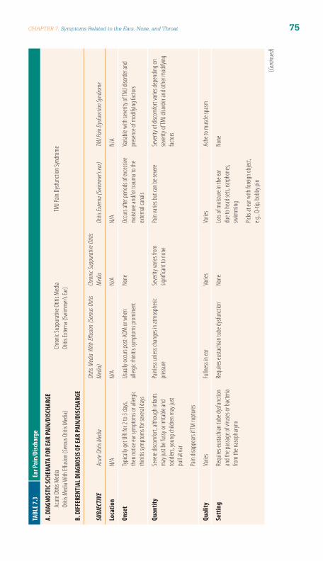
There are multiple causes for ear pain or discomfort, purulent discharge from an ear, and even associated acute hearing loss including acute otitis media, serous otitis me-dia (aka otitis media with effusion), otitis externa, chronic suppurative otitis media, and cerumen impaction. There are also some causes of ear pain that are not caused by ear problems. Examples of these include temporomandibular joint (TMJ) disorders (also called TMJ pain dysfunction syndrome and TMJ syndrome), dental disorders, and streptococcal pharyngitis. Table 7.3 provides the diagnostic schemata and the comparative presentations for the most common disorders related to the ear.
Acute Otitis MediaThe development of middle ear infections requires the transit of colonizing bacteria or vi-ruses from the nasopharynx or oropharynx, up into the eustachian tube followed by lack of normal patency or drainage from the tube. Several things are common causes of abnormal closure or dysfunction of the eustachian tube. These include any of the causes of swelling
08_Herrier_Ch07.indd 74 8/7/14 9:59 AM
CHAPTER 7: Symptoms Related to the Ears, Nose, and Throat 75TA
BLE 7
.3
Ear P
ain/
Disc
harg
e
A. D
IAGN
OSTI
C SCH
EMAT
A FO
R EA
R PA
IN/D
ISCH
ARGE
Acut
e Otit
is Me
dia
Chron
ic Su
ppur
ative
Otit
is Me
dia
TMJ P
ain D
ysfun
ction
Synd
rome
Otitis
Med
ia W
ith Eff
usion
(Sero
us O
titis
Media
) Ot
itis Ex
terna
(Swi
mm
er’s E
ar)
B. D
IFFE
RENT
IAL D
IAGN
OSIS
OF E
AR PA
IN/D
ISCH
ARGE
SUBJ
ECTI
VEAc
ute O
titis
Media
Otitis
Med
ia W
ith Eff
usion
(Sero
us O
titis
Media
)Ch
ronic
Supp
urativ
e Otit
is Me
diaOt
itis Ex
terna
(Swi
mmer’
s ear
)TM
J Pain
Dys
functi
on Sy
ndrom
e
Loca
tion
N/A
N/A
N/A
N/A
N/A
Onse
tTy
picall
y get
URI fo
r 2 to
3 da
ys,
then
notic
e ear
sym
ptom
s or a
llerg
ic rh
initis
sym
ptom
s for
seve
ral da
ys
Usua
lly oc
curs
post-
AOM
or w
hen
allerg
ic rh
initis
sym
ptom
s prom
inent
No
neOc
curs
after
perio
ds of
exce
ssive
m
oistu
re an
d/or
trau
ma t
o the
ex
terna
l can
als
Varia
ble w
ith se
verit
y of T
MJ di
sord
er an
d pr
esen
ce of
mod
ifying
facto
rs
Quan
tity
Seve
re dis
com
fort, a
lthou
gh in
fants
may
just
be fu
ssy or
irrita
ble an
d tod
dlers,
youn
g chil
dren
may
just
pull a
t ear
Painl
ess u
nless
chan
ges i
n atm
osph
eric
pres
sure
Seve
rity v
aries
from
signifi
cant
to no
nePa
in va
ries b
ut ca
n be s
evere
Seve
rity o
f disc
omfor
t vari
es de
pend
ing on
se
verit
y of T
MJ di
sord
er an
d oth
er m
odify
ing
factor
s
Pain
disap
pears
if TM
rupt
ures
Qual
ityVa
ries
Fulln
ess i
n ear
Varie
sVa
ries
Ache
to m
uscle
spas
m
Sett
ing
Requ
ires e
ustac
hian t
ube d
ysfun
ction
an
d the
passa
ge of
viru
ses o
r bac
teria
from
the n
asop
haryn
x
Requ
ires e
ustac
hian t
ube d
ysfun
ction
No
neLo
ts of
mois
ture
in th
e ear
due t
o hea
d sets
, earp
hone
s, sw
imm
ing
None
Picks
at ea
r with
forei
gn ob
ject,
e.g., Q
-tip,
bobb
y pin
(Cont
inued
)
08_Herrier_Ch07.indd 75 8/7/14 9:59 AM

76 PART 2: Assessment of Symptoms
Asso
ciate
d sy
mpt
oms
Puru
lent d
ischa
rge o
n the
pillo
w if
TM ru
pture
sEa
r pop
ping w
hen y
awns
Puru
lent d
ischa
rge o
n th
e pillo
wPu
rulen
t disc
harg
e on t
he
pillow
Head
ache
, grin
ding o
r pop
ping n
oise w
hen
open
mou
th. J
aw m
ay te
mpo
rarily
lock
whe
n op
ening
mou
th w
ideDe
creas
ed he
aring
Decre
ased
heari
ngDe
creas
ed he
aring
Poten
tially
decre
ased
heari
ng if
cana
l mos
tly oc
clude
dMa
y hav
e pere
nnial
aller
gic rh
initis
and
its sy
mpt
oms
Mod
ifyin
g fa
ctor
sNo
neNo
neNo
neYa
wning
, layin
g on t
he ea
r may
wo
rsen p
ainGr
inding
teeth
at ni
ght, c
hewi
ng gu
m m
akes
it w
orse
OBJE
CTIV
EAc
ute O
titis
Media
Otitis
Med
ia W
ith Eff
usion
(Sero
us O
titis
Media
)Ch
ronic
Supp
urativ
e Otit
is Me
diaOt
itis Ex
terna
(Swi
mmer’
s ear
)TM
J Pain
Dys
functi
on Sy
ndrom
e
Feve
rM
ild to
mod
erate
(38o C t
o 40o C)
None
Usua
lly no
neUs
ually
none
None
Ear e
xam
inat
ion
Ear c
anal
norm
al, m
ay ha
ve pu
rulen
t dis
charg
e if T
M ru
pture
sEa
r can
al no
rmal.
TM no
rmal
to ret
racted
wi
th pr
omine
nt sh
ort p
roces
s and
m
alleu
s
Ear c
anal
norm
al un
less
disch
arge
Pain
on pi
nna t
ractio
n or t
ragus
pr
essu
reNo
rmal
Red,
bulgi
ng TM
with
loss
of lan
dmark
s (lig
ht re
flex,
mall
eus,
shor
t proc
ess)
Fluid-
air le
vels
or ai
r bub
bles b
ehind
TMLa
rge c
entra
l TM
perfo
ration
with
pus,
red m
iddle
ear t
issue
, or
chole
steato
ma
Exter
nal c
anal
red sw
ollen
, with
pu
rulen
t disc
harg
e, pic
tures
que
grow
ths
TM-re
duce
d mob
ility
TM-re
duce
d mob
ility
TM no
rmal
if visu
alize
d
Othe
rPa
lpatio
n of T
MJ re
veals
crep
itus w
hen
open
ing m
outh
and/
or m
uscle
spas
m
Unde
rbite
or ot
her m
alocc
lusion
Mout
h doe
s not
open
strai
ght v
ertic
ally
Usua
l cau
sativ
e ag
ents
Viral,
Strep
tococ
cus p
neum
oniae
, H.
influe
nzae
, Mora
xella
catar
rhali
sNo
ne us
ually
Staph
yloco
ccus s
p, Ps
eudo
mona
s, fun
giSta
phylo
coccu
s sp,
Pseu
domo
-na
s, fun
giNo
ne
TABL
E 7.3
Ea
r Pai
n/Di
scha
rge
(Con
tinue
d )
08_Herrier_Ch07.indd 76 8/7/14 7:13 PM
CHAPTER 7: Symptoms Related to the Ears, Nose, and Throat 77
or enlargement of posterior pharyngeal lymphoid tissue, like a cold (viral URI) or upper respiratory manifestations of allergies. Roughly half are caused by respiratory viruses; the remaining are caused by the respiratory tract bacteria, Streptococcus pneumoniae, Hae-mophilus influenzae, and Moraxella catarrhalis. Acute otitis media (AOM) occurs mostly in children under 6 years of age who have relatively short straight eustachian tubes. By 6 years of age, the eustachian tube is considerably longer and is curved, making the retro-grade transit of microorganisms more difficult. Bottle propping in infants and toddlers who are lying on their back promotes passage of the ingested fluid with bacteria into the middle ear. In most cases, the onset of AOM symptoms is preceded by a 2- to 3-day history of viral URI or allergic rhinitis. In children who have not yet developed significant verbal skills, the ear pain is manifested by pulling at the affected ear or generalized fussiness or irritability. AOM is usually accompanied by a fever and decreased hearing in the affected ear. The natural course of AOM leads to increasing pain and pressure, finally resulting in a pinpoint perforation in the tympanic membrane (TM) with immediate cessation of severe pain due to the release of pressure. Over two-thirds of the cases resolve spontaneously without sequelae. This is true of virtually all viral causes and many cases caused by Haemophilus and Moraxella. While the cases caused by Streptococcus pneumoniae are less likely to resolve spontaneously, many still do. In less than 1% of the cases due to more virulent organisms such as S. pneumoniae, the mastoid bone is invaded, causing mastoiditis.
Objectively, most patients have a fever. On otoscopic examination, the TM is usually red. If tympanometry or pneumatic otoscopy is done, the TM is nonmobile. It may be bulging, and a pinpoint perforation may be visible. Normal landmarks are not visible. There may be a purulent discharge coming from the pinpoint perforation, and/or it may be noticed on the floor of the external canal. If the TM ruptures during sleep, the discharge can stain the pillow and the patient awakens to find the ear pain much better or gone. Current guidelines recommend that for most children 2 or over, only analgesic/antipyretics be given for 48 hours. Almost 75% will resolve without an-tibiotic therapy. Even for those patients referred to a primary care provider for severe symptoms for antibiotic therapy (whether < 2 years old or not), OTC analgesics/anti-pyretic products are appropriate. However, follow-up visits are required to check for the potential development of otitis media with effusion.
Otitis Media With Effusion/Serous Otitis MediaThe term otitis media with effusion (OME) more accurately reflects our knowledge of the inflammatory nature of the disease. Serous otitis media (SOM), an older but still frequently used term, reflected the belief (at the time) that there was only fluid behind the ear not caused by inflammation. It is now thought that there are two causes of OME: allergic rhinitis and post-AOM. In children who have had AOM, more than 60% will have some effusion remaining up to 8 weeks later. Like AOM, eustachian tube blockage/dysfunction is required for the development of OME. Usually OME resolves spontaneously without sequelae, but in some cases the inflammation creates bubbles of CO2 in the fluid, which diffuse out into the blood stream, creating a vacuum that pulls the TM back against the middle ear bones, causing them to touch each other. Un-treated, this can eventually cause permanent hearing loss when the bones fuse. Gen-erally, OME is painless unless there are changes in atmospheric pressure as occurs during takeoff and landing of an aircraft. Patients may notice a sensation of fullness, decreased hearing, and ear popping when yawning. Otoscopic examination reveals a nonmobile TM, possibly with the presence of an air fluid level or air bubbles behind the TM. The landmarks, particularly the malleus and its short process, become very prominent because the TM is retracted against the middle ear bones in more severe cases. The TM may be somewhat bluish in appearance. Treatment of severe OME
08_Herrier_Ch07.indd 77 8/7/14 9:59 AM

78 PART 2: Assessment of Symptoms
with a retracted TM requires the placement of pressure equalization tubes (PE tubes) through the TM. The tubes function to equalize the pressure, relieving the retraction induced contact with the bones of the middle ear, while allowing the inflammation to run its course. OME does not require antibiotic therapy. The efficacy of decongestants or corticosteroids is controversial.
Chronic Suppurative Otitis MediaChronic suppurative otitis media (CSOM) differs considerably from AOM. First, it requires the presence of a large central perforation in the TM. The cause of this cen-tral perforation is unclear. Trauma, placement of PE tubes, high fever, and repeated episodes of AOM with perforation have all been identified as causes. Rather than re-spiratory tract bacteria, the pathogens come from the flora of the external canal with Staphylococcus species and Pseudomonas aeruginosa predominating as causative agents. Other gram-negative bacteria can also be involved. Bacteria migrate through the central perforation, often facilitated by water from swimming or showering. Once inside the middle ear, they infect tissue and with accompanying inflammation, cause a purulent discharge. If untreated, this process can destroy important middle ear tissue, and invade the mastoid and other bony structures of the cranium. In chronic cases, a whitish cyst made up of epithelial tissue called a cholesteatoma may form. This tissue can enlarge, become infected, and eventually destroy middle ear bones. The two ma-jor symptoms of CSOM are decreased hearing and a purulent discharge for >14 days. Pain is generally not a prominent feature except in more widespread disease. On oto-scopic examination, there is a large central TM perforation with a purulent discharge. In long-standing disease, there may be a cholesteatoma seen as a whitish cyst in the middle ear. Patients with suspected CSOM should be referred to an otorhinolaryn-gologist, as soon as possible, for definitive treatment.
Otitis Externa (Swimmer’s Ear)Otitis externa (OE) is an infection of the external ear canal. Trauma from cleaning ears with foreign objects, wearing earpieces chronically, and constant moisture from sweat (especially while wearing earphones), or frequent immersion of the head in water (swimming) are all predisposing factors. Common pathogens include normal external ear canal flora including Staphylococcus sp, Pseudomonas aeruginosa, and multiple fun-gi. Patients generally have one or more predisposing factors and present with pain/dis-comfort and a purulent discharge. Hearing may or may not be affected depending on the amount of swelling and debris. Complete otoscopic examination may not be possible. However, to the extent that it is possible, usual findings include a red, swollen external canal with purulent material. With fungal infections, picturesque growths with colorful spores or hyphae may be seen. The TM is mobile on pneumatic ostoscopy, but swelling, debris, and especially pain in the external canal may prevent complete visualization of the TM. Since the canal is swollen and painful, the cardinal diagnostic finding is pain with a firm pull on the earlobe (pinna) or pressure on the tragus. Eliciting pain on pinna pull is generally diagnostic since patients with other ear disease will not experience any pain with this maneuver. In addition, there may be peri- or post-auricular lymphade-nopathy. Finally, if you suspect OE, be careful during the otoscopic examination. That is gently pull the pinna and possibly use a small diameter disposable otoscope speculum.
Ear Pain With a Normal Otoscopic ExaminationOccasionally, a patient complaining of ear pain will have a normal otoscopic examina-tion. When that occurs, think of referred pain from a dental problem, pharyngitis, or TMJ pain dysfunction syndrome. Specific problems may include a tooth abscess or
08_Herrier_Ch07.indd 78 8/7/14 9:59 AM

CHAPTER 7: Symptoms Related to the Ears, Nose, and Throat 79
streptococcal pharyngitis. A normal otoscopic examination in this setting warrants a mouth and throat examination by applying pressure on each tooth with a tongue de-pressor to detect abscessed teeth. Also, palpation over the TMJ and observation of the mouth opening should be conducted.
TMJ Pain Dysfunction SyndromeTMJ pain dysfunction syndrome can cause pain in the area of the ear due to muscle spasms caused by grinding the teeth (bruxism), or structural and/or functional abnor-malities of the TMJ. Patients may complain of ear pain, headache, or tinnitus. These patients may admit to a grinding, clicking, popping, snapping sensation or noise when they open and close their mouth. Ask if they grind their teeth at night and how frequently they chew gum, as both are typical in TMJ pain dysfunction syndrome. A quick check for TMJ problems can be done when examining the peri- and post- auricular lymph nodes. Press gently against the TMJ (just in front of the ears) and have the patient open their mouth slowly. Any clicking, grinding, popping sensations pal-pated may indicate TMJ problems. Also, palpable muscle spasms may be felt over the joint, which is typical. Watch the opening of the mouth carefully. If it does not open smoothly straight up and down, there may be TMJ problems. Finally, check for dental malocclusions especially an underbite. Generally, patients with TMJ pain dysfunction syndrome will have multiple suspicious findings, e.g., an underbite, a history of chew-ing gum frequently, an irregular mouth opening, and the presence of grinding, click-ing, popping over the TMJ on palpation. Patients suspected of TMJ problems should be initially referred to a dentist for further evaluation.
SORE THROAT/HOARSENESS
There are multiple causes of sore throats, but most are due to either infections (bacte-rial or viral) or irritant/allergic disorders. The most common bacterial infectious cause of sore throat is Streptococcus pyogenes (Group A β-hemolytic strep or GABHS) also known as strep throat. Numerous other bacteria have been implicated as causes of sore throat, but are less common (including Fusobacterium necrophorum, Arcanobac-terium haemolyticum, Group C and G streptococci, Mycoplasma pneumoniae, Chla-mydophila pneumoniae, N. gonorrhoeae, and even Corynebacterium diphtheriae). There are many viral causes, but the most common are those associated with viral URIs, which create pharyngitis either directly or by post-nasal drip. Other viral causes of pharyngitis include influenza viruses, coxsackieviruses (including those that cause herpangina and hand, foot, and mouth disease), Epstein-Barr virus (EBV), which is the cause of mononucleosis, and cytomegalovirus (CMV). Since primary HIV infections of-ten cause a syndrome like mononucleosis, with pharyngitis as a common component, depending on their individual risk factors, patients should have this diagnosis consid-ered. Another common cause of sore throat, usually by a post-nasal drip mechanism, is allergic rhinitis. Distinguishing between causes of hoarseness is also important. Viral laryngitis is a minor self-limiting disorder, whereas acute epiglottitis and carcinoma of the larynx can both be fatal if not diagnosed early and accurately. Table 7.4 provides the diagnostic schemata and comparative presentations of sore throat.
Streptococcal PharyngitisStrep throat is caused by a Group A β-hemolytic strep (Streptococcus pyogenes). It rep-resents between 10% and 30% of all patients reporting a sore throat as the primary or only symptom. It occurs most frequently in elementary school-age children (ages 5 to 11) and those in contact with them. Most cases occur during the school year, peaking during
08_Herrier_Ch07.indd 79 8/7/14 9:59 AM

80 PART 2: Assessment of Symptoms
TAB
LE 7
.4 S
ore
Thro
atB.
DIF
FERE
NTIA
L DIA
GNOS
IS O
F SOR
E THR
OAT
SUBJ
ECTI
VEStr
eptoc
occa
l Pha
ryngit
isVir
al Ph
aryn
gitis
Mono
nucle
osis
Herp
angin
aHF
MDPN
D
Loca
tion
May h
ave r
eferre
d ear
pain
Onse
tRa
pid on
set o
f sev
ere sy
mpt
oms
lastin
g 72 h
ours
Usua
lly sl
ower
onse
t and
las
t 5 to
10 da
ysSlo
w on
set, l
astin
g 5 to
7 d
ays m
ay be
prec
eded
by
fatig
ue/m
alaise
Rapid
onse
t lasts
7 to
10 da
ys12
to 36
hour
s ons
et, la
sts 7
to 10
days
Slow
onse
t, las
ts as
long
as
nasa
l sym
ptom
s
Quan
tity
Seve
re wi
th di
fficu
lty sw
allow
ing
all da
yW
ide ra
nge o
f sym
ptom
s fro
m PN
D to
strep
, but
us
ually
mild
er
Mild
to m
odera
te las
ting
all da
ySe
vere
pain
lastin
g all d
ay
difficu
lty sw
allow
ingSe
vere
pain
lastin
g all d
ay
difficu
lty sw
allow
ingM
ildly
painf
ul up
on
awak
ening
gets
bette
r du
ring t
he da
y
Qual
ityN/
AN/
AN/
A N/
A N/
A N/
A
Sett
ing
Occu
rs in
schoo
l-age
child
ren be
gin-
ning i
n elem
entar
y sch
ool a
nd th
ose
in co
ntac
t with
that
age g
roup
All a
ges
Adole
scent
s/you
ng ad
ults
(kiss
ing di
seas
e)Mo
stly c
hildr
enAl
l age
sAl
l age
s
Occu
rs pr
imari
ly du
ring s
choo
l yea
r in
fall/w
inter
Naus
ea vo
mitin
g in c
hildr
en
Asso
ciate
d sy
mpt
oms
Abse
nce o
f cou
gh or
othe
r nas
al sy
mpt
oms
Many
also
have
coug
h an
d URI
sym
ptom
sMa
rked f
atigu
e/m
alaise
Malai
seMa
laise
, skin
man
ifesta
tions
Sym
ptom
s of U
RI/
allerg
ic rh
initis
Mod
ifyin
g fa
ctor
sAc
idic o
r carb
onate
d drin
ks m
ake
it wor
seCe
rtain
form
s can
mim
ic str
epAc
idic o
r carb
onate
d drin
ks
mak
e it w
orse
Acidi
c or c
arbon
ated d
rinks
m
ake i
t wor
se
TABL
E 7.4
So
re Th
roat
A. D
IAGN
OSTI
C SCH
EMAT
A FO
R SO
RE TH
ROAT
Strep
tococ
cal P
haryn
gitis
Mono
nucle
osis
Hand
, Foo
t, and
Mou
th D
iseas
e (HF
MD)
Viral
Phar
yngit
is He
rpan
gina
Post-
nasa
l Drip
due t
o Alle
rgic
Rhini
tis/U
RI (P
ND)
TABL
E 7.4
So
re Th
roat
(Con
tinue
d)
OBJE
CTIV
EStr
eptoc
occa
l Pha
ryngit
isVir
al Ph
aryn
gitis
Mono
nucle
osis
Herp
angin
aHF
MDPN
D
Feve
r38
.4o C to 4
0o CUs
ually
low
grad
eLo
w-gr
ade f
ever
38.4o C t
o 40o C
Usua
lly lo
w gr
ade
None
Thro
at
exam
inat
ion
Beefy
red p
oster
ior ph
aryn
x with
sw
ollen
tons
ils w
ith ex
udate
(pus
) on
tonsil
s or p
oster
ior ph
aryn
x
Varia
ble fro
m m
ild to
se
vere
inflam
mati
onM
ildly
inflam
ed po
sterio
r ph
aryn
x, sw
ollen
tons
ilsMu
ltiple
vesic
les an
d/or
ulcera
tions
with
red b
order
on po
sterio
r pha
rynx a
nd
tonsil
s
Simila
r lesio
ns to
herp
an-
gina o
nly di
stribu
ted al
l ov
er ora
l cav
ity an
d thr
oat
Mild
ly infl
amed
poste
rior
phar
ynx w
ith cl
ear t
o muc
oid
nasa
l muc
ous d
raina
ge
Abou
t half
have
pus.
A few
with
shee
ts of
pus
Frenu
lum of
tong
ue m
ay
be ja
undic
ed
Othe
rPa
inful
anter
ior ce
rvica
l lym
phad
enop
athy
Many
have
viral
URI
findin
gsEle
vated
AST
/ALT
50%
have
trun
cal ra
sh67
% ha
ve pa
inful
bliste
rs on
palm
s, so
les, a
nd
butto
cks
Findin
gs of
aller
gic rh
initis
or
viral
URI
Fetid
brea
thPo
sitive
Mon
ospo
t tes
t
Posit
ive th
roat c
ulture
or ra
pid
strep
test
Painf
ul an
terior
/pos
terior
ce
rvica
l lymp
hade
nopa
thy
Lym
phad
enop
athy m
ay
exten
d to a
rm pi
t
Usua
l cau
sativ
e ag
ents
β-he
moly
tic St
reptoc
occu
s pyo
gene
sSa
me a
s URI
Epste
in-Ba
rr vir
usCo
xsac
kievir
us, e
ntero
virus
Coxs
ackie
virus
, ent
erovir
us
08_Herrier_Ch07.indd 80 8/7/14 7:13 PM

CHAPTER 7: Symptoms Related to the Ears, Nose, and Throat 81TA
BLE 7
.4
Sore
Thro
at (C
ontin
ued)
OBJE
CTIV
EStr
eptoc
occa
l Pha
ryngit
isVir
al Ph
aryn
gitis
Mono
nucle
osis
Herp
angin
aHF
MDPN
D
Feve
r38
.4o C to 4
0o CUs
ually
low
grad
eLo
w-gr
ade f
ever
38.4o C t
o 40o C
Usua
lly lo
w gr
ade
None
Thro
at
exam
inat
ion
Beefy
red p
oster
ior ph
aryn
x with
sw
ollen
tons
ils w
ith ex
udate
(pus
) on
tonsil
s or p
oster
ior ph
aryn
x
Varia
ble fro
m m
ild to
se
vere
inflam
mati
onM
ildly
inflam
ed po
sterio
r ph
aryn
x, sw
ollen
tons
ilsMu
ltiple
vesic
les an
d/or
ulcera
tions
with
red b
order
on po
sterio
r pha
rynx a
nd
tonsil
s
Simila
r lesio
ns to
herp
an-
gina o
nly di
stribu
ted al
l ov
er ora
l cav
ity an
d thr
oat
Mild
ly infl
amed
poste
rior
phar
ynx w
ith cl
ear t
o muc
oid
nasa
l muc
ous d
raina
ge
Abou
t half
have
pus.
A few
with
shee
ts of
pus
Frenu
lum of
tong
ue m
ay
be ja
undic
ed
Othe
rPa
inful
anter
ior ce
rvica
l lym
phad
enop
athy
Many
have
viral
URI
findin
gsEle
vated
AST
/ALT
50%
have
trun
cal ra
sh67
% ha
ve pa
inful
bliste
rs on
palm
s, so
les, a
nd
butto
cks
Findin
gs of
aller
gic rh
initis
or
viral
URI
Fetid
brea
thPo
sitive
Mon
ospo
t tes
t
Posit
ive th
roat c
ulture
or ra
pid
strep
test
Painf
ul an
terior
/pos
terior
ce
rvica
l lymp
hade
nopa
thy
Lym
phad
enop
athy m
ay
exten
d to a
rm pi
t
Usua
l cau
sativ
e ag
ents
β-he
moly
tic St
reptoc
occu
s pyo
gene
sSa
me a
s URI
Epste
in-Ba
rr vir
usCo
xsac
kievir
us, e
ntero
virus
Coxs
ackie
virus
, ent
erovir
us
08_Herrier_Ch07.indd 81 8/7/14 9:59 AM

82 PART 2: Assessment of Symptoms
the winter months. Circumstances associated with crowding of people, like a closed classroom, facilitate transmission of the organism. Recently, due to the large number of children in organized daycare or preschool, the age of suspicion for strep throat is con-siderably lower than previously seen. Concern for an accurate diagnosis is based on the possibility of nonsuppurative (autoimmune) inflammatory sequelae, acute rheumatic fever, in untreated patients. Acute rheumatic fever occurs between 2 and 5 weeks after the sore throat and presents with fever, carditis, migratory polyarthritis, and/or chorea. The carditis can result in permanent valvular damage and risk for developing bacterial endo-carditis. Due to careful attention to its diagnosis and prompt treatment, the incidence has been drastically reduced since the late 1940s to just one per 1 million population.
Subjectively, patients present with sudden onset of fever and severe sore throat, difficulty swallowing with pain worsened when swallowing acid liquids such as citrus juices and carbonated beverages. The pain is severe and constant throughout the day. Cough and nasal symptoms are uncommon. Malaise is common, but arthralgias and myalgias are not. Gastrointestinal symptoms including nausea, vomiting, and abdomi-nal pain may occur, but are more common in children. Generally, symptoms markedly improve by 72 hours after onset. Symptoms lasting longer than that should raise the suspicion of other causes such as viral pharyngitis and mononucleosis. A recent history of exposure to someone with a severe sore throat or diagnosed strep throat is common.
Objectively, examination of the throat and tonsils will reveal beefy red color, usually with purulent exudates and in many cases fetid (foul smelling) breath. Achieving com-plete visualization of the posterior pharynx and/or obtaining a culture or rapid strep test usually requires inducing the gag reflex with a tongue depressor. However, in a significant number of patients having them open their mouth wide, sticking their tongue out, and “panting like a dog” may preclude the need for a tongue depressor. Tonsillar tissue may be swollen. A marked fever (39°C to 40.5°C) is a significant feature. Painful anterior cervi-cal adenopathy is typical. A small number of patients will have a skin rash comprised of fine red papules on the truck that spread to the extremities but not the palms and soles of the feet. Typically, it has a sandpaper-like feel and blanches upon pressure. This mani-festation is called scarlet fever or scarlatina. The classic confirmation for diagnosis is con-sidered to be the throat culture. However, due to the 48-hour delay in obtaining culture results and the need to start therapy as soon as possible, the Rapid Antigen Detection Test (RADT) for S. pyogenes is usually preferred. Results of these tests are available in minutes. Unfortunately, they have a relatively high rate of false-positive findings because as many as one-fourth of patients are carriers of β-hemolytic Streptococcus. There is also a signifi-cant number of false negatives, mostly a function of poor sampling technique. Therefore, practically speaking, the clinical diagnosis requires a combination of typical features plus bacteriologic confirmation to accurately make the diagnosis. More technically, to rule out the carrier state, a definitive diagnosis requires typical signs and symptoms, plus some confirmation of the presence of the organism in the pharynx by standard culture or an RADT, and a positive result of acute and convalescent serologic tests for GABHS (i.e., antistreptolysin O or ASO test). If one tonsillar pillar or the uvula is swollen and displaced and the patient has difficulty opening their mouth without pain, suspect the suppurative complication peritonsillar abscess, which requires immediate referral.
Viral PharyngitisGenerally, viral pharyngitis presents with a wide range of symptoms and degrees of severity depending on the specific virus. Rhinovirus, adenovirus, coronavirus, herpes simplex, parainfluenza and RSV are common causes. Symptoms tend to have a slower onset, longer duration, and less pain than strep throat. Most patients with viral phar-yngitis have additional symptoms such as nasal symptoms, cough, and conjunctivitis
08_Herrier_Ch07.indd 82 8/7/14 9:59 AM

CHAPTER 7: Symptoms Related to the Ears, Nose, and Throat 83
that are very uncommon in patients with strep throat. However, in some cases viral pharyngitis can mimic streptococcal pharyngitis in every aspect except the positive bacteriological findings.
MononucleosisMononucleosis, caused by EBV, occurs most frequently in adolescents and young adults, hence its nickname the “kissing disease.” The onset of the sore throat is slow and it lasts 5 to 7 days with mild to moderate pain. In addition, most patients experi-ence significant fatigue and malaise that may last for 4 to 8 weeks. Sometimes it is the primary presenting symptom. Since the incubation period is 4 to 6 weeks, few patients remember any potential exposures. Objectively, examination of the throat reveals redness usually not as severe as in strep throat. About half the patients will have purulent exudates, with some having profuse exudates that is continuous over the posterior pharynx and tonsils. Painful anterior and posterior cervical lymph-adenopathy is common, with some patients having swollen, painful lymph nodes as remote from the throat as the underarms. Patients have a low-grade fever and 90% have mildly elevated AST and ALT levels. Jaundice develops in less than 5% of patients. All patients develop some degree of splenomegaly that may not be evident upon physical examination. A CBC reveals lymphocytosis with the presence of >10% atypical lymphocytes. A positive Monospot test and elevated EBV antibody levels are diagnostic for the disease. Unfortunately, neither may be positive during the first 2 weeks of the disease and may need to be repeated to confirm the diagnosis.
Herpangina/Hand, Foot, and Mouth DiseaseThese two disorders are caused by viruses in the enterovirus group, which includes coxsackieviruses and others. Some sources actually consider them to be different manifestations along the same disease spectrum. Both diseases usually present with severe throat pain, malaise, and difficulty swallowing and symptoms last for 7 to 10 days. Herpangina has a rapid onset and occurs most frequently in children, whereas hand, foot, and mouth disease (HFMD) has a slightly slower onset of 12 to 36 hours and occurs in patients of all ages. Objectively, both have dermatological manifesta-tions in more than 50% of patients. In herpangina, a maculopapular, vesicular rash appears on the trunk. In HFMD, the vesicular lesions with erythematous borders typi-cally occur on the palms of the hands and soles of the feet, as well as the buttocks in some cases. In HFMD, the fever is generally low grade, where herpangina presents with temperatures of 38.4°C to 40°C. Examination of the throat reveals varying degrees of erythema with red vesicles that ulcerate and have a red border. In herpangina, le-sions are limited to the posterior pharynx and tonsillar pillars, while in HFMD they occur all over the oral cavity including tongue and gingivae. Painful cervical lymph-adenopathy is common in both disorders.
Pharyngitis Due to Post-Nasal DripPatients with both allergic rhinitis and viral URIs can have sore throats. However, they differ in several ways from other forms of pharyngitis. Rarely is sore throat the primary complaint. While the patient may awaken with the sore throat, it usually goes away after several hours. In contrast, with most other causes of pharyngitis the pain is con-stant throughout the day and night. Also, the sore throat when due to post nasal drip lasts only as long as the rhinitis does. Objectively, examination of the throat reveals drainage or discharge, often in two tracks, on the posterior pharynx. When present, this drainage is often of similar consistency and appearance as the nasal discharge. There may be some mild inflammation on the soft palate or posterior pharynx.
08_Herrier_Ch07.indd 83 8/7/14 9:59 AM
84 PART 2: Assessment of Symptoms
Loss of Voice/HoarsenessLoss of voice or hoarseness at times accompanies sore throat or it can occur by itself. The most common cause of hoarseness is acute laryngitis. Respiratory viruses such as rhinovirus, adenovirus, and coronavirus are the most frequent cause. Allergies and voice strain due to overuse are other common causes. Smokers as well as patients with GERD may also experience the symptom. Onset of the hoarseness is slow and is accompanied by other symptoms of the causative disorder. Symptoms lasting longer than 2 weeks, especially in smokers, should be referred to an ENT specialist to look for more serious causes such as laryngeal carcinoma.
Croup, also known as viral laryngotracheobronchitis, is a disease of infants and young children in which the structures implicated in the name of the disorder become inflamed due to common respiratory viruses. Occurring mostly in the winter months, the hallmark signs are abrupt onset of a nocturnal cough that sounds like a seal barking along with inspiratory stridor and trouble breathing. Most cases are mild and require no treatment other than providing humidified air for the child to breath during attacks. This can be accomplished by taking the child into the bathroom, closing the door, and turning on the hot water in the shower to fill the room with steam. The warm moist air relieves the cough, stridor, and breathing difficulties. Severe difficulty breathing, continuous stridor at rest, retractions when breathing, early cyanosis, and lethargy are all signs of severe dis-ease that may require immediate treatment and/or hospitalization. In short, if the steam does not work well, then the child needs to be taken to a facility for definitive care.
Finally, acute epiglottitis, while rare, deserves mention. The incidence has drastically decreased in countries where routine immunization of children against Haemophilus influenzae type b and Streptococcus pneumoniae has been implemented. These are/were the two most common causes of the disease, although many other bacterial (and even some viral and fungal) causes have been documented. However, the rate in adults has re-mained constant with the average occurrence at age 45 with a gender ratio of 3:1 for males to females. It is a life-threatening infectious disease, with a 7% mortality rate in adults. It typically presents with an acute onset of sore throat, and difficult or painful swallowing. The classic sign is the sudden loss of voice as opposed to the slow onset of hoarseness and laryngitis seen in viral conditions. Patients generally present with a high fever and in later stages may experience difficulty breathing and stridor. This is a medical emergency and may require tracheostomy to prevent asphyxia and death. Therefore, patients (espe-cially older males) presenting with a severe sore throat and sudden loss of voice must be immediately referred.
KEY REFERENCES 1. Slavin RG, Spector SL, Bernstein IL, et al. The diagnosis and treatment of sinusitis: a practice parameter.
J Allergy Clin Immunol. 2005;116(6 suppl):S13-S47. 2. Anon JB. Upper respiratory infections. Am J Med. 2010;123(4 suppl):S16-S25. 3. Dykewicz MS, Hamilos DL. Rhinitis and sinusitis. J Allergy Clin Immunol. 2010;125(2 suppl):S103-115. 4. Neilan RE, Rolan PS. Otalgia. Med Clin North Am. 2010;94(5):961-971. 5. Ely JW, Hanson MR, Clark EC. Diagnosis of ear pain. Am Fam Physician. 2008;77(5):621-628. 6. Coker TR, Chan LS, Newberry SJ, et al. Diagnosis, microbial epidemiology and antibiotic treatment of
otitis media in children: a systematic review. JAMA. 2010;304(19):2161-2169.7. Schafer P, Baugh RF. Acute otitis externa: an update. Am Fam Physician. 2012;86:1055-61. 8. Wessels MR. Streptococcal pharyngitis. N Engl J Med. 2011;364(7):648-655. 9. Chan TV. The patient with sore throat. Med Clin North Am. 2010;94(5):923-943.
08_Herrier_Ch07.indd 84 8/7/14 9:59 AM

CHAPTER 7: Symptoms Related to the Ears, Nose, and Throat 85
SM, a regular customer at your store, comes in to pick up her mother’s prescription. As she pays for the prescription, she asks: “Is there anything better than Actifed for this cold I’ve got? I’m tired of being stuffed up. It’s been six days!”
a. Based on the information above, what are three likely causes for SM’s symptoms? Explain your rationale.
b. List 10 questions you would ask, physical examinations you would conduct, or lab tests you would order to identify the etiology of SM’s symptoms.
CASE 7.1
08_Herrier_Ch07.indd 85 8/7/14 9:59 AM

86 PART 2: Assessment of Symptoms
Iloff Medkem, a first-year pharmacy student, has the “ 2014 Crud,” which began 8 days ago with fever, rhinorrhea, facial fullness, myalgias, and arthralgia. After 5 days he began to feel better. However, his rhinorrhea returned 2 days ago as a mucoid discharge. Today he presents with pain under both eyes and the discharge has markedly changed.
a. List three questions you would ask to clarify his problem.
b. List three physical examinations you would perform to clarify the diagnosis.
c. What is the most likely diagnosis if the questions and examinations you listed above are positive?
CASE 7.2
08_Herrier_Ch07.indd 86 8/7/14 9:59 AM

CHAPTER 7: Symptoms Related to the Ears, Nose, and Throat 87
Howican Paddle presents to the pharmacy with a 3-day history of right ear discomfort with decreased hearing. This morning he woke up with a small yellow stain on his pillow.
a. What are the two most likely causes of his symptoms? Explain your rationale.
b. List two questions you would ask to help identify the cause. Explain your rationale.
c. For each of the diagnoses listed in question a above, list expected findings on ear examination.
CASE 7.3
08_Herrier_Ch07.indd 87 8/7/14 9:59 AM

88 PART 2: Assessment of Symptoms
FF, a 33-year-old fourth-grade teacher asks what would be good for this bad sore throat he has had for the last 48 hours.
List 10 questions/physical examinations/lab tests you would want to ask, conduct or order to confirm your assessment.
CASE 7.4
08_Herrier_Ch07.indd 88 8/7/14 9:59 AM