Assessment of Antibiotics Effect on Planktonic and …ijvm.org.il/sites/default/files/malik.pdf ·...
11
Israel Journal of Veterinary Medicine Vol. 72 (4) December 2017 3 Antibiotics Effect on Campylobacter Isolates Assessment of Antibiotics Effect on Planktonic and Biofilm Forms of Campylobacter Isolates Malik, H., 1 Rajagunalan, S., 2 Kumar, M. S., 1 * Kataria, J. L., 1 Anjay, P., 3 Sachan, S., 4 Upadhyay, A.K. 5 and Kumar, A. 6 1 Division of Veterinary Public Health, IVRI, Izatnagar 243122, Uttar Pradesh, India. 2 Department of Veterinary Public Health and Epidemiology, VCRI, TANUVAS, Tirunelveli 627001, Tamil Nadu, India. 3 Department of Veterinary Public Health and Epidemiology, Bihar Veterinary College, Patna 800014, Tamil Nadu, India. 4 Division of Veterinary Immunology, IVRI, Izatnagar 243122, Uttar Pradesh, India. 5 Department of Veterinary Public Health and Epidemiology, Veterinary College, GBPUAT, Pantnagar, U. S. Nagar – 263145, Uttarakhand, India. 6 Assistant Director General (Animal Health), Indian Council of Agricultural Research, Krishi Bhawan, Pusa, New Delhi 110001, India. * Corresponding Author: Dr. M. Suman Kumar, Division of Veterinary Public Health, IVRI, Izatnagar, India 243122. Tel: +91 581 2301602; Fax: +91 581 2301602. Email: [email protected] ABSTRACT Campylobacter spp. bacteria are one of the leading causes of food borne illness worldwide and has the ability to form biofilms. ese biofilms have been reported to confer resistance against antibiotics. In this study, the effect of five different antibiotics has been compared on the planktonic and biofilm forms of Campylobacter isolates by determining their minimum inhibitory concentration (MICs). A total of 55 isolates (11 Campylobacter jejuni and 41 Campylobacter coli) were subjected to flaA typing and cluster analysis. On the basis of flaA typing, 23 isolates, comprising 17 C. coli and 6 C. jejuni representing each cluster were chosen and analyzed for their biofilm forming potential at two different temperatures (37°C and 42°C) under both aerobic and microaerobic conditions. e biofilm production was higher at 37°C in comparison to 42°C, and it was enhanced under aerobic conditions compared to microaerobic conditions at both temperatures. MICs of gentamicin, kanamycin, tetracycline, erythromycin and carbenicillin were determined for both planktonic and biofilm forms of campylobacter bacteria grown in 96 well microtitre plates containing Muller Hinton (MH) broth. All the isolates in the planktonic form showed absolute resistance against carbenicillin. e MIC values of gentamicin, kanamycin, tetracycline and erythromycin for planktonic form were found to be 0.032 µg/ml, 2 µg/ml, 0.1 µg/ml and 0.0125 µg/ml, respectively; while, for biofilm forms the same were found to be 1.025 µg/ml, 8 µg/ml, 0.8 µg/ml and 0.2 µg/ml, respectively. e findings revealed 32, 16, 8 and 4 fold higher resistance by biofilm associated campylobacter bacteria against gentamicin, erythromycin, tetracycline and kanamycin, respectively. Keywords: Campylobacter jejuni; Campylobacter coli; flaA Typing; Biofilm; MIC; Antibiotic Resistance. INTRODUCTION Campylobacter spp. bacteria are among the leading cause of food borne illness worldwide (1). ey have the ability to form biofilms not only in the intestinal tract of their hosts but also on industrial surfaces which provide them a favour- able niche. ese biofilms have also been reported to offer resistance against antibiotics and disinfectants (2). e two predominant causative agents of Campylobacter related in- Research Articles
Transcript of Assessment of Antibiotics Effect on Planktonic and …ijvm.org.il/sites/default/files/malik.pdf ·...

Israel Journal of Veterinary Medicine Vol. 72 (4) December 2017 3 Antibiotics Effect on Campylobacter Isolates
Assessment of Antibiotics Effect on Planktonic and Biofilm Forms of Campylobacter IsolatesMalik, H.,1 Rajagunalan, S.,2 Kumar, M. S.,1* Kataria, J. L.,1 Anjay, P.,3 Sachan, S.,4 Upadhyay, A.K.5 and Kumar, A.6
1 Division of Veterinary Public Health, IVRI, Izatnagar 243122, Uttar Pradesh, India.2 Department of Veterinary Public Health and Epidemiology, VCRI, TANUVAS, Tirunelveli 627001, Tamil Nadu, India.3 Department of Veterinary Public Health and Epidemiology, Bihar Veterinary College, Patna 800014, Tamil Nadu, India.4 Division of Veterinary Immunology, IVRI, Izatnagar 243122, Uttar Pradesh, India.5 Department of Veterinary Public Health and Epidemiology, Veterinary College, GBPUAT, Pantnagar, U. S. Nagar – 263145, Uttarakhand, India.
6 Assistant Director General (Animal Health), Indian Council of Agricultural Research, Krishi Bhawan, Pusa, New Delhi 110001, India.
* Corresponding Author: Dr. M. Suman Kumar, Division of Veterinary Public Health, IVRI, Izatnagar, India 243122. Tel: +91 581 2301602; Fax: +91 581 2301602. Email: [email protected]
ABST RACT Campylobacter spp. bacteria are one of the leading causes of food borne illness worldwide and has the ability to form biofilms. These biofilms have been reported to confer resistance against antibiotics. In this study, the effect of five different antibiotics has been compared on the planktonic and biofilm forms of Campylobacter isolates by determining their minimum inhibitory concentration (MICs). A total of 55 isolates (11 Campylobacter jejuni and 41 Campylobacter coli) were subjected to flaA typing and cluster analysis. On the basis of flaA typing, 23 isolates, comprising 17 C. coli and 6 C. jejuni representing each cluster were chosen and analyzed for their biofilm forming potential at two different temperatures (37°C and 42°C) under both aerobic and microaerobic conditions. The biofilm production was higher at 37°C in comparison to 42°C, and it was enhanced under aerobic conditions compared to microaerobic conditions at both temperatures. MICs of gentamicin, kanamycin, tetracycline, erythromycin and carbenicillin were determined for both planktonic and biofilm forms of campylobacter bacteria grown in 96 well microtitre plates containing Muller Hinton (MH) broth. All the isolates in the planktonic form showed absolute resistance against carbenicillin. The MIC values of gentamicin, kanamycin, tetracycline and erythromycin for planktonic form were found to be 0.032 µg/ml, 2 µg/ml, 0.1 µg/ml and 0.0125 µg/ml, respectively; while, for biofilm forms the same were found to be 1.025 µg/ml, 8 µg/ml, 0.8 µg/ml and 0.2 µg/ml, respectively. The findings revealed 32, 16, 8 and 4 fold higher resistance by biofilm associated campylobacter bacteria against gentamicin, erythromycin, tetracycline and kanamycin, respectively.
Keywords: Campylobacter jejuni; Campylobacter coli; flaA Typing; Biofilm; MIC; Antibiotic Resistance.
INTRODUCTIONCampylobacter spp. bacteria are among the leading cause of food borne illness worldwide (1). They have the ability to form biofilms not only in the intestinal tract of their hosts
but also on industrial surfaces which provide them a favour-able niche. These biofilms have also been reported to offer resistance against antibiotics and disinfectants (2). The two predominant causative agents of Campylobacter related in-
Research Articles

Israel Journal of Veterinary Medicine Vol. 72 (4) December 2017Malik, H.4
fections are Campylobacter jejuni and Campylobacter coli (3, 4), which reside asymptomatically as commensals in many animal species. Consumption of undercooked contaminated poultry or cross-contamination of other food products with raw poultry products is acknowledged to be a major source of infection. In spite of its fastidious laboratory growth re-quirements, Campylobacter acquire mechanisms allowing it to survive a range of in vivo and ex vivo conditions, one such mechanism being the ability to form biofilms.
Biofilms consist of growing microorganisms intimately associated with each other, producing an extracellular poly-meric substance (ESP) of carbohydrate or exopolysaccharide adhering to synthetic or biological surfaces (5). Studies have shown that Campylobacter can survive in unfavorable condi-tions such as low level of nutrient and increased oxygenation when integrated with biofilms (6). Biofilms may provide the perfect niche for campylobacter survival as there are micro-environments in the biofilm that provide ideal conditions for its growth and maintenance (7). There exists a state of continuous attachment and detachment of bacteria from the matrix, making biofilms an enduring source of bacterial dis-charge. The presence of biofilms in water distribution system is thought to be a possible source for colonization of the bac-teria in the poultry flocks (8). Grant et al. (9) demonstrated ex vivo adhesion of abundant campylobacter to the mucus overlying the human gut tissue. It has been suggested that different strains of Campylobacter differ in their survival in nutrient poor environments (10), ability of biofilm formation and structural and dynamic properties of biofilm (11).
The most important property of the biofilm forms in clinical medicine is the enhanced resistance to antimicro-bial agents, through protection by the ESP (12), leading to multidrug resistance and therapeutic failure. Although the mechanisms are poorly understood, there is evidence that they may be related to the slow diffusion of drug and the modified nutrient environments leading to suppression of growth rate within the biofilm.
In view of the dearth of information on epidemiology of Campylobacter in relation to its survival in the environment, the present study was undertaken with the aim to study the biofilm forming ability among different strains of campy-lobacter and to compare the antimicrobial susceptibility of planktonic versus biofilm attached bacteria in terms of minimum inhibitory concentration (MIC) against a range of antibiotics.
MATERIALS AND METHODSA total of 55 Campylobacter isolates comprising of 11 C. jejuni and 44 C. coli isolated from poultry caeca were used in this study. The isolates were maintained in 15% glycerol broth and stored at -80°C. Bacteria were grown on Mueller Hinton agar for 48 h at 42°C under microaerophilic conditions (BD GasPakTMEZ, USA). The genomic DNA of the isolates was extracted using DNeasy Blood and Tissue Kit (Qiagen, USA) following the manufacturer’s protocol. The flaA gene was am-plified as per the protocol of CAMPYNET (13). The PCR was performed in a total volume of 50 µl, containing 4 µl of DNA, 5 µl of 10X PCR buffer, 20 ρmol of each forward and reverse primers, 5 µl of dNTP (2 mM), and 1 U of Dream Taq polymerase (ThermoFischer) and nuclease-free water to make up the final volume. Amplification was obtained with 35 cycles following an initial denaturation step at 94°C for 5 min. Each cycle involved denaturation at 94°C for 45 s, annealing at 45°C for 45 s and extension at 72°C for 105 s followed by a final extension step at 72°C for 5 min. After amplification, 5 µl of the PCR product was electrophoresed with 2% agarose (SRL, India) in 0.5X Tris-borate-EDTA (TBE) buffer containing 0.5 µg of ethidium bromide/ml to determine the presence or absence of the flaA amplicon.
A 10 µl sample of the PCR product was digested with 0.2 µl (2 U) of the restriction enzyme HpyF3I (DdeI) and using 2 µl of 10X buffer, nuclease free water was used to make up the volume up to 30 µl. The reaction tubes were incubated at 37°C for 4 h and stored at 4°C till further examination. A 100 bp plus DNA ladder (Fermentas, USA) was used as the standard for molecular size determinations. Gels were run at 75 V for 90 min in 0.5X TBE buffer. The DNA bands were visualized and documented using the gel documentation system (UVP, USA)
Dendrograms were constructed on the basis of obtained RFLP patterns using GelCompar II (BioNumerics, Belgium) by the unweighted pair group mathematic average method (UPGMA). Similarities between the profiles based on the band patterns were derived using the Dice’s coefficient (14).
The isolates to be assayed for biofilm forming ability were chosen after subjecting the isolates to flaA typing. One isolate was chosen from each cluster, formed at 70% similarity level. The protocol for bioassay formation was followed as per Merritt et al. (15) with slight modifications. Selected Campylobacter isolates were grown overnight in Mueller
Research Articles

Israel Journal of Veterinary Medicine Vol. 72 (4) December 2017 5 Antibiotics Effect on Campylobacter Isolates
Hinton broth at 42°C under microaerophilic conditions. The wells of 96 well microtitre plates (Nunc, Denmark) were inoculated with 200 µl of 48 h old cultures of C. jejuni and C. coli isolates with turbidity adjusted to 3 McFarland standard (~9 x 10⁸ CFU) and sterile broth as negative control. Plates were incubated at 42°C and 37°C under microaerophilic as well as aerobic conditions for 48 h. Following incubation, the supernatant was removed and wells were dried for 30 min at 55°C. To the wells 125 µl 0.1% aqueous crystal violet (CV) was added and incubated for 10 min at room temperature. The unbound CV was removed, and the wells were washed twice with sterile distilled water. The wells were air dried and bound CV was extracted into 200 µl of 95% ethanol. One hundred twenty five microliters of this solution was removed from each well and placed in a new 96 well plate. The optical density was observed at 620 nm (OD620) using a microplate reader to determine biofilm formation. By taking 42°C and microaerophilic conditions as standard, strains were classified into three categories: weak, moderate and strong biofilm producers as per Stepanovic et al. (16) as described in Table 1. Based on these criteria, optical density cut-off value (ODc) is the average OD of negative control + 3 × SD (standard deviation) of negative control. Experiments were performed in triplicate.
Statistical analysisThe effect of each variable (species and atmosphere) was as-sessed using the student’s t-test. Statistical significance was defined at p<0.05.
Table 1: Classification of isolates as per biofilm forming abilityCategories Cut off level C. jejuni C. coliNegative OD ≤ ODC - -Weak OD ≤ 2 X ODC 1 1Moderate 2 X ODC ≤ OD ≥ 4X ODC 5 14Strong OD ≥4X ODC - 2
ODc = 3 SD above OD of Negative control; OD: Optical Density
Minimum inhibitory concentrationThe minimal inhibitory concentrations (MICs) of gentamicin (32.8 µg/ml), kanamycin (128 µg/ml), tetracycline (25.6 µg/ml), erythromycin (25.6 µg/ml) and carbenicillin (408 µg/ml) for planktonic forms of C. jejuni and C. coli were determined using the broth microdilution assay. Two fold serial dilutions of antibiotics were prepared in Mueller Hinton (MH) broth.
The wells of microtitre plates, each containing 100 µl of serially diluted antibiotics, were inoculated with 100 µl of overnight grown Campylobacter culture with an OD620 value of 0.046 and incubated at 42°C for 4 days. The growth was estimated by recording the OD620 at the end of 4 days incubation period. The MIC was the lowest concentration of antibiotic, for which no significant increase in OD620 was noted (17, 18).
The MICs of antibiotics for biofilm forms were deter-mined against the aforementioned antibiotics using microtitre plate containing a 48 h preformed biofilm in each well. After aspiration of the culture supernatants, 200 µl of fresh MH broth containing two fold serially diluted antibiotics were added to each well and the plate was incubated for further 48 h at 42°C. The biofilm was then suspended by pipetting and growth was estimated by recording the OD620 at the end of the 48 h incubation period. The MIC was considered as the lowest concentration at which, no measurable growth was observed. All results were read as per “The Clinical & Laboratory Standards Institute” (CLSI), 2010 guidelines (19)
Scanning electron microscopyThe morphology of the Campylobacter biofilm was studied using scanning electron microscopy. The study was carried out using C. jejuni isolate C19 of poultry caecal origin. Biofilm for microscopy were cultured at 37°C under aerobic condi-tions. However, in this case, sterile coupons (3 mm diameter), cut out of polystyrene Petri dish were used as the surface for biofilm formation. Once the biofilm had been cultured, the coupons were removed from the growth media, washed and immediately fixed in 2.5% glutaraldehyde in phosphate buffer saline (pH 7.0) for 48 h at 4˚C. The sample was dehydrated in a series (30%, 50%, 80%, 90% and 100%) of ethanol solu-tions. The samples were critical point dried with liquid carbon dioxide and sputter coated with a thin layer of gold using gold coater ( JFC, 1600). Scanning electron microscope ( Jeol, Japan) was used at high vacuum in the secondary electron imaging mode to observe the biofilms at 1600X, 3300X, 3500X and 4300X magnifications. Representative views of the biofilms were digitally imaged.
RESULTSOut of the 55 isolates subjected to PCR assay for amplifica-tion of flaA gene of Campylobacter only 52 isolates (94.5%) produced a ~1725 bp amplicon. Appropriate amplification
Research Articles

Israel Journal of Veterinary Medicine Vol. 72 (4) December 2017Malik, H.6
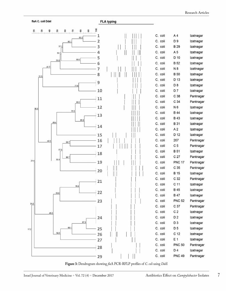
was not observed for 3 C. coli isolates, which were considered as untypeable and were not included for further studies. The amplified flaA products of both C. jejuni and C. coli were sub-jected to restriction digestion with HpyF3I (an isoschizomer of DdeI) restriction enzyme and banding patterns visualized on 2% agarose gel (Fig. 1). Out of 11 C. jejuni subjected to flaA typing, 9 flaA types (1-9) were found on the basis of number of bands obtained and their molecular weight (Fig. 2). Among 41 C. coli isolates, a total of 29 flaA types were observed (Fig. 3).
Selection of isolates for further study was done on the basis of percent similarity by flaA typing. A cut off level of 70% was set and one isolate from each cluster was selected. Those isolates which were not found to form any cluster were selected individually. Accordingly, 6 C. jejuni and 19 C. coli isolates were selected for further studies. Out of 19 C. coli, 2 isolates did not survive during the subculture, and only 17 C. coli were further characterized. Overall, a diverse profile was observed for C. jejuni isolates from similar and different geographical regions except for flaA types 5 and 6 (Fig. 3). Among C. coli, 5 flaA types (18, 20, 21, 24 and 28) revealed similar flaA genetic profiles irrespective of their location of isolation. However, 5 flaA types (9, 11, 13, 14 and 22) revealed similar genetic profiles of isolates from the same location of isolation. C. jejuni isolates obtained were genetically more diverse compared to that of C. coli based on the number of fla types obtained.
The biofilm forming ability of the isolates were evalu-ated under aerobic and microaerophilic conditions at two
different temperatures i.e., 37°C and 42°C. The 6 C. jejuni isolates under study revealed significant influence of aerobic and microaerobic conditions on biofilm formation with all the 6 isolates showing higher O.D. under aerobic conditions. The two incubation temperatures 37°C and 42°C reflected minor variation and did not show any statistically significant effect (p<0.05) within the similar atmospheric conditions for C. jejuni (Fig. 4). Amongst the 17 C. coli isolates, a sig-nificant effect was observed for different strains of C. coli with regards to both atmospheric conditions as well as the temperatures of incubation. Among the isolates tested, 13 showed higher O.D. values at 37°C under aerobic conditions, while only 4 isolates showed highest O.D. values at 37°C under microaerobic conditions. It was observed that biofilm formation was higher under aerobic conditions as compared to microaerobic conditions and at 37°C as compared to 42°C by most of the isolates (p>0.05) (Fig. 5).
Out of 6 C. jejuni, 5 isolates (N1, A1, C19, PNC77 and C40) were found to be moderate biofilm producers and only 1 isolate (C7) was classified as weak biofilm producer. Out of 17 C. coli isolates, 14 (PNC49, PNC52, A5, B10, N8, B50, 207, A2, D12, E1, C35, C32, C37 and PNC50) were found to be moderate biofilm producers, 2 (C27, and B29) strong biofilm producers and only one (D7) weak biofilm producer (Table 1).
Figure 1: Agarose gel showing fla PCR-RFLP profiles of Campylobacter spp. using restriction enzyme DdeI
Lane M: 100 bp ladder; Lane 1: PNC52; Lane 2: PNC49; Lane 3: B31; Lane 4: C38; Lane 5: A2; Lane 6: B44; Lane 7: B45; Lane 8: B47; Lane
9: B43; Lane 10: C34; Lane 11: PNC50Figure 2: Dendrogram showing flaA PCR-RFLP profiles of C. jejuni
using DdeI
Research Articles
Israel Journal of Veterinary Medicine Vol. 72 (4) December 2017 7 Antibiotics Effect on Campylobacter Isolates
Figure 3: Dendrogram showing flaA PCR-RFLP profiles of C. coli using DdeI
Research Articles

Israel Journal of Veterinary Medicine Vol. 72 (4) December 2017Malik, H.8
Table 2: MIC values for different antibiotics on planktonic and biofilm bacteria.
Sr. No. Isolate Species Gentamicin Kanamycin Tetracycline ErythromycinP B P B P B P B
1 N1 C. jejuni 0.513 4.1C 4 8A 0.1 1.6D 0.025 0.2C
2 A1 C. jejuni 1.025 1.025 4 8A 0.2 3.2D 0.025 1.6F
3 C19 C. jejuni 0.513 2.05B 4 16A 0.2 1.6C 0.05 0.2B
4 C7 C. jejuni 0.513 4.1C 8 16A 0.2 3.2D 0.05 0.2B
5 PNC77 C. jejuni 1.025 2.05A 4 8A 0.4 1.6B 0.0125 0.2D
6 C40 C. jejuni 1.025 2.05A 4 8A 0.4 0.8A 0.1 1.6D
7 C27 C. coli 1.025 2.05A 8 16A 0.2 1.6C 0.1 0.8C
8 PNC49 C. coli 1.025 4.1B 4 8A 0.2 3.2D 0.2 0.4A
9 PNC52 C. coli 0.257 1.025B 8 16A 0.1 0.8C 0.1 0.4B
10 B29 C. coli 1.025 2.05A 2 8B 1.6 3.2A 0.0125 0.8F
11 A5 C. coli 1.025 4.1B 8 16A 0.1 0.8C 0.2 1.6C
12 D10 C. coli 0.513 1.025A 8 16A 0.1 0.8C 0.1 0.2A
13 N8 C. coli 0.032 2.05F 4 8A 0.1 0.8C 0.05 0.2B
14 B50 C. coli 1.025 2.05A 8 32B 0.8 1.6A 0.0125 0.2D
15 207 C. coli 1.025 4.1B 4 16B 0.2 3.2D 0.1 1.6D
16 A2 C. coli 1.025 4.1B 4 8A 0.1 0.8C 0.1 1.6D
17 D12 C. coli 0.257 2.05C 8 16A 0.2 0.8B 0.05 1.6E
18 D7 C. coli 0.513 2.05B 8 32B 0.1 0.8C 0.05 0.4C
19 E1 C. coli 0.257 1.025B 4 8A 0.8 1.6A 0.0125 0.2D
20 C35 C. coli 0.257 2.05C 8 16A 0.1 0.8C 0.0125 1.6G
21 C32 C. coli 1.025 2.05A 4 16B 0.4 1.6B 0.1 0.4B
22 C37 C. coli 0.513 2.05B 8 16A 0.2 1.6C 0.1 1.6D
23 PNC50 C. coli 1.025 2.05A 4 8A 0.4 1.6B 0.1 0.8C
P: Planktonic cell; B: Biofilm; Superscript denotes increase in concentration: A (2-fold), B (4-fold), C (8 fold), D (16 fold), E (32 fold), F (64 fold), G (128 fold)
Figure 4: Graph showing biofilm formation by Campylobacter jejuni isolates C. jejuni biofilm formation under aerobic/microaerophilic conditions at either 37°C/42°C.
Error bars represent one standard deviation from the mean.
Research Articles

Israel Journal of Veterinary Medicine Vol. 72 (4) December 2017 9 Antibiotics Effect on Campylobacter Isolates
MICs of gentamicin, kanamycin, tetracycline, erythromy-cin and carbenicillin on planktonic C. jejuni and C. coli were determined. MICs of gentamicin, kanamycin, tetracycline and erythromycin for planktonic Campylobacter were found to be 0.032 µg/ml, 2 µg/ml, 0.1 µg/ml and 0.0125 µg/ml, re-spectively. However, all the isolates showed resistance against carbenicillin even at the highest concentration. The number of isolates showing particular MIC values are presented in table 3. All the isolates were found sensitive against gentami-cin, kanamycin, erythromycin and tetracycline while all were resistant to carbenicillin (CLSI, 2010) (Table 2).
The MICs of the listed antibiotics were determined for Campylobacter biofilms using microtitre plate containing a 48 h preformed biofilm in each well. The different antibiot-ics showed significant effect on biofilm forms. The results are presented in table 2. MICs of gentamicin, kanamycin, tetracycline and erythromycin for Campylobacter in biofilm were found to be 1.025 µg/ml, 8 µg/ml, 0.8 µg/ml and 0.2 µg/ml, respectively. MIC for carbenicillin was not recorded, as all the isolates showed resistance for the highest concentration. The distribution of MIC pattern is presented in table 3.
Scanning electron microscopyScanning Electron Microscopy was undertaken to examine the biofilm microstructure. The C. jejuni isolate C19 produced
Table 3: Number of times of occurrence of MIC values (µg/ml)
Conc. (µg/ml)
Gentamicin Kanamycin Tetracycline ErythromycinP B P B P B P B
0.0125 50.025 20.032 10.05 40.1 8 90.2 8 2 80.257 40.4 4 40.513 60.8 2 9 31.025 12 41.6 1 9 82.0 12.05 133.2 54.0 124.1 68.0 10 1016.0 1132.0 12
P: Planktonic cell; B: Biofilm; R: Resistant
Figure 5: Graph showing biofilm formation by Campylobacter coli isolates C. coli biofilm formation under aerobic/microaerophilic conditions at either 37°C/42°C. Error bars represent one standard deviation from the mean.
Research Articles

Israel Journal of Veterinary Medicine Vol. 72 (4) December 2017Malik, H.10
a mature biofilm in which a large number of cell clusters were entwined to form a dense net and the organism was found to be embedded in extensive matrix. Clusters were found enclos-ing abundant numbers of bacterial cells intimately associated with a fibrous cord like material. Morphology of the bacteria comprising the biofilm was relatively homogeneous, with the bacteria present as spiral or curved rod forms (Fig. 6).
DISCUSSIONThe flaA-RFLP typing revealed 94.5% typeability as reported by Harrington et al. (20) who found 93% typeability of PCR-RFLP using DdeI enzyme. The C. jejuni isolates obtained were genetically more diverse compared to that of C. coli based on the number of flaA types obtained, as reported by Khoshbakht
et al. (21) and Rajagunalan et al. (22). Overall, flaA typing was able to discriminate C. jejuni and C. coli isolates from similar as well as different geographical regions and sources. DdeI proved to be useful for the initial grouping of the strains (23).
It has been reported that under stressful conditions Campylobacter spp. undergo biofilm formation to remain viable in the environment and serve as a continuous source of contamination to the flocks in poultry houses as well as to the food in processing plants (24). The production of bio-films by most of the isolates was seen at 37˚C under aerobic conditions followed by 42˚C under aerobic, 37˚C under microaerobic and at 42˚C under aerobic conditions. These findings in this study were in accordance with Sulaeman et al. (25) and Reuter et al. (26) who reported that oxygen rich
Figure 6: Scanning electron micrograph of Campylobacter jejuni biofilm
Research Articles
Israel Journal of Veterinary Medicine Vol. 72 (4) December 2017 11 Antibiotics Effect on Campylobacter Isolates
conditions promote the biofilm formation by microaerophilic campylobacter bacteria. But on the contrary, Reeser et al. (27) has reported a higher biofilm production by campylobacter bacteria under microaerophilic conditions.
According to Stepanovic et al. (16) classification of bio-film producing ability of microorganisms, most of the isolates of Campylobacter were found to produce moderate biofilms with a few isolates showing strong and weak biofilm produc-tion. Out of 6 C. jejuni isolates, 5 (83.3%) were classified as moderate biofilm producers and the remaining isolate were weak biofilm producers. However, out of 17 C. coli isolates, 14 (82.3%) were moderate, 2 (11.7%) were strong and one (5.9%) isolate was a weak biofilm producer. Comparable findings were reported by Teh et al. (28), who reported that most Campylobacter spp. produce moderate biofilm with a few strains showing strong biofilm producing ability. Biofilm forming abilities of different bacterial strains have been re-ported to exhibit considerable difference when incubated at same temperature and atmospheric conditions (29, 30).
Bacteria in a biofilm are relatively resistant to antimicro-bial agents and host immune response (31). Sessile bacteria have proved to be less susceptible to antimicrobial agents as compared to their non-attached planktonic counterparts (32). In the present study, bacteria in biofilm were found to be several fold more resistant to antibiotics than planktonic bacterial cells. MIC of gentamicin for biofilm was 2-4 fold higher than MIC for planktonic cells. Sepandj et al. (33) observed only 12.5% sensitivity of gentamicin for biofilm associated bacteria. Low penetrating ability of gentamicin for biofilm may be a reason for increased MIC values. Gentamicin has been shown to have less penetrating ability in biofilm since less than 25% of this antibiotic was found in biofilm after 24 h of incubation (34).
In case of kanamycin, a four-fold increase in MIC was observed for the biofilm forms. Although kanamycin is known to rapidly kill growing cells, sessile cells in biofilm are perhaps the reason for its resistance (35). MIC of tetra-cycline was found to increase by 8 fold in biofilm associated bacteria, tetracycline is found to show good penetration for biofilm (36) and possibly there are other unknown reasons for its resistance. The MIC value for biofilm associated bac-teria for erythromycin, considered as drug of choice against campylobacter bacteria, increased by 16 folds. In our study, planktonic as well as biofilm bacteria were found to have absolute resistance against carbenicillin. Findings of Spoering
and Lewis (37) are in agreement with the results of this study, showing 100% resistance of biofilm bacteria for carbenicillin. Production of β-lactamases by bacteria or presence of slow growing bacteria in biofilm may possibly be the one of the reasons for its tolerance.
Chen et al. (38) has reported several fold increase in MIC value of biofilm compared to planktonic bacteria. Comparable results were presented by Grenier et al. (17) who have reported an increase in MIC of various antibiotics by 32 to 256 fold. Sepandj et al. (33) stated that the gram negative bacteria in biofilm state are much less susceptible to the antibiotics than their planktonic counterparts.
Scanning electron microscopy was performed to un-derstand the morphology of biofilm grown on polystyrene coupons. Campylobacter spp. has been reported to develop biofilm on various surfaces viz. glass fibre (39), glass cov-erslips (40), plastic coupons (41) and microtitre plates (27). Reeser et al. (27) has reported a higher degree of biofilm formation on polystyrene surface. Similar coupons used in the present study gave comparable results. Biofilm was observed with large number of cell clusters intertwined to form a thick mesh like structure. In an earlier study, similar results for campylobacter biofilm formation were reported by Joshua et al. (11). Bacteria comprising the biofilm were spiral or curved, comparable to the results of Svensson et al. (42), indicating the preservation of normal morphological features in biofilms during the normal incubation period of 48 hours. On the contrary, Gunther and Chen (30) reported both spiral and coccoid forms of the bacteria in roughly similar proportions. The variation in findings may be due to difference in the incubation time and other related parameters.
This study concluded that under aerobic or stressful conditions, Campylobacter spp. adapted to a biofilm lifestyle, allowing survival under detrimental conditions. The biofilm leads to increased resistance for antibiotics, and can function as a reservoir of viable planktonic cells in environment. The increased level of biofilm formation under aerobic conditions is likely to be an adaptation contributing to the infectiious existence of Campylobacter species in the environment.
ACKNOWLEDGEMENTS
The authors are thankful to the Director, Indian Veterinary Research Institute for providing facilities to conduct this research. This research programme was carried out under the ICAR funded-project ‘Outreach Programme on Zoonotic Diseases’.
Research Articles
Israel Journal of Veterinary Medicine Vol. 72 (4) December 2017Malik, H.12
REFERENCES1. Wilson, D. J., Gabriel, E., Leatherbarrow, A. J., Cheesbrough, J.,
Gee, S., Bolton, E., Fox, A., Fearnhead, P., Hart, C. A. and Dig-gle, P. J.: Tracing the source of campylobacteriosis. PLoS Genet-ics 4:e1000203, 2008.
2. Siringan, P., Connerton, P. L., Payne, R. J. H. and Connerton, I. F.: Bacteriophage-Mediated Dispersal of Campylobacter jejuni Biofilms. Appl. Environ. Microbiol. 77:3320-3326, 2008.
3. Moore, J. E., Corcoran, D., Dooley, J. S., Fanning, S., Lucey, B., Matsuda, M. and Whyte, P.: Campylobacter. Vet. Res. 36:351-382, 2005.
4. Kulkarni, S. P., Lever, S., Logan, J. M. J., Lawson, A. J., Stanley, J. and Shafi, M. S.: Detection of Campylobacter species: a comparison of culture and polymerase chain reaction based methods. J. Clin. Pathol. 55:749-753, 2002.
5. Nachamkin, I., Szymanski, C. M. and Blaser, M. J.: Campylobacter. ASM Press, 2008.
6. Fux, C. A., Costerton, J. W., Stewart, P. S. and Stoodley, P.: Survival strategies of infectious biofilms. Trends Microbiol. 13:34-40, 2005.
7. Donlan, R. M. and Costerton, J. W.: Biofilms: Survival mecha-nisms of clinically relevant microorganisms. Clin. Microbiol. Rev. 15:167-193, 2002.
8. Haddock, G., Mullin, M., MacCallum, A., Sherry, A., Tetley, L., Watson, E. and Everest, P.: Campylobacter jejuni 81-176 forms distinct microcolonies on in vitro-infected human small intestinal tissue prior to biofilm formation. Microbiol. 156:3079-3084, 2010.
9. Grant, A. J., Woodward, J. and Maskell, D. J.: Development of an ex vivo organ culture model using human gastro‐intestinal tissue and Campylobacter jejuni. FEMS Microbiol. Lett. 263:240-243, 2006.
10. Jones, D. M., Sutcliffe, E. M. and Curry, A.: Recovery of viable but non-culturable Campylobacter jejuni. J. Gen. Microbiol. 137:2477-2482, 1991.
11. Joshua, G. P., Guthrie-Irons, C., Karlyshev, A. V. and Wren, B. W.: Biofilm formation in Campylobacter jejuni. Microbiol. 152:387-396, 2006.
12. Stewart, P. S. and Franklin, M. J.: Physiological heterogeneity in biofilms. Nat. Rev. Microbiol. 6:199-210, 2008.
13. CAMPYNET: A network for the harmonization and stand-ardization of molecular typing methods for campylobacter. [up-dated 2001 Dec 21; cited 2015 June 22]. Available from: http://CAMPYNET.vetinst.dk., 2001.
14. Zorman, T., Heyndrickx, M., Uzunović-Kamberović, S. and Možina S. S.: Genotyping of Campylobacter coli and C. jejuni from retail chicken meat and humans with campylobacteriosis in Slovenia and Bosnia and Herzegovina. Inter. J. Food Micro-biol. 110:24-33, 2006.
15. Merritt, J. H., Kadouri, D. E. and O’Toole G. A.: Growing and analyzing static biofilms. Curr. Proto. Microbiol. Chapter 1:Unit 1B.1, 2005
16. Stepanović, S., Ćirković, I. and Ranin, L.: Biofilm formation by Salmonella spp. and Listeria monocytogenes on plastic surface. Lett. Appl. Microbiol. 38:428-432, 2004.
17. Grenier, D., Grignon, L. and Gottschalk, M.: Characterization of
biofilm formation by a Streptococcus suis meningitis isolate. Vet. J. 179:292-295, 2009.
18. Chen, X., Naren, G. W., Wu, C. M., Wang, Y., Dai, L., Xia, L. N. and Shen, J. Z.: Prevalence and antimicrobial resistance of Campylobacter isolates in broilers from China. Vet. Microbiol. 144:133-139, 2010.
19. CLSI. 2010.: Methods for Antimicrobial Dilution and Disk Sus-ceptibility Testing of Infrequently Isolated or Fastidious Bacteria; Approved Guideline - Second Edition (M45- A2). Wayne, PA.
20. Harrington, C. S., Moran, L., Ridley, A. M., Newell, D. G. and Madden, R. H.: Inter laboratory evaluation of three flagellin PCR/RFLP methods for typing Campylobacter jejuni and C. coli: the CAMPYNET experience. J. Appl. Microbiol. 95:1321-1333, 2003.
21. Khoshbakht, R., Tabatabaei, M., Hosseinzadeh, S., Shirzad Aski, H. and Seifi, S.: Genetic Characterization of Campylobacter Je-juni and C. coli isolated from broilers using flaA PCR-Restriction Fragment Length Polymorphism Method in Shiraz, Southern Iran. Jundishapur J. Microbiol. 8: e18573, 2015.
22. Rajagunalan, S., Bisht, G., Pant, S., Singh, S., Singh, R. and Dhama, K.: Prevalence and molecular heterogeneity analysis of Campylobacter jejuni and Campylobacter coli isolated from human, poultry and cattle, in Pantnagar, India. Veter. Archiv, 84:493-504, 2014.
23. Cardarelli-Leite, P., Blom, K., Patton, C. M., Nicholson, M. A., Steigerwalt, A. G., Hunter, S. B. and Swaminathan, B.: Rapid identification of Campylobacter species by restriction fragment length polymorphism analysis of a PCR-amplified fragment of the gene coding for 16S rRNA. J. Clin. Microbiol. 34:62-67, 1996.
24. Trachoo, N. and Brooks, J. D.: Attachment and heat resistance of Campylobacter jejuni on Enterococcus faecium biofilm. PJBS. 8:599-605, 2005.
25. Sulaeman, S., Hernould, M., Schaumann, A., Coquet, L., Bolla, J. M., Dé, E. and Tresse, O.: Enhanced Adhesion of Campylobacter jejuni to Abiotic Surfaces is mediated by Membrane Proteins in Oxygen-Enriched Conditions. PloS One. 7:e46402, 2012.
26. Reuter, M., Mallett, A., Pearson, B. M. and van Vliet, A. H.: Bio-film formation by Campylobacter jejuni is increased under aerobic conditions. Appl. Environ. Microbiol. 76:2122-2128, 2010.
27. Reeser, R. J., Medler, R. T., Billington, S. J., Jost, B. H. and Joens, L. A.: Characterization of Campylobacter jejuni biofilms under defined growth conditions. Appl. Environ. Microbiol. 73:1908-1913, 2007.
28. The, K. H., Flint, S. and French, N.: Biofilm formation by Campy-lobacter jejuni in controlled mixed-microbial populations. Int. J. Food Microbiol. 143:118-124, 2010.
29. Chemielewski, R. A. N. and Frank, F. J.: A predictive model for heat inactivation of Listeria monocytogenes biofilm on buna-N rub-ber. Food Sci. Technol. 39:11-19, 2006.
30. Gunther, N. W. and Chen, C. Y.: The biofilm forming potential of bacterial species in the genus Campylobacter. Food Microbiol. 26:44-51, 2009.
31. Hall-Stoodley, L., Costerton, J. W. and Stoodley, P.: Bacterial bio-films: from the natural environment to infectious diseases. Nat. Rev. Microbiol. 2:95-108, 2004.
Research Articles

Israel Journal of Veterinary Medicine Vol. 72 (4) December 2017 13 Antibiotics Effect on Campylobacter Isolates
32. Nickel, J. C., Ruseska, I., Wright, J. B. and Costerton, J. W.: To-bramycin resistance of Pseudomonas aeruginosa cells growing as a biofilm on urinary catheter material. Antimicrob. Agents Chem-other. 27:619-624, 1985.
33. Sepandj, F., Ceri, H., Gibb, A., Read, R. and Olson, M.: Minimum inhibitory concentration versus minimum biofilm eliminating con-centration in evaluation of antibiotic sensitivity of gram-negative bacilli causing peritonitis. Periton. Dialysis Int. 24:65-67, 2004.
34. Shigeta, M., Tanaka, G., Komatsuzawa, H., Sugai, M., Suginaka, H. and Usui, T.: Permeation of antimicrobial agents through Pseudomonas aeruginosa biofilms: a simple method. Chemother. 43:340-345, 1997.
35. Ito, A., Taniuchi, A., May, T., Kawata, K. and Okabe, S.: Increased antibiotic resistance of Escherichia coli in mature biofilms. Appl. Environ. Microbiol. 75:4093-4100, 2009.
36. Stone, G., Wood, P., Dixon, L., Keyhan, M. and Matin, A.: Tetra-cycline rapidly reaches all the constituent cells of uropathogenic Escherichia coli biofilms. Antimicrob. Agents Chemother. 46:2458-2461, 2002.
37. Spoering, A. L. and Lewis, K.: Biofilms and planktonic cells of Pseudomonas aeruginosa have similar resistance to killing by anti-microbials. J. Bacteriol. 183:6746-6751, 2001.
38. Chen, H., Yu, S., Hu, M., Han, X., Chen, D., Qiu, X. and Ding, C.: Identification of biofilm formation by Mycoplasma gallisepticum. Vet. Microbiol. 161:96-103, 2012.
39. Kalmokoff, M., Lanthier, P., Tremblay, T. L., Foss, M., Lau, P. C., Sanders, G., and Szymanski, C. M.: Proteomic analysis of Campylobacter jejuni 11168 biofilms reveals a role for the motility complex in biofilm formation. J. Bacteriol. 188:4312-4320, 2006.
40. Corcoran, A. T. and Moran, A. P.: Influence of growth conditions on diverse polysaccharide production by Campylobacter jejuni. FEMS Immunol. Med. Microbiol. 49:124-132, 2007.
41. Trachoo, N. and Frank, J. F.: Characteristics of biofilms associated with enhanced survival of Campylobacter jejuni. J. Food Protect. 65:1110-1116, 2002.
42. Svensson, S. L., Frirdich, E. and Gaynor, E. C.: Survival strate-gies of Campylobacter jejuni: stress responses, the viable but non-culturable state, and biofilms. Campylobacter. 3:571-590., 2008.
Research Articles