Assessment and Management of Patients With Endocrine Disorders
78
By Linda Self Assessment and Management of Patients with Endocrine Disorders
description
nsg
Transcript of Assessment and Management of Patients With Endocrine Disorders

By Linda Self
Assessment and Management of Patients with Endocrine Disorders

Glands of the Endocrine SystemHypothalamusPosterior PituitaryAnterior PituitaryThyroidParathyroidsAdrenalsPancreatic isletsOvaries and testes

HypothalamusReleasing and inhibiting hormonesCorticotropin-releasing hormoneThyrotropin-releasing hormoneGrowth hormone-releasing hormoneGonadotropin-releasing hormoneSomatostatin-=-inhibits GH and TSH

Anterior PituitaryGrowth Hormone--Adrenocorticotropic hormoneThyroid stimulating hormoneFollicle stimulating hormone—ovary in female, sperm
in malesLuteinizing hormone—corpus luteum in females,
secretion of testosterone in malesProlactin—prepares female breasts for lactation

Posterior PituitaryAntidiuretic Hormone
Oxytocin—contraction of uterus, milk ejection from breasts

Adrenal CortexMineralocorticoid—aldosterone. Affects sodium
absorption, loss of potassium by kidney
Glucocorticoids—cortisol. Affects metabolism, regulates blood sugar levels, affects growth, anti-inflammatory action, decreases effects of stress
Adrenal androgens—dehydroepiandrosterone and androstenedione. Converted to testosterone in the periphery.

Adrenal MedullaEpinephrine and norepinephrine
serve as neurotransmitters for sympathetic system

ThyroidFollicular cells—excretion of triiodothyronine (T3)
and thyroxine (T4)—Increase BMR, increase bone and calcium turnover, increase response to catecholamines, need for fetal G&D
Thyroid C cells—calcitonin. Lowers blood calcium and phosphate levels

ParathyroidParathyroid hormone—regulates serum calcium

Pancreatic Islet cellsInsulin
Glucagon—stimulates glycogenolysis and glyconeogenesis
Somatostatin—decreases intestinal absorption of glucose

Kidney1, 25 dihydroxyvitamin D—stimulates calcium
absorption from the intestineRenin—activates the RAASErythropoietin—Increases red blood cell production

OvariesEstrogenProgesterone—inportant in menstrual cycle,*maintains
pregnancy,

TestesAndrogens, testosterone—secondary sexual
characteristics, sperm production

ThymusReleases thymosin and thymopoietinAffects maturation of T lymphocetes

PinealMelatoninAffects sleep, fertility and aging

ProstaglandinsWork locallyReleased by plasma cellsAffect fertility, blood clotting, body temperature

AssessmentHealth history—energy level, hand and foot size
changes, headaches, urinary changes, heat and cold intolerance, changes in sexual characteristics, personality changes, others
Physical assessment—appearance including hair distribution, fat distribution, quality of skin, appearance of eyes, size of feet and hands, peripheral edema, facial puffiness, vital signs

Diagnostic EvaluationSerum levels of hormonesDetection of antibodies against certain hormonesUrinary tests to measure by-products (norepinephrine,
metanephrines, dopamine)Stimulation tests—determine how an endocrine gland
responds to stimulating hormone. If the hormone responds, then the problem lies w/hypothalmus or pituitary
Suppression tests—tests negative feedback systems that control secretion of hormones from the hypothalamus or pituitary.

Disorders of the PituitaryPituitary TumorsEosinophilic tumors may result in gigantism or in
acromegaly. May suffer from severe headaches, visual disturbances, decalcification of the bone, endocrine disturbances
Basophilic tumors may cause Cushing’s syndrome w/features of hyperadrenalism, truncal obesity, amenorrhea, osteoporosis
Chromophobic tumors—90% of pituitary tumors. Present with lowered BMR, obesity, somnolence, scant hair, low body temp, headaches, visual changes

Growth hormone deficiency in childhood will result in primary dwarfism.

Pituitary Tumors—Assessment and Diagnostic Findings
H&PVision testsCT, MRISerum levels of pituitary hormones, others

Diabetes InsipidusDeficiency of ADHExcessive thirst, large volumes of dilute urineCan occur secondary to brain tumors, head
trauma, infections of the CNS, and surgical ablation or radiation
Nephrogenic DI—relates to failure of the renal tubules to respond to ADH. Can be related to hypokalemia, hypercalcemia and to medications (lithium demeocycline)

ManifestationsExcessive thirstUrinary sp. gr. of 1.001.1.005

Assessment and Diagnostic FindingsFluid deprivation test—withhold fluids for 8-12 hours.
Weigh patient frequently. Inability to slow down the urinary output and fail to concentrate urine are diagnostic. Stop test if patient is tachycardic or hypotensive
Trial of desmopressin and IV hypertonic salineMonitor serum and urine osmolality and ADH levels

Pharmacologic Tx and Nursing Management
DDAVP—intranasal bidCan be given IM if necessary. Every 24-96h. Can
cause lipodystrophy.Can also use Diabenese and thiazide diuretics in mild
disease as they potentiate the action of ADHIf renal in origin—thiazide diuretics, NSAIDs
(prostaglandin inhibition) and salt depletion may helpEducate patient about actions of medications, how to
administer meds, wear medic alert bracelet

SIADHExcessive ADH secretion Retain fluids and develop a dilutional hyponatremiaOften non-endocrine in origin—such as bronchogenic
carcinomaCauses: Disorders of the CNS like head injury, brain
surgery, tumors, infections or medications like vincristine, phenothiazines, TCAs or thiazide diuretics
Meds can either affect the pituitary or increase sensitivity to renal tubules to ADH
Management: eliminate cause, give diuretics (Lasix), fluid restriction, I&O, daily wt., lab chemistries

SIADHRestoration of electrolytes must be gradualMay use 3% NaCl in conjunction with Lasix

ThyroidT3 and T4Need iodine for synthesis of hormones—excess will
result in adaptive decline in utilization called the Wolf-Chaikoff mechanism
Thyroid is controlled by TSH Cellular metabolism, brain development, normal
growth, affect every organ in the bodyT3 is five times as potent as T4Calcitonin—secreted in response to high levels of
serum calcium, increases deposition in the bone

ThyroidInspect glandObserve for goiterCheck TSH, serum T3 and T4T3 resin uptake test useful in evaluating thyroid
hormone levels in patients who have received diagnostic or therapeutic dose of iodine. Estrogens, Dilantin, Tagamet, Heparin, amiodarone, PTU,steroids and Lithium can cloud the accuracy
T3 more accurate indicator of hyperthyroidism according to text

ThyroidAntibodies seen in Hashimoto’s, Grave’s and other
auto-immune problems. Radioactive iodine uptake test measures rate of iodine
uptake. Patients with hyperthyroidism exhibit a high uptake, hypothyroidism will have low uptake
Thyroid scan—helps determine the location, size, shape and size of gland. “Hot” areas (increased function) and “cold” areas (decreased function) can assist in diagnosis.

Nursing ImplicationsBe aware of meds patient is taking (see list in text) that
can affect accuracy of testingAlso be aware if patient is taking multivitamins and
food supplements

HypothyroidismMost common cause is Hashimoto’s thyroiditisCommon in those previously treated for hyperthyroidismAtrophy of gland with agingMedications like lithium, iodine compounds, antithyroid
meds can causeRadiation treatments to head and neckInfiltrative diseases like amyloidosis, sclerodermaIodine deficiency and excessHypothalamic or pituitary abnormalityMore common in women, especially over age 50

ManifestationsFrom mild symptoms to myxedemaMyxedema –accumulation of mucopolysaccharides in
sc and interstitial tissues. Is the extreme form of hypothyroidism. Can progress to shock.
S/S—fatigue, hair loss, dry skin, brittle nails, numbness and tingling of the fingers, amenorrhea, weight gain, decreased heart rate and temperature, lassitude, cognitive changes, elevated cholesterol levels, constipation, hypotension

Pharmacologic Management of hypothyroidism
Levothyroxine is preferred agentDosage is based on TSHDesiccated thyroid used infrequently due to
inconsistent dosingAngina can occur when thyroid replacement is
initiated as it enhances effects of cardiovascular catecholamines (in pt. w/pre-existent CAD). Start at low dose.
Hypnotics and sedatives may have profound effects on sensorium

Management in MyxedemaCautious fluid replacementGlucose to restore to normal glycemic levelsAvoid rapid overheating due to increased oxygen
demands but keep warmMay give levothyroxine intravenously
With recovery,Modify activityHigh fiber foodsHome health for follow-up

HyperthyroidismExtreme form is Grave’s diseaseCaused by thyroiditis, excessive amount thyroid
hormone, abnormal output by immunoglobulinsIs more common in women

Manifestations of hyperthyroidismThyrotoxicosis—nervousness, irritable, apprehensive,
palpitations, heat intolerance, skin flushing, tremors, possibly exophthalmos
Have an increased sensitivity to catecholaminesCan occur after irradiation or presence of a tumor

Assessment and DiagnosisThyroid thrill and or bruit may be present Thyroid may be enlargedDecreased TSH, increased free T4 and an increased
radioactive iodine uptake

ManagementReduce thyroid hyperactivity—usually use radioactive
iodine, antithyroid meds or surgery)Beta blockersCan be relapse with antithyroid meds

Pharmacologic TherapyIrradiation with administration of radioisotope iodine
131—initially may cause an acute release of thyroid hormones. Should monitor for thyroid storm
S/S of thyroid storm—high fever. Tachycardia, delirium, chest pain, dyspnea, palpitations, weight loss, diarrhea, abdominal pain
Management of thyroid storm—oxygen, IV fluids with dextrose, hypothermic measures, steroids to treat shock or adrenal deficiency, iodine to decrease output of T4, beta blockers, PTU or Tapazole impedes formation of thyroid hormone and blocks conversion of T4 to T3

Antithyroid MedicationsPTU—propylthiouracil—blocks synthesis of
hormones Tapazole (methimazole)—blocks synthesis of
hormones. More toxic than PTU.Sodium Iodide-suppresses release of thyroid hormoneSSKI (saturated solution of potassium chloride)–
suppresses release of hormones and decreases vascularity of thyroid. Can stain teeth
Dexamethazone—suppresses release of thyroid hormones

Surgical ManagementReserved for special circumstances, e.g. large goiters,
those who cannot take antithyroid meds, or who need rapid normalization
Subtotal thyroidectomyBefore surgery, give PTU until s/s of hyperthyroidism
have disappearedIodine may be used to decrease vascularity

Nursing ManagementReassurance r/t the emotional reactions experiencedMay need eye care if has exophthalmosMaintain normal body temperatureAdequate caloric intakeManaging potential complications such as
dysrhythmias and tachycardiasEducate about potential s/s of hypothyroidism
following any antithyroid tx.

Parathyroid GlandsParathormone maintains sufficient serum calcium
levelsExcess calcium can bind with phosphate and
precipitate in various organs, can cause pancreatitisHyperparathyroidism will cause bone decalcification
and development of renal calculiMore common in womenSecondary hyperparathyroidism occurs in those with
chronic renal failure and renal rickets secondary to excess phosphorus retention (and increased parathormone secretion)
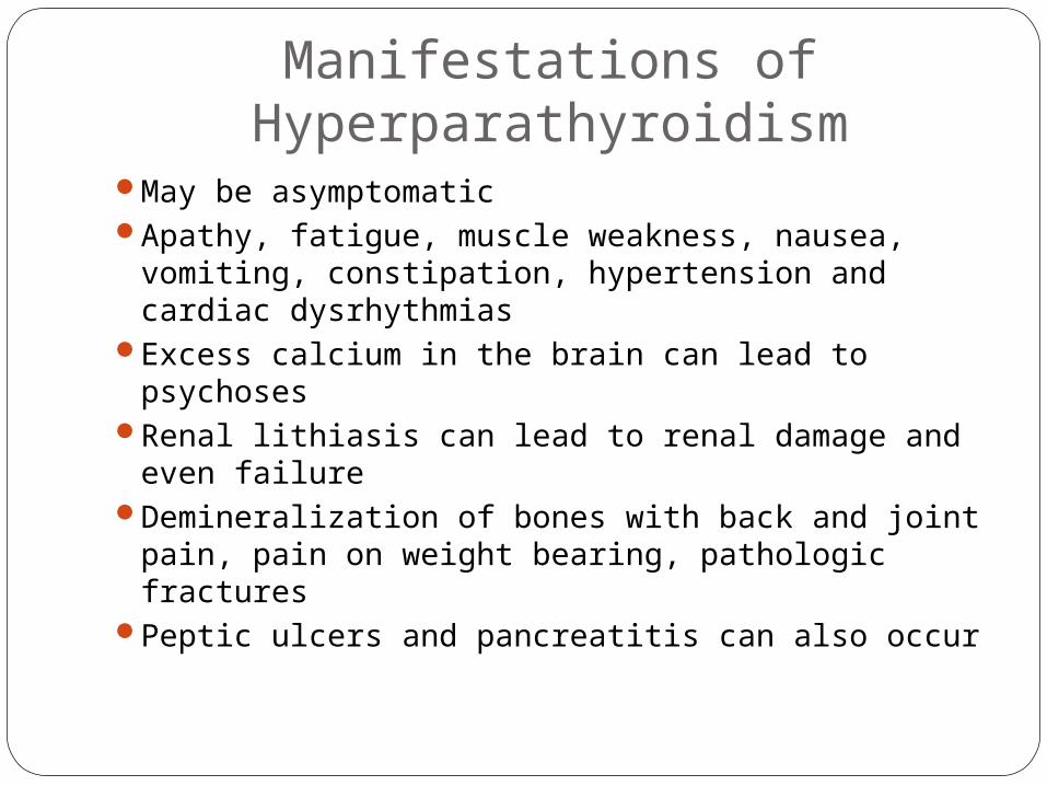
Manifestations of Hyperparathyroidism
May be asymptomaticApathy, fatigue, muscle weakness, nausea, vomiting,
constipation, hypertension and cardiac dysrhythmiasExcess calcium in the brain can lead to psychosesRenal lithiasis can lead to renal damage and even
failureDemineralization of bones with back and joint pain,
pain on weight bearing, pathologic fracturesPeptic ulcers and pancreatitis can also occur

Assessment and Diagnostic FindingsPersistent elevated calcium levelsElevated serum parathormone levelBone studies will reveal decreased densityDouble antibody parathyroid hormone test is used to
distinguish between primary hyperparathyroidism and malignancy
Ultrasound, MRI, thallium scan, fine needle biopsy also can be used to localize cysts, adenomas, or hyperplasia

ManagementRecommended treatment for hyperparathyroidism is
surgical removalHydration therapy necessary to prevent renal calculiAvoid thiazide diuretics as they decrease renal excretion of
calciumIncrease mobility to promote bone retention of calciumAvoid restricted or excess calcium in the dietFluids, prune juice and stool softeners to prevent
constipationWatch for s/s of tetany postsurgically (numbness, tingling,
carpopedal spasms) as well as cardiac dysrhythmias and hypotension

Hypercalcemic crisisSeen with levels greater than 15mg/dLCan result in life-threatening neurologic,
cardiovascular and renal symptomsTreatments include: hydration, loop diuretics to
promote excretion of calcium, phosphate therapy to promote calcium deposition in bone and reducing GI absorption of calcium
Give calcitonin or mithramycin to decrease serum calcium levels quickly

HypoparathyroidismSeen most often following removal of thyroid gland,
parathyroid glands or following radical neck surgeryDeficiency of parathormone results in increased bone
phosphate and decreased blood calcium levelsIn absence of parathormone, there is decreased
intestinal absorption of dietary calcium and decreased resorption of calcium from bone and through kidney tubules
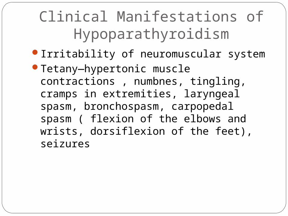
Clinical Manifestations of Hypoparathyroidism
Irritability of neuromuscular systemTetany—hypertonic muscle contractions , numbnes,
tingling, cramps in extremities, laryngeal spasm, bronchospasm, carpopedal spasm ( flexion of the elbows and wrists, dorsiflexion of the feet), seizures

Assessment and Diagnostic FindingsTrousseau’s sign—can check with a BP cuffChvostek’s sign—tapping over facial nerve causes
spasm of the mouth, nose and eyeLab studies may reveal calcium levels of 5-6 mg/dL or
lowerSerum phosphate levels will be decreased

Management of HypoparathyroidismRestore calcium level to 9-10 mg/dLMay need to give IV calcium gluconate for immediate
treatmentUse of parathormone IV reserved for extreme
situations due to the probability of allergic reactionsMonitor calcium levelsMay need bronchodilators and even ventilator
assistanceDiet high in calcium and low in phosphorus; thus,
avoid milk products, egg yolk and spinach.

Management of HypoparathyroidismKeep calcium gluconate at bedsideEnsure has IV accessCardiac monitoringCare of postoperative patients who have undergone
thyroid surgery, parathyroidectomy or radical neck surgery. Be watchful for signs of tetany, seizures, and respiratory difficulties

Adrenals--PheochromocytomaUsually benign tumorOriginates from the chromaffin cells of the adrenal
medullaAny age but usu. Between 40-50 years oldCan be familial10% are malignantMay be associated with thyroid carcinoma or
parathyroid hyperplasia or tumor

Clinical ManifestationsHeadache, diaphoresis, palpitations, hypertensionMay have hyperglycemia related to excess epinephrine
secretionTremors, flushing and anxiety as wellBlurring of visionFeeling of impending doomBPs exceeding 250/150 have occurred

Assessment and Diagnostic FindingsAssociated with the 5 H’s—hypertension, headache,
hyperhidrosis, hypermetabolism and hyperglycemiaUrinary catecholamines and metanephrine are direct and
conclusive testsSerum epinephrine and norepinephrine levels will be
elevatedUrinary vanillymandelic acid also diagnosticMust avoid coffee, tea, bananas, chocolate, vanilla and
ASA, nicotine, amphetamines, decongestants before 24h urine testing
Clonidine suppression test—in normal individual, would block catecholamine release
Imaging studies

ManagementBedrestElevated HOBICUNiprideCalcium channel blockers and Beta blockers Surgical management (manipulation of the tumor can
cause excessive release of catecholamines)Steroid therapy if adrenalectomy performedHypotension and hypoglycemia can occur post-op
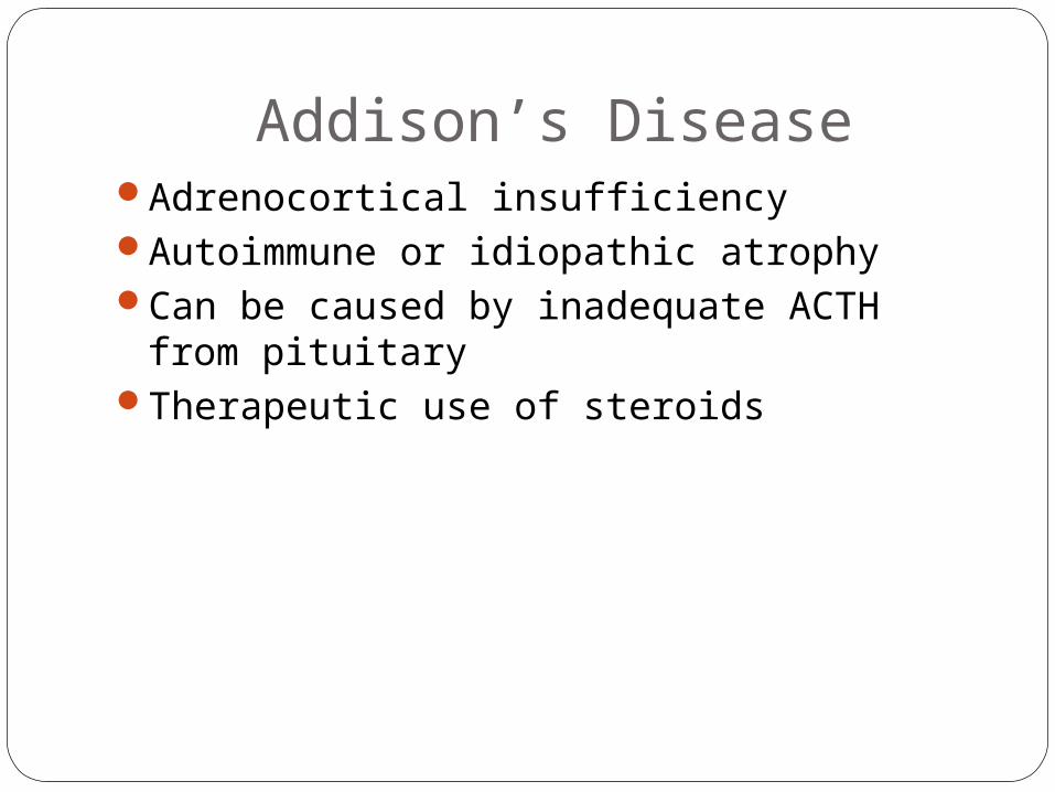
Addison’s DiseaseAdrenocortical insufficiencyAutoimmune or idiopathic atrophyCan be caused by inadequate ACTH from pituitaryTherapeutic use of steroids

ManifestationsMuscle weaknessAnorexiaDark pigmentationHypotensionHypoglycemiaLow sodium levelsHigh potassium levelsCan result in Addisonian crisis

Addisonian crisisCirculatory shockPallor, apprehension, weak&rapid pulse, rapid
respirations and low blood pressureHeadache, nausea, abdominal pain and diarrheaCan be brought on by overexertion, exposure to cold,
acute infection, decrease in salt intake

Assessment and Diagnostic FindingsEarly morning serum cortisol and plasma ACTH are
performed. Will distinguish between primary and secondary adrenal insufficiency. In primary, will have elevated ACTH levels and below normal cortisol levels.
If the adrenal cortex is not stimulated by the pituitary, a normal response to doses of exogenous ACTH (see text)
Blood sugar levels and electrolyte values

ManagementRestore circulatory status—fluids, steroidsMay need antibiotics if infection precipitated crisisMay need lifelong steroid therapy and
mineralocorticoid therapyMay need additional salt intakeCheck orthostaticsDaily weightsAware that stressors can precipitate crisesMedic alert bracelet or similar identification of history

Cushing’s SyndromeResults from excessive adrenocortical activityMay be related to excessive use of corticosteroid
medications or due to hyperplasia of the adrenal cortexOversecretion of corticosteroids can also be caused by
pituitary tumorCan be caused by bronchogenic carcinoma or other
malignancy

Manifestations of Cushing’s syndrome
Cataracts, glaucomaHypertension, heart failureTruncal obesity, moon face, buffalo hump, sodium
retention, hypokalemia, hyperglycemia, negative nitrogen balance, altered calcium metabolism
Decreased inflammatory responses, impaired wound healing, increased susceptibility to infections
Osteoporosis, compression fracturesPeptic ulcers, pancreatitisThinning of skin, striae, acneMood alterations

Assessment and Diagnostic FindingsOvernight dexamethasone suppression test frequently
used for diagnosisAdministered at 11pm and cortisol level checked at
8amSuppression of cortisol to less than 5mg/dL indicates
normal functioningMeasurement of plasma ACTH (radioimmunoassay) in
conjunction with dexamethasone suppression test helps distinguish pituitary vs. ectopic sites of ACTH.
MRI, CT and CT also help detect tumors of adrenal or pituitary

Medical ManagementIf pituitary source, may warrant transphenoidal
hypophysectomyRadiation of pituitary also appropriateAdrenalectomy may be needed in case of adrenal
hypertrophyTemporary replacement therapy with hydrocortisone or
FlorinefAdrenal enzyme reducers may be indicated if source if
ectopic and inoperable. Examples include: ketoconazole, mitotane and metyrapone.
If cause is r/t excessive steroid therapy, tapering slowly to a minimum dosage may be appropriate.

Primary Aldosteronism or Conn’s Syndrome
Excessive aldosterone secondary to adrenal tumor retain sodium and excrete potassiumResults in alkalosisHypertension—universal sign of hyperaldosteronismInability of kidneys to concentrate the urineSerum becomes concentratedExcessive thirstHypokalemia interferes with insulin secretion thus will
have glucose intolerance as well

Assessment and Diagnostic FindingsHigh sodiumLow potassium levelHigh serum aldosterone levelLow renin levelAldosterone excretion rate after salt loading is
diagnostic for primary aldosteronismRenin-aldosterone stimulation test

Management
Surgical removal of tumorCorrect hypokalemiaUsual postoperative care with abdominal surgeryAdminister steroidsFluidsMonitoring of blood sugarControl of hypertension with spironolactone
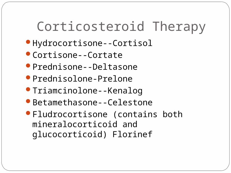
Corticosteroid TherapyHydrocortisone--CortisolCortisone--CortatePrednisone--DeltasonePrednisolone-PreloneTriamcinolone--KenalogBetamethasone--CelestoneFludrocortisone (contains both mineralocorticoid and
glucocorticoid) Florinef

IndicationsRAAsthmaMSCOPD exacerbationsLupusOther autoimmune disordersDermatologic disorders

DosingLowest doseLimited durationBest time to give dose is in early morning between 7-8
amNeed to taper off med to allow normal return of renal
function

Side Effects of SteroidsHypertension, thrombophlebitis, accelerated
atherosclerosisIncreased risk of infectionGlaucoma and corneal lesionsMuscle wasting, poor wound healing, osteoporosis,
pathologic fracturesHyperglycemia, steroid withdrawal syndromeMoon face, weight gain, acne

Case Study 135 year old male presents with BP of 188/112 at a
yearly physical exam. Previous exams noted blood pressures of 160/94 and 158/92. On questioning, patient admits to twice a month episodes of apprehension, severe headache, perspiration, rapid heartbeat, and facial pallor. These episodes had an abrupt onset and lasted 10-15 minutes.
Routine hematology and chemistry studies are wnl and chest xray and ECG are normal.
What is your impression?What labs would you draw?

Case Study 250 year old woman presents with enlargement of left
anterior neck. She has noted increased appetite over the past month with no weight gain, and more frequent bowel movements over the same period. Patient feels jittery at times, experiences palpitations and feels “hot” a lot recently.
She is 5’8” tall and weighs 150#. Heart rate is 110 and blood pressure is 110/76.
What might be this patient’s problem?What lab tests might you draw?

Case study 348 year old woman with a past history of mental
illness presents with a new onset of bizarre psychotic behavior. She had been well over the past two years.
She is 5’5” tall and weighs 138#. Her heart rate is 65, irreg and BP is 130/75. Exam is normal except that she is confused to place, time and year. Patient c/o joints aching and of feeling fatigued.
Lab tests reveal serum calcium level of 13.8mg/dL (reference range is 8.4-10.1)
Phosphorus is 2.4 (reference range is 2.5-4.5)What is your diagnosis?

Case Study 440 year old deeply tanned woman presents with a 6
month history of increasing fatigue. For the past three months she has suffered from recurrent URIs, poor appetite, abdominal cramps, fatigue and diarrhea. She has lost 25#. She has noted joint pains, muscle weakness, and has not menstruated for the past 3 months.
Labs reveal blood glucose of 59, Na+ 130, K+ 6.0.What disorder do you expect?

Case Study #527 year old woman presents with depression, insomnia,
increased facial fullness and recent increase in acne. She had an episode of depression and acute psychosis following uncomplicated delivery of normal baby boy 9 months previously. Her menses have been irregular since their resumption after the birth (she is not breast feeding). Patient relates has had several vaginal yeast infections recently.
Heart rate is 90bpm, BP is 146/100. Her face is puffy and has acne vulgaris. Thin extremities and with truncal obesity.
What are your suspicions?What labs will you draw?