
ashtma
51
View with images and charts Asthma a Common Disease INTRODUCTION Background Bronchial asthma is a chronic inflammatory disorder of airways. It is a major public health problem and important cause of morbidity and mortality. It is widely distributed but variable in the prevalence. Around 300 million people in the world currently have asthma. It is estimated that there may be an additional 100 million people with asthma by 2025 (Masoli, et al. 2004). In Bangladesh, according to First National Asthma Prevalence Study (NAPSI 1999) about 7 million- people (5.2% of - population) are suffering from current asthma (at least three episodes of asthma attack in last 12 months). Unfortunately, majority of these patients are in 1-15 years of age group that is 7.4% of total pediatric population of our country is suffering from asthma. The disease causes physical, emotional and financial sufferings for patients leading to deleterious effect on the overall socio-economic structure of the country. Asthma accounts for about 1 in every 250 deaths worldwide although modern management, which obviously includes patient education, can prevent 80% of such death. The economic cost of asthma is considerable both in terms of direct medical costs (such as hospital admissions and cost of pharmaceuticals) and indirect medical costs (such as loss of work-time and premature death). Over 18 million working days are lost due to asthma each year (Masoli, et al. 2004). The diagnosis of asthma is a clinical one; there is no confirmatory diagnostic blood test, radiographic or histopathlogical investigation. In some people the diagnosis can be corroborated by suggestive changes in lung function tests. The clinical diagnosis of asthma is not always simple. Some of the symptoms of asthma are shared with diseases of other systems. People with asthma may suffer form a variety of
-
Upload
regan-rose -
Category
Documents
-
view
213 -
download
1
description
Â
Transcript of ashtma
View with images and charts
Asthma a Common Disease
INTRODUCTION
BackgroundBronchial asthma is a chronic inflammatory disorder of airways. It is a major public health problem and important cause of morbidity and mortality. It is widely distributed but variable in the prevalence. Around 300 million people in the world currently have asthma. It is estimated that there may be an additional 100 million people with asthma by 2025 (Masoli, et al. 2004).
In Bangladesh, according to First National Asthma Prevalence Study (NAPSI 1999) about 7 million- people (5.2% of - population) are suffering from current asthma (at least three episodes of asthma attack in last 12 months). Unfortunately, majority of these patients are in 1-15 years of age group that is 7.4% of total pediatric population of our country is suffering from asthma.
The disease causes physical, emotional and financial sufferings for patients leading to deleterious effect on the overall socio-economic structure of the country. Asthma accounts for about 1 in every 250 deaths worldwide although modern management, which obviously includes patient education, can prevent 80% of such death.
The economic cost of asthma is considerable both in terms of direct medical costs (such as hospital admissions and cost of pharmaceuticals) and indirect medical costs (such as loss of work-time and premature death). Over 18 million working days are lost due to asthma each year (Masoli, et al. 2004).
The diagnosis of asthma is a clinical one; there is no confirmatory diagnostic blood test, radiographic or histopathlogical investigation. In some people the diagnosis can be corroborated by suggestive changes in lung function tests. The clinical diagnosis of asthma is not always simple. Some of the symptoms of asthma are shared with diseases of other systems. People with asthma may suffer form a variety of symptoms, none of which is specific for asthma. These are: wheeze, chest tightness, shortness of breath and cough.
The aims of treatments are, to abolish symptoms, to restore normal or best possible long-term airway function, to reduce the risk of severe attacks, to enable normal growth to occur in children, to minimize absence from school or, employment, patient and family participation, avoidance of identified causes where possible, use of lowest effective doses of convenient medications minimizing short-term and long-term side effects (Compton et al 2000).
Acute asthma represent 4% of all emergency department visit involving about 2 million people to 25% of the emergency department visit for acute asthma result in hospitalization. About 20-30% of the patient initially managed and discharged from emergency department have relapse. Globally an increase in the asthma mortality has been noticed over the past 15 years. From 1984 the national hospitalization in USA rate for asthmatic children increased

by 17%. The national death rate for asthma in children and adult are more than doubled from 1975 to 1995 (MMWR 1998). In UK, death from asthman rose steadily during 1980s, peaked in 1988 and have been falling during 1990s. However admission to hospital for acute severe asthma has not decreases and studies of asthma deaths, near total asthma and asthma admission reveal potentially preventable or avoidable factor in more than 75% of patient. Currently the cornerstone of the therapy for acute asthma is the rapid reversal of the patients airways obstruction. The main stay therapy for acute exacerbation is nebulized B2 agonist therapy repeatedly every 20 minute for one hour (serial 3 nebulisation) as initital therapy (National Asthma Education and Prevention Program, EPR-2, 1997). In the emergency department theophylline is not recommended because it appear to provide no additional benefit to optimum inhaled B 2 agonist therapy and steroid and increase the adverse effect (National Asthma Education & Prevention Program EPR-2, 1997). It has narrow therapeutic index and frequently associated with adverse effect even in therapeutic dose. Patient dose requirement varies to keep plasma level therapeutic (Seaton et al 2000).
Intravenous theophyllin is less effective than nebulized B 2 agonist and should therefore be reserved for the few patients who fail to respond to B 2 agonist. Patient like severe acute asthma is hypoxaemic and struggling to breath as a result of severe bronchoconstriction. Despite the refinement in therapeutic strategy for acute asthma emergency department visit and hospitalization continue to account for predominant proportion of health care costs for asthma. These facts stress the need for the innovative emergency department based intervention. RATIONALEPatients with acute severe asthma, the main target of treatment is to rapid relief of symptoms, rapid improvement of pulmonary function, to reduce the rate of hospital admissions, substantial reduction in costs of treatment and load on health care delivery system. Beta2
agonists have been shown to be effective and are recommended as first-line treatment for tese acute episodes Salbutamol is a B2 agonist and manufactured as a mixture containing ‘R’ salbutamol and ‘S’ salbutamol. Clinical studies with lev (R) Salbutamol have demonstrated that it binds with B2 adrenoceptor, promoting formation of cAMP and bronchodilation whereas ‘S’ salbutamol has been considered to be less effective. (Templeton et al 1998). It has been shown to exert paradoxical reactions in airway and may decrease asthma control. Furthermore metabolic clearance rate for ‘S’ salbutamol is ten fold slower than that for ‘R’ salbutamol, which may result in accumulation of ‘S’ salbutamol and subsequent potential adverse effects in asthma control.
Internationally there are a number of studies comparing various parameters of levsalbutmol and salbutamol. Nowak at al (2004) carried out a prospective, open lebel, non randomized pilot study to evaluate efficacy and tolerability of levsalbutamol in acute asthma. Asthmatics (FEV1, 20-55% predicted) were sequentially enrolled in this study .63, 1.25, 2.5, 3.75 or 5.0 mg levsalbutamol and 2.5 or 5 mg racemic salbutamol administered 3 times every 20 minutes interval. After the first dose FEV1 changes were 56% for 1.25 mg levsalbutomal and 6% and 14% for 2.5 mg and 5mg salbutamol respectively. Levsalbutamol greater than 1.25mg did not
further improve bronchodilation. There were a number of limitations associated with this study, most notably the open-label design and the small number of patients in each cohort. At the time the study was designed, high, cumulative doses of levsalbutamol administered as the single isomer in an acute setting had not been tested. Parameters like oxygen saturation, heart rate, respiratory rate were not considered. Truitt et al. (2003) conducted a retrospective study at Halifax regional Hospital to compare clinical efficacy, patient outcomes and medical costs in patients treated with levsalbutamol to those treated with racemic salbutamol. Clinical effcacy was evaluated by the number of nebulizer treatments, improvement in symptoms and objective clinical findings, the length of hospital stay, and hospital dischanrge disposition. Hefound that levsalbutamol treated patients required significantly fewer treatments, the mean length of hospital stay in the levsalbutamol group was almost 1 day less than racemic salbutamol group. Significantly more patients were readmitted to the hospital within 30 days in the racemic salbutamol group compare with levsalbutamol group. This was a review type of study and insitutional board approval was not obtained for this retrospective chart review.
In a study carl et al (2003) showed that levsalbutamol resulated in few hospital admission than racemic salbutamol when used for treatment of acute asthma. He carriedout a randomized, double blind, controlled trail study in the emergency department of an urban tertiary hospital. Patients received a nebulized solution of either 2.5 mg racemic salbutamol or 1.25mg levsalbutamol every 20 minutes interval. He showed that hospitalization rate was significantly lower in the levsalbutamol group (36%) than in the racemic salbutamol group (45%, P = .02). In this study hospitalization rate was the primary outcome. Other parameters like bronchodilation, oxygen saturation, heart rate respiratory rate were not included in this study. The study population was children (1 to 18 years).
Milgrim et al (2001) carried out a multicenter, randomized, double blinded study and showed that levsalbutomol results in improved safety and efficacy in children. Asthmatic children aged 4 to 11 years participated in this study and received nebulized levsalbutamol (.63 mg), racemic salbutamol (1.25 or 25mg) or placebo. The primary endpoint was FEV1. All active treatments significantly imporved FEV1 in comparison with placebo ( P <.001). This study had some limitations. The study population was children ( 4 to 11 years) The dose of levsalbutamol was small (.63 mg). Primary end point and vital signs might not be similar to adult patients. It demands a further study.
In another study schreck et al (2005) compared emergency department admission rates of patients presenting with acute asthma who were treated with either levsalbutamol or racemic salbutamol. It was a retrospective observational case review type of study. Patients presenting with acute asthma at 2 different sites were reviewed over 9 and 3 month conscutive periods. Outcome measures included emergency department hospital admission rate, length of stay and treatment costs. He found that significantly fewer admissions ( P = .0016) were observed in the levsalbutamol vs racemic salbutamol group. Treatment costs were lower with levsalbutamol mainly because of a decrease in hospital admissions. Limitations of this study include its retrospective design, lack of standardization in treatment regimens, smaller number of patients in the levsalbutamol group relative to the racemic salbutamol group, and lack of postdosing effiecacy comparisons regarding parameters such as peak expiratory flow rates and respiratory rates. Because of the retrospective design, no cause nad effect conclusuions can be drawn from the data.
Nelson et al. (1998) compared 2 doses of single enantiomer, levsalbutamol and equivalent amounts of levsalbutamol administered as racemic salbutamol with placebo in patients with moderate to severe asthma and he found in that the changes in peak FEV1 response to the first dose in levsalbutamol group was significantly greater compared with the racemic salbutamol with similar but non-significant results after 4 weeks and he concluded that levsalbutamol appears to provide a better therapeutic index that the standard dose of racemic saluutamol. in this study both. children and adult patients were included. Though there are a number of study internationaly regarding levsalbutamol and salbutamol in acute severe asthma there is no study in our perspective. Here, I went to investigate a multidose protocol of levsalbutamol and salbutamol inhalation in adult patients with acute asthma (PEF 35-50%). This multi-dose protocol will be cheap, easy to administer by the patient, reduce the load on health care delivery system and effectively control symptom and pulmonary function.
HYPOTHESIS
Levsalbutamol is superior to salbutamol for adult patient and with acute asthma.
OBJECTIVES
General objective:
To formulate an alternative effective first line therapy for adult patients with acute asthma.
Specific objective:
To compare effectiveness of multi-dose regime of levsalbutamol to salbutamol as a first-line therapy in adult patients with acute asthma.
REVIEW OF LITERATURE
ASTHMA
Asthma is a chronic inflammatory disorder causing hyper-responsiveness of the airways to certain stimuli resulting in recurrent variable airflow limitation, at least partly reversible, presenting as wheezing, breathlessness, chest tightness and coughing (National guideline-Asthma, Bronchiolitis and COPD, 2005).
HISTORY
The word asthma is derived from the Greek aazein, meaning “sharp breath”. The word first appears in Homer’s Iliad (Markato et al. 1982); Hippocrates was the first to use it in reference to the medical condition. Hippocrates thought that the spasms associated with asthma were more likely to occur in tailors, and metal workers.
Six centuries later, Galen wrote much about asthma, noting that it was caused by partial or complete bronchial obstruction. Moses Maimonides, an influential medical rabbi, philosopher, and physician, wrote a treatise on asthma, describing its prevention, diagnosis, and treatment (Rosner et al. 2002). In the 17 th century, Bernardino Ramazzini noted a connection between asthma and organic dust.

The use of bronchodilators started in 1901, but it was not until the 1960s that the inflammatory component of asthma was recognized, the anti-inflammatory medications were added to the regimen (Wikipedia, 2006).
ACUTE ASTHMA
Acute asthma is characterized by a progressive increase in shortness of breath, cough, wheezing or chest tightness, and by a decrease in expiratory airflow that can be quantified by simple measures of pulmonary function such as the peak expiratory flow rate (PEFR) and FEV1 (Rodrigo and Rodrigo, 2004).
Epidemiology and cost
Acute asthma is a common medical emergency faced by emergency department (ED) and intensive care specialists. In the United States, asthma represents the 11 th most frequent ED diagnosis nationwide, and adolescents and young adults are the most likely age groups to visit the ED for treatment. Women visit the ED and are hospitalized for acute asthma twice as often as men. Previous data suggested that 40% of these hospitalizations occur during the premenstrual phase of the cycle. Men are less likely than women to report severe asthma symptoms and activity limitations in the presence of airway obstruction. Data from Australia (Mellis et al. 1991), Canada (Krahn et al. 1996) and Spain reported that acute asthma accounted for 1 to 12% of all adult ED visits.
Of the 1.5 million ED visits by asthma patients in 1995 in United States, 20 to 30% of patients required hospital admission. Rates of hospital admission for female patients and blacks were consistently higher than for male patients whites. However, in the past decade there has been a decline in the number of patients with acute severe asthma requiring ICU admission, and a trend toward less advanced presentations with reduced level of respiratory acidosis and decreased ICU length of stay. In one large tertiary care hospital, only 4% of asthma admissions required ICU care over a 10-year period. In another report, 7% of adult acute asthma patients who presented to 37 EDs in France were transferred to an ICU. Probably, most hospitalizations, including those requiring ICU care, are preventable (Salmeron et al. 2001).
Developed economies might expect 1 of total health-care expenditures to be spent on asthma. The US studies estimated that the total burden to asthma was approximately $6 billion per year (Weiss et al. 1992). Direct costs (providing health care to asthmatics) rather than indirect costs (missed work, additional child care, etc.) represented the greater part (almost 90%) of the total societal cost. Together, hospitalizations and ED visits represent the single greatest cost category, accounting for almost 50% of the total cost overall. The average annual cost per patient who had an attack was $600, compared with $170 for those who did not, an increase of >3.5 times (Hoskins et al. 2000). Only approximately 20% of asthmatics have ever been admitted to an ED or hospital, yet these patients account for >80% of total direct costs (“high-cost patients”). The estimated annual per patient cost for those high-cost patients was $2,500, in contrast with $140 for the rest. These estimations indicate that hospitalization and ED visits account for the largest proportion of costs, and represent the area with the principal potential for savings.
PATHOPHYSIOLOGY

Different triggers cause asthma exacerbations by inducing airway inflammation or provoking acute bronchospasm or both. Triggers vary from person to person and from time to time. Exposure to indoor and outdoor allergens, air pollutants, respiratory tract infections (primarily viral), exercise, weather changes, foods, additives, drugs and extreme emotional expressions are the main triggers identified clinically. Other factors that may cause exacerbations are rhinitis, bacterial sinusitis, polyposis, menstruation, gastro-esophageal reflux, and pregnancy (Rodrigo and Rodrigo, 2004).
The mechanisms of acute airflow limitation vary according to the stimulus. Allergen-induced brochoconstriction results from the IgE-dependent release from airway mast cells of mediators, including histamine, prostaglandins, and leukotrienes, that contract the smooth muscle (Holgate et al. 1993). Acute airflow limitations may also occur because airways in asthma are hyper-responsive to a wide variety of stimuli. In this case, the mechanisms for causing bronchoconstriction consist in combinations of release of mediators from inflammatory cells and stimulation of local and central neural reflexes. Finally, airflow limitation results from edematous swelling of the airway wall with or without smooth-muscle contraction. The increase in microvascular permeability and leakage leads to the mucosal thickening and swelling of the airway outside the smooth muscle.
Progressive airway narrowing due to airway inflammation and/or increased bronchiolar smooth-muscle tone is the hallmark of an asthma attack, and leads to increased flow resistance, pulmonary hyperinflation, and ventilation/perfusion mismatching. Without correction of the airway obstruction, respiratory failure is a consequence of increased work of breathing, gas exchange inefficiency, and respiratory muscle exhaustion (Rodrigo and Rodrigo, 2004).
Lung mechanisms and cardiopulmonary interactions associated with airflow obstruction
Airflow obstruction is the most important physiologic disturbance in acute asthma. It inhibits airflow during both inspiration and expiration and can be quantities by simple pulmonary function testing such as the PEFR and FEV1. When expiratory airflow obstruction is sufficiently severe relative to minute ventilation to prevent return of alveolar pressure to atmospheric, dynamic hyperinflation is presents. The magnitude of hyperinflation can be assessed by the degree of increase in functional residual capacity and residual volume. This phenomenon may also be apparent when the thorax is imaged, typically as large lung volumes and flattened diaphragms seen on the chest radiograph. If passive expiration is incomplete, the end-expiratory recoil pressure of the respiratory system results in a positive end-expiratory pressure (PEFPi) analogous to positive end-expiratory pressure (PEEP) set by the ventilator-dependent patients (Pepe & Marine 1982). While the level of PEEPi correlates to the magnitude of hyperinflation, it is also influenced by the compliance of the respiratory system and hence can be increased by active expiratory muscle contraction, and increased elastic lung recoil.
Dynamic hyperinflation, particularly coupled to increased respiratory muscle activity, may profoundly affect cardiovascular performance (Rossi et al. 2000). Thus, lung hyperinflation increases after load on the right ventricle by increasing the length of pulmonary vessels and by direct compressive effects. With extreme inspiratory and efforts against obstructed airways, venous return and right ventricular filling increase, and this increase may be so pronounced that the intra-ventricular septum may shift toward the right ventricle, creating a conformational change of the left ventricle that functionally results in diastolic dysfunction of
left ventricular filling. Large negative pleural pressure swings may also impair left ventricular function by increasing after load. The aggregate effect of these cyclical respiratory events is to accentuate inspiratory increases in stroke volume and expiratory decreases in stroke volume. This can be measured as an increase in the pulsus paradoxus (PP), the difference between the maximal and minimal systolic arterial BP during the respiratory cycle. Finally, if the surrounding pressure of the heart continues to rise with progressive hyperinflation, there can also mechanical compression of the heart and coronary vessels that can lead to myocardial ischemia and deterioration in cardiac function (Pinsky. 2000).
Gas exchange
Mild-to-moderate hypoxemia, along with hypocapnia and respiratory alkalosis, are common arterial blood gas (ABG) findings in severe acute asthma (McFadden and Lyons. 1968). If airflow obstruction is severe and unrelieved, there may be progression to hypercapnia and metabolic acidosis, the former as a result of lactate production by the respiratory muscles exceeding clearance mechanisms. Analysis of blood gases in three published reports showed that only 13% of patients had PaCO2 between 45 mmHg and 60 mmHg and 4% had values >60 mmHg. Studies of patients with respiratory failure secondary to acute severe asthma using multiple inert gas-elimination techniques have shown that bimodality of distributions with little shunt is a characteristic feature of their gas exchange; these studies demonstrated a substantial fraction of perfusion is associated with areas of lung with low ratio. Thus, regional V/Q inequality are the most important mechanism hypoxemia. Carbon dioxide retention during AA also can be associated with V/Q inequality and alveolar hypoventilation due to respiratory muscle weakness and fatigue. Despite the amplitude and severity of V/Q mismatch, intrapulmonary shunt is marginal even in the most severe conditions. This may reflect three pathophysiologic factors: (1) airway occlusion is not complete, (2) collateral ventilation preserves ventilation in distal alveolar units, and (3) hypoxemia. These observations carry an important implication for management, since V/Q mismatch is the predominant gas exchange abnormality. Patients with even severe asthma can be readily oxygenated with supplemental oxygen at concentrations of 28 to 32%. When patients with AA appear refractory to supplemental oxygen, other abnormalities such as pneumonia should be sought. Multiple mediators are implicated in the V/Q mismatch. Recently, platelet-activating factor has been postulated as a cause that could disturb pulmonary gas exchange (Acuna et al. 2002).
When patients become asymptomatic, FEV1 tends to be at least 40 to 50% of predicted; when physical signs disappear, FEV1 is typically 60 to 70% of predicted or higher (McFedden et al. 1973). Because pulmonary function and ABGs assess two different pathophysiologic mechanisms, it is not surprising that correlations between FEV1 and PaCO2 or PaO2 are poor. The observation that signs and symptoms of acute asthma may resolve and spirometric measures may improve significantly while hypoxemia persists is consistent with the notion that bronchodilator therapy achieves early relief of bronchospasm of large central airways, while small airway inflammation persist with associated V/Q mismatching and hypoxemia. Finally, the combination of acute hypercapnia and high intrathoracic prssures in the patient with severe AA can produce a significant rise in intracranial pressure. Thus, there are several published clinical reports of patients who showed neurological signs such as unilateral or bilateral mydriasis and quadriparesis during an acute episode, and subarachnoid and subconjunctival hemorrhage have been described as well.
ASTHMA ATTACK EVOLUTION

There are two different pathogenic scenarios involved in the asthma attack progression. When airway inflammation is predominant, patients show a progressive (over many hours, days, or even weeks) clinical and functional deterioration (type 1 or slow-onset acute asthma). Data from different cohort studies (Kolbe et al. 1998) showed that the prevalence of this type of asthma progression is between 80% and 90% of adults with acute asthma who presented to an ED. Upper respiratory tract infections were frequent triggers, and these patients exhibited a slow therapeutic response. Also, they may have allergic inflammation with eosinophils in the airways. In the less common asthma progression scenarior, bronchospasm is predominant and patients presenting with a sudden-onset asthma attack (type 2 or asphyxic or hyperacute asthma) characterized by rapid development of airway obstruction (<3 to 6 h after the onset of the attack).
Respiratory allergens, exercise, and psychosocial stress are the most frequent triggers. Surprisingly, these patients show a more rapid and complete response to treatment.
FATAL ASTHMA
In many countries, asthma mortality increased from the 1960s to the second half of the 1980s, butt reached a plateau and has subsequently declined. This recent downward trend may reflect better management of this condition in primary care. Asthma has a low mortality rate compared with other lung diseases, butt mortality does occur, typically in patients with poorly controlled disease whose condition gradually deteriorates over a period of days or even weeks before the fatal attack. Occurrence of this type of asthma progression is between 60% and 90% of adults with acute asthma who have fatal and near-fatal crises (Plazza et al. 2002). This observation suggests many patients have a window of opportunity for recognition and reversal of this period of deterioration. Infrequently, death occurs suddenly. Accordingly, most deaths are preventable, and a useful practice is to assume that every exacerbation is potentially fatal (McFadden et al. 1997).
Two hypotheses have been postulated for the cause of asthma-related deaths. Cardiac arrhythmias may contribute to some of the observed mortality, particularly in adults. The risk is theoretically increased by hypokalemia and prolongation of the QTc interval coupled to the use of -agonists in high doses (Rodrigo and Rodrigo, 2002). However, in a series of patients with near-fatal attacks, few arrhythmias other than sinus tachycardias and bradycardias were found. A more likely hypothesis is that deaths occur as a result of asphyxia due to severe limitation of airflow and hypoxemia. This hypothesis has received support from the pathologic evidence indicating that patients with fatal asthma almost invariably have extensive airway obstruction, with mucous plugging and dynamic hyperinflation apparent even at autopsy. Data suggest it is very uncommon to die without substantial luminal obstruction. Smooth-muscle contraction and production of inflammatory mucus exudates are important mechanisms for fatal attacks in young and old individuals with asthma.
EMERGENCY DEPARTMENT MANAGEMENT
Assessment
Acute asthma is a medical emergency that must be diagnosed and treated urgently. The assessment of an asthma exacerbation constitutes a process with two different dimensions: (1) a static assessment to determine the severity of attack, and (2) a dynamic assessment to evaluate the response to treatment (Rodrigo and Rodrigo, 2000a).

Medical History:
A brief history pertinent to any exacerbation should be obtained. The objectives are to determine time of onset and severity of symptoms, especially compared with previous exacerbations, all current medications, prior hospitalizations and ED visits, prior episodes of respiratory failure (intubations, mechanical ventilation), and psychiatric or psychological disorder. The existence of such events has been associated with poor outcomes, but their absence does not ensure low risk (Molfino et al. 2000).
A number of conditions may mimic or complicate the diagnosis of AA. The absence of a history of asthma, particularly in an adult, should alert the ED physician to an alternative diagnosis (Rodrigo and Rodrigo et al. 2000b). Congestive heart failure, particularly predominant left ventricular failure or mitral stenosis, occasionally may present with episodic shortness of breath accompanied by wheezing. Perhaps the most common and most difficult diagnostic problem in asthma is its differentiation from COPD. In subjects >40 years of age, a distinction between COPD and asthma is often difficult, if not impossible. Laryngeal/tracheal/bronchial obstruction resulting from any of a number of causes may produce shortness of breath, localized wheezing, inspiratory stridor localized over the trachea, or unilateral hyperinflation noted on chest radiography, which often mimics asthma. Recurrent small pulmonary emboli may be manifested by attacks of shortness of breath and very rarely, wheezing heard on careful auscultation. Finally, recurrent attacks of shortness of breath at rest may be due to the hyperventilation syndrome.
Examination
Particular attention should be paid to the patient’s general appearance. Patients with the most severe conditions will be sitting upright. The use of accessory muscles has received attention as an indicator of severe obstruction, and the presence of sternocleidomastoid retractions or supra-sternal retraction correlated with impairment in lung function. In consequence, accessory muscle use can be considered a useful sign of severe airflow obstruction.
Respiratory rate (RR) >30 breaths/min, tachycardia >120 beats/min, or pulse pressure (PP) >12 mmHg have been described as vital signs of acute severe asthma. However, composite data from large clinical studies demonstrated that >50% of patients with acute severe asthma have heart rates ranging between 90 beats/min and 120 beats/min, with only 15% exceeding this value. In general, successful treatment of airflow obstruction is associated with a decrease in heart rate, although some improving patients remain tachycardic because of the chronotropic effects of bronchodilators. While distinguishing between asthma-related tachycardia and treatment-related tachycardia can be difficult, patients who note subjectively improved breathing but exhibit a fine tremor are likely receiving excessive -agonist dosing. Specifically, older patients tend toward treatment-related tachycardia. Respiratory rates ranges between 20 breaths/min and 30 breaths/min in >50% of patients, and are -30 breaths/min in <20% of patients (Smith et al. 2000). In severe airflow obstruction, Pulse pressure is greater than the normal value of 10 mmHg and typically >15 mmHg; however, only severe PP (>25 mmHg) was a reliable indicator of severe asthma. Finally, wheeze and dyspnea are present in virtually all patients with AA, and they correlated poorly with the degree of airflow limitation (Shim et al. 1983).
Spirometry or Peak flowmetry

The major cause of respiratory failure and fatal asthma is an underestimation of the severity of a given attack. Nevertheless, the severity of airflow obstruction cannot be accurately judged by means of symptoms and physical examination by themselves. The measurement of lung function provides a more objective assessment of obstruction, but does depend on good technique and adequate patient effort. On presentation, after the initial treatment, and at subsequent frequent intervals, it constitutes an integral part of the assessment of disease severity (static assessment) and the response to therapy (dynamic assessment) in any patient >5 years of age. Measurement of airflow obstruction should be made using one of following techniques. PEFR measured with a peak flowmeter, or FEV1 determined by spirometry. Many studies (Rodrigo and Rodrigo, 2000c) have found satisfactory correlations between both measures among healthy and stable patients or patients with acute asthma. PEFR value tended to have more variability when pulmonary function was more impaired and to underestimate the degree of pulmonary impairment. Even though spirometry is the “gold standard”, in most asthma patients, it is easier to measure PEFR than FEV1; PEFR measurement is common in the ED because it is inexpensive, portable, and safe. The typical asthmatic patient who presents for care to an ED will exhibit a broad range of PEFR and FEV1 values. Approximately 55% of patients with have values <40% of normal and one fifth will range between 40% and 60% of normal.
Pulse Oximetry
Measurement of oxygen saturation by pulse saturation by pulse oximetry (SO2) is necessary in all patients with Acute asthma to exclude hypoxemia; it allows monitoring of SO2 on a continuous basis. The measure of SO2 indicates which patients may be in respiratory failure and therefore in need of more intensive management. The goal of treatment should be to maintain Spo2 at -92% (Kelly et al. 2001).
Arterial Blood Gas Analysis
ABG determination is rarely necessary before the initiation of treatment. Because of the accuracy and utility of pulse oximetry, only patients whose oxygenation is not restored to >90% with oxygen therapy require ABG determination. When adequate oxygenation remains a problem despite supplemental oxygen, additional complicating conditions such as pneumonia should be considered. Repeated ABG sampling usually is not needed to determine whether a patient is deteriorating or improving. In most cases, valid judgments can be based on serial physical examinations and PEFR determinations (Nowak et al. 1983). In fact, the decision to proceed with endotracheal intubation and mechanical ventilation is a clinical assessment and should not be overly influenced or await ABG analysis (Tuxen et al. 2000).
Chest Radiography
Chest radiography plays only a small role in the assessment and management of patients with AA. Many studies (Findley et al. 1980) have demonstrated that the incidence of specific abnormalities on chest radiography in adults with uncomplicated acute asthma is low, and have suggested that the information obtained is rarely helpful in ED management. On the basis of these data, chest radiographs are indicated only in patients who present with signs or symptoms of pneumothorax (pleuritic chest pain, mediastinal crunch, subcutaneous emphysema, cardiovascular instability, or asymmetric breath sounds), in patients with clinical findings suggestive of pneumonia, or in an asthmatic patient who after 6 to 12 h of intensive treatment does not respond to therapy.

Cardiac Rhythm Monitoring
ECGs need not be routinely obtained, but continual monitoring is appropriate in older patients (Shim et al. 1983) and in those with coexisting heart disease. The usual rhythm is sinus tachycardia, although supra-ventricular arrhythmias are not uncommon. Frequent transient ECG findings include right-axis deviation, clockwise rotation, and evidence of right ventricular strain. If due to asthma alone, reversal within hours of response to therapy is to be expected.
Response to Therapy
Measurement of the change in PEFR or FEV1 over time may be one of the best ways to assess patients with acute asthma and predict the need for hospital admission. The response to initial treatment in the ED is better predictor of the need for hospitalization than is the severity of an exacerbation at presentation (Stein et al, 1990). Early response to treatment (PEFR or FEV1 at 30 min) is the most important predictor of outcome (Rodrigo GJ et al. 1999). PEFR variation over baseline >50 L/min and PEF>40% of normal, both measured at 30 min after beginning of treatment, are predictors of good outcome (Rodrigo and Rodrigo, 1997).
In summary, symptoms and signs guide treatment decisions, but repeated measurement of PEFR or FEV1 compared to baseline joined with continuous monitoring of Spo2 is critical to evaluate the severity of airway obstruction, the adequate of gas exchange, andd the response to treatment (Rodrigo and Rodrigo, 2004).
Treatment
The severity of asthma exacerbations determines the treatment. The goals of treatment may be summarized as maintenance of adequate arterial oxygen saturation with supplement oxygen, relieve airflow obstruction with repetitive administration of rapid acting inhaled bronchodilators (-agonists and anticholinergics) and reduce airway inflammation and to prevent future relapses with early administration of systemic corticosteroids.
Oxygen
Because hypoxemia is produced by mismatch, it is usually fully corrected with modest increase in fraction of inspired oxygen (e.g., 1 to 3 L/min by nasal cannula or mask). In spite of this, the use of high-flow oxygen has been assumed to be harmless and recommended to all patients with acute asthma (Inwald et al.). Uncontrolled oxygen has been postulated to correct the effects of hypoxemia and to compensate and trend for PaO2 to fall with -agonist therapy. Recently, the first randomized controlled study on the effect of administration of two oxygen concentrations (28% vs 100%) on gas exchange in acute asthma showed that patients receiving 28% oxygen had a fall in Paco2; opposite, patients who received 100% oxygen treatment >40 mmHg. Hyperoxia associated with hypercarbia occurring in asthma exacerbations had been explained by the treatment guidelines should be based on achieving target PaO2 and SpO2 rather than on administering predetermined concentrations or flow rates of inspired oxygen. Efforts should be made to confirm the presence of hypoxia by pulse oximetry before administering oxygen. In this way, normoxic patients will not require treatment with oxygen. The goal of treatment should be to maintain Spo2 at _92% (Kelly et al. 2001).

Humidification of the inspired air/oxygen mixtures is not currently recommended in the guidelines for the treatment of acute asthma. However, in a new small but interesting trial (Moloney et al. 2002), the authors demonstrated that significant dehydration of expired air is present in patients with an asthma attack, and that bronchoconstriction triggered by dry-air challenge in the laboratory can be prevented by humidifying the inspired air. Thus, these findings suggest that full humidification of the inspired gases should be recommended for patients with AA.
2-Adrenergic Agonist
Specific short-acting inhaled 2-agonists are the drugs of choice to treat acute asthma. Their onset of action is rapid, and their side effects are well tolerated. Salbutamol (albuterol in North America), the most frequently used drug in the EDs around the world, has an onset of action of 5 min and a duration of action of 6 h. Other used drugs are metaproterenol, terbutaline, and fenoterol. Long-acting drugs cannot be recommended for emergency treatment. Levsalbutamol (R-albuterol) may prove to be more efficacious and less toxic than racemic albuterol (Handley et al. 2000). Finally, subcutaneous epinephrine has become up to date because of its marked cardiac side effects compared with inhaled agents and should be reserved only when patients are not benefiting from inhaled medicines.
With regard to 2-agonist use, there are four areas of debate: IV vs inhaled therapy, doses and intervals of administration, pressurized metered-dose inhalers (pMDIs) with spacer vs small-volume nebulizers, and continuous vs intermittent nebulization. The inhaled route has a faster onset, fewer adverse effects, and is more effective than systemic routes. Although popular in some countries, available evidence does not support the use of IV 2-agonists in the treatment of patients with severe AA. Therefore, they should be considered only if the response to the inhaled drug is poor or if the patient is coughing excessively, is moribund, or becomes so despite inhalation therapy (Rodrigo and Rodrigo, 2004).
Doses and dosing intervals should be individualized using objective measures of airflow obstruction as guides (American Respiratory Care Foundation and American Association of Respiratory Care, 1991). A substantial body of evidence supports the use of high and repeated doses (Rodrigo and Rodrigo 2004). The aim of treatment is to induce maximal stimulation of 2 receptors without causing significant side effects. Approximately two thirds of patients will be sensitive to inhaled salbutamol, and optimal treatment for this group is 2.4 to 3.6 mg delivered by pMDI and spacer (four fuffs each 10 min) or 5 to 7.5 mg delivered by jet nebulizer. For the remainder of patients, albuterol even in high doses has little effect. A single dose of 7.5 mg of nebulized salbutamol or sequential doses of 2.5 mg of nebulized salbutamol are clinically equivalent in the treatment of patients with moderate-to-severe acute asthma; however, twice as many patients in the single-dose group experienced side effects. A more slowly resolving asthma attack likely is demonstrative of significant airway inflammation. When this is a predominant component, bronchodilator therapy tends to be ineffective and the patient will require a longer period of intensive treatment. So, if an attack does not resolve within a relatively brief period of time (2 to 3 h), it is unlikely to do so.
More rapid and profound bronchodilatation with fewer side effects and less time in the ED can be achieved when sufficient doses of 2-agonists are administered using pMIDs and large-volume spacers (valved spacer device) than when conventional doses are administered with a jet nebulizer (Newman et al. 2002). This is true particularly in patients with the most severe obstruction. Each 2-agonist treatment with a pMDI and spacer takes 1 to 2 min, as compared with 15 to 20 min for each treatment with a jet nebulizer. In our experience, the

pMDI plus spacer constitutes the only way to deliver quickly high doses of bronchodilators to patients with acute severe asthma or life-threatening asthma with reduced level of consciousness.
Continuous nebulization is thought to be more beneficial than intermittent therapy; however, a recently published meta-analysis of randomized controlled trials (Rodrigo et al. 2002) of adults with acute asthma found no significant differences between the two methods in terms of pulmonary function improvement or hospital admission; nevertheless, continuous nebulization was associated with side effects. These findings argue against the routine use of continuous nebulization in the ED treatment of patients with acute asthma.
Finally, side effects are dose dependent and can occur with all routes of administration, but are more pronounced with oral and IV routes than with inhalational delivery methods. For selective 2-adrenergic agonists, the principal side effects are mediates via receptors on vascular smooth muscle (tachycardia and tachyarrhythmia), skeletal muscle (tremor, hypokalemia due to potassium entry into muscle cells), and cells involved in lipid and carbohydrate metabolism (increase in blood-free fatty acids, insulin, glucose and pyruvate). The stimulation of 2-adrenoreceptor also seem to contribute to the pathogenesis of lactic acidosis during severe acute asthma which more commonly develops in patients receiving IV 2-agonists. It has been hypothesized that catecholaminergic stimulation of increased glycolysis and glycogenolysis is associated with increased pyruvate generation and increased conversion to lactate because of either overwhelmed or coincidentally disturbed oxidative metabolism (James et al. 1999).
Anticholinergic Drugs
The rationale for the use of anticholinergic therapy is the presumption of incrased airway vagal tone in patients with acute asthma, but the role of anticholinergics has been less well defined than 2-agonists. The role of anticholinergic bronchodilators, including ipratropium, however, in the management of acute exacerbations is less clear. Stoodley et al (1999) conducted a meta-analysis of all treatment of acute asthma exacerbation in adults. Combination therapy appeared to have greater absolute positive effects (improvement in FEV1, of 7.3 percent and an improvement in PEF rate of 22.1 percent) in patients with more severe outflow obstruction at baseline.
Corticosteroids
Systemic corticosteroids should be considered in the management of all but the mildest exacerbations of asthma. Theses agents are not bronchodilators but are extremely effective in reducing airway inflammation present in virtually all asthmatics. Despite controversy about their efficacy, route of delivery and dosage, data summarized in two systematic reviews (Rodrigo and Rodrigo, 1999; Harrison et al. 1986 and Rowe et al. 2002) suggest the following : (1) systemic corticosteroids probably require >6 to 24 h to improve pulmonary function (2) IV and oral corticosteroids appear to have equivalent effects in most patients with acute asthma (3) while precise dose-response relationships are not well described, there is a tendency toward greater and more rapid improvement in pulmonary function with medium and high doses, although these effects likely plateau without additional benefit at very high dosing; 800 mg of hydrocortisone or 160 mg of methylprednisolone in four divided doses per day are generally adequate for most patients.
The benefit of therapy, however, with systemic corticosteroids for reducing the number of relapses in patients following an acute attack is conclusive. Hence, oral corticosteroids in a dose equivalent of 40 to 60 mg prednisone or prednisolone per day during a period of 7 to 14 days is effective, cheap, and safe. However, there is some evidence that high-dose inhaled corticosteroid therapy alone may be as effective as oral corticosteroids when used in patients with mild asthma on ED discharge (Edmond et al. 2002).
Theophylline
As monotherapy, theophylline is inferior to 2-agonists in the ED treatment of acute asthma (Rossing et al. 1980) and the addition of IV aminophylline to inhaled 2-agonists does not confer significant benefit but does increase the incidence of complicating tremor, nausea, anxiety, and tachyarrhythmia (Parameswarad et al. 2002). These observations have resulted in consensus statements and guidelines that do not recommended the routine use of theophylline in the treatment of acute asthma (Inwald et al. 2001). The use of theophylline/aminophylline should be reserved only for those patients not responding to standard therapy. In these circumstances, a loading dose of 6 mg/k over 30 min followed by an infusion of 0.5 mg/kg/h with measured of theophylline blood levels is recommended (8 to 12 g/mL). In patients already receiving theophylline on presentation to the ED, a serum level should be measured and approximate dosing continued if deemed necessary.
Magnesium Sulfate
The use of magnesium for the treatment of AA was first reported in 1936 by Uruguayan physicians. One suggested mechanism of action is that by inhibiting smooth-muscle cell calcium channels magnesium blocks muscle contraction. In general, this drug is safe and inexpensive in the usual clinical dose of 1.2 to 2 gm IV over 20 min. However, three meta-analyses of trials (Rodrigo and Rodrigo 2000). Bourdon et al. 1999 and Koepsell et al. 1999) assessing the magnesium administration for acute asthma do not support routine use. Inhaled magnesium preparations have also been evaluated but show no, or clinically insignificant effects. Finally, a new and large multi-center study (Silverman et al. 2002) demonstrated that IV magnesium sulfate only improves pulmonary function when administered as an adjunct to standard therapy (nebulized 2-agonists and IV corticosteroids) in a very select subgroup of patients (FEV1 <20% of predicted).
Heliox
Under most breathing conditions, airflow in humans is largely laminar; at high gas velocities and with severe airway narrowing, increasingly turbulent airflow may occur. Turbulence has two complications in the context of acute asthma; (1) the airway resistance for any given degree of anatomic obstruction will be greater, leading to a greater work of breathing and the potential for more dynamic hyperinflation; and (2) turbulence makes the delivery of aerosolized particles to the lower airways more difficult, since gas streams impacting on airway walls can result in droplet deposition above the intended site of delivery. Turbulent airflow can be reduced by the application gases with a lower density and higher viscosity than air. Heliox (helium and oxygen) is such a gas mixture and is available for administration to patients with AA. The benefits of heliox are lost when large amounts of supplement oxygen are introduced into the heliox breathing circuit; concentrations of helium much below 70% are not likely to confer significant benefit. Fortunately, most patients with acute asthma do not require large amounts of supplemental oxygen to maintain adequate So2 (-92%). Additionally, research using heliox mixtures has demonstrated a greater percentage of lung

particle retention and a large delivery of albuterol from both pMDIs and jet nebulizers. This suggests that one of the beneficial effects of heliox use may include improved deposition of aerosolized bronchodilators. Studies have shown that heliox administered to patients with severe airflow obstruction can reduce both inspiratory and expiratory airway resistance as judged by a decrease in the PP and increase in the PEFR independent of the action of bronchodilators (Hess et al. 1999).
Leukotriene Antagonists:
There is some evidence to suggest that leukotriene modifiers, often used in the management of chronic asthma, may have a role in the treatment of AA as well. A study (Silverman et al. 2000) of two doses of oral zafirlukast (20 mg and 160 mg) in the ED demonstrated an improved in pulmonary function and dyspnea score. Also, a rapid but small improvement in FEV1 with IV montelukast vs placebo was observed in patients with AA refractory to initial 2-agonists (Camargo et al. 2003). However, additional studies are necessary to determine if these agents offer a significant benefit over and above that derived from routine therapy.
Other Therapies
Other agents have been suggested to be of benefit for AA therapy, including inhaled general anesthetics (Echevarria et al. 1986), lidocaine (Hunt et al. 1996) and inhaled furosemide (Karpel et al. 1994) however, there are inadequate data to support these therapies as anything other than experimental. Inhaled mucolytic drugs have not been shown to benefit treatment of exacerbations and they may worsen cough or airway obstruction. Finally, sedation should be strictly avoided during AA because of the respiratory depressant effect of these drugs.
Discharge or Hospitalization
Spirometry and clinical assessment are used to establish disposition decisions. “The patient should be hospitalized if, despite 2 to 3 hours of intensive treatment in the ED he or she still has significant wheezing, accessory muscle use, permanent requirement for oxygen to maintain Spo2-92%, and a persistent reduction in lung function (FEV1 or PEF -40% predicted), in much the same way that the presence of factors indicating high risk of asthma-related death (inadequate access to medical care and medications, difficult home conditions, and difficult-to-obtain transport to hospital in the event of further deterioration) would lead to a decision to hospitalize the patient (Rodrigo and Rodrigo, 1997 and Rodrigo and Rodrigo, 1999).
However, if a patient is free of symptoms and has lung functions (FEV1 or PEFR) _60 of predicted, the patient can be discharged unless other mitigating circumstances exist. Finally, patients with post-treatment lung function between these two extremes (40 to 60% of normal) require continued treatment. These patients can potentially be discharged, assuming adequate time to determine if patients with acute asthma can improve for safe discharge from the ED. Most patients discharged to home should be placed on at least a 7- to 14-day course of prednisolone while contact is made with the next tier of care providers. 2-agonist use should be able to be decreased over this time; if increasing use of “rescue” 2-agonist use is undertaken, the patient should return immediately to the ED or to primary care provider. Patients receiving long-term corticosteroid treatment should resume corticosteroids upon discharge from the Ed (Rodrigo and Rodrigo, 2004).
Admission to the ICU

Patients with findings of severe airflow obstruction who improve minimally or deteriorate despite therapy should be admitted to an ICU. Clinical markers for this include respiratory distress, high PP or a falling pulse in a patient with fatigue, or the patient’s subjective sense of impending respiratory failure. Other indications for ICU admission include respiratory arrest, altered mental status, Spo2 <90% despite supplemental oxygen, and a rising Paco2
coupled to clinical evidence of no resolution (Tuxen et al. 2000).
ICU management
Many patients admitted to the ICU with acute asthma simply require additional time for the therapies instituted in the ED to be continued and for respiratory function to improve. These patients often require a relatively brief period of time in the ICU; when improvement is clear, they can be discharged to the regular ward. Since it is not clear on ICU admission which patients will pursue such a course, the benefit of the ICU is largely one of careful observation and rapid response to those patients who do not improve and eventually require mechanical ventilatory support. The other role of the ICU for patients with acute asthma is to care for those patients with respiratory arrest out of the hospital or shortly after arrival in the ED.
Non-invasive Positive Pressure Ventilation
While non-invasive positive pressure ventilation (NIPPV) has revolutionized the management of patients with diverse forms of respiratory failure (Brochard et al. 2002). In one study of feasibility (Martin et al. 1982), continuous positive airway pressure (CPAP) at a level of approximately 12 cm H2O was administered to asthmatic subjects in whom bronchospasm was induced by aerosolized histamine. Under these controlled circumstances, CPAP was demonstrated to raise residual decrease intrathoracic pressure swings associated with tidal breathing and diminish the inspiratory work. This potentially beneficial effect of positive pressure in spontaneously breathing asthmatics was thought to result from a reduction in the increased work of breathing associated with initiation of inspiratory flow under conditions of dynamic hyperinflation, e.g., a reduction in inspiratory threshold load. Meduri and colleagues reported 17 episodes of NIPPV use in asthma. NIPPV was associated with a reduction in Paco2 and improvement in dyspnea over the early hours of use. Two patients required intubation and no complications related to NIPPV were noted.
A course of NIPPV seems warranted in patients at risk for evolution of respiratory failure or as an alternative to intubation. Based on experience with this modality of treatment for other forms of respiratory failure, early use when the patient is first identified as at high risk, including the ED, is appropriate. Successful use of NIPPV depends on patient education and coordination with the breathing circuit, things that are more easily achieved when the patient is not in extremis. Uncooperative and obtunded patients can rarely if ever be stabilized with NIPPV and are at risk for complications such as aspiration. Patients should be given a well-fitting mask and initially instructed to hold it to their face before securing it with straps to provide for brief accommodation. Initial ventilator settings should be an expiratory positive airway pressure, CPAP, or PEEP of approximately 5 cm H2O and inspiratory pressure (or pressure support) of approximately 8 cm H2O. If the patient has difficulty during inspiration triggering breaths, expiratory positive airway pressure can be gradually increased. If tidal volumes (VTS) are shallow (<7 mL/kg), inspiratory pressure can be increased. Rarely will patients tolerate total pressures >15 to 20 cm H2O without mas leaks or a sense of claustrophobia or mask discomfort. When improvement is dramatic and has persisted for hours, episodic removal of the mask or reduction in support pressures can be attempted. If patients do not clearly improve or appear to be of marginal status with NIPPV, it should be
appreciated that removal of NIPPV may precipitate rapid deterioration and all means for intubation should be readily and personnel available to proceed to this next level treatment.
Intubation and Postintubation Stabilization
When patients deteriorate despite pharmacologic intervention with or without NIPPV, intubation should be performed electively. Intubation may be performed awake or with rapid induction. In general, nasal intubation should be avoided because of the higher incidence of sinusitis in asthmatics and the possible presence of nasal polyps. Intubation should be accomplished with the largest possible endotracheal tube for two reasons. Since the tube represents a resistance in series with the obstructed airways, dynamic hyperinflation tends to worsen intubation and this can be avoided to some extent by selecting a larger tube. Also, it is common for patients intubated for AA to mobilize large mucous plugs during recovery, which has a greater potential for causing acute obstruction in small endotracheal tubes.
Mechanical ventilation follows cardiorespiratory collapse in approximately 20% of episodes (Tuxen et al. 2000). Causative factors are pulmonary hyperinflation, hypovolemia, and sedation. In the postintubation period, dangerous levels of pulmonary hyperinflation can develop if patients are “bagged” excessively in a misguided attempt to stabilize, or resuscitate. With severe airflow obstruction, even delivery of normal minute ventilation may cause substantial gas trapping that reduces venous return and hence cardiac output. In the same patients, hypovolemia related to previous dehydration, sedation, and muscle relaxation all act to decrease mean systemic vascular pressure, further decreasing venous return to the heart.
If the patient doest not improve with slow manual bagging, consideration should be given to a possible tension pneumothorax, and chest radiography should be performed immediately if time permits. If the more common hyperinflation is the cause of hypotension, the patient should be slowly bagged, intravascular volume administered (1 to 2 L or more is often required) and sedatives given to synchronization of the patient with the ventilator (Rodrigo and Rodrigo, 2004).
Levsalbutamol, Salbutamol and Bronchial wall
Racemic salbutamol is an equimolar mixture of (R) salbutamol and (S) salbutamol. Clinical studies with R-salbutamol have demonstrated it binds with to the 2 adrenoceptor, promoting formation of cAMP and bronchodilation whereas (S) salbutamol has been considered to be less effective agonists, (Templeton et al. 1998). It has been shown to exert biological effects on airway smooth muscle paradoxical reactions in airway and may decrease asthma control, although the mechanism whereby (S) salbutamol evokes hyperresponsiveness appears to be unrelated to be 2-adrenoceptor activation. Furthermore the metabolic clearance rate for ‘S’ salbutamol is ten fold slower than that for ‘R’ salbutamol, which may result in accumulation of (s) salbutamol and subsequent potential adverse effects in asthma control. Specifically ‘S’ salbutamol increases intracellular calcium, enhances experimental airway hyperresponsiveness to sparmogens and may have proinflammatory effects as demonstrated by eosinophil superoxide production in response to interleukin (IL-5).
In spite of many recent comparative studies regarding the effect of ‘S’ salbutamol in asthma, the role of ‘S’ salbutamol in mast cell mediator release has not been well established, mast cells are important effector cells in asthma by virtue of their capacity to respond to IgE dependent activation with release of proinflammatory mediators, including histamine,

leukotriene and cytokines. Seong H. Cho et al. 2001 shows that (s) salbutamol activates, mast cells and enhances important mast all mediators such as histamine and IL-4 and out data suggest that (s) salbutamol may have adverse effects on asthma control by increasing the production of mast cell mediators.
Levsalbutamol inhibited cell proliferation at low concentrations. the growth-inhibitory effect of Levsalbutamol occurs via activation of the cAMP/PKA pathway (Basil et al. 2006). On the other hand, in four-week study equivalent amounts of pure-levosalbutamol provided greater bronchodilatation than similar amounts of Levsalbutamol given in racemic mixtures (Nelson et al. 1999). Furthermore after 4 weeks the baseline morning FEV1 was lower in those receiving the racemate than in those receiving placebo or Levsalbutamol.
MATERIALS AND METHODS
Type of study
This is a single blind, randomized prospective study.
Place of study
The study was carried out in the Emergency Department of National Institute Diseases of Chest and Hospital, Mohakhali, Dhaka.
Period of study
The study was done during the period of December 2006 to November 2007.
Study population
Adult patient with acute asthma attending the emergency department of NIDCH during the above mentioned period were the study population.
Selection of patients
In this study of one hundred adult (18 to 50 years) patients with acute asthma attending the emergency department of NIDCH were selected. The diagnosis of acute asthma was done from history, examination and previous records of investigations (according to National Guideline for Asthma, Bronchiolitis and COPD).
The inclusion criteria for these patients were-
1. Established cases of bronchial asthma.
2. PEF 35%-50% of predictive value.
3. SO2>90%
4. Adult patient (18-50 years)
5. Informed written consent of the patients.
The exclusion criteria were-
1. Long history of smoking, COPD. Chronic cough, old TB, fever (>380C).
2. Co-morbidity-CCF, CRF, CLD malignancy.
3. Age <18 years.
4. Pregnancy.
5. Unable to blow PEF meter.
6. SO2<90% inspite of treatment with O2 and drugs.
Sample size
Total 100 patients were selected after adequate evaluation in emergency department to include in this study.
Sampling technique
Adult patients with acute asthma were consecutively evaluated to include in this study and were arranged to the two groups randomly from a computer generated random number table.
Ethical issues
This study was done in a well equipped emergency department of a super-specialized chest hospital in presence of all the remedies of emergency management. Very severe asthma defined as PEF less than 30% were excluded and only patients with PEF 35-50% were included after taking proper consent from the patient. Moreover this protocol was approved by a thesis committee.
Study design
1. In this single-blind, randomized controlled study, adult patients with acute asthma was selected consecutively from emergency department.
2. After selection of patients, a written consent was taken from the participants.
3. Patient’s demographic data, total duration of acute exacerbation total asthma suffering period and pre-medication taken to control asthma were noted in a protest questionnaire.
4. These patients were divided into two groups ‘A’ and ‘B’ with the use of a computerized random number table. Randomly selected group ‘A’ was enrolled in study and group ‘B’ was enrolled as control.
5. Study group received levosalbutamol delivered by a metered dose inhaler (MDI) into a volumetric uptech spacer device in a dose of five puffs at 10 minutes interval. The randomly selected another group received five puffs of salbutamol (100 gm/puff) at 10 minutes interval by the same measure and same device.
6. High flow oxygen was given when SO2 had fallen below 92%
7. Variables were measured immediately before starting treatment and at 20 minutes interval thereafter for one hour in each patient.

8. Peak expiratory flow (PEF, saturation of oxygen (SO2), respiratory rate (R/R), heart rate (HR), accessory muscles used, dyspnoea, wheeze were the variables that were recorded in a preformed questionnaire form (appendix-I)
9. At the end, each patient was asked for nausea, palpitation, tremor, anxiety, headache, dry mouth and if present was noted.
10. At the end every patient was assessed and those improved and PEFR > 60% of predictive value were discharged with oral prednisolone and bronchodilators.
Checklist of outcome measures
Pre-treatment 20 min 40 min 1 hour1. Res. rate2. Heart rate 3. SO24. PEFR
Data collection
All data were collected and compiled in a preformed questionnaire form (appendix-I).
Statistical analysis
All data were analyzed with a Statistical Package for Social Sciences (SPSS PC plus) software package and unpaired ‘t’ test was done during analysis to find out differences in different variables between these two groups.
RESULTS
Out of 100 patients 50 patients were enrolled in levosalbutamol group and 50 patients were in salbutamol group.
Age of Levsalbutamol (study) group was 31.911.3 (meanSD) and control group was 32.57 (meanSD). The difference was not statistically significant (P=0.82). Among the study group the highest percentage of patients (50%) was in the age group 21-30 years followed by 32% in the age group 31-40 years, 18% in the age group 41-50 years and below 20 age group. Similar age pattern was found in the control group patients with highest percentage (52%) in the age group 31-40 years followed by 28% in the age group 21-30 years and also below 20 age group, the lowest in the age group 41-50 years (16%).
Among study group patients, male was 31 (62%) and female was 19 (38%). In control group patients male was 30 (60%) and female was 20(40%). No statistically significant sex difference was found between the two groups of patients (P>0.05).
Among the studied patients, the highest percentage had below SSC level 51% followed by SSC passed level education (28%), higher secondary level of education (18%) and graduate and above were 4%. Analysis found no statistically significant difference of education between two groups of patients (P>0.05).

Mean duration of acute exacerbation of acute asthma was 118.877.8 hrs (meanSD) in study group and 104.760.1 hrs (meanSD) in control group. The P value was 0.31 which was not statistically significant.
Mean duration of sufferings from asthma was 10.2 years (SD5.6) in study group and 11.2 years (SD4.8) in control group with a P value of 0.315 which was not significant statistically.
Premedication taken by the patients before coming to the emergency department of NIDCH revealed 96% and 100% patients had inhaled salbutamol, 56.9% and 67.3% had inhaled steroid, 61.3% and 53.6% took oral steroid, 28.2% & 26.3% patients took oral aminophyllene or theophyllene in study and control groups respectively.
Table-I: Sex distribution of the patients.
Levosalbutamol group Salbutamol group SignificanceNo. % No. %
Male 31 62. % 30 60%Not SignificantFemale 19 38. % 20 40%
Table-II: Educational status in two groups of patients in study population.
Level of education Group I Group II Total SignificanceNo % No. % No. %
Below SSC 28 56 32 64 60 60
Not SignificantSSC passed 10 20 11 22 21 21HSC passed 10 20 5 10 15 15Graduate & above 2 4 2 4 4 4Mean pretreatment respiratory rate was in study group 31.20 /min (SD1.56) and in control group was 31.20 /min (SD1.49). The difference of pretreatment respiratory rate values was not statistically significant. After intervention mean respiratory rate were 28.80 /min, 1.32, 26.8 /min 1.32, 25.73 /min 1.23 in study group and 28.93 /min 1.95, 27.40 /min 2.11 & 26.131.85 in control group at 20 min at 40 min & at 60 min respectively. The difference in respiratory rate improvement is not significant (P>0.05) at all the three stage.
Table-III: Changes of Respiratory rate before and after intervention in two groups of patients in studied population.
Group A Group BMean ResSD Mean ResSD
Pre-treatment 31.201.56 31.201.49NS
After 20 minutes 28.801.32 28.931.95NS
After 40 minutes 26.831.49 27.402.11NS
After 60 minutes 25.731.23 26.131.85NS
NS - Not significantP<0.05 in unpaired ‘t’ test
Heart rate- mean pretreatment heart rate was in study 123.13 /min (SD5.76) and in control group was in 125.13 / min (SD5.36). The difference of pretreatment heart rate was not statistically significant (P>0.05). After intervention mean heart rate were 124.87 /min 5.32,
125.93 /min 5.27, 27.60 /min 5.21 in study group and 126.27 /min 5.11, 128.13 /min 4.82, 129.93 /min 4.64 in control group at 20 min, 40 min and 60 min respectively. The difference in heart rate improvement is not significant.
Table-IV: Changes of Heart rate before and after intervention in two groups of patients in studied population.
Group A Group BMean HRSD Mean HRSD
Pre-treatment 123.135.76 125.135.36NS
After 20 minutes 124.875.32 126.275.11NS
After 40 minutes 125.935.27 128.134.82NS
After 60 minutes 127.605.21 129.934.64NS
NS - Not significantP<0.05 in unpaired ‘t’ test
Saturation of oxygen-Mean pretreatment SO2 was in study group 93.80% (SD1.28) and in control group 93.15% (SD1.04). The difference of pretreatment SO2 values was not statistically significant. After intervention mean SO2 were 95.33%1.54, 97.10%0.88, 97.80%0.53 in study group and 94.07%1.03, 95.00%1.15, 96.60%0.84 in study group at 20 min, 40 min and 60 min respectively. The difference in SO2 improvement is not significant at all three stage.
Table-V: Changes of SO2 before and after intervention in two groups of patients in studied population.
Group A Group BMean SO2SD Mean SO2SD
Pre-treatment 93.801.28 93.151.04NS
After 20 minutes 95.331.54 94.071.03NS
After 40 minutes 97.100.88 95.001.15NS
After 60 minutes 97.800.53 96.600.84NS
NS - Not significantP<0.05 in unpaired ‘t’ test
Peak expiratory flow rateMean pretreatment PEFR was in study group 205.83 /min (SD40.6) and in control group 203.17 L/min (SD31.91). The difference of pretreatment PEFR values was not statistically significant. After intervention, mean PEFR were 235.67 L/min (41.14), 264.50 L/min (45.04), 287.00 L/min (44.38) in study group and 214.50 L/min (31.4L), 229.67 L/min (35.21) and 253.17 L/min 36.23 in control group. The difference in PEFR improvement is significant at all the 3 stages (p=<.05).

Table-VI: Changes of PEFR before and after intervention in two groups of patients in studied population.
Group A Group BMean PEFRSD Mean PEFRSD
Pre-treatment 205.8340.61 203.1731.91NS
After 20 minutes 235.6741.14 214.5031.47*
After 40 minutes 264.5045.04 229.6735.21*
After 60 minutes 287.0044.38 253.1736.23*
NS - Not significant; *SignificantP<0.05 in unpaired ‘t’ test
At the end of protocol, every patient was assessed clinically & PFT and those improved and PEF >60% of predictive value was discharged with oral prednisolone and bronchodilators. 6% patients in levosalbutamol group and 20% patients in control group don’t fulfilling the criteria for discharge were admitted into the hospital. This difference is statistically significant (P<0.05).
At the end of study, each patient was asked for any adverse effect and 24% patients in control group 16% patients in study group had tremor. Palpitation nausea and headache occur in almost equal number of patients in both groups. This side-effect profile had no significant statistical difference between these two groups.
DISCUSSION
Bronchial asthana is a major public health problem. Around 300 million people in the world have current asthma. An effective management is assential to control the disease and as well as disease prevention. The aim of the present study was to findout an effective therapy for adult patients with acute asthma. This prospective study was conducted in National institute of diseases of the chest and Hospital, Mohakhali, Dhaka, for a period of one year starting from December 2006 to November 2007. Total 100 patients were treated during this period, and two groups were made from a computrised random table. Socio-demographic and clinical variables were measured in each patient. The present study demonstrated that levsal butamol produced significant bronchodilatiation in asthmatic patients. This study failed to demonstrate signaficant differences in heart rate, respiratory rate and oxygen saturation.
Sociodemographic data of the study subjects were evaluated. Mean age of the study subject was 31.9 + 11.3 years and that of the control group was 32.5 + 13.7 years and the highest proportion of the study subjects (50%) were below 30 years. Analysis revealed that no statistically significant mean age difference was found between patients of study and control group ( P > 0.05). This meen age of patients was consistent with the finding of Nowalk et al (2004) who conducted a pilot study comparing the effect of levsalbutamol and racemic salbutamol.
Analysis of the patients in respect to sex showed male predominance with a male : female ratio 1.6:1 in study group and 1.5:1 in control group and there was no statistically significant sex difference in between the study and control group ( P > 0.05). This finding correlated with the finding of NAPS (1999), where male : female ratio was 1.03:1. The male
predominance may be explained by greater prevalence rate of male for the disease and differences in health care seeking behavior between men and women.
Educational status of the patients were evaluated. The highest number of patients in the study group had below S.S.C level of education (56%), S.S.C and H.S.C passed subjects were 40%. In control group below S.S.C level was 60%, S.S.C and H.S.C passed subjects were 36%. This educational status of the patients was consistent with the study of Kabir et al (1999). He showed that 67.7% asthmatic patients received below S.S.C level of education and 21.06% patients were S.S.C passed. This reflects the educational status of the people of Bangladesh.
Analysis of the duration of acute attack was evaluated. The mean duration of acute asthma attack in study group was 118.8+ 77.8 hours. In control group it was 104.7 + 60.1 hours. Analysis revealed. That there was no statistically significant difference in two groups (P > 0.05). This analysis is consistent with the study Kabir et al (1999) of Bangladesh. He showed that mean duration of acute attack was 108.6 + 65.3 hours.
Premedications taken by asthmatic patients before attending the emergency department of National Institute of Diseases of the chest and Hospital was evaluated. In study group 96% patients were treated with inhaled salbutamol and 56% treated with inhaled steroid. In the control group 98% patients recived inhaled salbutamol and 68% patients received inhaled steroids. This scenario differs from Kabir et al (1999). In National Asthma Prevalence study of Bangladesh (NAPS) he showed that 22% asthmatic people in the community used inhalation therapy. This difference may be explained that before attending Emergency Department, they received inhalation therapy in different places.
Analysis of heart rate of the patients were evaluated. The base line mean heart rate was 4.47 in study group and 4.6 in control group. Analysis revealed that there was no statistically significant difference in heart rate (P > 0.05). The mean change of heart rate was consistent with the finding of Gawchik et al (1999) who showed that mean change of heart rate after study was 10.6 in levsalbutamol group and 10.8 in salbutamol group. This was not statistically significant ( P>0.05).
Respinatory rate was analysed. In study group mean reduction in respipinatory was 5.47 in study group and in control group it was 5.07. The analysis was not statistically significant. Analysis of respiratory rate was consistent with Schreck et all (2005). He carriedout a study comparing levsalbatomol and salbutamol in the Emergency Department of a teaching hospital. He showed that mean change of respiratory rate in levsalbutamol group was 4.6 and in salbutomol group it was 5.4. It was not significant (P > 0.05).
Analysis of percentage saturation of oxygen was evaluated. In study group the mean difference before and after treatment was 4.0 and in control group it was 3.45. The result was not significant. Schreck et al (2005) in the previously mentioned study showed the mean change of percentage saturation of oxygen was 3.2 in levsalbutamol group and 3.1 in salbutomol group. It was not statistically significant. (P > 0.05).
Analysis of expiratory flow rate (PEFR) was evaluated in study group mean PEFR was 81.39 and in control group it was 50.0. The analysis was statistically significant ( P <0.05). Ford et al (2000) showed that the average increase in PEF for 1.25 mg of levsalbutamol was 88% grater than 2.5mg of racemic salbutamol. The difference was statistically significant.

Hospitalization rate after treatment was evaluated and this study demonstrated that hospitalization following treatment with levsalbutamol was 6% and after treatment with salbutamol was 20%. The difference was statistically significant. Carl et al (2003) demonstrated hospitalization rate was significantly lower in levsalbutamol group (36%) than in the racemic salbutamol group (45%, P<0.05). This finding was consistent with the present study.
The present study demonstrated that there side effects like palpitation, nausea, vomiting, dry mouth nervousness were similar in both groups. Nelson et al (1998) showed that palpitation, nausea, vomiting, dry mouth nervousness were similar following administration of levsalbutamol 1.25 mg and salbutamol 2.5 mg. These finding were similar to present study.
Nowak (2004) carried out a prospective open label non-randomized pilot study to evaluate efficacy and tolerability to levsalbutamol in acute asthma. Asthmatics (FEV1 20-55% predicted) were sequentially enrolled into cohorts and received 0.63, 1.25, 2.5, 3.75 or 5.0 mg levsalbutamol or 2.5 or 5.0 mg racemic salbutamol every 20 minutes3. After the first dose, FEV1 changes were 56% (0.6L) for 1.25 mg levsalbutamol and 6% (0.07 L) and 14% (0.21L) for 2.5 mg and 5 mg racemic salbutamol respectively. After three doses, FEV1
changes were 74%(0.9L), 0.39% (0.5) and 37% (0.6L) for levsalbutamol 1.25 mg , 2.5 mg salbutamol and 0.63 mg levsalbutamol respectively. Levsalbutamol greater than 1.25 mg did not further improve bronchodilation. Levsalbutamol at a dose of 1.25 mg produced effective bronchodilation, that was greater than both doses of racemic salbutamol doses. He also showed that changes in B. mediated side effects between the two groups was not significant. These finding was consistent to present study. Which showed significant improvement in bronchodilatation following levsalbutatmol treatment but no significant differences in side effects. This is similar to present study.
Schreck et al. (2005) conducts retrospective observational study to compare emergency department admission rates of patients presenting with acute asthma who were treated with either salbutamol or levsalbutamol. Outcome measures included ED hospital admission rate, arrival acuity and treatment costs. He found that among 736 cases significantly fewer admission 4.7% vs 15.1% respectively (P=0.0016) were observed in the levsalbutamol vs salbutamol group. Treatment costs were lower with levsalbutamol mainly because of a decrease in hospital admissions. In present study 6% patients in levsalbutamol group and 20% patients in control group don’t fulfilling the criteria for discharge were admitted into the hospital. The result was statistically significant (P <0.05) and it was similar to the previously mentioned study.
Nelson et al. (1998) compared 2 doses of a single enantiomer, levsalbutamol of (0.63 & 1.25 mg) and equivalent amounts of levsalbutamol administered as racemic salbutamol with placebo in patients with moderate to severe asthma. He carried out a randomized, double blind study and over three hundred and sixty patients of 12 years of age or older were treated with study drugs by means of nebulizations 3 times daily for 28 day. The primary end point was peak change in FEV1. He found that the changes in peak FEV1 after 1st dose response was significantly greater in the levosalbutamol group than in racemic salbutamol group (0.92 & 0.82 L respectively). The greatest increase in FEV1 was seen after levsalbutamol 1.25 mg especially in patients with severe asthma. With similar but non-significant results after 4

works. The difference was statistically significant and was more evident in patients who were not receiving inhaled corticosteroids. This findings were consistent with the present study.
Truitt et al. (2003) conducted a study to compare clinical efficiency, patient outcomes in hospitalized patients treated with levsalbutamol to those treated with racemic salbutamol. This was a -retrospective chart review study and he found that patients treated with levsalbutamol required less medication, had shorter lengths of hospital stay had decreases cost for nebulizer therapy and hospitalization and appearance to have a more prolonged therapeutic benefit. This study was consistent with present study. As there was less hospitalization, so the treatment cost was less.
Results:
Levsalbutamol group Salbutamol group Signif-icance
Pre-treatment
20 min 40 min 1 hour Pre-treatment
20 min 40 min 1 hour
Res. rate
31.2+1.59
28.8+1.32
26.8+1.49
25.7+1.23
31.2+1.49
28.9+1.9
27.4+2.1
26.1+1.85
NS
Heartrate
123.1+5.7
124.8+5.3
125.9+5.2
127.6+5.21
125.1+5.3
126.2+5.1
128.1+4.8
129.9+4.6
NS
SO2 93.8+1.2
95.3+1.5
97.1+.88
97.8+.53
93.1+1.04
94.07+1.03
95+1.15
96.6+84 NS
PEFR
205.8+40.6
235.6+41.1
264.5+45.0
287+44.38
203.1+31.9
214.5+31.47
229.6+35.2
253.1+36.2
S
NS- Not significant; S-significant
P<0.05 in unpaired ‘t’ test
In spite of all these facts the study had some limitations such as studied population was small. Only 100 patients were included in this study. So, study results might be reevaluated by further large-scale study. FEV1 was not measured, though it was an important parameter. Pediatric patients those were most frequently affected by asthma but these patients were not considered here. Very sever acute asthmatics (PEFR <35%) were excluded from this study for ethical issue. Baseline theophylline and steroid level were not measured here due to lack of logistics. So, these may influence study results. This was a single blind study. So, there was a chance of bias in spite of taking all types of measure to prevent it.
This study had some strength. The variables measured here were quantitative and important parameters such as PEFR, SO2, in assessing patients with acute asthma were included.
All these evidences reveal that levsalbutamol in multi-dose inhalation form is effective in bronchodialation and reduction of hospital admission of acute asthmatics in adults, thereby reduce the overall cost of treatment and reduce loss of working period of the productive age group patients which is very important for a developing country like Bangladesh. This is first

time in Bangladesh that levsalbutamol was used in inhaler form with multiple dose regime in emergency department for treatment of severe acute asthma. It demands further evaluation regarding its doses schedule through a large scale double blind study.
SUMMARY
This study was conducted to compare effectiveness of multidose regimen of levosalbutamol and salbutamol inhaler through a volumetric spacer device in adult patients with acute asthma, which could be more effective, user friendly and cheap.
The study was a randomized, single-blind, prospective study and was carried out in the emergency department of National Institute of Diseases of the Chest and Hospital (NIDCH), Mohakhali, Dhaka over a period of twelve months total 100 patients fulfilling the criteria of inclusions were included in this study and two groups are done in randomized manner. Study group received a multi-dose regimen of levosalbutamol by a metered dose inhaler through a spacer device and control group received salbutamol by metered dose inhaler through an identical spacer device.
Clinical parameters, pulmonary functions, and side effects profile were recorded and results were analyzed by “unpaired ‘t’ test”.
The study demonstrated significant improvement in expiratory flow chart rate (PEFR) and better oxygenation and fewer side effects and less hospital admission rate in levosalbutamol group to salbutamol to salbutamol in multidose inhalation form.
The multidose regime of levosalbutamol in this study by a metered dose inhaler through a spacer device which is very user-friendly cheap and can be used at home. Thus patient will be very complaint with the regimen.
As the young, productive age group patients are mostly affected by acute asthma, this regimen will be very helpful in reducing their sufferings, saving their earnings and working periods which is very important for a developing country like Bangladesh.
RECOMMENDATION
On the basis of the findings of this study the following recommendations are made for implementation to minimize the morbidity and economic burden of asthma patients.
1. Knowledge regarding the effectiveness of Levsalbutamol may be disseminated among doctors by organizing regular training programs. So that they can update their knowledge and can give proper treatment asthma patients.
2. Use of MDI along with spacer may be familiarized through Medias like Radio/TV and also through Video in all asthma centers and chambers of all chest Physicians and all Medical College Hospitals OPD in the country.
3. Series of panel discussion can be made on concept about the use of Levsalbutamol by spacer device.
4. Person to person counseling to be given more importance. Training on person to person counseling and demonstration of Levsalbutamol inhaler use may be initiated for all practicing physicians in the country in phases, including all Internees from all Medical College
Hospitals in order to motivate them to train their asthma patients about the technique of inhaler use with different type of devices.
CONCLUSION
Repetitive doses of Levsalbutamol delivered by MDI through spacer device is an effective modality for management of patient with severe acute asthma.
It is well tolerated by most of the patients. It is convenient and user-friendly procedure easily understood by patients and their attendants.
It is a cheap method which does not involve technical equipment like nebulizer and is not dependent on external source of energy.
The procedure is easily applicable in domestic environment and low facility emergency or outpatient department particularly in rural settings. In addition, it can play a crucial role till arrival of rescue services like ambulance as well as during transport of the patient to hospital.
BIBLIOGRAPHY
Acuna AA, Gabrijeleie J, Uribe EM, Robinovich R, Roca J et al. (2002), Fluticasone propionate attenuates platelet-activating facotr-induced gas exchange defects in mild asthma. Eur Respir J 19: 872-878.
American Respiratory Care Foundation and The American Association for Respiratory Care. (1991), Aerosol consehsus statement. Chest 100: 1106-1109
Asmus MJ, Hendeles L. (2000), Levalbuterol nebulizer solution: Is it wroth five times the cost of albuterol? Pharmacotherapy 20: 123-129.
Basil O Ibe, Ada Mae Portugal, J Usha Raj. (2006), Levalbuterol inhibits human airway smooth muscle cell proliferation: Therapeutic implications in the management of asthma. International Archives of Allergy and Immunology 139: 225-236.
Bourdon C, Camargo C, Bretzlaff J, Rowe BH, Bota GW et al. (1999), Intravenous magnesium sulfate treatment for acute asthma in te emergencydepartment: a systematic review of the literature. Ann Emerg Med 36: 181-190
British Thoracic Society and others. (1993), Guidelines for the management of asthma: a summary. Thorax 48: S1-S24.
British Thoracic Society and others. (2003), Guidelines on the management of acute asthma. Thorax 58: 132-150.
Brochard L. (2002), Noninvasive ventilation for acute respiratory failure. JAMA 288: 932-935.
Camargo CA, Smithline HA, Malice MP, David S. (2003), Randomized controlled trail of intravenous montelukast in acute asthma. Am J Respir Crit Care Med 166: 528-533
Carl JC, Myers TR, Kirchner HL, Kercsmar CM. (2003), Comparison of racemic albuterol and levalbuterol for treatment of acute astuma. J Pediatr 143: 731-736.

Choudhury AY, Choudhury S, Roul A, Chakarborty R, Ahmed S, Hossain AKM, et al. (2004), therapeutic response patterns of high and repetitive doses of salbutamol in patients with severe acute asthma. Chest & Heart 28: 37-45.
Echevarry a M, Gelb AW, Wexler Hr, Gordon H. (1986), Influrane and halothane in status asthmaticus. Chest 89: 153-154.
Edmonds ML, Camargo CA, Brenner BE, Rowe BH. (2002), Replacement of oral corticosteroids with inhaled corticosteroids in the treatment of acute asthma following emergency department discharge: a meta-analysis. Chest 121: 1798-1805
Emerman CL, Cydulka RK, Skobeloff E. (1996), Survey of asthma practice among emergency physicians. Chest 109: 708-712
Global strategy for asthma management and prevention. NIH Publication 02-3659, 2002. Available at. http://www.ginasthma.com Accessed February 18, 2004.
Handley DA. Tinkelman D, Noonan M, Rollin TE, Snider MF et al. (2000), Dose response evaluation of levalbuterol versus racemic albuterol in patients with Asthma. J Asthma 37: 319-327
Harold S. Nelson. (1999), Clinical experience with levalbuterol. J. Allergy clin Immunol 104: S77-S84.
Harrison BDW. Hunt GS, Ali NJ stokes TC, Vaughan DA et al. (1999), Need for intravenous hydrocortisone in addition to oral prednisolone in patients admitted in hospital with severe asthma without ventilatory failure. Lancet 1: 181-184.
Hassan MR, Kabir MRML, Mahmud AM, Banoor S, Hossain MA et al. (2002), Self-reported asthma symptoms in children and adults Bangladesh: Finding of National Asthma Prevalence Study. International Journal of Epidemiology 31: 483-488.
Holgate S. (1993), Mediator and cytokine mechanisms in asthma. Mayo Clin Proc 69: 495-496
Hoskins G, McCowan C, Neville RG, Thomas GE, Smith B et al. (2000), Risk factors and costs associated with an asthma attack. Thorax 55: 19-24.
Hunt LW. Swedlund HA, Gleich GJ. (1996), Effect of nebulized lidocaine on severe glucocorticoid-dependent asthma. Mayo Clin Proc 71: 361-368
Idris. H.A., Ahamed HI, Michael F, Johan C, Morrabel A, McGorray S, Hendeles L. (1993), Emergency department treatment of severe asthma: Metered-Dose inhaler plus Holding chamber is equivalent in effectiveness to nebulizer. Chest 3: 665-672.
Inwald D, Ronald M, Kuitert S. (2001), Oxygen treatment for acute severe asthma. BMJ 325: 98-100
James JH, Luchette FA, McCarter FD, Fischer JE. (1999), Lactate is an unreliable indicator of tissue hypoxia in injury or sepsis Lancet 354: 505-508
Karpel JP, Dworkin F, Hager D, Hess JE. (1994), Inhaled furosemide is not effective in acute asthma. Chest 106: 1396-1400
Kabir, Hasan A.R.M.L., Rahman M.R. Mahmud A.K.M.F. et al (1999), ‘First International Asthma Prevalence Study (NAPS) in Bangladesh. 1st International Conference on Asthma and Chest Disease. Asthma Association, Bangladesh.
Kelly AM, McAlpine R, Kyle E. (2001), How accurate are pulse oximeters in patients with acute exacerbations of chronic obstructive airways disease? Respir Med 95: 336-340
Koble J, Fergusson W, Garrett J. (1998), Rapid onset asthma: a severe but uncommon manifestation. Thorax 53: 241-247.
Koepsell T, Alter H. Hilty W. (1999), Intravenous magnesium as an adjuvant in acute bronchospasm: a meta-analysis. Ann Emerg Med 36: 191-197
Krahn MD, Berka C, Langlois P, Detsky AS. (1996), Direct and indirect costs of asthma in Canada, 1990. Can Med Assoc J 154: 821-831.
Marketos SG, Ballas CN. (1982), Bronchial asthma in the medical literature of Greek antiquity. J Asthma 19: 263-9.
Martin JG. Shore s, Engel LA. (1982), Effect of continuous positive airway pressure on respiratory mechanics and pattern of breathing in induced asthma. Am Rev Respir Dis 126:812-817.
McFadden ER, Lyons H. (1968), Arterial blood gas tension in asthma. N EngI J Med 278: 1027-1032.
Mc Fadden ER, Kiser R, deGroot WJ (1973). Acute bronchial asthma: relatIONS between clinical and physiologic manifestations. N Engl J Med. 288:221-225.
McFadden ER, Warren EL. (1997), Observations on asthma mortality. Ann Intern Med 127: 147.
Mellis CM, Peat JK, Bauman AE, Woolcock AJ. (1991), The cost of asthma in New South Wales. Med J 155: 522-528.
National guidelines. (2005), Asthma, Bronchiolitis, and COPD; Asthma Association; Dhaka, Bangladesh 6-8.
Nelson HS, Bensch G, Pleskow WW, DiSantostefano R, DeGraw S, Reasner DS, Rollins TE, Rubin PD. (1998), Improved bronchodilation with levalbuterol compared with racemic albuterol in patients with asthma. J Allergy Clin Immunolo 102: 943-952.
Newman KB, Milne S, Hamilton C, Hall K. (2002), A comparison of albuterol administered by metered-dose inhaler and spacer with albuterol by nebulizer in adults presenting to an urban emergency department with acute asthma. Chest 121: 1036-1041.
Nowak RM, Emerman CL, Schaefer K, Disantostefano RL, Vaickus L, Roach JM. (2004), Levalbuterol compared with recemic albuterol in the treatment of acute asthma: Results of a pilot study. Am J Emerg Med 22: 29-36.
Nowak RM, Tomlanovich MC, Sarkar DD. (1983), Arterial blood gases and pulmonary function testing in acute bronchial asthma. JAMA 249:2043-2046
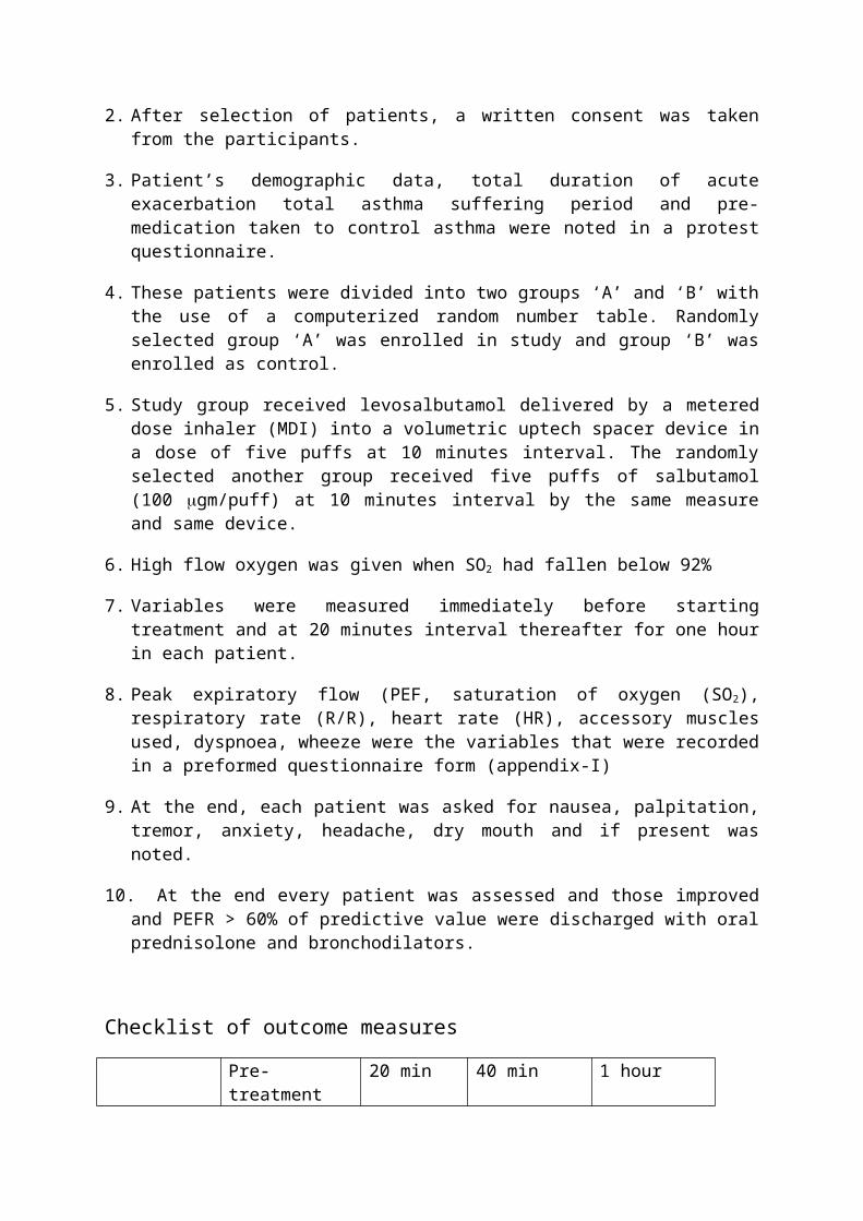
Parameswaran K, Belda J, Rowe BH. (2002), Addition of intravenous aminophylline to 2- agonists in adults with acute asthma (Cochrane Review). The Cochrane Library, Issue 4. Oxford. UK: Update Software
Pepe PE, Marini JP. (1982), Occult positive end-expiration pressure in mechanically ventilated patients with airflow obstruction.Am Rev Respir Dis 126: 166-170
Peter Canady, Asthma. (2006), eMedicine.com.
Pinnock H, Johnson A, Young P, Martin N. (2001), Are doctors still failing to asses and treat asthma attacks? An audit of the management of acute attacks in a health district. Respir Med 93: 397-401.
Pinsky MR. (2000) Cardiopulmonary interactions associated with airflow obstruction. In: Hall JB, Corbridge T, Rodrigo C, et al. eds. Acute asthma: assessment and management. New York. NY: McGraw-Hill 105-123.
Rodrigo GJ and Rodrigo C. (2000a) Rapid-onset asthma attack: a prospective cohort study about characteristics and response to the emergency department treatment. Chest 118:1547-1522.
Rodrigo GJ, Rodrigo C. (1997), A new index for early prediction of hospitalization in patients with acute asthma. Am J Emerg Med 15: 8-13
Rodrigo GJ, Rodrigo C. (1999) Early prediction of poor response acute asthma patients in the emergency department Chest 114: 1016-1021.
Rodrigo GJ, Rodrigo C. (1999), Corticosteroids in the emergency department therapy of acute adult asthma: an evidence based evaluation. Chest 116:285-295.
Rodrigo GJ, Rodrigo C. (2000b) Emergency department assessment: severity and outcome prediction. In: Hall JB, Corbridge T, Rodrigo C, et al. eds. Acute asthma: assessment and management. New York. NY: McGraw-Hill 125-138.
Rodrigo GJ, Rodrigo C. (2000c) First-Line Therapy for Adult Patients with Acute Asthma Receiving a Multiple-Dose Protocol of Ipratropium Bromide Plus Albuterol in the Emergency Department. Am. J. Respir. crit. Care Med 161: 1862-1868
Rodrigo GJ, Rodrigo C. (2002a), Continuous vs intermittent 2-agonists in the treatment of acute adult asthma: a systematic review with meta-analysis. Chest 122: 160-165
Rodrigo GJ, Rodrigo C. (2002b), The Role of Anticholinergics in aute athma treatment. An Evidence-Based Evaluation. Chest 121: 1977-1987.
Rodrigo GJ, Rodrigo C. (2004), Acute Asthma in Adults; A Review. Chest 125: 1081-1102.
Rosner F. (2002), Moses Maimonides treatise on asthma. Thorax 36: 245-251
Rossi A, Ganassini A, Brusasco V. (2000), Airflow obstruction and dynamic pulmonary hyperinflation. In Hall JB, Corbridge T, Rodrigo C, et al, eds. Acute asthma: assessment and management. New York, NY: McGraw-Hill 57-82
Rossing TH, Fanta CH, Goldstein DH, Snapper JR, McFadden ER. (1993) Emergency therapy of asthma: comparison of the acute effects of asthma: Rodrigo G, Rodrigo C.

Assessment of the patient with acute asthma in the emergency department: a factor analytic study. Chest 104:1325-1328
Salmeron S. Liard R, Elkharrat D, Muir J, Neukirch F et al. (2001), Asthma severity adequacy of management in accident and Emergency Departments in France: a prospective study. Lancet 358: 629-635.
Schreck DM, Babin S. (2005), Comparison of recemic albuterol and levalbuterol in the treatment of acute asthma in the ED. American Journal of Emergency Medicine 23: 842-847.
Shim CS, Williams MH. relationship of wheezing to the severity of obstruction in asthma. Arch Intern Med 1983; 143:890-892
Silverman RA, Osborn H, Bunge J, Gallagher EJ, Chiang W et al. (2002), IV magnesium sulfate in the treatment of acute severe asthma: a multicenter randomized controlled trial. Chest 122:489-497
Silverman RA. Chen Y, Bonucelli CM. (2000), The Zafirlukast Acute Asthma study Group: zafirlukast improves emergency department outcomes after an acute asthma episode. Ann Emerg Med 35: S10
Smith DH, Weiss K, Sullivan SD. (2000), Epidemiology and costs of acute asthma. In: Hall JB, Corbridge T, Rodrigo c, et al, eds. Acute asthma: assessment and management. New York, NY: McGraw-Hill 1-10
Stein LM, Coie RP. (1990), Early administration of corticosteroids in emergency room treatment of acute asthma. Ann Intern Med 112: 822-827.
Templeton AGB, Chamman ID, Chilvers ER, Morley J, Handley DA. (1998), Effects of S-salbutamol on human isolated bronchus. Pulmonary Pharmacology & Terapeutics 11: 1-6.
Terrance Truitt, MD, FCCP; James Witko, MD. FCCP; and Michael Halpern, MD, PhD. (2003). Levalbuterol Compared to racemic Albuterol. Efficacy and Outcomes in Patients Hospitalized with COPD or Asthma. CHEST: 123:128-135.
Tuxen DV, Anderson MB, Scheinkestel CD. (2000), Mechanical ventilation for severe asthma. In: Hall JB, Corbridge T, Rodrigo C, et al, eds. Acute asthma: assessment and management. New York, NY: McGraw-Hill 209-228.
Weiss KB, Gergen PJ, Hodgson TA (1992). An economic evaluation of asthma in the United States. N Engl J Med. 326: 862-866.
Wikipedia, (2006). the free encyclopedia.htm. Asthma.


