ASE Guidelines on Aortic Regurgitation What Do I...
23
2/18/2018 1 Mitral Regurgitation The New ASE Guidelines: Role of 2D/3D and CMR ASE Guidelines on Aortic Regurgitation What Do I Measure? Case Studies William A. Zoghbi MD, FASE, MACC Professor and Chairman, Department of Cardiology Elkins Family Distinguished Chair in Cardiac Health Houston Methodist Hospital Released The same day in March 2017! JASE 30: 303, 2017
Transcript of ASE Guidelines on Aortic Regurgitation What Do I...
2/18/2018
1
Mitral Regurgitation
The New ASE Guidelines: Role of 2D/3D and CMR
ASE Guidelines on Aortic Regurgitation
What Do I Measure?Case Studies
William A. Zoghbi MD, FASE, MACCProfessor and Chairman, Department of Cardiology
Elkins Family Distinguished Chair in Cardiac Health
Houston Methodist Hospital
ReleasedThe same day
in March 2017!JASE 30: 303, 2017

2/18/2018
2
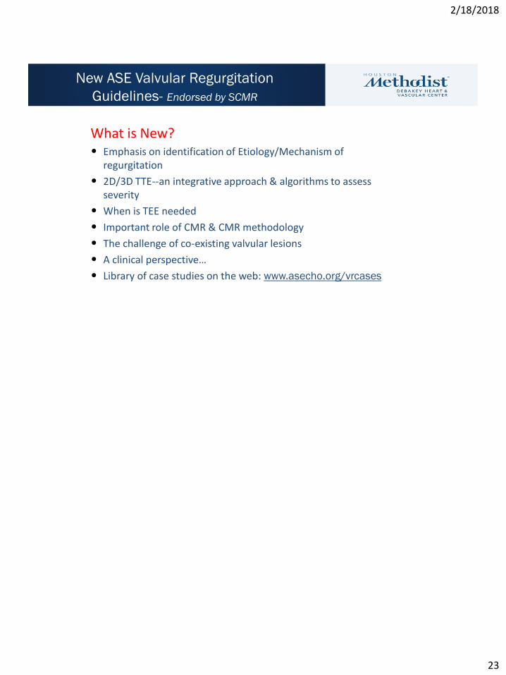
New ASE Valvular Regurgitation
Guidelines- Endorsed by SCMR
General Considerations
What is New?• Emphasis on identification of Etiology/Mechanism of
regurgitation
• 2D/3D TTE--an integrative approach & algorithms to assess severity
• When is TEE needed
• Important role of CMR & CMR methodology
• The challenge of co-existing valvular lesions
• A clinical perspective…
• Library of case studies on the web: www.asecho.org/vrcases
Zoghbi W et al. JASE 30: 303, 2017
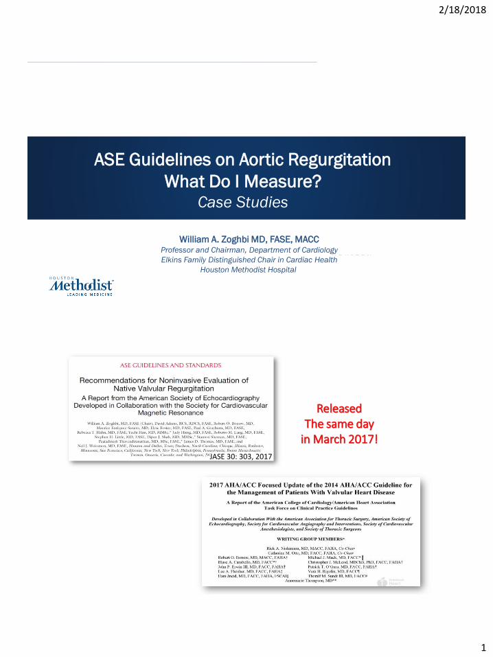
Aortic Regurgitation
2/18/2018
3
Aortic Regurgitation
Zoghbi W et al. JASE 30: 303, 2017
Assessment of AR Severity
Echo/Doppler Indicators of Severity
• Aortic Valve/ Root/Mechanism
• LV enlargement
• Color Doppler: jet width; vena Contracta
• Pressure half-time
• Regurgitant Volume/Fraction
• Diastolic retrograde flow in aorta

2/18/2018
4
MildAR
SevereAR
Color Doppler CW Doppler Desc Aorta - PW
Aortic Regurgitation- Color Doppler

2/18/2018
5
VC
FlowConvergence
JetWidth
LA
Central AR Jet Eccentric AR Jet
LV
AR Severity- Color Doppler
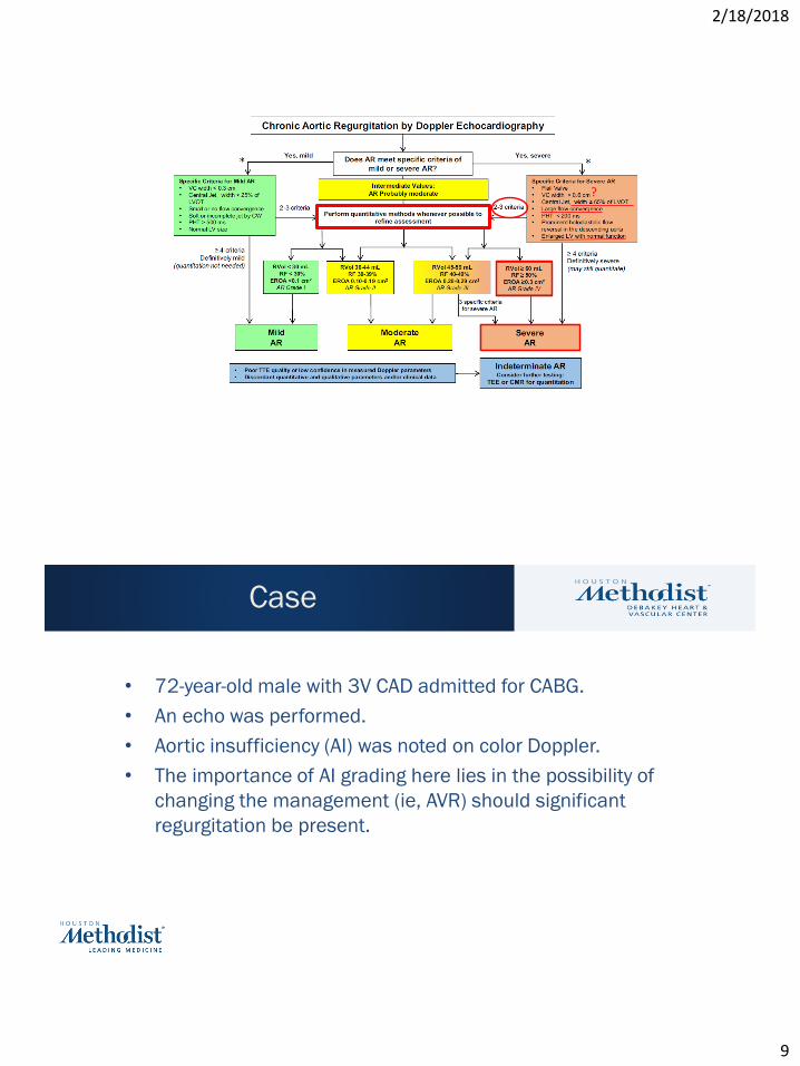
Does AR meet specific criteria of
mild or severe AR?
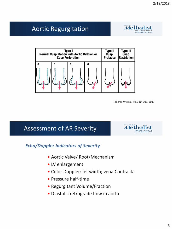
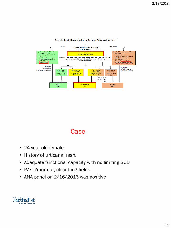
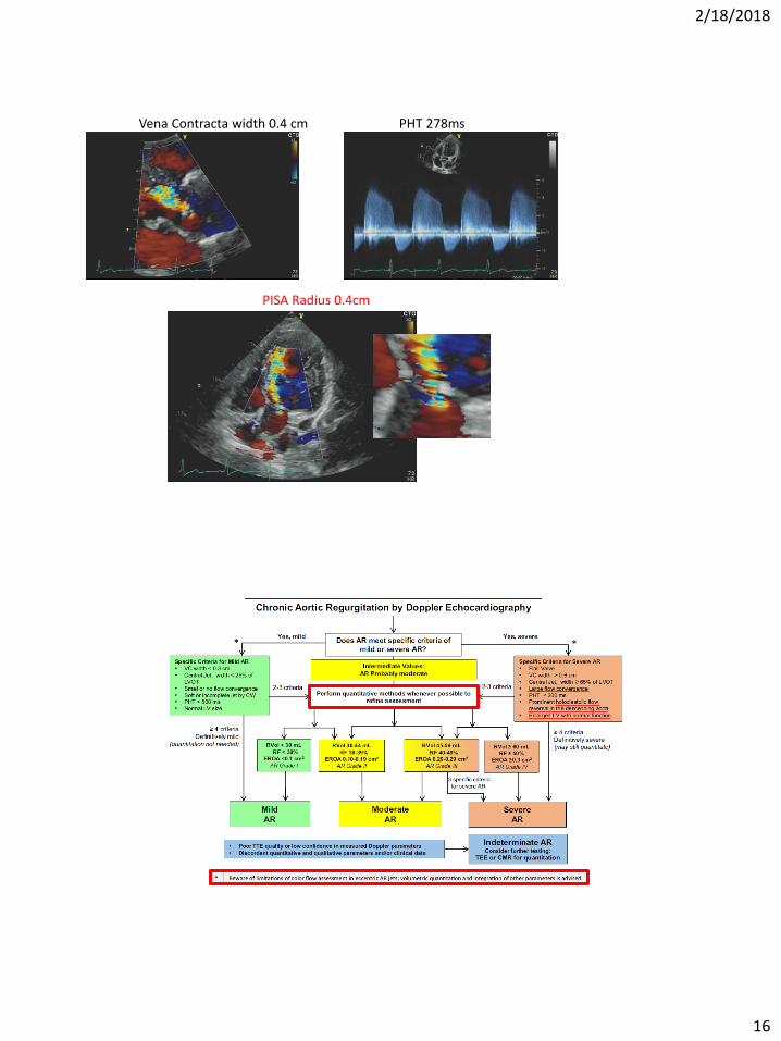
Chronic Aortic Regurgitation by Doppler Echocardiography
• Poor TTE quality or low confidence in measured Doppler parameters
• Discordant quantitative and qualitative parameters and/or clinical data
Indeterminate ARConsider further testing:
TEE or CMR for quantitation
* Beware of limitations of color flow assessment in eccentric AR jets; volumetric quantitation and integration of other parameters is advised
Intermediate Values:
AR Probably moderate
Perform quantitative methods whenever possible to
refine assessment
Severe
AR
Yes, severe
≥ 4 criteria
Definitively severe
(may still quantitate)
*Specific Criteria for Severe AR
• Flail Valve
• VC width > 0.6 cm
• Central Jet, width ≥ 65% of LVOT
• Large flow convergence
• PHT < 200 ms
• Prominent holodiastolic flow
reversal in the descending aorta
• Enlarged LV with normal function
Mild
AR
Yes, mild
≥ 4 criteria
Definitively mild
(quantitation not needed)
*
Specific Criteria for Mild AR
• VC width < 0.3 cm
• Central Jet, width < 25% of
LVOT
• Small or no flow convergence
• Soft or incomplete jet by CW
• PHT > 500 ms
• Normal LV size
Moderate
AR
2-3 criteria 2-3 criteria
RVol 30-44 mL
RF 30-39%
EROA 0.10-0.19 cm2
AR Grade II
RVol ≥ 60 mL
RF ≥ 50%
EROA ≥0.3 cm2
AR Grade IV
3 specific criteria
for severe AR
RVol 45-59 mL
RF 40-49%
EROA 0.20-0.29 cm2
AR Grade III
RVol < 30 mL
RF < 30%
EROA <0.1 cm2
AR Grade I

2/18/2018
6
Case
• 59-year-old male with a PMH significant for IV drug abuse
• He presented to the ED with a recent history of chest
pain, SOB, fever & chills
• BP 158/66, HR 56, RR 16, SpO2 97% RA
• Loud 3/6 diastolic murmur heard at LLSB
• Bibasilar rales on lung auscultation
Parasternal Long Axis SAX View at AV Level A4C View
Parasternal Long Axis With Color Doppler SAX View with Color A5C View with Color
2/18/2018
7
Faint Doppler reversal signal in the descending aortic arch
Diminished RVOT flow EDD 6.4 cm
EDV 254 mL (143 ml/m2) = Severely enlarged
LVEF 62%
PHT 412 ms
?

2/18/2018
8
SV METHOD (LVOT SV – RVOT SV)Pulsed Doppler RVOT
RVOT diam 2.3 cm
RVOT TVI 14 cm
RVOT SV = 0.785*2.32*14 = 58 mL
Pulsed Doppler LVOT
LVOT diam 2.2 cm
LVOT TVI 41 cm
LVOT SV = 0.785*2.22*41 = 156 mL
Rvol = 156 – 58 = 98 mLR F = 98/156 = 63%
Internal Check of VolumesPulsed Doppler LVOT
LVOT diam 2.2 cm
LVOT TVI 41 cm
LVOT SV = 0.785*2.22*41 = 156 mL
LV SV = EDV – ESV = 254 – 95 = 159 mL
RVol = ~90-95 mLRF = RVol/SVLVOT = ~60%
2/18/2018
9
?
Case
• 72-year-old male with 3V CAD admitted for CABG.
• An echo was performed.
• Aortic insufficiency (AI) was noted on color Doppler.
• The importance of AI grading here lies in the possibility of
changing the management (ie, AVR) should significant
regurgitation be present.

2/18/2018
10
A4C View
A5C View with Color
Parasternal Long Axis
Parasternal Long Axis With Color Doppler
SAX View at AV Level
SAX View with Color
Based on these views only…
Is AR mild, moderate, severe or
Indeterminate?

2/18/2018
11
Faint Doppler reversal signal in the descending arch
Dense AI jet signal
Prominent RVOT VTI EDD 4.5 cm
EDV 139 mL (79 ml/m2) = upper limit
of normal size
LVEF 63%
Jet/LVOT 0.4
PHT 311 ms
EDD 4.5 cm
Vena Contracta width 0.4 cm
PISA radius 0.5 cm

2/18/2018
12
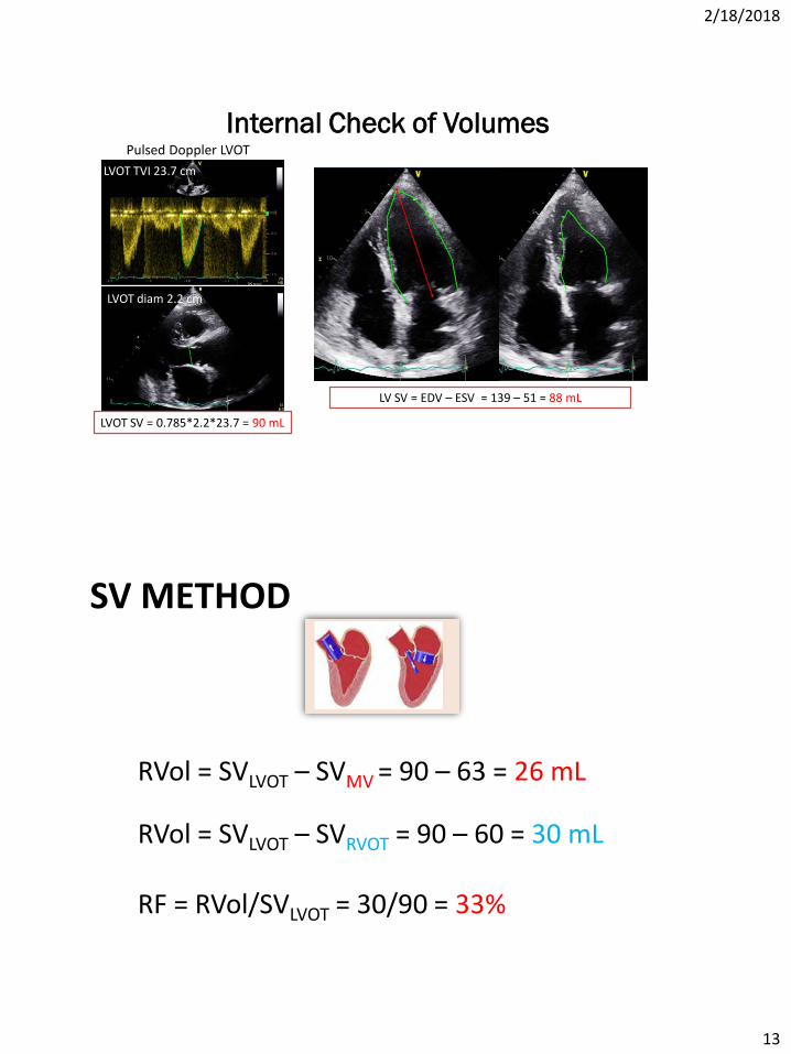
SV METHOD
Pulsed Doppler RVOT
RVOT diam 2.4 cm
RVOT TVI 13.3 cm
RVOT SV = 0.785*2.4*13.3 = 60 mL
Pulsed Doppler LVOT
LVOT diam 2.2 cm
LVOT TVI 23.7 cm
LVOT SV = 0.785*2.2*23.7 = 90 mL
Mitral Annulus
MV annulus inflow VTI 9 cm
MV annulus diam 3 cm
MV SV = 0.785*3*9 = 63 mL
2/18/2018
13
Internal Check of VolumesPulsed Doppler LVOT
LVOT diam 2.2 cm
LVOT TVI 23.7 cm
LVOT SV = 0.785*2.2*23.7 = 90 mL
LV SV = EDV – ESV = 139 – 51 = 88 mL
SV METHOD
RVol = SVLVOT – SVRVOT = 90 – 60 = 30 mL
RF = RVol/SVLVOT = 30/90 = 33%
RVol = SVLVOT – SVMV = 90 – 63 = 26 mL
2/18/2018
14
Case
• 24 year old female
• History of urticarial rash.
• Adequate functional capacity with no limiting SOB
• P/E: ?murmur, clear lung fields
• ANA panel on 2/16/2016 was positive

2/18/2018
15
PLAX Mmode AV
SAX color Doppler
PLAX PLAX zoom in aortic valve
PLAX color Doppler SAX
Faint Doppler reversal signal in the descending arch
Dense AI jet signal
EDD 5.4 cm
EDV 141 mL (80 ml/m2) =
Dilated
LVEF 63%
2/18/2018
16
PISA Radius 0.4cm
PHT 278msVena Contracta width 0.4 cm

2/18/2018
17
SV method PISA method
SV METHOD
Pulsed Doppler RVOT
RVOT TVI 13.3 cm
RVOT SV = 0.785*2.3^2*14.3 = 59 mL
Pulsed Doppler LVOT
LVOT diam 2.2 cm
LVOT TVI 23.7 cm
LVOT SV = 0.785*2.4^2*19.3 = 87 mL
Mitral Annulus
MV annulus inflow VTI 9 cm
MV annulus diam 3 cm
MV SV = 0.785*3^2*8.1 = 57 mL
2/18/2018
18
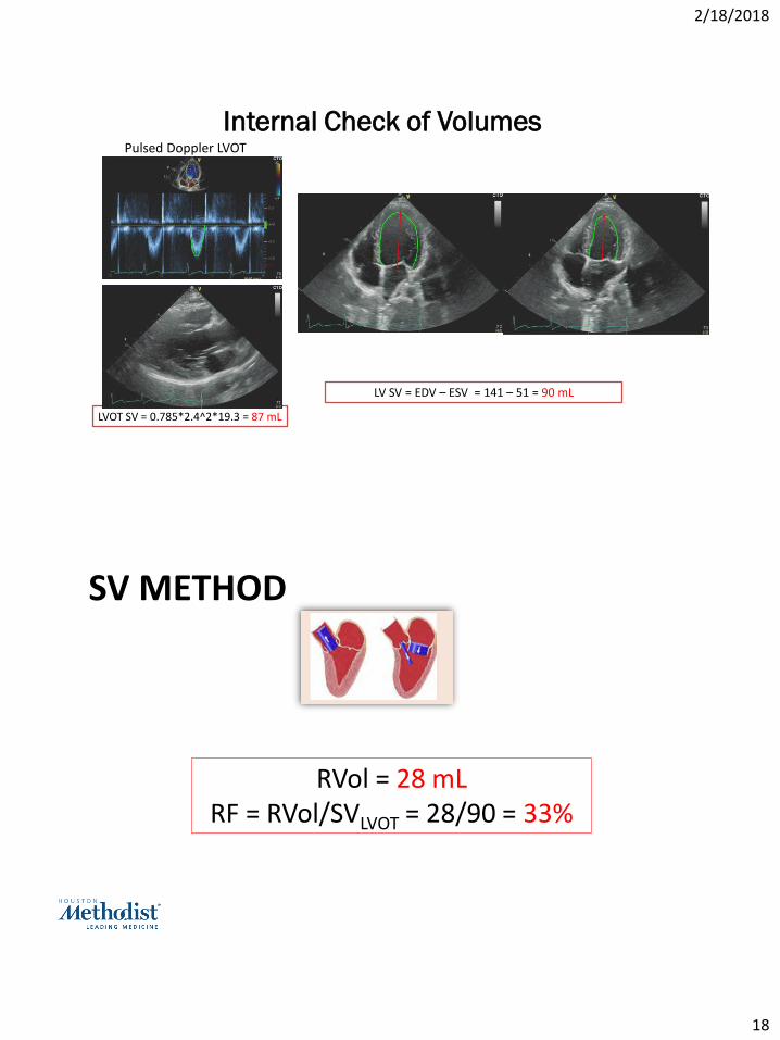
Internal Check of VolumesPulsed Doppler LVOT
LVOT diam 2.2 cm
LVOT TVI 23.7 cm
LVOT SV = 0.785*2.4^2*19.3 = 87 mL
LV SV = EDV – ESV = 141 – 51 = 90 mL
SV METHOD
RVol = 28 mLRF = RVol/SVLVOT = 28/90 = 33%

2/18/2018
19

2/18/2018
20
What is your best initial assessment of Severity of Aortic regurgitation?
A. Mild
B. Mild to moderate
C. Moderate
D. Moderate to severe
E. Severe
Eccentric AI jets
Reliable indicators of severity
• Vena Contracta- if clearly defined• Regurgitant flow and regurgitant fraction• Flow reversal in aorta• LV size –always look at the scale!
Less reliable indicators of severity:
• Jet width/LVOT diameter• Area of jet in Short axis • Adequate CW jet recording may be difficult- “bidirectional”
2/18/2018
21
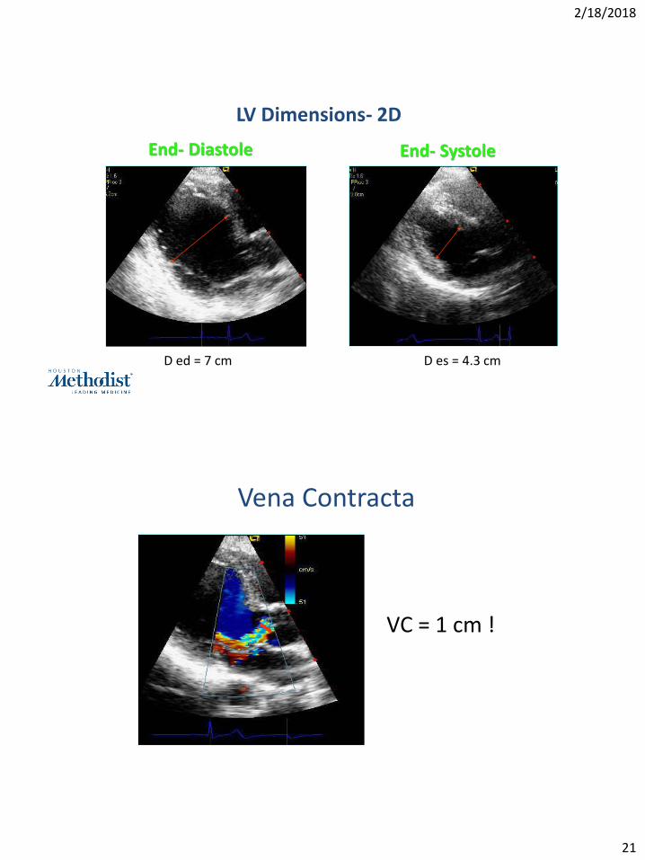
D ed = 7 cm D es = 4.3 cm
LV Dimensions- 2D
End- Diastole End- Systole
Vena Contracta
VC = 1 cm !

2/18/2018
22
Aortic diastolic Flow Reversal
LVOT Flow
RVOT Flow
D= 3cm
TVI = 34 cm
SVLVOT = 240 ml
SVRVOT = 69 ml
D=2.5cm
TVI = 14 cm
Reg V = 240-69=171mL
RF = 171/240= 71%
2/18/2018
23
New ASE Valvular Regurgitation
Guidelines- Endorsed by SCMR
General Considerations
What is New?• Emphasis on identification of Etiology/Mechanism of
regurgitation
• 2D/3D TTE--an integrative approach & algorithms to assess severity
• When is TEE needed
• Important role of CMR & CMR methodology
• The challenge of co-existing valvular lesions
• A clinical perspective…
• Library of case studies on the web: www.asecho.org/vrcases