ASCERTAINING DRUG RESISTANCE BACTERIA FROM HOSPITAL ...
16
www.wjpps.com Vol 6, Issue 12, 2017. 801 Padmalochana et al. World Journal of Pharmacy and Pharmaceutical Sciences ASCERTAINING DRUG RESISTANCE BACTERIA FROM HOSPITAL DRAINAGE WATER AND EVALAUTING ITS SUSCEPTIBILITY WITH LEAF EXTRACT OF BARLERIA PRIONITIS K. Padmalochana* and R. Saranya Department of Biochemistry, Sri Akilandeswari Women’s College, Wandiwash – 604408, TN, India. ABSTRACT To study the identification of drug resistant bacteria from hospital sewage water especially from drainage canal. The sample was processed for enumerating the total bacterial count and showed a total population of 2.3×10 4 . The BOD level of the sample as it is a liquid was evaluated using titration method. The major part of the work denoted by isolating the drug resistant bacteria by cultivating the bacteria in media containing different class of drug. The antibiotic is applied in the drug resistant bacteria were Vancomycin, Amoxicillin, Cephalexin, Amikacin. The identified bacteria S. aureus, Klebsiella species, Pseudomonas species was determined for its multidrug resistant property by antibiogram using different generation antibiotic and zone integration. The antibiogram of the isolated bacteria expression of antibiotic resistant characters by producing beta lactamase (penicillin, cephalexi), 6’-N. acetyl transferases (Amikacin), D-alanine TD-alanine transpeptidase (vancomycin), OXA-1 beta- lactamase (amoxicillin). The enzyme activity was determined by nitrocefin test. The drug resistant bacteria is treat with Barleria prionitis methanolic leaf extract which yield 13.93% of the phytoconstituent by qualitatively. The B.prionitis was resistant to the drug resistant bacteria. KEYWORDS: Drug resistant bacteria, Barleria prionitis, Antibiotics, Enzymes. WORLD JOURNAL OF PHARMACY AND PHARMACEUTICAL SCIENCES SJIF Impact Factor 6.647 Volume 6, Issue 12, 801-816 Research Article ISSN 2278 – 4357 *Corresponding Author Dr. K. Padmalochana Department of Biochemistry, Sri Akilandeswari Women's College, Wandiwash - 604408, TN, India. Article Received on 29 Sept. 2017, Revised on 20 October 2017, Accepted on 09 Nov. 2017 DOI: 10.20959/wjpps201712-10545
Transcript of ASCERTAINING DRUG RESISTANCE BACTERIA FROM HOSPITAL ...
www.wjpps.com Vol 6, Issue 12, 2017.
801
Padmalochana et al. World Journal of Pharmacy and Pharmaceutical Sciences
ASCERTAINING DRUG RESISTANCE BACTERIA FROM HOSPITAL
DRAINAGE WATER AND EVALAUTING ITS SUSCEPTIBILITY
WITH LEAF EXTRACT OF BARLERIA PRIONITIS
K. Padmalochana* and R. Saranya
Department of Biochemistry, Sri Akilandeswari Women’s College, Wandiwash – 604408,
TN, India.
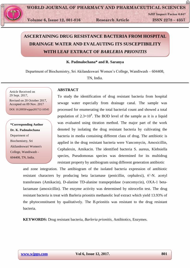
ABSTRACT
To study the identification of drug resistant bacteria from hospital
sewage water especially from drainage canal. The sample was
processed for enumerating the total bacterial count and showed a total
population of 2.3×10⁴. The BOD level of the sample as it is a liquid
was evaluated using titration method. The major part of the work
denoted by isolating the drug resistant bacteria by cultivating the
bacteria in media containing different class of drug. The antibiotic is
applied in the drug resistant bacteria were Vancomycin, Amoxicillin,
Cephalexin, Amikacin. The identified bacteria S. aureus, Klebsiella
species, Pseudomonas species was determined for its multidrug
resistant property by antibiogram using different generation antibiotic
and zone integration. The antibiogram of the isolated bacteria expression of antibiotic
resistant characters by producing beta lactamase (penicillin, cephalexi), 6’-N. acetyl
transferases (Amikacin), D-alanine TD-alanine transpeptidase (vancomycin), OXA-1 beta-
lactamase (amoxicillin). The enzyme activity was determined by nitrocefin test. The drug
resistant bacteria is treat with Barleria prionitis methanolic leaf extract which yield 13.93% of
the phytoconstituent by qualitatively. The B.prionitis was resistant to the drug resistant
bacteria.
KEYWORDS: Drug resistant bacteria, Barleria prionitis, Antibiotics, Enzymes.
WORLD JOURNAL OF PHARMACY AND PHARMACEUTICAL SCIENCES
SJIF Impact Factor 6.647
Volume 6, Issue 12, 801-816 Research Article ISSN 2278 – 4357
*Corresponding Author
Dr. K. Padmalochana
Department of
Biochemistry, Sri
Akilandeswari Women's
College, Wandiwash -
604408, TN, India.
Article Received on
29 Sept. 2017,
Revised on 20 October 2017,
Accepted on 09 Nov. 2017
DOI: 10.20959/wjpps201712-10545

www.wjpps.com Vol 6, Issue 12, 2017.
802
Padmalochana et al. World Journal of Pharmacy and Pharmaceutical Sciences
INTRODUCTION
In recent years, bacterial resistance to antibiotics has raised to the forefront of world health
issues. Overuse of antibiotics has been identified as a leading cause for the development of
bacterial antibiotic resistance. A unique feature of enzymes that physically modify antibiotics
is that these mechanisms alone actively reduce the concentration of drugs in the local
environment; therefore, they present a unique challenge to researchers and clinicians
considering new approaches to anti-infective therapy (Gerard D. Wright et al, 2005).
Understanding the mechanisms of resistance is important in order to define better ways to
keep existing agents useful for a little longer but also to help in the design of better
antimicrobial agents that are not affected by the currently known, predicted, or unknown
mechanisms of resistance (Denis K. Byarugaba et al, 2009). Aminoglycosides modified at
amino groups by AAC enzymes or at hydroxyl groups by ANT or APH enzymes lose their
ribosome-binding ability and thus no longer inhibit protein synthesis. Besides
aminoglycoside-modifying enzymes, efflux systems and rRNA mutations have been
described (Quintiliani and Courvalin, 1995). Multidrug resistance among many organisms
has become a big challenge to infectious disease management. It is increasingly being
reported in bacteria and is often mediated by genetic mobile elements such as plasmids,
transposons, and integrons (Dessen et al., 2001).
Kingdom : Plantae.
Subkingdom : Tracheobionta.
Division : Magnoliophyta.
Class : Magnoliopsida.
Subclass : Asteridae.
Order : Scrophulariales.
Family : Acanthaceae.
Genus : Barleria.
Species : prionitis.
Common Name : Bajradantip.
Tamil Name : Cemmulli.
Barleria prionitis is used to treat fever, respiratory diseases, toothache and joint pains. A
mouthwash made from root tissue is used to relieve toothache and treat bleeding gums. The
leaves are used to healing of wounds and to relieve joint pains. Extracts of the plant are

www.wjpps.com Vol 6, Issue 12, 2017.
803
Padmalochana et al. World Journal of Pharmacy and Pharmaceutical Sciences
incorporated into herbal cosmetics and hair products to cure skin and scalp problems (Abha
Khare et al., 2016, Patel Bharatkumar et al., 2015, Sattya Narayan et al., 2015). In this
present investigation we have collected the sewage from hospital drainage, determination of
Biological Oxygen Demand of the water sample, isolation of antibiotic resistance bacteria,
determination of bacteria for its multidrug resistant property, extraction of Total
Phytochemcials from Barleria prionitis leaf, screening for potential phytoconstituents from
the B. prionitis leaf extract, antibacterial activity using the multi drug resistant bacteria
towards plant derivative and enzyme activity measurement assay from the drug resistant
bacteria along with the antibiotics and the leaf extract.
MATERIALS AND METHODS
COLLECTION OF SAMPLE
The sample was collected from Government General Hospital, Pondicherry. The location
defined in sample collection was from inpatient room wash area and the drained water was
collected in the day hour’s. The seepage water was collected in a aseptic container and
immediately transported to the laboratory and stored in the refrigerator (Cruickshankk 1980,
Radha 2009). The sample was evaluated for its Biological Oxygen Demand. The procedure is
done for the all the BOD bottles before and after the incubation. For the determination of
BOD of the given beverage sample the readings are tabulated as.
Table 1: BOD groundwork.
Sl. No. Day Volume of sample
(ml)
Burette Reading (ml) Volume of Titrant
(Na2S2O3 used)
DO
(mg/L) Initial Final
Blank (A) 0 100 0 *** *** ***
Sample (A) 0 100 0 ***
INCUBATION FOR 5 DAYS IN B.O.D INCUBATOR
Blank (B) 5 100 0 *** *** ***
Sample (B) 5 100 0 ***
Therefore,
Biological Oxygen Demand =
Determination of total bacterial count
The sample was evaluated for its total bacterial population by serial dilution and spread plate
method.
www.wjpps.com Vol 6, Issue 12, 2017.
804
Padmalochana et al. World Journal of Pharmacy and Pharmaceutical Sciences
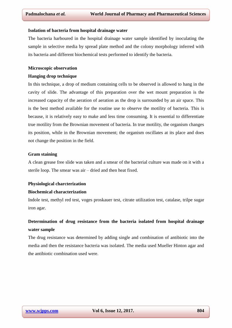
Isolation of bacteria from hospital drainage water
The bacteria harboured in the hospital drainage water sample identified by inoculating the
sample in selective media by spread plate method and the colony morphology inferred with
its bacteria and different biochemical tests performed to identify the bacteria.
Microscopic observation
Hanging drop technique
In this technique, a drop of medium containing cells to be observed is allowed to hang in the
cavity of slide. The advantage of this preparation over the wet mount preparation is the
increased capacity of the aeration of aeration as the drop is surrounded by an air space. This
is the best method available for the routine use to observe the motility of bacteria. This is
because, it is relatively easy to make and less time consuming. It is essential to differentiate
true motility from the Brownian movement of bacteria. In true motility, the organism changes
its position, while in the Brownian movement; the organism oscillates at its place and does
not change the position in the field.
Gram staining
A clean grease free slide was taken and a smear of the bacterial culture was made on it with a
sterile loop. The smear was air – dried and then heat fixed.
Physiological charcterization
Biochemical characterization
Indole test, methyl red test, voges proskauer test, citrate utilization test, catalase, trilpe sugar
iron agar.
Determination of drug resistance from the bacteria isolated from hospital drainage
water sample
The drug resistance was determined by adding single and combination of antibiotic into the
media and then the resistance bacteria was isolated. The media used Mueller Hinton agar and
the antibiotic combination used were.
www.wjpps.com Vol 6, Issue 12, 2017.
805
Padmalochana et al. World Journal of Pharmacy and Pharmaceutical Sciences
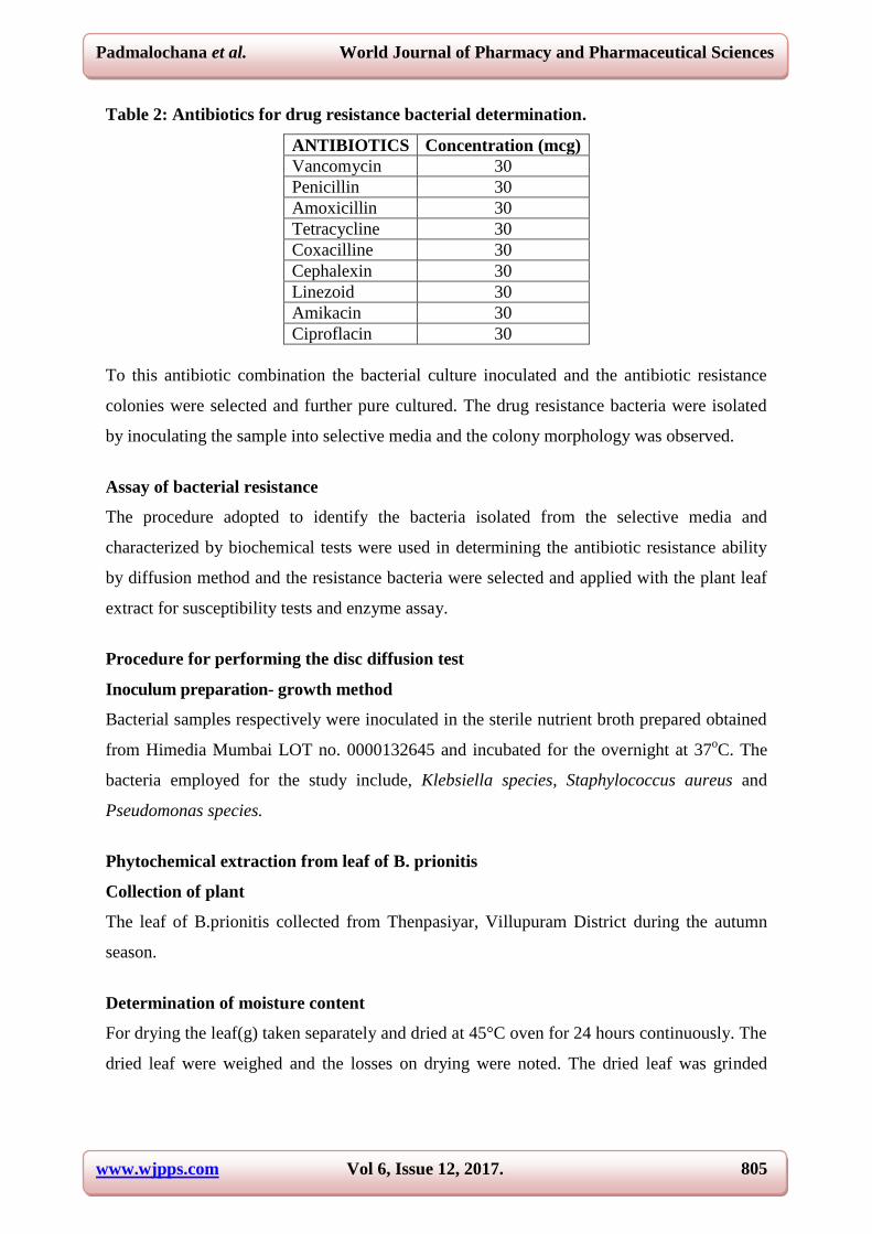
Table 2: Antibiotics for drug resistance bacterial determination.
ANTIBIOTICS Concentration (mcg)
Vancomycin 30
Penicillin 30
Amoxicillin 30
Tetracycline 30
Coxacilline 30
Cephalexin 30
Linezoid 30
Amikacin 30
Ciproflacin 30
To this antibiotic combination the bacterial culture inoculated and the antibiotic resistance
colonies were selected and further pure cultured. The drug resistance bacteria were isolated
by inoculating the sample into selective media and the colony morphology was observed.
Assay of bacterial resistance
The procedure adopted to identify the bacteria isolated from the selective media and
characterized by biochemical tests were used in determining the antibiotic resistance ability
by diffusion method and the resistance bacteria were selected and applied with the plant leaf
extract for susceptibility tests and enzyme assay.
Procedure for performing the disc diffusion test
Inoculum preparation- growth method
Bacterial samples respectively were inoculated in the sterile nutrient broth prepared obtained
from Himedia Mumbai LOT no. 0000132645 and incubated for the overnight at 37oC. The
bacteria employed for the study include, Klebsiella species, Staphylococcus aureus and
Pseudomonas species.
Phytochemical extraction from leaf of B. prionitis
Collection of plant
The leaf of B.prionitis collected from Thenpasiyar, Villupuram District during the autumn
season.
Determination of moisture content
For drying the leaf(g) taken separately and dried at 45°C oven for 24 hours continuously. The
dried leaf were weighed and the losses on drying were noted. The dried leaf was grinded

www.wjpps.com Vol 6, Issue 12, 2017.
806
Padmalochana et al. World Journal of Pharmacy and Pharmaceutical Sciences
using a mechanical blender and made to a fine powder. The powdered sample’s were used for
extraction.
Extraction of total phytoconstituents
The powdered leaf of B.prionitis added with the solvent methanol along with ethanol ie in the
ration 8: 2 (80% Methanolic Ethanol v/v) was used for the extraction of leaf. 20g each of leaf
powder was separately taken in a beaker and soaked it in 100 ml methanol and the beaker
was covered using aluminum foil to avoid evaporation and kept undisturbed for 24 hours at
room temperature. After the incubation period the extract was then filtered using filter paper
and was followed by filtration using filter paper. The filtrate was collected and then the 80%
methanolic ethanol kept for evaporation in a water bath at 65°C until complete saturation
occurs. The crude extract obtained evaluated for its total content gravimetrically.
Preparation of phytochemical concentration
The concentrated extract of each was diluted using 25 ml of 80% methanolic ethanol and the
diluted extract was used for further studies. The concentration of the extract was noted in
table: The extract was screened for its total phyto-nutrient by qualitatively.
Phytochemical screening
1. Test for Alkaloids (Test): Treat the jujube seed extract with few drops of s reagents.
Formation of reddish brown precipitate indicates presence of alkaloids.
2. Test for phenols/Tannins (ferric chloride Test): Mix the crude extract with 2ml of 2%
solution of Fecl3. Blue-green or black coloration indicates presence of phenols & tannins
3. Test for Terpenoids (salkowski Test): To the seed extract few drops of con.H2SO4 and
2ml chloroform and shaken then allow standing appearance of golden yellow colour
indicates the presence of triterpenoids.
4. Test for flavonoids (Ferric Chloride): To the extract a pinch of magnesium ribbon
added and followed by concentrated Hydrochloric acid which gives a magenta color with
effervescence.

www.wjpps.com Vol 6, Issue 12, 2017.
807
Padmalochana et al. World Journal of Pharmacy and Pharmaceutical Sciences
5. Test for fixed oils (paper Test): Few drop of the extract applied over the surface of
Whattman filter and allowed to air dry for 10 minutes. If the filter paper retains the oil
stain it confirms the presence of Fixed oil.
Antibacterial activity using the multi drug resistant bacteria towards plant derivative
Testing the antibacterial activity by well diffusion method
Muller Hinton agar was use to check antimicrobial activity by well diffusion method.
Autoclaved medium was poured in to petriplates in the laminar air flow hood. On cooling the
medium within petriplates the microorganism from 24hrs old broth were spread then wells
were made on the petriplates with the help of stainless steel borer of diameter 6- 8 mm. Three
wells were made on entire surface of medium; one is for control to the test organism and
remaining for different concentration of the sample. Now increasing volume (μl) of
phytochemicals preparations was poured in the first well and subsequently for the other well.
These plates were incubated for 24-48hrs and the diameter of zone of inhibition was
measured with the help of scale.
The freshly prepared inoculum was swabbed all over the surface of the MHA plate using
sterile cotton swab. Five wells of 6mm diameter were bored in the medium with the help of
sterile cork-borer having 6mm diameter and were labeled properly and fifty micro-liters of
the working suspension/solution of different medicinal plant extract and same volume of
extraction solvent for control was filled in the wells with the help of micropipette. Plates were
left for some time till the extract diffuse in the medium with the lid closed and incubated at
37°C for 24 hour and measured using scale and mean were recorded after incubation, plates
were observed for zone of inhibition.
Preparation of culture broth
Bacterial samples respectively were inoculated in the sterile nutrient broth prepared obtained
from Himedia Mumbai, India and incubated for the overnight at 37oC.
Preparation of the agar lawns
The sterile pre incubated bored plates were taken for the analysis and the lawn was made by
using the sterile cotton swabs and lawn was made.

www.wjpps.com Vol 6, Issue 12, 2017.
808
Padmalochana et al. World Journal of Pharmacy and Pharmaceutical Sciences
Preparation of the test solution
Test solution was prepared by the equivalent of 200 mg/ml concentration and used for
analysis. The secondary metabolites were added into the well at a concentration of 50μl and
kept for incubation. And the results were recorded by measuring the zone using calibrated
reader scale.
The plate incubated were observed for the zone of inhibition and the zone measurement was
made using standard measuring scale- HiAntibiotic Zone Scale (HIMEDIA Laboratories)and
the readings were noted in mm. Note: The plant material used were leaf of B.prionitis extract
obtained from 80% Methanolic Ethanol (v/v).
Enzyme assay
The bacteria showed susceptible and resistant in both diffusion assays were evaluated for its
total enzyme activity spectrophotometrically.
Bacterial cultivation
The isolated bacterial culture was inoculated in sterile nutrient broth and kept for incubation
at 37⁰C for 24 hours. The broth culture as sonicated for 5 minute as top release the resistance
enzyme present in the periplasmic membrane of the cell. After sonication the suspension was
centrifuged at 10000 rpm for 10 minute and the supernatant was collected.
Total enzyme precipitation
The supernatant was added with 500 µl of 100% Acetone (ice cold) to precipitate the
enzyme. The mixture was kept for incubation in refrigerator for 15 minute. The suspension
was centrifuged to collect the precipitate. The collected precipitate was washed with 0.01M
phosphate buffer and finally the pellet was suspended in phosphate buffer.
Determination of enzyme activity
The enzyme activity was determined by applying antibiotics as a substrate. The following
table shows the combination of preparations,
Table 3: Antibiotic with enzyme extract for Activity assay.
BLANK CONTROL T1 T2 T3 T4 T5
Dis.H2O 3ml 3ml 3ml 3ml 3ml 3ml 3ml
ENZYME 20µl 20µl 20µl 20µl 20µl 20µl 20µl
ANTIBIOTICS 0 0 k.linezoid k.cephalexin P.Amikacin P.Cloxacillin S.Vancomycin

www.wjpps.com Vol 6, Issue 12, 2017.
809
Padmalochana et al. World Journal of Pharmacy and Pharmaceutical Sciences
RESULTS AND DISCUSSION
The aim of the study describe the identification of drug resistance bacteria from seepage of
hospital sewage system. The drainage water sample collected from the hospital during the
day hours without any prior bleaching and transported to the laboratory. The sample was
processed for enumerating the total bacterial count which showed a total population of 2.366
x 104..The BOD level of the sample as it is a liquid was evaluated using titremtric method and
found to be 6 mg/l for day 0 and 12 mg /l for day 5 of the hospital drainage water which falls
in the range prescribed by BSI, as the sewage sample was by nature its flowing water. The
major part of the work denoted by isolating the drug resistance bacteria by cultivating the
bacteria in media containing different class of drug. The anitibiogram study put forth the
resistant bacteria by formation of no zone formation. The antibiotic applied in detecting the
drug resistant bacteria were vancomycin, penicillin, amoxicillin, coxacillin, cephalexin,
Linezoid, amikacin by the bacteria obtained from the hospital drainage water.
Figure 1: Hospital Drainage water.
Figure 2: BOD (A) preparation – Day 0, (B) Titration – Day 0, (C) Titration – Day 5.
www.wjpps.com Vol 6, Issue 12, 2017.
810
Padmalochana et al. World Journal of Pharmacy and Pharmaceutical Sciences
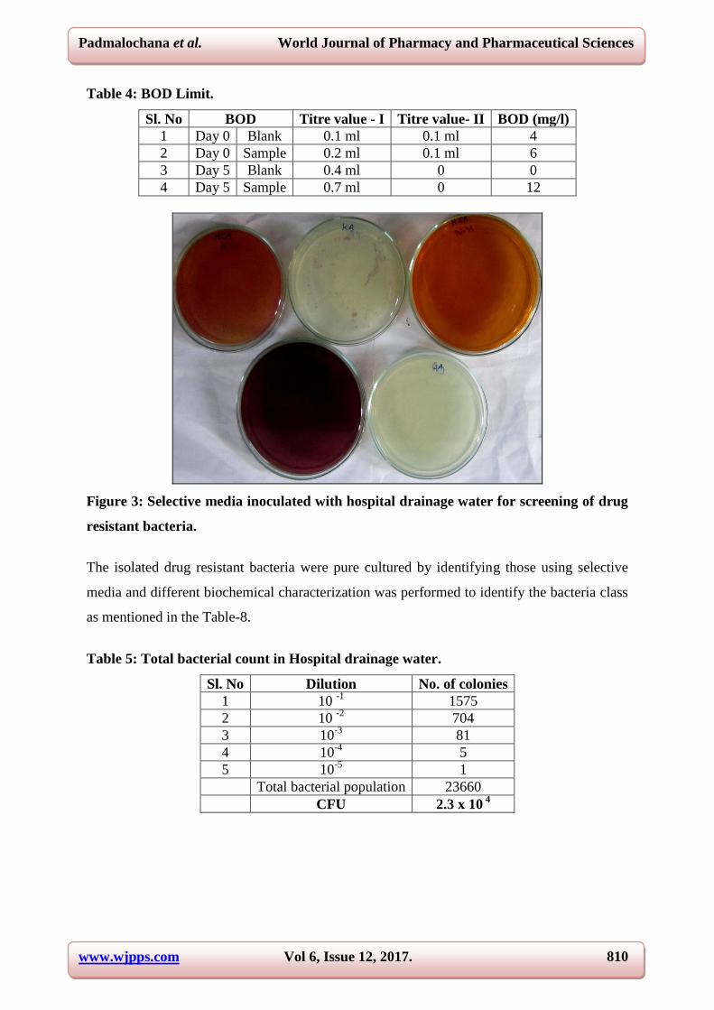
Table 4: BOD Limit.
Sl. No BOD Titre value - I Titre value- II BOD (mg/l)
1 Day 0 Blank 0.1 ml 0.1 ml 4
2 Day 0 Sample 0.2 ml 0.1 ml 6
3 Day 5 Blank 0.4 ml 0 0
4 Day 5 Sample 0.7 ml 0 12
Figure 3: Selective media inoculated with hospital drainage water for screening of drug
resistant bacteria.
The isolated drug resistant bacteria were pure cultured by identifying those using selective
media and different biochemical characterization was performed to identify the bacteria class
as mentioned in the Table-8.
Table 5: Total bacterial count in Hospital drainage water.
Sl. No Dilution No. of colonies
1 10 -1
1575
2 10 -2
704
3 10-3
81
4 10-4
5
5 10-5
1
Total bacterial population 23660
CFU 2.3 x 10 4
www.wjpps.com Vol 6, Issue 12, 2017.
811
Padmalochana et al. World Journal of Pharmacy and Pharmaceutical Sciences
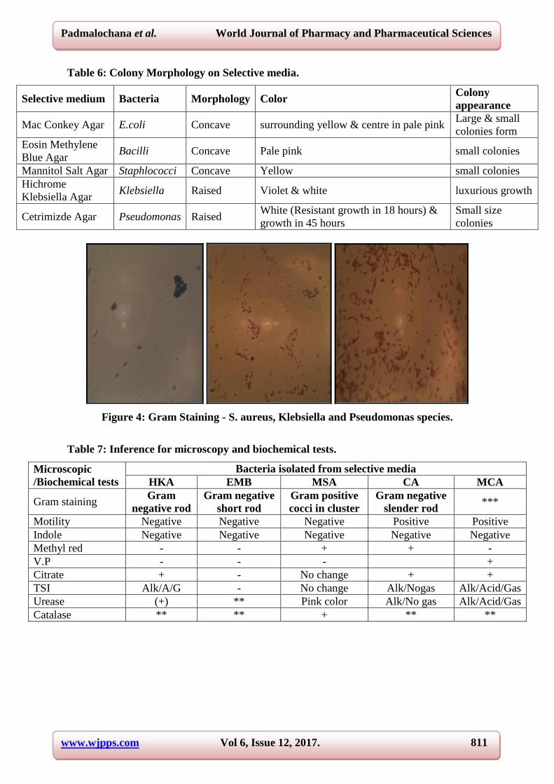
Table 6: Colony Morphology on Selective media.
Selective medium Bacteria Morphology Color Colony
appearance
Mac Conkey Agar E.coli Concave surrounding yellow & centre in pale pink Large & small
colonies form
Eosin Methylene
Blue Agar Bacilli Concave Pale pink small colonies
Mannitol Salt Agar Staphlococci Concave Yellow small colonies
Hichrome
Klebsiella Agar Klebsiella Raised Violet & white luxurious growth
Cetrimizde Agar Pseudomonas Raised White (Resistant growth in 18 hours) &
growth in 45 hours
Small size
colonies
Figure 4: Gram Staining - S. aureus, Klebsiella and Pseudomonas species.
Table 7: Inference for microscopy and biochemical tests.
Microscopic
/Biochemical tests
Bacteria isolated from selective media
HKA EMB MSA CA MCA
Gram staining Gram
negative rod
Gram negative
short rod
Gram positive
cocci in cluster
Gram negative
slender rod ***
Motility Negative Negative Negative Positive Positive
Indole Negative Negative Negative Negative Negative
Methyl red - - + + -
V.P - - - +
Citrate + - No change + +
TSI Alk/A/G - No change Alk/Nogas Alk/Acid/Gas
Urease (+) ** Pink color Alk/No gas Alk/Acid/Gas
Catalase ** ** + ** **

www.wjpps.com Vol 6, Issue 12, 2017.
812
Padmalochana et al. World Journal of Pharmacy and Pharmaceutical Sciences
Figure 5: Antibiotic Resistant assay A. Pseudomonas B. S. aureus C. Klebsiella.
The identified bacteria S.aureus, Klebsiella species, Pseudomonas species was determined for
its multidrug resistant property by antibiogram assay using different generation antibiotic and
the zone interpretation was noted in table 10.The antibiogram of the isolated bacteria defines
its expression of antibiotic resistance character by producing beta lactamase (Penicillin,
Coxacillin, Cephalexin), 6’-Nacetyltransferases (Amikacin), aminoglycoside
phosphoryltransferase (Amikacin), D-alanine-D-alanine transpeptidase (Vancomycin), OXA-
1 beta-lactamase (Amoxycillin) exhibiting extended spectrum of beta lactamase and beta
metallo lactamase activity. The drug resistance bacteria showed susceptible character towards
the antibiotic used represented in table-4.
Table 8: Antibiotic resistant bacteria detection.
ANTIBIOTICS S. aureus Pseudomonas Klebseilla
Vancomycin 17mm Resistant Resistant
Penicillin 30mm Resistant Resistant
Amoxicillin 26mm Resistant Resistant
Tetracycline 28mm 15mm 13mm
Coxacilline 29mm Resistant Resistant
Cephalexin 32mm Resistant Resistant
Linezoid 33mm Resistant 22mm
Amikacin 30mm 30mm Resistant
Ciproflacin 31mm 17mm 27mm

www.wjpps.com Vol 6, Issue 12, 2017.
813
Padmalochana et al. World Journal of Pharmacy and Pharmaceutical Sciences
Figure 6: Fresh and dried leaf of Barleria prionitis and extract.
Table 9: Moisture content in B. prionitis.
Fresh leaf (g) Dried leaf (g) Moisture %
108.7 38.9 69.8
Table 10: Extract yield from leaf of B. prionitis.
Final weight (g) Initial weight (g) Extract quantity (g) Yield %
118.3509 114.3378 4.0131 13.93
Table 11: Result for Phytochemical screening.
S. No. Test Inference
1 Alkaloid +
2 Flavanoid -
3 Tannin -
4 Phenol +
5 Turpenoid -
Figure 7: Antibacterial activity of B. prionitis leaf extract with drug resistant bacteria
(isolated).
The drug resistance was aimed to treat with the herbal extract by preparing the leaf extract of
B. prionitis and the total quantity of the phytochemical and its constituents were evaluated as
recorded in table 11,12 and 13.The drug resistance bacteria was screened for susceptible

www.wjpps.com Vol 6, Issue 12, 2017.
814
Padmalochana et al. World Journal of Pharmacy and Pharmaceutical Sciences
character with that of plant secondary materials rather using antibiotic which presently show
sensitive but they obtain resistance in the due course. As a experimental trial the bacteria
applied for antibacterial assay using plant materials from leaf extract of B. prionitis. As with
no surprise the bacteria was susceptible with the plant materials .As the study can be further
extended with what bioactive molecule has the tendency to kill the bacteria as natural
products show a wide property as an antibacterial agent. The effect of the plant extract exhibit
a potent bactericidal agent as denoted in table 14.
Table 12: Susceptibility of Drug resistant bacteria with B. prionitis leaf extract.
Drug resistant bacteria
Extract Concentration (mg) MIC (mg)
6.02 12.03 18.05 27.07
Zone Inhibition (mm)
Pseudomonas species 21 27 30 36 9.87
S.aureus 0 0 12 13 9.11
Klebsiella species 0 0 0 16 10.09
The enzyme activity was qualitatively determined by performing Nitrocefin test which
indicated by the presence of red color and similarly Acidimetric test by tube method which
was identified by the formation of yellow color (table-15).
Table 13: Enzyme activities by Antibiotic resistance bacteria.
Control Absorbance Sample Absorbance % Activity
c-Linezoid(klebsiella) 0.378 T1-Linezoid(klebsiella) 1.009 63.1
c-Cephalexin(klebsiella) 0.476 T2-Cephalexin(klebsiella) 1.009 53.3
c-Amikacin(pseudo) 0.01 T3-Amikacin(pseudo) 0.033 2.3
c-Coxacillin(pseudo) 0.008 T4-Coxacillin(pseudo) 0.028 2
c-Vancomycin(s.aureus) 0.018 T5-Vancomycin(s.aureus) 1.71 169.2
c-Plant extract 0.727 ESA 1.71 98.3
Figure 8: Graphical representation of Enzyme Activity.

www.wjpps.com Vol 6, Issue 12, 2017.
815
Padmalochana et al. World Journal of Pharmacy and Pharmaceutical Sciences
CONCLUSION
Antibiotic resistance is a serious threat and challenge to human treatment procedures
adopting with different microbial ailments that continues to challenge the countries with poor
sanitation. In particular, MDR is common with prominent pathogens such as Staphylococcus
aureus and Mycobacterium tuberculosis, as well as emerging pathogens such as
Acinetobacter baumannii, Escherichia coli, and Pseudomonas aeruginosa. Sewage from
hospitals contains high number of resistant bacterial strains and high antibiotic residues
which contaminate the environment and thus spreads the contagious resistant diseases was
mainly contributed by enzyme. Several studies indicate that sewage from hospital provide an
excellent environment for the development of antibiotic resistant’s and contains high number
of pathogens. Thus the present study was investigated to isolate and characterize the
antibiotic resistant bacteria from hospital waste especially from drainage canal. Though there
is expanding diversity in antibiotic resistance, there is always a continuous need of effort in
discovering new therapeutic strategies to address this challenge. This piece of report provides
an insight about the prominent resistant bacteria exhibiting enzyme activity.
REFERENCES
1. Gerard D. Wright, Bacterial resistance to antibiotics: Enzymatic degradation and
Modification, Advanced Drug Delivery Reviews, 2005; 57: 1451–1470.
2. Denis K. Byarugaba, A. de J. Sosa Mechanisms of Antimicrobial Resistance,
Antimicrobial Resistance in Developing Countries, DOI 10.1007/978-0-387-89370-9_2, _
Springer Science Business Media, LLC, 2009.
3. Quintiliani, R. and Courvalin, P. Mechanisms of resistance to antimicrobial agents, In
Manual of Clinical Microbiology, ed, 1995.
4. Dessen, A., Di Guilmi, A. M., Vernet, T., and Dideberg, O. Molecular mechanisms of
antibiotic resistance in gram-positive pathogens. Curr. Drug Targets Infect. Dis., 2001; 1:
63–77.
5. Abha Khare, Phytochemical and antimicrobial activity of Barleria prionitis leaves,
International Journal of Applied Research, 2016; 2(10): 95-97.
6. Patel Bharatkumar K., Chandel B. S., Chauhan H. C., Patel Kirit B., Parth Falguni M.,
Patel Manoj V., Patel Sanjiv I., Pandya R. P. and Shah Jignesh D. Evaluation of
antibacterial activities of Barleria Prionitis Linn, 29 July, 2015; 9(30): 1840-1848. DOI:
10.5897/AJMR2015.7545.
www.wjpps.com Vol 6, Issue 12, 2017.
816
Padmalochana et al. World Journal of Pharmacy and Pharmaceutical Sciences
7. Sattya Narayan Talukdar, Md. Bokhtiar Rahman and Sudip Paul, A Review on Barleria
prionitis: Its Pharmacognosy, Phytochemicals and Traditional Use Journal of Advances in
Medical and Pharmaceutical Sciences, 2015; 4(4): 1-13. Article no.JAMPS.20551 ISSN:
2394-1111.
8. R. Cruickshankk, “Medical Microbiology” 12th
eds. (revised reprint) Edinburg: Churchill
Livingstone, 1980; 170–189.
9. Radha KV. A Case Study of Biomedical Waste Management in Hospitals, Global J
Health Sci., 2009; 1(1): 82-88.