As featured in - RSC Publishing Home
22
As featured in: See Pascal Jonkheijm et al., Chem. Soc. Rev., 2014, 43, 4449. Featuring work from the research group of Prof. Pascal Jonkheijm at the MESA + Institute for Nanotechnology, Enschede, Netherlands. Bioinspired molecular engineering. About supramolecular systems for dynamically probing cells Achieving bioactive and dynamic systems is crucial to further our understanding of cellular processes with temporal and spatial control. Consequently, these systems are currently at the heart of many medical applications. Registered charity number: 207890 www.rsc.org/chemsocrev
Transcript of As featured in - RSC Publishing Home
As featured in:
See Pascal Jonkheijm et al.,Chem. Soc. Rev., 2014, 43, 4449.
Featuring work from the research group of
Prof. Pascal Jonkheijm at the MESA+ Institute for
Nanotechnology, Enschede, Netherlands. Bioinspired
molecular engineering.
About supramolecular systems for dynamically probing cells
Achieving bioactive and dynamic systems is crucial to further our
understanding of cellular processes with temporal and spatial
control. Consequently, these systems are currently at the heart of
many medical applications.
Registered charity number: 207890
www.rsc.org/chemsocrev

This journal is©The Royal Society of Chemistry 2014 Chem. Soc. Rev., 2014, 43, 4449--4469 | 4449
Cite this: Chem. Soc. Rev., 2014,
43, 4449
About supramolecular systems for dynamicallyprobing cells
Jenny Brinkmann, Emanuela Cavatorta, Shrikrishnan Sankaran, Bettina Schmidt,Jasper van Weerd and Pascal Jonkheijm*
This article reviews the state of the art in the development of strategies for generating supramolecular
systems for dynamic cell studies. Dynamic systems are crucial to further our understanding of cell
biology and are consequently at the heart of many medical applications. Increasing interest has
therefore been focused recently on rendering systems bioactive and dynamic that can subsequently be
employed to engage with cells. Different approaches using supramolecular chemistry are reviewed with
particular emphasis on their application in cell studies. We conclude with an outlook on future
challenges for dynamic cell research and applications.
1. Introduction
Cellular plasma membranes possess spatially organized arraysof receptors serving as fingerprints. These receptor fingerprintsregulate information transfer into the cell thereby initiatinga plethora of intracellular signaling processes.1–3 Also whencells interact with other cells or materials, these fingerprintsregulate a number of cellular processes. Although receptorclustering has been recognized to play an important role incell function, it remains largely unknown how the collectiveinteraction between populations of different receptors andligands in two opposed fluid membranes occurs, how it is main-tained and regulated. Most intriguing is the potential association ofthese receptors to lipid platforms, known as rafts, to supposedlyassist the formation of larger functional complexes.4–6 Oftendifferent ligands activate exactly the same receptors of a certainsignal chain. However, temporal variability of protein activitiesis not the only factor that allows cells to manage many vitalprocesses with comparatively few components. Even the spatialdistribution of the proteins within the cells plays a major role.7
Insight into the mechanisms that control and regulate cellfunction will be gained by assembling synthetic architecturesdecorated with ligands and using those architectures to engagewith receptor fingerprints, mimic and modulate the interactionthereof. So far, the organization of receptors and their mutualinteraction have been difficult to address in living cells. Equallychallenging is the manipulation of this well-defined organiza-tion and monitoring with sufficient spatial and temporal
resolution the functional consequences of an altered distributionof receptors. Tailored supramolecular systems with biologicalligands will provide an extremely flexible platform (in termsof ligand density, different ligands, separation, etc.). Suchsystems would allow for analyzing the complexity of the cellmembrane and for correlating organization and structure withbiological function.
In this review the focus lies on surveying different strategiesthat have been used to fabricate supramolecular bioactivesystems for exploring their interaction with cells. Often integratedstrategies combining supramolecular assembly with fabricationtechniques, modern molecular biology strategies and imagingtechniques are employed. We strongly believe that supramolecularand adaptive chemistry combined with fabrication methodsprovide excellent tools to construct dynamic biological systems,which are to be employed for cell manipulation experiments.Making use of reversible chemical strategies is a rewarding taskin developing functional materials and devices.8,9 Knowing thelimitations involved in ordering peptides and proteins at differentlength scales will surely hasten the development of future applica-tions, e.g. tissue engineering and chemical biology.10,11 Self-assemblyis an attractive tool and an efficient bottom-up strategy to governthermodynamic control aiming to position ligands at predefinedlocations and the modulation thereof. Dynamic bioactive systemsopen exciting possibilities in fundamental and applied research,and eventually supramolecular cell manipulation.
We start with a review of polymer systems in which bioactiveligands can be dynamically presented through hydrogen-bondedand host–guest driven supramolecular interactions and their proper-ties are illustrated in the context of cell studies. Reorganizationof polymers or hydrogels can also be accomplished by real-timeswitching by e.g. light, temperature, pH, electric field, hydrolysis orenzymolysis methods for which the reader is referred to elsewhere.12
MESA+ Institute for Nanotechnology and Department of Science and Technology,
Laboratory Group of Bioinspired Molecular Engineering, University of Twente,
P.O. Box 217, 7500 AE, Enschede, The Netherlands.
E-mail: [email protected]; Tel: +31 534892987; Web: www.jonkheijm.org
Received 20th January 2014
DOI: 10.1039/c4cs00034j
www.rsc.org/csr
Chem Soc Rev
REVIEW ARTICLE
Ope
n A
cces
s A
rtic
le. P
ublis
hed
on 3
1 M
arch
201
4. D
ownl
oade
d on
1/6
/202
2 9:
25:5
2 PM
. T
his
artic
le is
lice
nsed
und
er a
Cre
ativ
e C
omm
ons
Attr
ibut
ion
3.0
Unp
orte
d L
icen
ce.
View Article OnlineView Journal | View Issue

4450 | Chem. Soc. Rev., 2014, 43, 4449--4469 This journal is©The Royal Society of Chemistry 2014
Then, we continue with the description of work in which variouspeptide based systems, such as a-helices, b-sheets and definedprotein scaffolds, and nucleotide based systems are employed incellular environments. We carry on reviewing a range of amphiphilicsystems, which are formed through hydrophilic and lipophilicproperties, that dynamically present bioactive ligands and areevaluated in cellular studies. Finally we review dynamic self-assembled monolayers that have been used in a cellular contextfocusing on real-time switching by external triggers.
1.1 Interfacing cells with hydrogen-bonded and host–guestdriven supramolecular polymers
Hydrogen-bonded biopolymers are promising candidates forconstruction of cell-interactive matrices. An excellent exampleexploiting the directionality and reversibility of hydrogen bondingis given by 2-ureido-4[1H]-pyrimidinone (UPy) quadruple hydrogen
bonding units.13 The reversible nature of these hydrogen-bonding interactions (with lifetimes between 0.1 and 1 s) createsresponsive materials and allows for a modular approach. Forexample, Meijer and coworkers attached this UPy unit to eitherend of oligocaprolactam or oligo(trimethylene carbonate) andalso to one end of a bioactive peptide such as cell-adhesive RGDpeptides. RGD is a fibrinogen derived peptide that binds withintegrins and so promotes cell adhesion, making it an idealcandidate to probe integrin mediated cell adhesion. When bothUpy building blocks are mixed together, a bioactive supramole-cular scaffold was fabricated through Upy–Upy interactionswhile electrospinning (Fig. 1A).13 In vitro studies showed goodcell adhesion and spreading when the Upy-materials containedthe Upy–RGD peptides.13 The in vivo behaviour was studied bythe subcutaneous implantation of solution-cast supramolecularpolymer films in rats. It was observed that the peptide bearing
Jenny Brinkmann (far left), Emanuela Cavatorta (second fromleft), Shrikrishnan Sankaran (third from left), Pascal
Jonkheijm (middle), Bettina Schmidt (second from right) andJasper van Weerd (far right)
Jenny Brinkmann is currently doing her PhD focusing on thedevelopment of dynamic bioactive surfaces for cell studies makinguse of supramolecular host–guest interactions. She received her BScin Biomedical Engineering in 2009, a program offered jointly by theTechnical University of Denmark and the Faculty of Health Sciencesat Copenhagen University. In 2011 she graduated with an MSc inBiomedical Engineering with a focus on Molecular Bioengineering,from the Federal Swiss Institute of Technology Zurich (Switzerland).Emanuela Cavatorta received her MSc degree in Chemistry in 2012from the University of Parma (Italy). During her master’s assign-ment she worked on surface-immobilized peptide nucleic acids forsensory systems under the supervision of Prof. R. Corradini. Thesame year she started her PhD research in the group of PascalJonkheijm on work that focuses on supramolecular functionalizationof cell membranes.Shrikrishnan Sankaran obtained his BE (Hons) in Electronics andInstrumentation and MSc (Hons) in Biological Sciences from the
Birla Institute of Technology and Science (BITS), Pilani (India). He did his master’s thesis at the Raman Research Institute, Bangalore,under Prof. V. Lakshminarayanan on Electrochemical Analysis of Biomolecular Interactions. He is currently pursuing his PhD at theUniversity of Twente, working on developing dynamic bacterial systems using supramolecular interactions.Bettina Schmidt studied chemistry at the Technische Universitat Dresden (Germany), where she received her MSc degree in 2011. Sheconducted the research for her master’s thesis under the direction of Prof. K. Gloe working on the synthesis and characterization offlavonoid-based ligands and the study of their transition metal complexes. Since October 2011 she is a PhD candidate at the University ofTwente under the supervision of Pascal Jonkheijm. The aim of her project is the development of porphyrin-based supramolecularassemblies and their functionalization with biomolecules for targeting studies under physiological conditions.Jasper van Weerd received his BA in Biology and Medical Laboratory Research from the Saxion University of Applied Science in 2007(Netherlands). Afterwards, he obtained his MSc in Biomedical Engineering from the University of Twente in 2010, focusing on molecular,cellular and tissue engineering. From 2008 till 2009 he was working as a part-time researcher for ZebraBioscience BV on modulation-encoded multiplex DNA detection in lab-on-a-chip devices. Since 2010 he has been pursuing his PhD at the University of Twente onsupported lipid bilayers for directing cell fate, membrane electrophoresis and surface gradient formation.Pascal Jonkheijm earned his PhD in macromolecular chemistry from the University of Eindhoven (Netherlands) with Prof. E. W. Meijer andProf. A. P. H. J. Schenning and he performed his post-doctoral studies at the Max Planck Institute of Molecular Physiology in Dortmund(Germany) with Prof. H. Waldmann. He has recently been appointed at the MESA+ Institute for Nanotechnology and the University ofTwente as Adjunct Professor and is heading research in the so-called Bioinspired Molecular Engineering. He received an NWO InnovationVENI and VIDI Grant, the Young Investigator Award of the Biomedical Materials Program and a Starting ERC Grant. His group aims todevelop dynamic chemical strategies to understand, direct and manipulate cellular processes with temporal and spatial control (densities,specificities, separation). Insight into the mechanisms that direct and regulate cellular function will be used to make a new generation ofsmart biomaterials, to fabricate multifunctional biochips and to renew synthetic biology.
Review Article Chem Soc Rev
Ope
n A
cces
s A
rtic
le. P
ublis
hed
on 3
1 M
arch
201
4. D
ownl
oade
d on
1/6
/202
2 9:
25:5
2 PM
. T
his
artic
le is
lice
nsed
und
er a
Cre
ativ
e C
omm
ons
Attr
ibut
ion
3.0
Unp
orte
d L
icen
ce.
View Article Online
This journal is©The Royal Society of Chemistry 2014 Chem. Soc. Rev., 2014, 43, 4449--4469 | 4451
polymer was able to induce signalling of cells and the physio-logical process resulting in the growth of new blood vesselsfrom pre-existing ones.13 An important consideration in usinghydrogen bonding in aqueous media is the competitive environ-ment. Hydrogen bonds are much weaker in water than in thebulk; however the hydrophobic shielding of this bond in theupper layer of the polymer film makes this binding strong butdynamic.13 Additional hydrogen bonding in the lateral directioncan be introduced by incorporating urea (U) moieties adjacent toboth Upy-moieties on either end of oligocaprolactone.14 WhenUpy-functionalized extracellular matrix (ECM) derived peptideswere mixed into these Upy-U-biomaterials, such bioactive bio-materials induced human primary tubular epithelial cells toform tight monolayers, which was not seen on the biomaterialslacking these Upy-ECM peptides.14
An alternative to supramolecularly end-cap polymeric chainswith bioactive ligands is to supramolecularly graft bioactiveligands onto linear co-polymers. Scherman and coworkersillustrated this concept using cucurbit[8]uril (CB[8]) to connectmannose-functionalized viologen to pendant naphthol-moieties ona methacrylate polymer through ternary complexation.15 Alterna-tively, simply mixing CB[6]-grafted hyaluronic acid (CB[6]-HA)with spermidine-functionalized bioactive peptides results in theformation of a stable in vitro and in vivo supramolecular host–guest system as described by Kim and coworkers (Fig. 1B).16 Forexample, when formyl peptide receptor like 1 (FPRL1) specificpeptide WKYMV was mixed in, its therapeutic signal transductionwith elevated Ca2+ and phosphor-extracellular signal-regulatedkinase (pERK) levels in FPRL1-expressing human breast adeno-carcinoma cells was observed.16 An interesting hydrogel system wasmade after mixing CB[6]-HA with HA carrying 1,6-diaminohexaneor spermine as pendant moieties (in their protonated forms)to make ultrastable host–guest complexes between the twoHA-polymers (Fig. 1B).17 The hydrogel could be further modularlymodified with for example a bioactive peptide-tagged CB[6],which can be anchored to (residual) diaminohexane moieties inthe hydrogel.17 When an RGD-tagged CB[6] was incorporatedinto the hydrogel, human fibroblast cells entrapped in the hydrogelproliferated approximately 5-fold in 14 days and showed a spreadmorphology.17 The results were thought to match well with thecharacteristic cell adhesion and proliferation behaviours inthe RGD environment.17 In contrast, when hydrogels lackedthe RGD-tagged CB[6], cell proliferation within the hydrogel net-work was relatively low and the cells retained a round morphologyshowing poor adhesion.16 Stupp and coworkers reported thecomplexation of RGD containing adamantane guest moleculesto the culture medium on b-cyclodextrin (bCD) host engraftedalginate gels.18 Such supramolecular host–guest gels inducedfocal adhesion formation and cell spreading.18 Similarly,Cooper-White and coworkers spatially assembled bCD-modifiedpeptides onto adamantane-terminated polystyrene–polyethylene-glycol films.19 Depending on the density of the surface boundpeptide, human mesenchymal stem cells showed increasedadhesion and variations in morphology ranging from rounded tohighly spread, with associated changes in cytoskeletal organizationfrom a disorganized actin cytoskeleton to well-defined and
highly aligned stress fibers.19 In another contribution from Kimand coworkers, a covalent polymerized network of side chainfunctionalized CB[6] was used as a host template.20,21 Throughhost–guest interactions of two spermidine conjugates, a typicalpolyamine guest of CB[6], bearing galactose20 or folate21 as targetingligands, has been introduced to the polymeric spherical nano-network of CBs for receptor mediated endocytosis.
Another strategy to create supramolecular polymers decoratedwith bioactive ligands makes use of threading ligand-functionalizedhost molecules on various polymers. For example Stoddart andcoworkers threaded lactoside-functionalized aCD22 and Kim andcoworkers threaded mannose-functionalized CB[6] onto thedecamethylene segments of a linear chain of polyviologen.23
The self-assembled lactoside-pseudorotaxanes were investigated fortheir ability to inhibit galectin-1-mediated T-cell agglutination.22
The lactoside-pseudopolyrotaxane exhibited a valency-corrected10-fold enhancement over native lactose in the agglutinationassay, which was greater than the enhancements observed forlactoside-bearing trivalent glycoclusters and a lactoside-bearingchitosan polymer tested using the same assay.22 The self-assembled mannose-pseudopolyrotaxanes not only effectivelyinduce bacterial aggregation, but also exhibit high inhibitoryactivity against bacterial binding to host urinary epithelialcells (Fig. 1C).23 The most potent inhibitor was the mannose-pseudorotaxane threaded with only three mannose-CB[6] showing300 times higher inhibitory potency as compared to free mannose,indicating that the density of bioactive ligands along the rotaxane iskey for optimizing interactions.23 The CD-based pseudorotaxanewill show appreciable dethreading due to weaker interactions withthe polyviologen–decamethylene copolymer when compared tothe CB-based pseudorotaxane. A polyrotaxane architecture wouldprevent unwanted dethreading from occurring. For example, Yuiand coworkers showed that the mobility of cationic aCDs along apolyethylene glycol (PEG) chain in a polyrotaxane architecture isfavorable for efficient polyplex formation with anionic DNA asexpected.24 Also, it is expected that this polyrotaxane will showsufficient cleavage of the disulfide linkages under reducing con-ditions, because the introduction of only two disulfide linkagesavoids the overstabilization of the polyplex. The cleavage of thedisulfide bond will trigger the pDNA release through the slowdissociation of the non-covalent linkages between aCDs and PEGas was studied in vitro using dithiothreitol.24 Also in vivo release ofthe pDNA was followed by microscopy.24 The polyplex was com-pletely removed from endosomes and/or lysosomes 90 min aftertransfection of fibroblast cells.24 Yui and coworkers also madepolyrotaxanes with RGD-functionalized aCDs on linear guestpolyethyleneglycol.25 As a result, a faster initial recognition processwas observed by quartz crystal microbalance-dissipation (QCM-D)measurements when RGD peptides were residing on the poly-rotaxane architecture in comparison to covalently immobilizedRGD systems.25 However, despite this faster initial recognitionprocess stable focal adhesion formation that is accompanied byactin polymerization was suppressed.25
Host–guest characteristics can also be used to create supra-molecular polymer nanoparticles (SNP). Tseng and coworkersdeveloped a molecular recognition system based on an adamantane
Chem Soc Rev Review Article
Ope
n A
cces
s A
rtic
le. P
ublis
hed
on 3
1 M
arch
201
4. D
ownl
oade
d on
1/6
/202
2 9:
25:5
2 PM
. T
his
artic
le is
lice
nsed
und
er a
Cre
ativ
e C
omm
ons
Attr
ibut
ion
3.0
Unp
orte
d L
icen
ce.
View Article Online

4452 | Chem. Soc. Rev., 2014, 43, 4449--4469 This journal is©The Royal Society of Chemistry 2014
guest and a bCD host to achieve self-assembly of SNPs (Fig. 1D).Three basic molecular building blocks were used to formthe SNPs: adamantane-grafted polyamidoamine dendrimer(Ad-PAMAM), bCD-grafted branched polyethylenimine (CD-PEI)
and adamantane-functionalized PEG (Ad-PEG).26,27 Ad-PEG func-tions as a capping reagent and solvates to improve the water solu-bility, structural stability and non-adhesiveness.26 When Ad-PEGfunctionalized with cell-adhesive RGD or cell-penetrating TAT
Fig. 1 Schematic representations of (A) the self-complementary hydrogen-bonding Upy moiety in a supramolecular polymer that allows for the incorporationof different Upy-functionalized bioactive ligands (green and red moieties) by simply mixing the components (reproduced with permission from ref. 13 providedby the Nature Publishing Group, 2014) and (B) the host–guest complexation between host CB[6]-conjugated biopolymers and guest polyamine-conjugatedbiopolymers to form a supramolecular hydrogel that allows simply mixing in of various bio-ligand-tagged CB[6] (reproduced with permission from ref. 17provided by the American Chemical Society, 2014). (C) Glyco-pseudopolyrotaxanes composed of various densities of CB[6]-based mannose wheels threadedon polyviologen showed the highest inhibitory potency for E. coli ORN178-induced hemagglutination relative to monomeric mannoside studies in the case ofthe lowest density of wheels per chain (reproduced with permission from ref. 23 provided by John Wiley and Sons, 2014). (D) Schematic representation of thehost–guest self-assembly approach for the preparation of supramolecular nanoparticles by simply mixing host CD-functionalized polymers with complexguest Ad-functionalized dendrimers and Ad-functionalized bioactive ligands. In the mixture pre-complexed Tf with DNA is present and can be included in theSNP via electrostatic interactions (reproduced with permission from ref. 28 provided by John Wiley and Sons, 2014).
Review Article Chem Soc Rev
Ope
n A
cces
s A
rtic
le. P
ublis
hed
on 3
1 M
arch
201
4. D
ownl
oade
d on
1/6
/202
2 9:
25:5
2 PM
. T
his
artic
le is
lice
nsed
und
er a
Cre
ativ
e C
omm
ons
Attr
ibut
ion
3.0
Unp
orte
d L
icen
ce.
View Article Online

This journal is©The Royal Society of Chemistry 2014 Chem. Soc. Rev., 2014, 43, 4449--4469 | 4453
peptides was blended in during the one-pot mixing process,SNPs are formed that can engage in specific cell-integrin inter-actions and subsequently can be internalized into cells.27 When atranscription factor (Tf) pre-complexed with a luciferase reportervector (DNA) was also blended in during the one-pot mixingprocess, uniform SNPs of 50 nm diameter were found thatwere internalized by HeLa cells after 12 h (Fig. 1D).28 Thebioactivity of Tf was quantified by measuring the biolumines-cence intensity from the DNA reporter.28 The results show thatthese SNP systems provide a method for manipulating cellularbehaviour.
1.2 Employing a-helices for cell studies
One widely studied naturally occurring structural motif that hasbeen exploited to dynamically interface with cells is the a-helix.An a-helix is a chain of amino acids forming a right-handed coilstabilized by hydrogen bonds. Coiled-coils consist of two or morea-helices wrapped around each other like strands of a rope. This isachieved by a patterned arrangement of hydrophobic and chargedresidues along the polypeptide chain of each a-helix. The mostexplored pattern is that of leucine zippers that contain seven-residuerepeats (heptads) (Fig. 2A).29 Having high binding affinities (lownM range), these leucine zippers prove to be effective, non-toxiclabels to study cellular processes at the membrane. Leucine zippershave been extensively used in vivo and in vitro to immobilizevarious biomolecules in close proximity to each other by fusingcomplementary coils to them. For example, Matsuzaki and
coworkers engineered them to selectively label receptors onthe cell membrane.30 In this work the protein receptors weregenetically fused with one coil and expressed in mammaliancells while the complementary coil was conjugated with afluorescent dye and mixed with the cells.30 Labeling of thereceptors on the membrane was witnessed within minutes.Receptor functions like internalization of the receptor andcalcium influx as a response to certain chemical stimuli werenot impaired and were monitored.30 Cholesterol-modifiedcoiled-coil forming peptides E and K were successfully employedby Kros and coworkers for insertion in the membranes of Chinesehamster ovary cells and the skin of zebrafish embryos.31 Fluor-escent microscopy was used to confirm the specific supramole-cular coiled-coil formation on the membranes.31 Futaki andcoworkers used the heterodimeric coiled-coil formation betweenE and K peptides for the artificial activation of epidermal growthfactor receptor (EGFR) by supramolecular dimerization.32 WhereasFamulok and coworkers previously employed extracellular con-stitutively dimerized (disulfide bridged) leucine-zipper EGFRfusion to analyze the mechanism of an intracellular EGFRactivating factor,33 in the report of Futaki and coworkers it isthe addition of dimeric K-coil peptides that triggered dimerizationof fusions of the E-coil peptide with EGFR receptors on plasmamembranes and thereby activated the receptor (Fig. 2B).32
Autophosphorylation of EGFR was confirmed by Western blotusing a phosphorylated Y1173-specific antibody.32
The self-assembling property of leucine zippers has enabledresearchers to further assemble them into microstructures.34
Microscopic fibers have been assembled using two syntheticcomplementary a-helical coils and some of the side chains havebeen functionalized with azides, alkenes and thiols by clickreactions in water.34 This was visualized by conjugating biotin to thefibers that were then allowed to bind with gold or rhodamine labeledstreptavidin (SAv).34 Through binding of biotin-functionalizedbiomolecules to SAv, these fibers can thus potentially be usedto develop multi-component functionalized systems. Similarly,fibrils have been constructed from synthetic leucine zipperswith RGD peptides fused to their N-terminal ends.35 Thepresence of numerous RGD peptides at the surface of thesefibrils promoted multivalent interactions with the integrins atthe surface of cells.35 These fibrils were shown to bind withmuch higher affinities than short RGD-containing peptides insolution by cell-adhesion inhibition assays. Also, by immobilizingthese fibrils on the surface, substrates promoting cell adhesioncomparable to that achieved with the commonly used fibronectinand vitronectin were designed.35 Going one step further, keratinbased a-helices have been used to fabricate biocompatiblehydrogels through lateral association of the coiled-coils as reportedby Iwata and coworkers (Fig. 2C).36 By integrating a laminin domain,which interacts with various types of integrins, the hydrogelwas developed to promote cell adhesion and proliferation.36
The hydrogels were shown to be a suitable platform for theadhesion and proliferation of neural progenitor cells.36 Sincethese hydrogels are biocompatible and biodegradable, they canbe implanted into brain tissue to potentially combat neuro-degenerative disorders like Parkinson’s disease.
Fig. 2 (A) Helical-wheel representation of a parallel dimer with a heptadrepeat of amino acids present in assemblies of coiled-coil peptide motifs.The heptad repeat positions are labeled from a to g and the a-helicespropagate into the page. The a, d surface is predominantly hydrophobic,and residues at positions e and g are often charged (reproduced withpermission from ref. 29b provided by Springer, 2014). (B) Scheme of artificialactivation of transmembrane receptors by a helical peptide through coiled-coil formation (reproduced with permission from ref. 32 provided by JohnWiley and Sons, 2014). (C) Schematic diagram showing the self-assembly ofkeratins in the presence of extracellular matrix protein domains (reproducedwith permission from ref. 36 provided by the American Chemical Society,2014). (D) Illustration of the molecular design of bioactive surfaces capableof dynamically and reversibly regulating immobilized ligands using theleucine zipper assembly (reproduced with permission from ref. 37 providedby the American Chemical Society, 2014).
Chem Soc Rev Review Article
Ope
n A
cces
s A
rtic
le. P
ublis
hed
on 3
1 M
arch
201
4. D
ownl
oade
d on
1/6
/202
2 9:
25:5
2 PM
. T
his
artic
le is
lice
nsed
und
er a
Cre
ativ
e C
omm
ons
Attr
ibut
ion
3.0
Unp
orte
d L
icen
ce.
View Article Online

4454 | Chem. Soc. Rev., 2014, 43, 4449--4469 This journal is©The Royal Society of Chemistry 2014
The reversible nature of binding between complementary coilshas also been used to produce functional solid supports for cellinteractions (Fig. 2D). Substrates for reversible cell adhesion havebeen designed by immobilizing leucine zipper coils on a surface.37
The coils on the surface (coil-A) have a RGD peptide fused to theirC-terminal end, accessible to cell receptors in solution.37 Thesesurfaces were made cell repellant by adding a high affinity com-plementary coil with a PEG linker (B) to the solution.37 The coilsbind and the PEG linkers mask the RGD peptides preventing cellsfrom adhering to the surface. This situation was then reversed byadding coil-A into solution that binds competitively to coil-B, thusreleasing it from the surface and unmasking the RGD peptide,allowing the cells to attach to it again.37 Surface immobilization ofcoiled-coils has enabled the generation of cell surface interactivegradients as studied by Tirrell and coworkers.38 A glass surfacewas modified by generating a monolayer of an a-helical coil(ZR) on it and fabricating a polydimethylsiloxane (PDMS) micro-channel to enclose it.38 The complementary coil (ZE) was fusedto a fibronectin protein domain which promotes cell adhesion.By passing unmodified ZE and fibronectin-fused ZE through amicrofluidic gradient generator and flowing this mixturethrough the ZR immobilized microchannel, a gradient of surfaceimmobilized fibronectin was produced.38 Cell adhesion assayson these surfaces revealed that the cell density increased withthe increasing concentration of fibronectin.38
1.3 Employing b-sheets for cell studies
Self-assembled nanostructures can also be fabricated using syn-thetic b-sheet forming peptides. Alternate placement of positivelycharged, hydrophobic and negatively charged residues gives
rise to electrostatic and solvophobic interactions between thepeptides and directs the b-sheet formation. Attaching variousgroups to the terminals of the peptides provides functionaliza-tion of these b-sheets and also promotes the formation ofparticular supramolecular structures like tapes, ribbons, fibrilsand fibers. By attaching carbohydrate conjugated coils of differentgeometries to these peptides, the effect of functional groups onthe length of assembled nanostructures was studied by Lee andcoworkers (Fig. 3A).39 Short linear coils resulted in micrometerscaled ribbons but long dendritic coils generated ribbons onlyabout 150 nm long due to steric crowding effects.39 The carbo-hydrates were selected to bind with receptors on pathogenicbacteria and it was shown that both nano-assemblies wereeffective in immobilizing particular bacterial strains.39 At highconcentrations, the long ribbons caused bacterial clusteringwhereas the shorter structures did not, thus showing that thesize of these assemblies elicits different responses from theliving entities binding to them.39
Another approach involved using triblock constructs made of acarbohydrate, a PEG linker and a b-sheet forming peptide.40 Bymodifying the length of the PEG linker, the stability of thenanoribbons formed was modified and the most stable structurefor bacterial motility inhibition and agglutination was identified.40
Using mannose as the carbohydrate, the ribbons were designed tobe specific towards a particular strain of pathogenic bacteria.40
The ribbons were further modified by encapsulating the fluores-cent probe, Nile red, into the hydrophobic interface formed bythe bilayer of b-tapes.40 This enabled the ribbons to be used forthe fluorescent detection of clustered pathogenic bacteria.These techniques can be extended to clinical screening and
Fig. 3 (A) Supramolecular building blocks representing b-sheet peptides with attached coils and carbohydrates. Negatively stained transmissionelectron micrographs show nanostructures from the peptides. GP1-peptides, with a small and linear coil, form nanostructures of several micrometerslong, whereas GP2-peptides, with a high-volume-fraction dendritic coil, form nanostructures of only about 150 nm long. Both nanostructures couldimmobilize bacteria to a similar degree; however, only the long nanostructures were shown to induce the formation of bacterial clusters (reproducedwith permission from ref. 39 provided by the American Chemical Society, 2014). (B) Representation of the nanoribbon self-assembly of a peptideconsisting of a random coil block (Tat CPP), a flexible-linker block (GSGG) and a b-sheet assembly block. After encapsulation of hydrophobic guestmolecules (Nile red), the nanoribbons can be internalized in cells as observed from the overlaid confocal laser scanning microscopy (CLSM) images(reproduced with permission from ref. 41 provided by John Wiley and Sons, 2014).
Review Article Chem Soc Rev
Ope
n A
cces
s A
rtic
le. P
ublis
hed
on 3
1 M
arch
201
4. D
ownl
oade
d on
1/6
/202
2 9:
25:5
2 PM
. T
his
artic
le is
lice
nsed
und
er a
Cre
ativ
e C
omm
ons
Attr
ibut
ion
3.0
Unp
orte
d L
icen
ce.
View Article Online
This journal is©The Royal Society of Chemistry 2014 Chem. Soc. Rev., 2014, 43, 4449--4469 | 4455
isolation of pathogens in food and water. Encapsulating dyes inthe hydrophobic domain of these structures potentially enablestherapeutic drugs to be transported into human cells.41 b-Sheetforming peptides were fused with cationic cell penetratingpeptides borrowed from the HIV-1 Tat protein.41 A green dyewas conjugated to the peptides and Nile red was incorporated intothe nanoribbons.41 These assemblies were shown to penetrate thecell membrane of HeLa cells and stain the cytoplasm whereas thepeptide conjugated dye entered the nucleus (Fig. 3B).41 This wasspeculated to be due to the disassembly and transport of differentcomponents of the structure within the cell due to variouscytoplasmic entities. This could facilitate multiple drug deliveryinto different regions of the cell.
1.4 Interfacing cells with protein scaffolds
One of the key aspects in developing supramolecular systemsthat interact with cells is identifying ligands that can specifi-cally bind with particular receptors on the membrane. Aminoacids confer a wide range of structural and functional proper-ties to the peptides they form. This enables researchers toidentify even short peptides that bind strongly with mostproteins. Scaffolding proteins have been developed as an effec-tive tool for identifying and displaying these peptide sequences.These proteins usually consist of an underlying stable structuralelement, termed the scaffold. A flexible and variable loopcontaining the required peptide extends from this scaffoldand is exposed to the solvent. Nature’s most successful proteinscaffolds for molecular recognition are antibodies, which canbind to countless biomedical targets with high specificity andaffinity. However, non-antibody protein scaffolds have generatedmuch interest because of limitations of antibodies, which generallyinclude expensive recombinant production in mammalian cellsand the inability to site-specifically incorporate chemical tags.Alternative protein scaffolds have been developed such as affi-bodies, ankyrin repeat proteins, kunitz domains, PDZ-domains,knottins and anticalins, which have been reviewed elsewhere.42
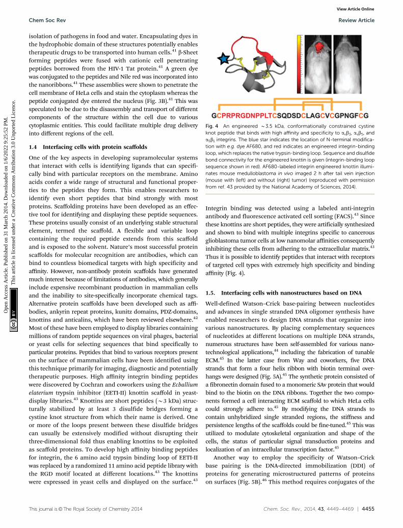
Most of these have been employed to display libraries containingmillions of random peptide sequences on viral phages, bacterialor yeast cells for selecting sequences that bind specifically toparticular proteins. Peptides that bind to various receptors presenton the surface of mammalian cells have been identified usingthis technique primarily for imaging, diagnostic and potentiallytherapeutic purposes. High affinity integrin binding peptideswere discovered by Cochran and coworkers using the Ecballiumelaterium trypsin inhibitor (EETI-II) knottin scaffold in yeast-display libraries.43 Knottins are short peptides (B3 kDa) struc-turally stabilized by at least 3 disulfide bridges forming acystine knot structure from which their name is derived. Oneor more of the loops present between these disulfide bridgescan usually be extensively modified without disrupting theirthree-dimensional fold thus enabling knottins to be exploitedas scaffold proteins. To develop high affinity binding peptidesfor integrin, the 6 amino acid trypsin binding loop of EETI-IIwas replaced by a randomized 11 amino acid peptide library withthe RGD motif located at different locations.43 The knottinswere expressed in yeast cells and displayed on the surface.43
Integrin binding was detected using a labeled anti-integrinantibody and fluorescence activated cell sorting (FACS).43 Sincethese knottins are short peptides, they were artificially synthesizedand shown to bind with multiple integrins specific to cancerousglioblastoma tumor cells at low nanomolar affinities consequentlyinhibiting these cells from adhering to the extracellular matrix.43
Thus it is possible to identify peptides that interact with receptorsof targeted cell types with extremely high specificity and bindingaffinity (Fig. 4).
1.5. Interfacing cells with nanostructures based on DNA
Well-defined Watson–Crick base-pairing between nucleotidesand advances in single stranded DNA oligomer synthesis haveenabled researchers to design DNA strands that organize intovarious nanostructures. By placing complementary sequencesof nucleotides at different locations on multiple DNA strands,numerous structures have been self-assembled for various nano-technological applications,44 including the fabrication of tunableECM.45 In the latter case from Way and coworkers, five DNAstrands that form a four helix ribbon with biotin terminal over-hangs were designed (Fig. 5A).45 The synthetic protein consisted ofa fibronectin domain fused to a monomeric SAv protein that wouldbind to the biotin on the DNA ribbons. Together the two compo-nents formed a cell interacting ECM scaffold to which HeLa cellscould strongly adhere to.45 By modifying the DNA strands tocontain unhybridized single stranded regions, the stiffness andpersistence lengths of the scaffolds could be fine-tuned.45 This wasutilized to modulate cytoskeletal organization and shape of thecells, the status of particular signal transduction proteins andlocalization of an intracellular transcription factor.45
Another way to employ the specificity of Watson–Crickbase pairing is the DNA-directed immobilization (DDI) ofproteins for generating microstructured patterns of proteinson surfaces (Fig. 5B).46 This method requires conjugates of the
Fig. 4 An engineered B3.5 kDa, conformationally constrained cystineknot peptide that binds with high affinity and specificity to avb3, avb5, anda5b1 integrins. The blue star indicates the location of N-terminal modifica-tion with e.g. dye AF680, and red indicates an engineered integrin-bindingloop, which replaces the native trypsin-binding loop. Sequence and disulfidebond connectivity for the engineered knottin is given (integrin-binding loopsequence shown in red). AF680-labeled integrin engineered knottin illumi-nates mouse medulloblastoma in vivo imaged 2 h after tail vein injection(mouse with (left) and without (right) tumor) (reproduced with permissionfrom ref. 43 provided by the National Academy of Sciences, 2014).
Chem Soc Rev Review Article
Ope
n A
cces
s A
rtic
le. P
ublis
hed
on 3
1 M
arch
201
4. D
ownl
oade
d on
1/6
/202
2 9:
25:5
2 PM
. T
his
artic
le is
lice
nsed
und
er a
Cre
ativ
e C
omm
ons
Attr
ibut
ion
3.0
Unp
orte
d L
icen
ce.
View Article Online

4456 | Chem. Soc. Rev., 2014, 43, 4449--4469 This journal is©The Royal Society of Chemistry 2014
protein-of-interest functionalized with short single-strandedoligonucleotides (ssDNA), e.g. via simple biotin–SAv conjugation,and surface substrates functionalized with capture oligonucleo-tides complementary to the DNA–protein conjugates employingvarious modern microstructuring techniques.47 For example,Heath and coworkers installed biotin on cell-surface proteins viaincubation with N-hydroxysuccinimide–biotin. The biotinylatedhuman primary neurons and astrocyte cells were then encodedwith ssDNA–SAv conjugates.48 The DNA-tagged cells were immo-bilized on the corresponding spots bearing the complementaryDNA.48 Various methods are available for the cell-surfaceattachment of ssDNA tags,46 for example membrane anchoringof oligonucleotides or the Staudinger ligation of phosphine-modified ssDNA to azide groups that are installed on the cellsurface through metabolic labeling.49,50 DDI of cells on glasssurfaces has been used to study cell sorting, cell adhesion andintercellular interactions or to grow small populations of cellswhich could be utilized for screening purposes.50–52 Niemeyer andDehmelt and coworkers fabricated protein ligand arrays withsubcellular dimensions (Fig. 5B).53,54 To this end, a DDI arraywas fabricated by dip-pen lithography to generate micrometer-sizedpatterns, typically 45 mm with a pitch of 12 mm.53 The DDIarrays represented either biotinylated epidermal growth factor
for culturing with carcinoma cells or different antibodies withbinding specificity for peptide epitopes on transmembranereceptor constructs such as the regulatory unit II-b.54 The latterrecognition led to specific recruitment and concentration of thetransmembrane proteins which was visualized by total internalreflection microscopy (TIRF).54 TIRF studies were used to furthervisualize the recruitment of the cytoplasmic catalytic subunit cat-aof protein kinase A to the DDI-areas.54
1.6. Employing peptide amphiphile nanostructures for cellstudies
When peptides are substituted with amphiphilic moieties, theybecome interesting building blocks to create self-assemblingbiofunctional nanostructures.55,56 Such peptide amphiphiles (PA)self-assemble in aqueous conditions into 1D nanostructures thatpossess a hydrophobic core and a hydrophilic shell. These nano-structures could easily be used to encapsulate small hydrophobicmolecules for drug delivery. Moreover, the fibrous character ofthe nanostructures allows their employment in 3D networks asbioactive scaffolds. The structure of the PAs that is required tocreate such high-aspect-ratio nanofibers is shown in Fig. 6A.One part of the PAs is a hydrophobic block, usually an alkylchain the length of which can be tuned. The hydrophobic partof the molecule is conjugated to a b-sheet forming segment coupledto a sequence with charged amino acids. The final element in thestructure is a bioactive peptide sequence. The charged amino acidsguarantee good solubility under physiological conditions. Betweenthem there is electrostatic repulsion which pushes the moleculesapart. The balance of all three forces, hydrophobic (alkyl chain),hydrogen bonding (b-sheet forming segment) and electrostaticrepulsion (charged amino acids), determines the size andshape of the final cylindrical assembly (Fig. 6B).55 Due to theircomposition the PAs can be considered biodegradable sincethey can be metabolized into amino acids and lipids.57 Essentialto the biofunctionality of the nanostructures are the epitopesthat are introduced that can interact with cells or proteins.This part is presented on the periphery of the assembled
Fig. 5 (A) A DNA/protein-based matrix (ECMDP) was constructed from aDNA ribbon (ECMD) surface-functionalized with proteins (ECMP) containingthe RGD domain of human fibronectin (reproduced with permission fromref. 45 provided by the American Chemical Society, 2014). (B) Schematicrepresentation of a protein-array inside living cells using surface-boundligand patterns, DDI and bait-presenting artificial receptor constructs(bait-proteins, orange, blue, green and purple surface bound ligands viaDDI). Interaction analysis of a cytosolic prey protein (yellow) fused to afluorescent protein (red) can be imaged by total-internal reflection fluores-cence microscopy (reproduced with permission from ref. 53 provided byJohn Wiley and Sons, 2014).
Fig. 6 (A) Molecular structure of a representative PA with four rationallydesigned chemical entities. (B) Molecular graphics illustration of an IKVAV-containing PA molecule and its self-assembly into nanofibers (reproducedwith permission from ref. 55 provided by John Wiley and Sons, 2014). (C)Scanning electron micrograph of the IKVAV nanofiber network formed byadding cell media to the PA aqueous solution (reproduced with permissionfrom ref. 55 provided by John Wiley and Sons, 2014).
Review Article Chem Soc Rev
Ope
n A
cces
s A
rtic
le. P
ublis
hed
on 3
1 M
arch
201
4. D
ownl
oade
d on
1/6
/202
2 9:
25:5
2 PM
. T
his
artic
le is
lice
nsed
und
er a
Cre
ativ
e C
omm
ons
Attr
ibut
ion
3.0
Unp
orte
d L
icen
ce.
View Article Online
This journal is©The Royal Society of Chemistry 2014 Chem. Soc. Rev., 2014, 43, 4449--4469 | 4457
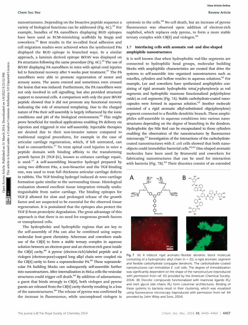
nanostructures. Depending on the bioactive peptide sequence avariety of biological functions can be addressed (Fig. 6C).55 Forexample, bundles of PA nanofibers displaying RGD epitopeshave been used as ECM-mimicking scaffolds by Stupp andcoworkers.58 Best results in the so-called focal adhesion andcell migration studies were achieved when the synthesized PAsdisplayed the RGD epitope in branched ways. In a similarapproach, a laminin derived epitope IKVAV was displayed onPA structures following the same procedure (Fig. 6C).59 The use ofIKVAV displaying PA nanofibers in mice with spinal cord injuriesled to functional recovery after 9 weeks post treatment.57 The PAnanofibers were able to promote regeneration of motor andsensory axons. The axons entered and sometimes even crossedthe lesion that was induced. Furthermore, the PA nanofibers werenot only involved in cell signalling, but also provided structuralsupport to neuronal cells. A comparison with only the pure IKVAVpeptide showed that it did not promote any functional recoveryindicating the role of structural templating. Due to the chargednature of PAs their self-assembly is largely influenced by the ionicconditions and pH of the biological environment.59 This mightprove beneficial for medical applications enabling PA delivery viainjection and triggered in vivo self-assembly. Injectable therapiesare desired due to their non-invasive nature compared totraditional surgical procedures, for example in the case ofarticular cartilage regeneration, which, if left untreated, canlead to osteoarthritis.57 To treat spinal cord injuries in mice apeptide domain with binding affinity to the transforminggrowth factor b1 (TGF-b1), known to enhance cartilage repair,is used.57 A self-assembling bioactive hydrogel prepared byusing two different PAs, a non-bioactive and the TGF-bindingone, was used to treat full thickness articular cartilage defectsin rabbits. The TGF-binding hydrogel induced de novo cartilageformation quite similar to the surrounding tissue. Histologicalevaluation showed excellent tissue integration virtually undis-tinguishable from native cartilage. The binding epitopes forTGF-b allowed for slow and prolonged release of the growthfactor and are suspected to be essential for the observed tissueregeneration. It is postulated that the epitopes also protect theTGF-b from proteolytic degradation. The great advantage of thisapproach is that there is no need for exogenous growth factorsor transplanted cells.
The hydrophobic and hydrophilic regions that are key tothe self-assembly of PAs can also be combined using supra-molecular host–guest chemistry. Scherman and coworkers madeuse of the CB[8] to form a stable ternary complex in aqueoussolution between an electron-poor and an electron-rich guest insidethe CB[8] cavity.60 A pyrene (electron-rich)-labeled peptide and aviologen (electron-poor)-capped long alkyl chain were coupled viathe CB[8] cavity to form a supramolecular PA.60 These supramole-cular PA building blocks can subsequently self-assemble furtherinto nanostructures. After internalization in HeLa cells the vesicularstructures could trigger cell death.60 By addition of adamantane,a guest that binds strongly to CB[8], both viologen and pyreneguests are released from the CB[8] cavity thereby resulting in a lossof the nanostructures.60 The release of pyrene was confirmed bythe increase in fluorescence, while uncomplexed viologen is
cytotoxic to the cells.60 No cell death, but an increase of pyrenefluorescence was observed upon addition of electron-richnaphthol, which replaces only pyrene, to form a more stableternary complex with CB[8] and viologen.60
1.7 Interfacing cells with aromatic rod- and disc-shapedamphiphile nanostructures
It is well known that when hydrophobic rod-like segments areconnected to hydrophilic head groups, molecular buildingblocks with amphiphilic characteristics are created that drive thesystems to self-assemble into organized nanostructures such asmicelles, cylinders and hollow vesicles in aqueous solutions.61 Forexample, Lee and coworkers have synthesized amphiphiles con-sisting of rigid aromatic hydrophobic tetra( p-phenylene)s as rodsegments and hydrophilic mannose functionalized poly(ethyleneoxide) as coil segments (Fig. 7A). Stable carbohydrate-coated nano-capsules were formed in aqueous solution.62 Another moleculeconsisted of a rigid aromatic alkyl-substituted oligo(phenylene)segment connected to a flexible dendritic branch. These amphi-philes self-assemble in aqueous conditions into various nano-structures depending on the degree of branching in the dendron.Hydrophobic dye Nile Red can be encapsulated in those cylindersenabling the observation of the nanostructures by fluorescencemicroscopy.63 Investigation of the interactions of the carbohydrate-coated nanostructures with E. coli cells showed that both nano-objects could immobilize bacterial cells.62,63 Disc-shaped aromaticmolecules have been used by Brunsveld and coworkers forfabricating nanostructures that can be used for interactionwith bacteria (Fig. 7B).64 Their discotics consist of an extended
Fig. 7 (A) A triblock rigid aromatic-flexible dendritic block moleculeconsisting of a hydrophobic alkyl chain (n = 21), a rigid aromatic segmentand flexible carbohydrate conjugate dendrons. The carbohydrate-coatednanostructures can immobilize E. coli cells. The degree of immobilizationwas significantly dependent on the shape of the nanostructure (reproducedwith permission from ref. 63 provided by the American Chemical Society,2014). (B) Discotic compounds functionalized with mannose ligands (R2)and inert glycol side chains (R1) form columnar architectures. Binding ofthese systems to bacteria result in their clustering, which was visualizedusing fluorescence microscopy (reproduced with permission from ref. 64provided by John Wiley and Sons, 2014).
Chem Soc Rev Review Article
Ope
n A
cces
s A
rtic
le. P
ublis
hed
on 3
1 M
arch
201
4. D
ownl
oade
d on
1/6
/202
2 9:
25:5
2 PM
. T
his
artic
le is
lice
nsed
und
er a
Cre
ativ
e C
omm
ons
Attr
ibut
ion
3.0
Unp
orte
d L
icen
ce.
View Article Online

4458 | Chem. Soc. Rev., 2014, 43, 4449--4469 This journal is©The Royal Society of Chemistry 2014
aromatic core of three 2,20-bipyridine-3,30-diamines linked to acentral benzene-1,3,5-tricarbonyl unit.64 These discotics canself-assemble in water to form an auto-fluorescent columnarpolymer.64 The peripheral hydrophilic ethylene glycol chainsprovide water-solubility and shield the hydrophobic core topromote hydrogen bonding and p–p stacking between adjacentdiscotics. Functionalization of the peripheral ethylene oxide tailswith mannose groups led to selective binding of the supramole-cular polymers to E. coli bacteria.64 The same discotic moleculeshave been adopted as cellular uptake carriers by Brunsveld andcoworkers.65 p–p-Stacked assemblies of discotic monomers thatfeature peripheral amine groups are easily taken up by the cellsvia endocytosis to the cytoplasm.65 As expected, when theseperipheral amine groups are absent, the supramolecular polymersare not taken up.65 Interestingly when non-cell permeablemonomers, which are functionalized with biotinylated ligands,are blended in supramolecular polymers of amine-functionalizedmonomers, successful uptake of the supramolecular polymer wasobserved through staining with a fluorescently labelled anti-biotinantibody after fixation of the cells.65
1.8 Interfacing cells with lipid bilayer architectures
The early notion that the cell membrane, which consists of alipid bilayer with associated proteins and carbohydrates, acts asa liquid in which its constituents can move freely is considerednowadays an oversimplification of reality. This so-called ‘fluidmosaic model’ has been updated to include variable patchiness,variable thickness and higher protein occupancy than was previouslyconsidered. Studying membrane associated processes forcedscientists to conceive model lipid bilayers that mimicked theamphiphilic lipid architecture ensuring that bilayer propertiesare comparable to those of natural cell membranes. Such modelmembranes can be subdivided into suspended, so-called blacklipid membranes (BLMs), and solid supported lipid bilayers(SLBs) and lipid vesicles (Fig. 8). Among the broad class of self-assembled amphiphilic lipids that are generally termed asvesicles, liposomes are unilamellar vesicles where the aqueousinterior of each vesicle is bound by a single lipid bilayer.66–69
Besides the aforementioned examples other lipid inspired structureshave also been studied like lipid monolayers and lipid derivedpolymers. Common, either natural or synthetic, lipid buildingblocks can be categorized according to their structure, e.g. fattyacids, phospholipids, glycerophospholipids, triacylglycerols,eicosanoids, waxes, sphingolipids, as well as steroids, isoprenoidsand terpenes. Composition and protocol of preparation canbe tailored to tune the physicochemical properties of the lipidsystem, either vesicular or supported, e.g. surface charge,bilayer fluidity and lamellarity (i.e. number of lipid bilayers).Fluidity, which refers to the lateral diffusion of the membraneconstituents, is affected by the gel–liquid transition tempera-ture of the lipids, which is determined by the alkyl tails and ismodulated in vivo by cholesterol. The lipid systems can befurther modified by direct covalent coupling of ligands to lipidbilayers, non-covalent interaction using chelating lipids andvia insertion of natural or synthetic lipid anchors. A detailedoverview of ligand-bilayer immobilization via covalent and
non-covalent chemistry is reviewed elsewhere.68 Modificationsof lipid bilayer structures can improve the limited stability incertain environments, such as in air or in vivo.
1.8.1 Interfacing cells with lipid vesicles. Interfacial cellularmembrane processes often involve lipid vesicles that traffic inand out of the cell and among its compartments. Exo- andendocytosis are examples of these phenomena. Although themolecular mechanism is not entirely clear, such lipid vesicles arehypothesized to dock to the membrane and eventually spatiallyand temporally rearrange their lipid bilayer to fuse with the cellmembrane.67 This fusion process has found two main applica-tions that we will further discuss in this section: upon fusion, thecargo of the vesicle is delivered into the cell; on the other hand,the rearrangement of the lipids occurring upon fusion can installartificial molecular functionalities on the outer surface of themembrane.66,69,70
Liposomes and liposome-based structures have matured forthe intracellular delivery of drugs and imaging agents over thelast decades and have found clinical application.69 The delivery
Fig. 8 An overview of artificial lipid bilayers and a few parameters are giventhat can be fine-tuned such as (A) phase transition temperature resulting inmobile (liquid) or immobile (gel) bilayers, (B) ligand immobilization throughe.g. non-covalent interactions (left), covalent reactions (middle) and post-insertion of ligand-modified lipids (right). (C) Various types of lipid-basedsolid-supported and solution architectures.
Review Article Chem Soc Rev
Ope
n A
cces
s A
rtic
le. P
ublis
hed
on 3
1 M
arch
201
4. D
ownl
oade
d on
1/6
/202
2 9:
25:5
2 PM
. T
his
artic
le is
lice
nsed
und
er a
Cre
ativ
e C
omm
ons
Attr
ibut
ion
3.0
Unp
orte
d L
icen
ce.
View Article Online

This journal is©The Royal Society of Chemistry 2014 Chem. Soc. Rev., 2014, 43, 4449--4469 | 4459
takes place by uptake of the particle and its cargo by the cell.The internalization can be mediated by non-specific electro-static recognition between positively charged vesicles and thenegatively charged lipids of cell membranes,71 as extensivelydemonstrated in the field of gene transfection where typicallyDNA and cationic lipids are combined in the so-called lipoplexes.71
Anderson and coworkers have adopted the use of lipidoids tofacilitate non-viral delivery of small interfering RNA to endo-thelial cells (Fig. 9A).72–74 Michael addition chemistry betweenalkylacrylate and acrylamide materials and amines was utilizedto create a structurally diverse library of lipid-like moleculestermed lipidoids, which were analyzed for their ability totransfect cells both in vitro and in vivo.72,73 The library allowedthe study of the role of the length and number of alkyl chains,the charge and the substitution of the amines as well as theamide bond. The lead candidates facilitate sequence-specificknockdown in a variety of cellular targets and animal species.72
Typically these lipidoids form cationic 200 nm spherical lipid-likeparticles.73 Improvement in delivery efficacy has been achievedthrough synthesizing lipidoids following ring-opening of alkyl-substituted epoxides by amine substrates.74 With these lipid-likeparticles siRNA-directed liver gene silencing at therapeuticallyrelevant doses was possible.74
Endocytotic pathways can also be exploited to enter thevesicle in the cytosol in the form of an endosome. To this end,vesicles can be decorated with specific ligands that recognizecomplementary cell receptors to enhance their uptake.75 Thelateral diffusion of such ligands on the surface of the vesicle
allows their pre-organization and thereby creates an array ofmultiple binding sites for cell receptors, which leads to a morefavorable interaction according to the concept of multivalency.76
However, in addition to a thermodynamically favorable bindingof the carrier vesicle to the membrane, the carrier vesicle has torelease its cargo to elicit cell responses. Moreover, especiallywhen considering in vivo applicability, vesicle features such aspersistence, clearance or accumulation of the vesicles in thebody have to be balanced by enzyme degradability into non-toxicbyproducts after the release of the load. Typically a decorationwith hydrophilic polymers such as polyethylene glycol improvesthe in vitro and in vivo stability of lipid vesicles. Vesicles basedon an amphiphilic CB[6] derivatized with EG6 at its peripheryallow for incorporation of spermine-modified targeting ligandsand imaging probes through specific host–guest interactionsbetween spermidine and the CB[6] host.77 When these supra-molecular lipid vesicles were loaded with doxorubicin, inter-nalization into cells by receptor mediated endocytosis andrelease of entrapped drugs was confirmed.77
Further developments have been demonstrated employing asilicate porous framework to decorate the surface of porphyrin-conjugated vesicles.78 These particles circulated in blood forprolonged time and reached the cytosol by endocytosis. Uponirradiation of the porphyrin units by light inside the cells, thesinglet oxygen caused cell death of cancer cells.78 Enhancedstability could also be achieved by using interbilayer-crosslinkedmultilamellar vesicles.79 Crosslinking was achieved by themaleimide modified head-groups of opposing bilayers using a
Fig. 9 (A) An example of the synthesis of lipidoids through the conjugate addition of amines to acrylamides. TEM images of lipidoid nanoparticlescomplexed with siRNA (scale bar 200 nm). Successful silencing of SHP-1 by siSHP-1 delivery in HUVECs (10% serum) as concluded from quantitative real-time PCR measurements to determine the SHP-1 expression in HUVECs at 2 days after siRNA transfection using the synthesized lipidoids (NA114, twodoses) and compared with lipofectamine 2000 (LIPO2000) and all siGFP-transfected groups (reproduced with permission from ref. 73 provided by JohnWiley and Sons, 2014). (B) Schematic showing formation of vesicles from biotinylated lipid and modification of cells with biotinylated lipid vesicles. Thevesicle modified MSCs are further modified with sialyl Lewis X using SAv and biotinylated SLeX. Rolling of SLeX modified hMSCs on a P-selectin coatedsurface was significantly improved (8 mm s�1 as compared to 61 mm s�1 for unmodified MSCs) from flow chamber assays (0.5 dyn cm�2) (reproduced withpermission from ref. 84 provided by Elsevier, 2014).
Chem Soc Rev Review Article
Ope
n A
cces
s A
rtic
le. P
ublis
hed
on 3
1 M
arch
201
4. D
ownl
oade
d on
1/6
/202
2 9:
25:5
2 PM
. T
his
artic
le is
lice
nsed
und
er a
Cre
ativ
e C
omm
ons
Attr
ibut
ion
3.0
Unp
orte
d L
icen
ce.
View Article Online

4460 | Chem. Soc. Rev., 2014, 43, 4449--4469 This journal is©The Royal Society of Chemistry 2014
dithiol linker. Residual maleimides were capped by reactionwith thiolated PEG and yielded an inert vesicular surface.Interbilayer-crosslinked vesicles stably carried antigenic proteinsin the hydrophilic vesicle core and apolar immunostimulatoryagents in the hydrophobic vesicle wall. Endosomal lipasescatalyzed the selective rupture and release of the cargo.79
Alternatively, fusion of vesicles to the cellular membrane caninstall chemical functionalities on the cell surface.80 Thisfusion leads to cell membranes that are transiently doped withartificial lipids without altering the cellular machinery.80 Thismethod serves as an alternative to e.g. antibody recognition,81
metabolic labeling82 and covalent modification83 methods. Transi-ent chemically modified membranes are of interest as they wouldpotentially transform cells in therapeutic agents as tumor vaccinesor building blocks for tissue regeneration. The surface of stem cells(hMSC) was chemically engineered by incubation with liposomesintegrally composed of an amphiphilic biotinylated lipid.84 Theartificial lipids were observed as rafts, i.e. locally clustered onthe membrane. The exposed biotin groups were accessible forbinding to SAv and subsequently to biotinylated homing ligandsialyl Lewis X (Fig. 9B).84 The engineered hMSCs with sialylLewis X showed improved rolling behavior on P-selectin surfacesunder flow conditions while not changing the cell phenotype.These results indicate that the transitory modification of cellsurfaces with lipid vesicles can be used to efficiently immobilizeadhesion ligands and potentially target systemically administeredcells to the site of inflammation.84 Again, size, charge and overallcomposition of the vesicle can be tailored, as well as the indivi-dual lipids in terms of their gel–liquid transition temperature andfusogenicity (i.e. propensity to fuse).67 Chemoselective cell-surface engineering based on electrostatic interaction betweenthe positively charged liposomes and negatively charged lipidsof the cellular membrane led to the display of bio-orthogonalfunctional groups at cellular membranes.85 This strategy, asreported by Yousaf and coworkers, allowed for the display ofketone or oxyamine groups to different populations of cells forsubsequent cell assembly via oxime ligation.85 The authors demon-strated several applications including the selective fluorescentlabeling of the cell surface, the formation of small spheroid cellassemblies that are yet able to differentiate, and the generationof large and dense, 3D multilayered tissue-like structures fortissue engineering applications.85 In another report thiol-reactive maleimide headgroups of the lipid bilayer surface ofdrug-loaded uni- or multilamellar liposomes were reacted withthe plasma membrane of lymphocytes.86 This strategy enabledcontinuous autocrine stimulation of donor cells in vivo.86 Fromthese examples it is evident that lipid vesicles can be employedto change native cell membranes, either by the tunable deliveryof active agents or by cell membrane functionalization, intoreactive surfaces for new biotechnological applications such astissue regeneration and cell-based therapies.
1.8.2 Interfacing cells with lipid bilayers. The formation ofSLBs is made possible by Langmuir–Blodgett and vesicle fusiontechniques on a wide variety of solid supports using differentkinds of lipids. Depending on the lipids used, the phasetransition temperature can be tuned resulting in either liquid
or gel state SLBs or inducing phase separation. Certain ternarylipid mixtures give rise to so-called lipid raft domains. Theseliquid-ordered patches, otherwise referred to as detergentresistant domains, are suggested to resemble mammalian cellmembrane organization and play a key role in receptor cluster-ing and signal transduction.87 The use of charged lipids resultsin SLBs possessing a certain surface charge. The importance ofthe surface charge in SLBs for fabricating biomimetic systemswas shown by neuronal cell culture on positively charged SLBs.Upon doping the bilayer with a cationic lipid (DOTAP, 1,2-dioleoyl-3-trimethylammonium-propane) neuronal cell adhesion waspromoted, an effect not observed on neutral, zwitterionic SLBs.88
Moreover, patterning of SLBs can be achieved by applicationof diffusion barriers, micro-contact printing and by the use ofmicro-fluidic systems. Interestingly, SLBs have also beenproposed as a separator medium for membrane proteins usinga process referred to as membrane electrophoresis.89,90 SLBsare particularly interesting to study membrane associatedproteins in their native form. Transmembrane proteins canbe reconstituted in SLBs without modification and studied withrespect to receptor clustering and biological activity. DopingSLBs with biotin modified lipids and subsequently assemblingfluorescently labeled SAv and biotinylated EGF (epidermalgrowth factor) allowed for simple fluorescent monitoring anddetection of local enrichment and clustering of EGF on the cellmembrane.91 Groves and coworkers were interested in theprotein sorting that occurs upon T-cell immunological synapse(IS) formation (Fig. 10A).92 To shed light on this process, SLBswere prepared presenting key proteins aimed at mimickingthe antigen presenting cell (APC) membrane. In vivo, APC andT cells interact to yield the hallmark IS where the peripheraland central supramolecular activation clusters, pSMAC andcSMAC, are formed. The reasons underlying why T-cell receptor(TCR) segregates to the cSMAC and the lymphocyte associatedantigen-1 (LFA-1) to pSMAC just microns apart were not under-stood.93 It was known that the cluster size of LFA-1 and TCRduring IS formation differed, i.e. a few receptors comparedto approximately 100, respectively. They chose to increase theLFA-1 cluster size to assess whether segregation to the cSMACwould occur. Crosslinking in the SLB of LFA-1 and its antigen,the intercellular adhesion molecule-1 (ICAM-1), was achievedby antibody interaction. To allow for TCR cluster formation theSLB was doped with the peptide-major histocompatibility complex(pMHC). It was observed that an increase in LFA-1 cluster sizemediated its translocation from the peripheral to central SMAC,confirming that cluster size influences membrane positioning ofLFA-1 under the influence of centripetal F-actin flow. The naturaland dynamic response of the T-cell induced by the SLB surface is aclear showcase of the biomimetic potential of artificial lipidbilayers. Intriguingly, receptor clustering can be tuned at biologicalrelevant length scales as shown in the aforementioned example.More recently, they investigated the threshold level of pMHCligands per TCR cluster by partitioning the SLB with Cr-barriers,thus limiting the total number of ligands available for TCR clusterformation.94 Besides for studying membrane processes, SLBs haveproven to be an interesting platform for cell culture as well.
Review Article Chem Soc Rev
Ope
n A
cces
s A
rtic
le. P
ublis
hed
on 3
1 M
arch
201
4. D
ownl
oade
d on
1/6
/202
2 9:
25:5
2 PM
. T
his
artic
le is
lice
nsed
und
er a
Cre
ativ
e C
omm
ons
Attr
ibut
ion
3.0
Unp
orte
d L
icen
ce.
View Article Online

This journal is©The Royal Society of Chemistry 2014 Chem. Soc. Rev., 2014, 43, 4449--4469 | 4461
Given the importance of cadherin for the formation of adherensjunctions and subsequent polarization of epithelial cells the useof cadherin functionalized SLBs was studied recently by Textorand coworkers.95 A biotinylated SLB was incubated sequentially
with SAv, biotinylated IgG and Fc tagged extracellular domainE-cadherin. E-cadherin presented in this format was laterallymobile when coupled to the SLB and allowed sufficient initiationof cell polarization to mimic cell–cell contacts.95
Fig. 10 (A) Schematic of a T-cell on a substrate patterned with diffusion barriers (reproduced with permission from ref. 92 provided by Elsevier, 2014). (B)Schematic of the method used to create peptide RGD-functionalized SLB glass surfaces for studying neural stem cell adhesion. Phase contrast images ofNSCs after incubation on RGD-SLBs (FGF2-containing media, 5 days, scale bar: 100 mm) (reproduced with permission from ref. 96 provided by Elsevier,2014). (C) Targeting and fusogenic peptides are chemically conjugated to maleimide–lipids and mixed in at 1–5 wt%. The vesicles composed of eitherfluid (DOPC) or non-fluid (DPPC) zwitterionic phosphatidylcholine lipids with 30 wt% cholesterol are further modified with 5 wt% PEG-2000 PE toenhance colloidal stability and decrease nonspecific interactions. The vesicles are loaded with cargo (chemotherapeutics (doxorubicin, 5-fluorouracil,cisplatin)), q-dots for imaging, diphtheria toxin A-chain and siRNA. The vesicles (1) bind to human hepatocellular carcinoma cells with high affinity owingto recruitment of the targeting peptides (magenta) to the cell surface, (2) become internalized by receptor-mediated endocytosis and (3) release theircargo into the cytosol upon endosome acidification and protonation of the fusogenic peptide (blue). (4) Cargos modified with a nuclear localizationsequence (NLS) are transported through the nuclear pore complex and become concentrated in the nucleus (reproduced with permission from ref. 104provided by the Nature Publishing Group, 2014).
Chem Soc Rev Review Article
Ope
n A
cces
s A
rtic
le. P
ublis
hed
on 3
1 M
arch
201
4. D
ownl
oade
d on
1/6
/202
2 9:
25:5
2 PM
. T
his
artic
le is
lice
nsed
und
er a
Cre
ativ
e C
omm
ons
Attr
ibut
ion
3.0
Unp
orte
d L
icen
ce.
View Article Online

4462 | Chem. Soc. Rev., 2014, 43, 4449--4469 This journal is©The Royal Society of Chemistry 2014
Owing to the lipid bilayers’ inherent non-fouling nature withrespect to protein adsorption and cell adhesion, specific cellresponses can be observed. Tirrell and coworkers designedhighly tunable cell culture surfaces using SLBs. One studyshowed the potential of SLBs to culture neuronal cells.96
SLBs were doped with cell adhesive RGD peptide amphiphilesconsisting of a hydrophobic anchor and a hydrophilic spacerconjugated to the peptide sequence (Fig. 10B).96 Having anaccessible RGD motif, cell responses could be fine-tuned byvarying the hydrophilic spacer. A similar approach was adoptedby Gold and coworkers that utilized SLBs decorated with anIKVAV peptide instead.97 This peptide sequence is derived fromthe ECM glycoprotein laminin-1 that is known to modulate celladhesion and neurite outgrowth activity. Instead of usingpeptide amphiphiles, incorporated during bilayer formation,they demonstrated in situ modification of the SLBs.97 Duringbilayer formation thiol-reactive maleimide-functionalized lipidswere included that at a later stage react with cysteine modifiedIKVAV peptides.64 Model neuronal cells (PC12) only adhered tothe peptide modified SLBs. In a follow-up study they were able toelucidate a positive, nonlinear correlation between the number ofattached rat neuronal cells (AHP) and the density of IKVAVpeptides tethered to the SLB. Also, a threshold of ligand concen-tration was found, required for cell binding.98 Adopting a similarin situ modification strategy, SLBs were prepared serving as acarrier for ECM components. Huang and coworkers demonstratedthat an SLB consisting of part of a carboxylic acid modified lipidcould easily be modified to express collagen type 1 or fibronec-tin.99 In their set-up, collagen and the underlying lipid bilayer area better representation of the cellular environment where lipid–collagen interactions are important. The system allowed forsustained A10 smooth muscle cell culture only on collagenmodified SLBs. Not only the presentation of certain bioactiveligands on a SLB dictate cellular responses but also their mobilityas was demonstrated by Reimhult and coworkers.100 Dependingon the phase transition temperature, the lateral mobility of lipidswas greatly affected. In a liquid state system diffusion coefficientsin the order of mm2 per second are found while gel state SLBsshow negligible diffusion.100 This in turn influences the mobilityof cadherin tethered to biotin-modified lipids. The bilayer stateinfluenced cell response dramatically; a more spread cell mor-phology was observed on gel state SLBs compared to liquid stateones.100 Within the rich field of planar lipid bilayer research,others chose to focus on the incorporation of functionally activeproteins into such model systems and to ensure proper function,often involving electrical signaling. Therefore lipid bilayer systemsshould meet certain electrical standards compatible with the useof electrochemical techniques. For instance, means of lipid bilayertethering must be considered. An overview of design criteriainvolved and past results are nicely presented in the review byNaumann and coworkers.101 Another aspect that deserves atten-tion is the lack of stability of SLBs when exposed to air. Bilayerstabilization can be achieved by the use of photo-cross-linkablelipids that bear apolar tails containing diacetylene bonds.102
These lipids readily crosslink under UV light to yield a solventresistant and air-stable SLB that is also applicable to prepare
polymerized lipid vesicles. However, such strategies do not yieldmobile SLBs. Several attempts in that direction have been made;for instance sterol tethering of SLB and the use of protein layerson top of SLB imparted air-stability while retaining lateral motionof lipids.103
Both liposomes, as discussed in the previous section, and SLBsare powerful systems for cell research due to the high degree ofsurface tunability and their multipurpose, dynamic nature andare proven bio-mimetic systems. Ashley and coworkers designeda multicomponent particle-supported lipid bilayer combiningboth systems.104 These so-called protocells consist of a carriersynthesized by liposome fusion onto spherical nanoporous silicaparticles, followed by modification of the resulting supportedlipid bilayer with PEG, targeting peptides and fusogenic peptides(Fig. 10C).104 Therapeutic and imaging agents could be simulta-neously loaded into the silica core with high capacity due to thenano-sized pores which increase the surface area. The rate of thecargo release could be optimized by affecting the particle dissolu-tion in the acidic endosomal environment through modifyingsilica composition and the protonation of the fusogenic peptide.In their study the protocell displayed a 10 000 fold higher affinityfor human hepatocellular carcinoma compared to hepatocytes,endothelial or immune cells, demonstrated the delivery of encap-sulated cargo upon cellular uptake and showed enhanced stabilityover traditional liposomes.104
1.8.3 Interfacing cells with other lipid inspired structures.Next to lipid bilayers, lipid monolayers and derivatives havebeen studied. In one non-covalent approach a photocleavablePEG–lipid including an oleyl group, which can bind to any celltype by non-covalent insertion into the cell membrane, wasused.105 Due to the lipophilic interior of the plasma membranethe oleyl chain can be anchored inside the membrane. Addition ofa PEG to the chain acts as a solubilizing unit for the hydrophobicoleyl in aqueous solutions. For dynamic studies a photocleavablelinker was inserted between the PEG and the oleyl moiety.105
Attachment of the PEG–lipid to the substrate was mediatedthrough the amino-reactive ester group added to the endgroupof the PEG. By irradiation with UV light at 365 nm the oleyl moietyis cleaved off, leaving only the cell repellent PEG layer on thesurface.105 They could furthermore show that tuning the lightdose can vary the degree of cellular release.105 Others have chosento include only the lipid headgroup in monolayers. For example,the use of zwitterion polymers, especially those bearing phos-phatidylcholine (PC) groups, has drawn attention. The PC groupcarries both positive and negative charge and is the mainconstituent of the outer layer of the erythrocyte membrane. Asa result, materials bearing PC groups on the surface reduce theamount of non-specific, irreversible protein adsorption. Such poly-mers are studied to enhance the biocompatibility of biomaterials,mostly used in biosensors. Takai and coworkers presented anoverview of PC–polymers and their application.106
1.9 Interfacing cells with dynamic self-assembled monolayers
The highly complex composition of the ECM and its interactionwith molecular fingerprints on the cell membrane make it ahighly challenging task to create model systems that allow us to
Review Article Chem Soc Rev
Ope
n A
cces
s A
rtic
le. P
ublis
hed
on 3
1 M
arch
201
4. D
ownl
oade
d on
1/6
/202
2 9:
25:5
2 PM
. T
his
artic
le is
lice
nsed
und
er a
Cre
ativ
e C
omm
ons
Attr
ibut
ion
3.0
Unp
orte
d L
icen
ce.
View Article Online

This journal is©The Royal Society of Chemistry 2014 Chem. Soc. Rev., 2014, 43, 4449--4469 | 4463
mimic cells’ spatial as well as temporal organization. Due totheir advantage in being well-ordered, permitting high interfacialcontrol of specific bioactive ligands and allowing great flexibilityin the creation of complex substrates with spatial precision, self-assembled monolayers (SAMs) have been a particularly attractivestrategy since their discovery in the early 80s. Analogous to self-assembling systems in solution, they are highly ordered nano-structures that spontaneously organize on surfaces. Morerecently, SAMs are also actively investigated as model substratesfor dynamic cell studies. This is desirable, as they not only allowthe spatial control of bioactive ligands, but additionally permitcontrol of their presence on the substrate in time. DynamicSAMs can change their surface properties in response to externalstimuli such as a change in pH, temperature, light or electro-chemical stimuli. However, since cells require steady physio-logical conditions, like pH and temperature, conversion ofsubstrate properties in response to light or electrochemicalstimuli has been used favourably for cell studies. To date, anumber of different types of substrate materials and chemicalstrategies to immobilize bioactive ligands have been exploitedfor use as dynamic substrates. Nevertheless, the majority ofsystems use SAMs of alkanethiols on gold for a number ofadvantages: e.g. being bio-inert, allowing simple and well-orderedfunctionalization and being available to various substrate analysistechniques. Alkanethiolates form densely packed well-orderedmonolayers on gold where the thiol head group binds with highaffinity to the underlying gold substrate. They can furthermore besimply adapted to hold several different functional groups in orderto modify the substrate with specific bioactive ligands. Conductivityof the gold substrate makes it suitable for even more analyticaltechniques to characterize interfacial interactions. We will heredescribe selected examples from the literature to illustrate thevarious strategies using dynamic SAMs for cell studies.
In a recent example, thiol interaction with gold acted asanchoring points for RGD peptides to achieve sub-cellularcontrol by functionalizing a gold electrode array.107 Modificationof the separating glass regions with PEG to make them cell-repellentresulted in selective cell-adhesion to the RGD-functionalizedelectrodes. Spatial design of sub-cellular distances betweenadjacent electrodes allowed cells to spread over multiple elec-trodes. When Searson and coworkers applied a negative voltagepulse on an individual gold electrode, selective release of thethiols was triggered and consequently retraction of that veryregion of the attached cell could be visualized by monitoringfluorescently labelled actin with fluorescence microscopy.107
Modulating cell detachment in this manner opens a new roadto gain significant insights into cellular pathway signalling.Flexibility in alkanethiol functionalization makes them a usefultool for creation of multi-purpose cell culture surfaces withmodular cell-repellent and cell-adhesive regions. The combi-nation of micropatterning technology with self-assembly ofdifferentially terminated thiols presents a powerful approachto generate spatially resolved micro-confinements to createmultifunctional SAM profiles on the substrate.47,108–110 Recentstudies have already demonstrated great potential of such patternedsystems for studying cell motility as well as cell–cell interactions.47
With the appropriate design of the SAM properties, such asspatial definition alongside chemical functionality, an electri-cal stimulus can for instance be used to trigger the release of aparticular section on the surface and allow cells to migrate intonewly available areas.108 This design makes it possible to studycell migration in a non-invasive manner. However, sincecoupling alkanethiols to a desired biomolecule can be labor-ious, other coupling strategies taking place directly at theinterface of a pre-assembled monolayer have been explored.Some of the frequently employed organic strategies includeredox-active hydroquinone (HQ) monolayers in combinationwith Diels–Alder conjugation,111,112 oxime reaction113–117 andCu(I)-catalysed Huisgen cycloaddition.118,119 The electrochemicalconversion of HQ-terminated monolayers on gold has been asuccessfully employed strategy to chemically regulate interfacialcell–substrate interactions. HQ can undergo oxidation upon anelectrochemical stimulus and convert to benzoquinone (BQ),which, in turn, can react with a number of functional groups.111
To ensure specific interactions, selective to the bioactive ligandof choice, a critical step is to block the background to unspecificbinding of proteins present in biological media. Short ethyleneglycol linkers have proven themselves as a very efficient choicefor that purpose, commonly used at 1 : 99% ligand to ethyleneglycol (EG) ratios. When HQ groups, present in a cell-inertbackground, are oxidized to BQ, they can readily form a covalentadduct via a Diels–Alder reaction with a cyclopentadiene(Fig. 11A).111 When such a cyclopentadiene is functionalized withRGD a reversible substrate is obtained that allows for controllingcell-repellent and cell-adhesive areas by changing the redox-potentials as described by Mrksich and coworkers.111 For spatialcontrol of cell adhesion and migration this strategy was extendedusing micropatterns. Patterns of hexadecanethiolate on goldwere reacted with fibronectin (Fn), onto which cells wereallowed to adhere.111 The backfilling of the non-patternedregions with a 1 : 99% HQ to EG mixture made this at first anarea unavailable for fibroblast cells.111 Upon electrically stimulatedoxidation of the HQs to BQ and following Diels–Alder mediatedRGD binding, these cells could consequently migrate out of theirpatterns into the surrounding areas.111 Instead of starting witha cell-inert HQ layer, Mrksich and coworkers used a silyl etherconjugated to HQ to create an O-silyl HQ functionality furthercoupled to an RGD.112 Upon electrical activation HQ converts toBQ and causes hydrolysis of the silyl ether, causing the releaseof the RGD. The resulting BQ functionality could as in theprevious example again react with a diene-coupled RGD ligandvia a Diels–Alder reaction, creating a dual-functional cell-adhesiveor repellent surface. In a similar fashion, the redox-activeHQ-terminated SAMs can, when oxidized to the resulting BQ,be coupled to aminooxy-terminated ligands by an oxime reactionas described by Yousaf and coworkers.113–117 Oxyamine reactscovalently with the ketone groups on BQ in high yield underphysiological conditions to form a stable oxyamine linkage. Thereacting aminooxy groups can furthermore be introduced in mostbiomolecules by using standard synthetic procedures. Complexsurfaces with well-defined spatial control were generated basedon this approach by improving the system with microarray or
Chem Soc Rev Review Article
Ope
n A
cces
s A
rtic
le. P
ublis
hed
on 3
1 M
arch
201
4. D
ownl
oade
d on
1/6
/202
2 9:
25:5
2 PM
. T
his
artic
le is
lice
nsed
und
er a
Cre
ativ
e C
omm
ons
Attr
ibut
ion
3.0
Unp
orte
d L
icen
ce.
View Article Online

4464 | Chem. Soc. Rev., 2014, 43, 4449--4469 This journal is©The Royal Society of Chemistry 2014
micropatterning techniques.113,114 In the latter case, the patternedareas were cell-adhesive and non-patterned areas at first cell-repellent. In this example, photo-induced conversion as well aselectrochemical activation was combined to selectively convert thenon-patterned areas.114 These regions initially presented a 1 : 99%mixture of HQ groups terminated with a photo-sensitive protec-tion group and cell-repellent EG4.114 UV illumination through agradient photomask could then selectively deprotect the HQgroups, which, followed by electrochemical conversion to BQ,could bind an aminooxy-terminated RGD ligand via an oximeconjugate (Fig. 11B).114 This caused the fibroblasts (adhering tothe mCP areas) to spread to the photo-activated RGD-gradient. Byapplying a reductive potential these aminooxy-RGD could then beselectively released. Consequently cells adhering to these non-patterned functionalized areas were released, while cells stillremained attached in the mCP areas.
In regenerative medicine a challenging and rewarding taskis to understand the underlying mechanisms driving stem celldifferentiation. Combining electroactive SAMs with microarraytechnology was demonstrated to be an elegant and promisingstrategy to immobilize a library of bioactive ligands on asubstrate to screen for optimal conditions.110,115 As in theexamples above, alkanethiol-gold substrates terminated withHQ groups or EG4 were used as the basic building blocks.Using microarray printing and mixing different ratios of HQ toEG4-terminated moieties, patterns of various ligand densitieswere obtained. A solution of alkanethiol-EG4 was then used toblock the remaining areas around the patterned areas. Creatinga library of different oxyamine-tethered ligands that could reactwith electrically oxidized HQ, BQ, arrays of precisely controlledspatial resolution and varying densities could be tailored on thesubstrate. With this versatile and complex platform at hand itwas possible to screen for the effect of ligand density andcomposition on the rate of stem cell (hMSC) differentiation.115
A similar platform has been used for studies of single cellpolarization.117 Cu(I)-catalyzed Huisgen cycloaddition betweenazides and alkynes is a chemoselective reaction that proceedsunder mild reaction conditions and therefore makes it anattractive approach for fabricating bioactive dynamic SAMs.
Fig. 11 (A) HQ immobilized to gold substrates oxidizes to BQ and under-goes Diels–Alder reaction with RGD–cyclopentadiene (Cp). Attachmentof RGD changes the substrate from cell-repellent to cell adhesive(reproduced with permission from ref. 111 provided by John Wiley andSons, 2014). (B) Combination of mCP and electrochemical as well asphoto-induced activation for complex cell substrates (reproduced withpermission from ref. 114 provided by John Wiley and Sons, 2014).114
(C) Electrochemical stimulated oxidative degradation of dicobalt hexa-carbonyl complexes to reveal alkynes on the monolayer and subsequentreaction with azides coupled to biomolecules via Cu(I)-catalyzed Huisgencycloaddition reaction (reproduced with permission from ref. 118 providedby the American Chemical Society, 2014). (D) Glass modified with PEGmoieties featuring a photocleavable linker. Upon light exposure thesephotocleavable groups undergo intermolecular oxidation and thus causePEG to be released. Whilst PEG is inert to cell adhesion, the photocleavageallows cells to spread to the newly available cell-adhesive areas (reproducedwith permission from ref. 119 provided by Elsevier, 2014).
Review Article Chem Soc Rev
Ope
n A
cces
s A
rtic
le. P
ublis
hed
on 3
1 M
arch
201
4. D
ownl
oade
d on
1/6
/202
2 9:
25:5
2 PM
. T
his
artic
le is
lice
nsed
und
er a
Cre
ativ
e C
omm
ons
Attr
ibut
ion
3.0
Unp
orte
d L
icen
ce.
View Article Online

This journal is©The Royal Society of Chemistry 2014 Chem. Soc. Rev., 2014, 43, 4449--4469 | 4465
Recently it was applied in combination with the oxime reactionas an in situ ‘‘hide-and-reveal’’ strategy of small biomolecules tocreate a dual chemoselective SAM, displaying switchable (HQ)as well as non-switchable azide functional groups.116 Bifunc-tional bioactive moieties were created by co-coupling the pep-tides with an oxyamine as well as an alkyne end group, whichare the respective chemical partners for the BQ and azide.When the moieties were switched from presenting linear tocyclic RGD, by application of a mild electrochemical potential,they could monitor real-time changes in cell behaviour such aschanges in cell adhesion and migration.116
Cu(I)-catalyzed Huisgen alkyne–azide cycloaddition has alsobeen reported by Yeo and coworkers as a direct strategy forelectroactive substrate dynamicity without use of the HQ link-age (Fig. 11C).118 Alkyne-terminated monolayers were maskedwith dicobalt hexacarbonyl complexes, making them inertto react with azides.118 Upon electrochemical activation thedicobalt hexacarbonyl complex was oxidatively degraded andthe thus exposed alkyne groups were available for reaction withazides.118 Coupling azides to a cell-adhesive peptide resulted insubstrates that were turned from cell-repellent to cell-adhesiveupon reaction with alkynes.118
Besides electrochemical stimulated release systems, the useof photo-induced activation has been explored.119,120 Siliconoxide substrates presenting free amine groups could provide areactive substrate for photo-cleavable linkers via carbamatebonding as described by del Campo and coworkers.120 Theselinkers contained an intercalated photolabile group, the o-nitro-benzyl derivative (NVOC), which upon UV irradiation underwentan intermolecular redox reaction, and hence, underwent cleavagefrom the substrate.120 Tethering this linker with a bio-ligandcreated a cell-adhesive substrate, that, upon light exposure, couldbe reversed to be cell-repellent. Similarly, a photo-cleavable2-nitrobenzyl-linked PEG monolayer formed on glass couldmake the substrate cell repellent, and upon light exposure causePEG-release and feature a cell-adhesive substrate (Fig. 11D).119
Spatial control of these systems can easily be achieved by aphotomask, thus regulating the desired areas of illumination.Yousaf and coworkers integrated a bioactive surface strategy withphotoelectroactive surface strategy to generate dynamic ligandsurface gradients for controlling cell adhesion, tissue shape
morphing and cell migration.121 To this end an NVOC-protectedHQ terminated alkanethiol was mixed in with the hydrophilicparts of a SAM that was otherwise hydrophobic.121 Irradiationrevealed the HQ group and fibroblasts were allowed to adhere tothe hydrophobic regions of the SAM.121 Only when the HQ wasconverted to the quinone group and oxyamine–RGD was coupledto the photo-deprotected regions, cells started to migrate into theRGD regions.121 Jiang and coworkers used SAMs of photorespon-sive azobenzenes conjugated to RGD on a background of EG6 tocontrol cell adhesion reversibly. Switching of azobenzenesbetween E isomer and Z isomer by UV light rendered the surfaceto be either adhesive by presenting the RGD or repellent byshielding the RGD within the inert EG6 layer.122
Since natural interfaces between the cell and its interactingligands build on non-covalent interactions, chemical systemsrelying on non-covalent binding such as hydrophobic, van derWaals, ion-dipole or hydrogen bonds are being explored andare promising to come yet a step closer to mimic natural cell–ECM interactions. A novel strategy to reversibly attach andrelease cells is the use of supramolecular host–guest chemistry.On a quartz substrate a host monolayer was formed by silane-terminated a-CD (Fig. 12A).123 a-CD can recognize multipledifferent guests, such as naphthalene, stilbene and azobenzene,and therefore holds potential as a multifunctional template forimmobilizing different biological ligands to one substrate. Inthis particular study azobenzene was used as a guest to form aninclusion complex with a-CD when present in the trans isomer.To make the substrate available for cell adhesion, a RGD-peptidewas coupled to azobenzene. UV irradiation of 365 nm triggeredtrans-to-cis isomerization of azobenzene which leads to therelease of the azobenzene complex and thus also the release ofthe cells from the substrate.123 Jonkheijm and coworkersrecently reported the use of a supramolecular CB[8]-host as aversatile system for fabricating dynamic SAMs (Fig. 12B).124,125
Electroactive viologen was used as a first guest to link the CB[8]to the gold substrate by modifying it with a thiol. Cell-adhesiveRGD could act as a second guest when terminated with thearomatic amino acid tryptophan to bind to CB[8] with highaffinity. When this ternary complex was assembled on goldsubstrates to form a monolayer, cells were able to spread onthe available RGD epitopes.125 Electrochemical activation of the
Fig. 12 Supramolecular host–guest assemblies of a (A) photo-responsive a-CD-host and azobenzene–RGD–guest (reproduced with permission fromref. 123 provided by the American Chemical Society, 2014) and (B) electrochemically responsive CB[8]-host with two guests, i.e. surface bound viologenand RGD-peptide functionalized tryptophan (reproduced with permission from ref. 125 provided by John Wiley and Sons, 2014).
Chem Soc Rev Review Article
Ope
n A
cces
s A
rtic
le. P
ublis
hed
on 3
1 M
arch
201
4. D
ownl
oade
d on
1/6
/202
2 9:
25:5
2 PM
. T
his
artic
le is
lice
nsed
und
er a
Cre
ativ
e C
omm
ons
Attr
ibut
ion
3.0
Unp
orte
d L
icen
ce.
View Article Online
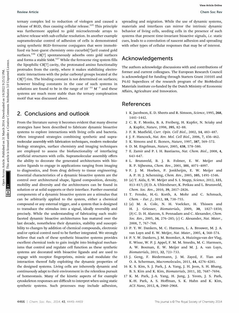
4466 | Chem. Soc. Rev., 2014, 43, 4449--4469 This journal is©The Royal Society of Chemistry 2014
ternary complex led to reduction of viologen and caused arelease of RGD, thus causing cellular release.125 This principlewas furthermore applied to gold microelectrode arrays toachieve release with sub-cellular resolution. In another examplesupramolecular control of adhesion of cells is demonstratedusing synthetic RGD–ferrocene conjugates that were immobi-lized via host–guest chemistry onto cucurbit[7]uril coated goldsurfaces.126 CB[7] spontaneously adsorbs onto gold surfacesand forms a stable SAM.127 While the ferrocene ring system fillsthe lipophilic CB[7] cavity, the protonated amino functionalityprotrudes from the cavity, where it makes stabilizing electro-static interactions with the polar carbonyl groups located at theCB[7] rim. The binding constant is not determined on surfaces;however binding constants in the case of such systems insolutions are found to be in the range of 10�11 M�1 and thesesystems are much more stable than the ternary complexationmotif that was discussed above.
2. Conclusions and outlook
From the literature survey it becomes evident that many diversestrategies have been described to fabricate dynamic bioactivesystems to explore interactions with living cells and bacteria.Often integrated strategies combining synthetic and supra-molecular assembly with fabrication techniques, modern molecularbiology strategies, surface chemistry and imaging techniquesare employed to assess the biofunctionality of interfacingartificial structures with cells. Supramolecular assembly offersthe ability to decorate the generated architectures with bio-active ligands to engage in applications ranging from imagingto diagnostics, and from drug delivery to tissue engineering.Essential characteristics of a dynamic bioactive system are thecontrol over architectural shape, ligand composition, density,mobility and diversity and the architectures can be found insolution or at solid supports or their interface. Further essentialcomponents of a dynamic bioactive system are a stimulus thatcan be arbitrarily applied to the system, either a chemicalcompound or any external trigger, and a system that is designedto transduce the stimulus into a signal, ideally reversibly andprecisely. While the understanding of fabricating such multi-faceted dynamic bioactive architectures has matured over thelast decade, nonetheless their dynamics, stability and suscepti-bility to changes by addition of chemical compounds, electronicand/or optical control need to be further integrated. We stronglybelieve that each of these synthetic bioactive systems providesexcellent chemical tools to gain insight into biological mechan-isms that control and regulate cell function as these syntheticsystems are decorated with bioactive ligands and are used toengage with receptor fingerprints, mimic and modulate theinteraction thereof fully exploiting the dynamic properties ofthe designed systems. Living cells are inherently dynamic andcontinuously adapt to their environment in the relentless pursuitof homeostasis. Many of the kinetic aspects of for examplecytoskeleton responses are difficult to interpret when using staticsynthetic systems. Such processes may include adhesion,
spreading and migration. While the use of dynamic systems,materials and interfaces can mirror the intrinsic dynamicbehavior of living cells, seeding cells in the presence of suchsystems that present time-invariant bioactive signals, i.e. staticcues, leads to a convolution of nascent adhesion and spreadingwith other types of cellular responses that may be of interest.
Acknowledgements
The authors acknowledge discussions with and contributions offormer and current colleagues. The European Research Councilis acknowledged for funding through Starters Grant 310105 andP4.02 Superdices of the research program of the BioMedicalMaterials institute co-funded by the Dutch Ministry of EconomicAffairs, Agriculture and Innovation.
References
1 K. Jacobson, E. D. Sheets and R. Simson, Science, 1995, 268,1441–1442.
2 C. R. F. Monks, B. A. Freiberg, H. Kupfer, N. Sciaky andA. Kupfer, Nature, 1998, 395, 82–88.
3 F. R. Maxfield, Curr. Opin. Cell Biol., 2002, 14, 483–487.4 J. F. Hancock, Nat. Rev. Mol. Cell Biol., 2006, 7, 456–462.5 K. Simons and E. Ikonen, Nature, 1997, 387, 569–572.6 D. M. Engelman, Nature, 2005, 438, 578–580.7 E. Zamir and P. I. H. Bastiaens, Nat. Chem. Biol., 2008, 4,
643–647.8 L. Brunsveld, B. J. B. Folmer, E. W. Meijer and
R. P. Sijbesma, Chem. Rev., 2001, 101, 4071–4097.9 F. J. M. Hoeben, P. Jonkheijm, E. W. Meijer and
A. P. H. J. Schenning, Chem. Rev., 2005, 105, 1491–1546.10 (a) T. Aida, E. W. Meijer and S. I. Stupp, Science, 2012, 335,
813–817; (b) D. A. Uhlenheuer, K. Petkau and L. Brunsveld,Chem. Soc. Rev., 2010, 39, 2817–2826.
11 T. Fenske, H.-G. Korth, A. Mohr and C. Schmuck,Chem. – Eur. J., 2012, 18, 738–755.
12 (a) M. A. Cole, N. H. Voelcker, H. Thissen andH. J. Griesser, Biomaterials, 2009, 30, 1827–1850;(b) C. D. H. Alarcon, S. Pennadam and C. Alexander, Chem.Soc. Rev., 2005, 34, 276–285; (c) C. Alexander, Nat. Mater.,2008, 7, 767–768.
13 P. Y. W. Dankers, M. C. Harmsen, L. A. Brouwer, M. J. A.van Luyn and E. W. Meijer, Nat. Mater., 2005, 4, 568–574.
14 P. Y. W. Dankers, J. M. Boomker, A. Huizinga-van der Vlag,E. Wisse, W. P. J. Appel, F. M. M. Smedts, M. C. Harmsen,A. W. Bosman, E. W. Meijer and M. J. A. van Luyn,Biomaterials, 2011, 32, 723–733.
15 J. Geng, F. Biedermann, J. M. Zayed, F. Tian andO. A. Scherman, Macromolecules, 2011, 44, 4276–4281.
16 K. S. Kim, S. J. Park, J. A. Yang, J. H. Jeon, S. H. Bhang,B. S. Kim and K. Kim, Biomaterials, 2011, 32, 7687–7694.
17 K. M. Park, J.-A. Yang, H. Jung, J. Yeom, J. S. Park,K.-H. Park, A. S. Hoffman, S. K. Hahn and K. Kim,ACS Nano, 2012, 6, 2960–2968.
Review Article Chem Soc Rev
Ope
n A
cces
s A
rtic
le. P
ublis
hed
on 3
1 M
arch
201
4. D
ownl
oade
d on
1/6
/202
2 9:
25:5
2 PM
. T
his
artic
le is
lice
nsed
und
er a
Cre
ativ
e C
omm
ons
Attr
ibut
ion
3.0
Unp
orte
d L
icen
ce.
View Article Online

This journal is©The Royal Society of Chemistry 2014 Chem. Soc. Rev., 2014, 43, 4449--4469 | 4467
18 J. Boekhoven, C. M. Rubert Perez, S. Sur, A. Worthy andS. I. Stupp, Angew. Chem., Int. Ed., 2013, 52, 12077–12080.
19 H. Li, J. Frith and J. J. Cooper-White, Biomacromolecules,2014, 15, 43–52.
20 E. Kim, D. Kim, H. Jung, J. Lee, S. Paul, N. Selvapalam,Y. Yang, N. Lim, C. Park and K. Kim, Angew. Chem., Int. Ed.,2010, 49, 4405–4408.
21 D. Kim, E. Kim, J. Kim, K. Park, K. Baek, M. Jung, Y. Ko,W. Sung, H. Kim, J. Suh, C. Park, O. Na, D.-k. Lee, K. Lee,S. Han and K. Kim, Angew. Chem., Int. Ed., 2007, 46,3471–3474.
22 A. Nelson, J. M. Belitsky, S. Vidal, C. S. Joiner, L. G. Baum andJ. F. Stoddart, J. Am. Chem. Soc., 2004, 126, 11914–11922.
23 J. Kim, Y. Ahn, K. M. Park, D.-W. Lee and K. Kim,Chem. – Eur. J., 2010, 16, 12168–12173.
24 N. Yui and T. Ooya, Chem. – Eur. J., 2006, 12, 6730–6737.25 J.-H. Seo, S. Kakinoki, Y. Inoue, T. Yamaoka, K. Ishihara
and N. Yui, J. Am. Chem. Soc., 2013, 135, 5513–5516.26 H. Wang, S. Wang, H. Su, K.-J. Chen, A. Armijo, W.-Y. Lin,
Y. Wang, J. Sun, K.-i. Kamei, J. Czernin, C. G. Radu and H.-R. Tseng, Angew. Chem., Int. Ed., 2009, 48, 4344–4348.
27 H. Wang, K. Liu, K.-J. Chen, Y. Lu, S. Wang, W.-Y. Lin,F. Guo, K. Kamei, Y.-C. Chen, M. Ohashi, M. Wang, X.-Z. Zhao, C. K.-F. Shen and H.-R. Tseng, ACS Nano, 2010, 4,6235–6243.
28 Y. Liu, H. Wang, K.-i. Kamei, M. Yan, K.-J. Chen, Q. Yuan,L. Shi, Y. Lu and H.-R. Tseng, Angew. Chem., Int. Ed., 2011,50, 3058–3062.
29 (a) H. Robson Marsden and A. Kros, Angew. Chem., Int. Ed.,2010, 49, 2988–3005; (b) J. Mason, K. Muller and K. Arndt,in Protein Engineering Protocols, ed. K. Arndt and K. Muller,Humana Press, 2007, vol. 352, pp. 35–70.
30 Y. Yano, A. Yano, S. Oishi, Y. Sugimoto, G. Tsujimoto,N. Fujii and K. Matsuzaki, ACS Chem. Biol., 2008, 3,341–345.
31 H. R. Zope, F. Versluis, A. Ordas, J. Voskuhl, H. P. Spainkand A. Kros, Angew. Chem., Int. Ed., 2013, 52, 14247–14251.
32 I. Nakase, S. Okumura, G. Tanaka, K. Osaki, M. Imanishiand S. Futaki, Angew. Chem., Int. Ed., 2012, 51, 7464–7467.
33 A. Bill, A. Schmitz, B. Albertoni, J.-N. Song, L. C. Heukamp,D. Walrafen, F. Thorwith, P. J. Verveer, S. Zimmer,L. Meffert, A. Schreiber, S. Chatterjee, R. K. Thomas,R. T. Ullrich, T. Lang and M. Famulok, Cell, 2010, 143,201–211.
34 Z. N. Mahmoud, S. B. Gunnoo, A. R. Thomson,J. M. Fletcher and D. N. Woolfson, Biomaterials, 2011, 32,3712–3720.
35 V. Villard, O. Kalyuzhniy, O. Riccio, S. Potekhin,T. N. Melnik, A. V. Kajava, C. Ruegg and G. Corradin,J. Pept. Sci., 2006, 12, 206–212.
36 T. Nakaji-Hirabayashi, K. Kato and H. Iwata, Biomacromo-lecules, 2008, 9, 1411–1416.
37 B. Liu, Y. Liu, J. J. Riesberg and W. Shen, J. Am. Chem. Soc.,2010, 132, 13630–13632.
38 K. Zhang, A. Sugawara and D. A. Tirrell, ChemBioChem,2009, 10, 2617–2619.
39 Y.-b. Lim, S. Park, E. Lee, H. Jeong, J.-H. Ryu, M. S. Lee andM. Lee, Biomacromolecules, 2007, 8, 1404–1408.
40 Y.-b. Lim, S. Park, E. Lee, J.-H. Ryu, Y.-R. Yoon, T.-H. Kimand M. Lee, Chem.–Asian J., 2007, 2, 1363–1369.
41 Y.-b. Lim, E. Lee and M. Lee, Angew. Chem., Int. Ed., 2007,46, 3475–3478.
42 R. J. Hosse, A. Rothe and B. E. Power, Protein Sci., 2006, 15,14–27.
43 (a) R. H. Kimura, A. M. Levin, F. V. Cochran andJ. R. Cochran, Proteins, 2009, 77, 359–369; (b) S. J. Moore,M. G. Hayden Gephart, J. M. Bergen, Y. S. Su, H. Rayburn,M. P. Scott and J. R. Cochran, Proc. Natl. Acad. Sci. U. S. A.,2013, 110, 14598–14603.
44 A. V. Pinheiro, D. Han, W. M. Shih and H. Yan, Nat.Nanotechnol., 2011, 6, 763–772.
45 F. A. Aldaye, W. T. Senapedis, P. A. Silver and J. C. Way,J. Am. Chem. Soc., 2010, 132, 14727–14729.
46 R. Meyer, S. Giselbrecht, B. E. Rapp, M. Hirtz andC. M. Niemeyer, Curr. Opin. Chem. Biol., 2014, 18, 8–15.
47 J. Voskuhl, J. Brinkmann and P. Jonkheijm, Curr. Opin.Chem. Biol., 2014, 18, 1–7.
48 U. Vermesh, O. Vermesh, J. Wang, G. A. Kwong, C. Ma,K. Hwang and J. R. Heath, Angew. Chem., Int. Ed., 2011, 50,7378–7380.
49 N. S. Selden, M. E. Todhunter, N. Y. Jee, J. S. Liu,K. E. Broaders and Z. J. Gartner, J. Am. Chem. Soc., 2012,134, 765–768.
50 R. A. Chandra, E. S. Douglas, R. A. Mathies, C. R. Bertozziand M. B. Francis, Angew. Chem., Int. Ed., 2006, 45, 896–901.
51 H. Schroeder, B. Ellinger, C. F. W. Becker, H. Waldmannand C. M. Niemeyer, Angew. Chem., Int. Ed., 2007, 46,4180–4183.
52 H. Onoe, S. C. Hsiao, E. S. Douglas, Z. J. Gartner,C. R. Bertozzi, M. B. Francis and R. A. Matthies, Langmuir,2010, 28, 8120–8130.
53 S. Gandor, S. Reisewitz, M. Venkatachalapahty, G. Arrabito,M. Reibner, H. Schroeder, K. Ruf, C. M. Niemeyer,P. I. Bastiaens and L. Dehmelt, Angew. Chem., Int. Ed.,2013, 52, 4790–4794.
54 G. Arrabito, S. Reisewitz, L. Dehmelt, P. I. Bastiaens,B. Pignataro, H. Schroeder and C. M. Niemeyer, Small,2013, 9, 4243–4249.
55 H. Cui, M. J. Webber and S. I. Stupp, Pept. Sci., 2010, 94, 1–18.56 J. D. Hartgerink, E. Beniash and S. I. Stupp, Science, 2001,
294, 1684–1688.57 R. N. Shah, N. A. Shah, M. M. Del Rosario Lim, C. Hsieha,
G. Nuber and S. I. Stupp, Proc. Natl. Acad. Sci. U. S. A., 2010,107, 3293–3298.
58 H. Storrie, M. O. Guler, S. N. Abu-Amara, T. Volberg,M. Rao, B. Geiger and S. I. Stupp, Biomaterials, 2007, 28,4608–4618.
59 V. M. Tysseling-Mattiace, V. Sahni, K. L. Niece, D. Birch,C. Czeisler, M. G. Fehlings, S. I. Stupp and J. A. Kessler,J. Neurosci., 2008, 28, 3814–3823.
60 D. Jiao, J. Geng, X. J. Loh, D. Das, T. C. Lee and O. A. Scherman,Angew. Chem., Int. Ed., 2012, 51, 9633–9637.
Chem Soc Rev Review Article
Ope
n A
cces
s A
rtic
le. P
ublis
hed
on 3
1 M
arch
201
4. D
ownl
oade
d on
1/6
/202
2 9:
25:5
2 PM
. T
his
artic
le is
lice
nsed
und
er a
Cre
ativ
e C
omm
ons
Attr
ibut
ion
3.0
Unp
orte
d L
icen
ce.
View Article Online

4468 | Chem. Soc. Rev., 2014, 43, 4449--4469 This journal is©The Royal Society of Chemistry 2014
61 J.-H. Ryu, D.-J. Hong and M. Lee, Chem. Commun., 2008,1043–1054.
62 B.-S. Kim, W.-Y. Yang, J.-H. Ryu, Y.-S. Yoo and M. Lee,Chem. Commun., 2005, 2035–2037.
63 J.-H. Ryu, E. Lee, Y.-b. Lim and M. Lee, J. Am. Chem. Soc.,2007, 129, 4808–4814.
64 M. K. Muller and L. Brunsveld, Angew. Chem., Int. Ed.,2009, 48, 2921–2924.
65 K. Petkau-Milroy, M. H. Sonntag, A. H. A. M. van Onzenand L. Brunsveld, J. Am. Chem. Soc., 2012, 134, 8086–8089.
66 L. Huang and R. E. Pagano, J. Cell Biol., 1975, 67, 38–48.67 H. R. Marsden, I. Tomatsu and A. Kros, Chem. Soc. Rev.,
2011, 40, 1572–1585.68 C. H. Yu and J. T. Groves, Med. Biol. Eng. Comput., 2010, 48,
955–963.69 D. S. Watson, A. N. Endsley and L. Huang, Vaccine, 2012,
30, 2256–2272.70 J. Nguyen and F. C. Szoka, Acc. Chem. Res., 2012, 45, 1153–1162.71 B. Yang, S.-Y. Geng, X.-M. Liu, J.-T. Wang, Y.-K. Chen,
Y.-L. Wang and J.-Y. Wang, Soft Matter, 2012, 8, 518–525.72 A. Akinc, A. Zumbuehl, M. Goldberg, E. S. Leshchiner,
V. Busini, N. Hossain, S. A. Bacallado, D. N. Nguyen,J. Fuller, R. Alvarez, A. Borodovsky, T. Borland, R. Constien,A. de Fougerolles, J. R. Dorkin, K. N. Jayaprakash,M. Jayaraman, M. John, V. Koteliansky, M. Manoharan,L. Nechev, J. Qin, T. Racie, D. Raitcheva, K. G. Rajeev,D. W. Y. Sah, J. Soutschek, I. Toudjarska, H.-P. Vornlocher,T. S. Zimmermann, R. Langer and D. G. Anderson, Nat.Biotechnol., 2008, 26, 561–569.
73 S.-W. Cho, M. Goldberg, S. M. Son, Q. Xu, F. Yang, Y. Mei,S. Bogatyrev, R. Langer and D. G. Anderson, Adv. Funct.Mater., 2009, 19, 3112–3118.
74 K. T. Love, K. P. Mahon, C. G. Levins, K. A. Whitehead,W. Querbes, J. R. Dorkin, J. Qin, W. Cantley, L. L. Qin,T. Racie, M. Frank-Kamenetsky, K. N. Yip, R. Alvarez,D. W. Y. Sah, A. de Fougerolles, K. Fitzgerald,V. Koteliansky, A. Akinc, R. Langer and D. G. Anderson,Proc. Natl. Acad. Sci. U. S. A., 2010, 107, 1864–1869.
75 E. Kluza, D. W. J. van der Schaft, P. A. Hautvast,W. J. Mulder, K. H. Mayo, A. W. Griffioen, G. J. Strijkersand K. Nicolay, Nano Lett., 2010, 10, 52–58.
76 J. J. Reczek, A. A. Kennedy, B. T. Halbert and A. R. Urbach,J. Am. Chem. Soc., 2009, 131, 2408–2415.
77 K. M. Park, D.-W. Lee, B. Sarkar, H. Jung, J. Kim, Y. H. Ko,K. E. Lee, H. Jeon and K. Kim, Small, 2010, 6, 1430–1441.
78 X. Liang, X. Li, X. Yue and Z. Dai, Angew. Chem., Int. Ed.,2011, 50, 11622–11627.
79 J. J. Moon, H. Suh, A. Bershteyn, M. T. Stephan, H. Liu,B. Huang, M. Sohail, S. Luo, S. H. Um, H. Khant,J. T. Goodwin, J. Ramos, W. Chiu and D. J. Irvine, Nat.Mater., 2011, 10, 243–251.
80 D. Dutta, A. Pulsipher, W. Luo, H. Mak and M. N. Yousaf,Bioconjugate Chem., 2011, 22, 2423–2433.
81 S. S. Agasti, M. Liong, C. Tassa, H. J. Chung, S. Y. Shaw,H. Lee and R. Weissleder, Angew. Chem., Int. Ed., 2012, 51,450–454.
82 J. A. Prescher, D. H. Dube and C. R. Bertozzi, Nature, 2004,430, 873–877.
83 S. C. Hsiao, B. J. Shum, H. Onoe, E. S. Douglas, Z. J. Gartner,R. A. Mathies, C. R. Bertozzi and M. B. Francis, Langmuir,2009, 25, 6985–6991.
84 D. Sarkar, P. K. Vemula, W. Zhao, A. Gupta, R. Karnik andJ. M. Karp, Biomaterials, 2010, 31, 5266–5274.
85 D. Dutta, A. Pulsipher, W. Luo and M. N. Yousaf, J. Am.Chem. Soc., 2011, 133, 8704–8713.
86 M. T. Stephan, J. J. Moon, S. H. Um, A. Bersthteyn andD. J. Irvine, Nat. Med., 2010, 16, 1035–1041.
87 D. Lingwood and K. Simons, Science, 2010, 327, 46–50.88 D. Afanasenkau and A. Offenhausser, Langmuir, 2012, 28,
13387–13394.89 E. T. Castellana and P. S. Cremer, Surf. Sci. Rep., 2006, 61,
429–444.90 J. van Weerd, S. O. Krabbenborg, J. C. T. Eijkel,
H. B. J. Karperien, J. Huskens and P. Jonkheijm, J. Am.Chem. Soc., 2014, 136, 100–103.
91 J. M. Nam, P. M. Nair, R. M. Neve, J. W. Gray andJ. T. Groves, ChemBioChem, 2006, 7, 436–440.
92 N. C. Hartman and J. T. Groves, Curr. Opin. Cell Biol., 2011,23, 370–376.
93 N. C. Hartman, J. A. Nye and J. T. Groves, Proc. Natl. Acad.Sci. U. S. A., 2009, 106, 12729–12734.
94 B. N. Manz, B. L. Jackson, R. S. Petit, M. L. Dustin andJ. T. Groves, Proc. Natl. Acad. Sci. U. S. A., 2011, 108, 9089–9094.
95 M. Charnley, R. Kroschewski and M. Textor, Integr. Biol.,2012, 4, 1059–1071.
96 B. Ananthanarayanan, L. Little, D. V. Schaffer, K. E. Healyand M. Tirrell, Biomaterials, 2010, 31, 8706–8715.
97 S. Svedhem, D. Dahlborg, J. Ekeroth, J. Kelly, F. Hook andJ. Gold, Langmuir, 2003, 19, 6730–6736.
98 D. Thid, K. Holm, P. S. Eriksson, J. Ekeroth, B. Kasemo andJ. Gold, J. Biomed. Mater. Res., Part A, 2008, 84A, 940–953.
99 C.-J. Huang, N.-J. Cho, C.-J. Hsu, P.-Y. Tseng, C. W. Frankand Y.-C. Chang, Biomacromolecules, 2010, 11, 1231–1240.
100 M. Andreasson-Ochsner, G. Romano, M. Håkanson,M. L. Smith, D. E. Leckband, M. Textor and E. Reimhult,Lab Chip, 2011, 11, 2876–2883.
101 R. L. C. Naumann, C. Nowak and W. Knoll, Soft Matter,2011, 7, 9535–9548.
102 K. Morigaki, H. Schonherr and T. Okazaki, Langmuir, 2007,23, 12254–12260.
103 Y. Deng, Y. Wang, B. Holtz, J. Li, N. Traaseth, G. Veglia,B. J. Stottrup, R. Elde, D. Pei, A. Guo and X.-Y. Zhu, J. Am.Chem. Soc., 2008, 130, 6267–6271.
104 C. E. Ashley, E. C. Carnes, G. K. Phillips, D. Padilla,P. N. Durfee, P. A. Brown, T. N. Hanna, J. Liu, B. Phillips,M. B. Carter, N. J. Carroll, X. Jiang, D. R. Dunphy,C. L. Willman, D. N. Petsev, D. G. Evans, A. N. Parikh,B. Chackerian, W. Wharton, D. S. Peabody and C. JeffreyBrinker, Nat. Mater., 2011, 10, 389–397.
105 S. Yamaguchi, S. Yamahira, K. Kikuchi, K. Sumaru,T. Kanamori and T. Nagamune, Angew. Chem., 2012, 124,132–135.
Review Article Chem Soc Rev
Ope
n A
cces
s A
rtic
le. P
ublis
hed
on 3
1 M
arch
201
4. D
ownl
oade
d on
1/6
/202
2 9:
25:5
2 PM
. T
his
artic
le is
lice
nsed
und
er a
Cre
ativ
e C
omm
ons
Attr
ibut
ion
3.0
Unp
orte
d L
icen
ce.
View Article Online

This journal is©The Royal Society of Chemistry 2014 Chem. Soc. Rev., 2014, 43, 4449--4469 | 4469
106 K. Ishihara and M. Takai, J. R. Soc., Interface, 2009, 6,S279–S291.
107 B. Wildt, D. Wirtz and P. C. Searson, Nat. Methods, 2009, 6,211–213.
108 S. Raghavan, R. A. Desai, Y. Kwon, M. Mrksich andC. S. Chen, Langmuir, 2010, 26, 17733–17738.
109 Z. Chen, Y. Li, W. Liu, D. Zhang, Y. Zhao, B. Yuan andX. Jiang, Angew. Chem., Int. Ed., 2009, 48, 8303–8305.
110 X. Jiang, R. Ferrigno, M. Mrksich and G. M. Whitesides,J. Am. Chem. Soc., 2003, 125, 2366–2367.
111 M. N. Yousaf, B. T. Houseman and M. Mrksich, Angew.Chem., Int. Ed., 2001, 40, 1093–1096.
112 W. S. Yeo, M. N. Yousaf and M. Mrksich, J. Am. Chem. Soc.,2003, 125, 14994–14995.
113 E. W. L. Chan and M. N. Yousaf, J. Am. Chem. Soc., 2006,128, 15542–15546.
114 E. W. L. Chan, S. Park and M. N. Yousaf, Angew. Chem., Int.Ed., 2008, 47, 6267–6271.
115 W. Luo, E. W. L. Chan and M. N. Yousaf, J. Am. Chem. Soc.,2010, 132, 2614–2621.
116 B. M. Lamb and M. N. Yousaf, J. Am. Chem. Soc., 2011, 133,8870–8873.
117 D. K. Hoover, E. W. L. Chan and M. N. Yousaf, J. Am. Chem.Soc., 2008, 130, 3280–3281.
118 I. Choi, Y. K. Kim, D. H. Min, S. W. Lee and W. S. Yeo,J. Am. Chem. Soc., 2011, 133, 16718–16721.
119 C. G. Rolli, H. Nakayama, K. Yamaguchi, J. P. Spatz,R. Kemkemer and J. Nakanishi, Biomaterials, 2011, 33,2409–2418.
120 M. Wirkner, J. M. Alonso, V. Maus, M. Salierno, T. T. Lee,A. J. Garcıa and A. del Campo, Adv. Mater., 2011, 23,3907–3911.
121 W. Luo and M. N. Yousaf, J. Am. Chem. Soc., 2011, 133,10780–10783.
122 D. Liu, Y. Xie, H. Shao and X. Jiang, Angew. Chem., Int. Ed.,2009, 48, 4406–4408.
123 Y.-H. Gong, C. Li, J. Yang, H.-Y. Wang, R.-X. Zhuo andX.-Z. Zhang, Macromolecules, 2011, 44, 7499–7502.
124 A. Gonzalez-Campo, M. Brasch, D. Uhlenheuer, A. Gomez-Casado, L. Yang, L. Brunsveld, J. Huskens and P. Jonkheijm,Langmuir, 2012, 28, 16364–16371.
125 A. Qi, J. Brinkmann, J. Huskens, S. Krabbenborg, J. de Boer andP. Jonkheijm, Angew. Chem., Int. Ed., 2012, 51, 12233–12337.
126 P. Neirynck, J. Brinkmann, Q. An, D. van der Schaft,L. Gustav Milroy, P. Jonkheijm and L. Brunsveld, Chem.Commun., 2013, 49, 3679–3681.
127 J. Young, H. D. Nguyen, L. Yang, J. Huskens, P. Jonkheijmand L. Brunsveld, ChemBioChem, 2010, 11, 180–183.
Chem Soc Rev Review Article
Ope
n A
cces
s A
rtic
le. P
ublis
hed
on 3
1 M
arch
201
4. D
ownl
oade
d on
1/6
/202
2 9:
25:5
2 PM
. T
his
artic
le is
lice
nsed
und
er a
Cre
ativ
e C
omm
ons
Attr
ibut
ion
3.0
Unp
orte
d L
icen
ce.
View Article Online