As Easy as Black & White: CXR Interpretation...
18
1 “I Can’t Breathe!” Rapidly Assessing & Intervening in Air Leak Syndromes 1 [email protected] www.cherylherrmann.com Methodist Medical Center of Illinois, Peoria Class Code 251 Objectives • Explain the causes of air leak syndromes • Describe the treatment strategies for air leak syndromes • Identify characteristics on CXR that indicate these air leak syndromes 2 Classifications of air leak syndromes 1. Primary pneumothorax 2. Secondary pneumothorax 3. Iatrogenic pneumothorax 4. Pneumomediastinum 5. Pneumopericardium 6. Hydropneumothorax 3 As Easy as Black & White: CXR Interpretation 101 Wednesday 12:30-1:45 4 5 Basics of Xrays • Photograph negative principle • White color indicates lack of exposure • Black color indicates intense exposure • Dense substances absorb all the rays and appear white on the film • Soft tissues and air absorb part of the beam and appear gray or black 6 Whitest • Bone: Ribs, Sternum, Spine, Clavicle • Barium • Calcium Deposits • Prosthetic valves • Surgical wires, clips
Transcript of As Easy as Black & White: CXR Interpretation...

1
“I Can’t Breathe!”Rapidly Assessing & Intervening in
Air Leak Syndromes
1
Methodist Medical Center of Illinois, PeoriaClass Code 251
Objectives
• Explain the causes of air leak syndromes
• Describe the treatment strategies for air leak syndromes
• Identify characteristics on CXR that indicate these air leak syndromes
2
Classifications of air leak syndromes
1. Primary pneumothorax
2. Secondary pneumothorax
3. Iatrogenic pneumothorax
4. Pneumomediastinum
5. Pneumopericardium
6. Hydropneumothorax
3
As Easy as Black & White: CXR Interpretation 101
Wednesday 12:30-1:45
4
5
Basics of Xrays
• Photograph negative principle
• White color indicates lack of exposure
• Black color indicates intense exposure
• Dense substances absorb all the raysand appear white on the film
• Soft tissues and air absorb part of thebeam and appear gray or black
6
Whitest
• Bone: Ribs, Sternum, Spine, Clavicle
• Barium
• Calcium Deposits
• Prosthetic valves
• Surgical wires, clips

2
7
Clinical Findings that show up White
• Pulmonary Edema
• Pneumonia
• Pleural Effusion
• Atelectasis
• Tumors
• ARDS
8
Off White -- Gray
• Fluid
• Blood
• Heart
• Veins/arteries
• Aorta
• Skin/fat
9
Blackest
• Air
• Lungs
• Trachea
• Stomach
• Bowel
Clinical Findings that show up BLACK
AIR LEAK SYNDROMES!
10
11
Normal Chest X-ray
• Pleural is only able to be identified if separated from the thoracic lining by fluid or air
12
Pneumothorax
• Air in the pleural space that inhibits complete lung expansion
• A thin, white line represents the displaced visceral pleura

3
13
• Left Pneumothorax on CT scan
14
Left Pneumo under fluro
15 16
Pneumothorax Clinical Presentation
• Diminished or absent lung sounds over the affected lung
• Dyspnea
• Tachypnea
• Acute pain on affected side of the chest
• Decreased Sp02 & p02
• Subcutaneous emphysema
• Black area over lung field with no lung markings on CXR
18
Pneumothorax
• Initial Treatment:– Chest tube insertion if greater than 10 – 15 %
– If tension pneumothorax ---- it is a medical EMERGENCY and needs immediate needle decompression

4
19
Tension Pneumothorax
• Distended neck veins
• Hypotension
• Tracheal deviation
Note compressed swan ganz
Classifications of air leak syndromes
1. Primary pneumothorax
2. Secondary pneumothorax
3. Iatrogenic pneumothorax
4. Pneumomediastinum
5. Pneumopericardium
6. Hydropneumothorax
20
Primary Spontaneous Pneumothorax (PSP)
• Occurs without a precipitating event in a person who does not have lung disease
• Actually, most individuals with PSP have unrecognized lung disease
21
Primary Spontaneous Pneumothorax
• Incidence
– 7.4 per 100,000
– Greater in men than women
• Risk Factors
– Smoking
– Family History
– Marfan’s Syndrome
– Homocystinuria
– Thoracic endometriosis 22
PSP Clinical Presentation
• Usually occurs at rest
• Sudden onset of dyspnea and pleuritic chest pain
• Symptoms related to the volume of air in the pleural space
• Hypoxemia
• Rarely hypercapnia – no underlying lung disease
• Acute respiratory alkalosis if pain, anxiety and hypoxemia
• Age = early 20’s, rare after 40
23
PSP Treatment
• Initial
– Removal of air from the pleural space
• Needle aspiration, if small
• Chest tube, if large
– Supplemental oxygen
• Subsequent
– Preventing reoccurrence
– Reoccurance is 35 - 54%
24

5
PSP Treatment
• If after 6 hours the pneumothorax reabsorbs, patient may be sent home
• Needs to live close to emergency medical center if d/c in 6 hours.
25
PSP Treatment: Supplemental Oxygen
• Air in the pleural space is reabsorbed when the communication between the alveoli and the pleural space (air leak) closes.
• Supplemental oxygen markedly increases the rate of reabsorption
26
PSP: Persistent Air leak after 3 days
1. Heimlich valve
2. Infusing autologous blood into the pleural space
3. Video-Assisted Thoracoscopy (VAT) to oversew the area of the leak and perform pleurodesis
27
Heimlich Valve
• One way valve
• Can be discharged
• Call 911 if sudden sharp chest pain and severe shortness of breathe
28
Autologous Blood Patch
• Withdrawal of blood from peripheral vein & infuse blood into the pleural space via chest tube.
• Infuse 24 – 200 ml
• After infusion, drape chest tube tubing over a hook 60 cm above the patient’s chest and then to the WSD chamber.
• Chest tube removed 24 hours after cessation of air leak.
29
Video Assisted Thoracosopy (VATS) Pleurodesis
• Pleurodesis:
– Mechanical or chemical irritation between the parietal and the visceral layers of the pleura to close off the space between them and prevent further air or fluid from accumulating
30

6
Pleurodesis
• Mechanical
– Parietal pleurectomy
– Laser abrasion of the parietal pleura
– Pleural abrasion with dry gauze
• Chemical
– Intrapleural instillation of a chemical irritant – usually tetracycline derivative or talc
31
Case Study #1
• 18 y/o female walking up a hill and felt a “pop” in chest
• Abruptly becomes SOB and severe stabbing pain in left chest area
• Couldn’t take deep breaths
• Pain eventually subsided and whole lung area felt weak and bruised
32
Next day
• Walking on college campus and had to stop 2 – 3 times during the walk
• Breathing was labored and pain was stabbing.
• Came to ED
33
Dx: Spontaneous Pneumothorax
• 90% collapse of left lung
• Chest tube inserted
• Resolved after several days
• No family history
34
PMH
• Looking back as a senior was running sprints on a really cold windy day. I felt something “pop” in my chest and couldn’t take deep breaths.
• Stopped running, went home, rested. Just felt “tight/bruised” feeling.
• Now questions if it was a small pneumothorax.
• Had a few more of these episodes in HS
35
Medical workup
• Found underlying asthma
36

7
A year later…
• Walking , Abruptly becomes SOB and severe right chest pain
• Dx: spontaneous right pneumothorax (90%)
• Chest tube inserted
• Took 10 days to resolve
• “There was just a moment when I just knew that it had closed”
37
Another year later --- age 20
• Tubing in the ocean waves
• Sudden stabbing pain in left lung
• Xray: 10% pneumothorax that resolved on it’s own.
38
Treatment
• Inhalers for asthma and steroid inhaler for next 10 years
• Kinesiologist: natural supplements to boost the adrenal system
• Now at age 42, off inhalers and has not had any further episodes
39
Secondary Spontaneous Pneumothorax (SSP)
• A pneumothorax that occurs as a complication of an underlying lung disease
• Can be a complication of any lung disease. Most often occurs with:
– COPD
– Pneumocystis jirovecii infection
– Cystic fibrosis
– Tuberculosis 40
SSP Clinical Presentation
• C/O of dyspnea and chest pain on the same side as the pneumothorax
• Symptoms more severe than with PSP as SSP patients have less pulmonary reserve due to the underlying lung disease.
• Persistent air leaks are more common and tend to persist longer than PSP
41
SSP Treatment
• Should be hospitalized: diminished pulmonary reserve increases their risk for adverse outcomes.
• Initial Treatment
– Chest tube insertion
– Chest tube should remain in place until a procedure if performed to prevent recurrent SSP
42

8
SSP: Prevention of recurrence
• Video-Assisted Thoracoscopy (VAT) with stapling of blebs and pleural abrasion.
• Chemical pleurodesis
• Pleural Blood Patch
• Heimlich valve
43 44
Case Study # 263 y/o white male (RK) comes to ED with SOB and left sided chest pain for the past hour
• Woke up “feeling weird” and felt very SOB
• The left sided chest pain, which does not radiate, started when the SOB started.
• The pain is mildly sharp and stabbing in quality
45
PMH
– COPD – wears continuous oxygen at home
– CHF
– AAA repair
– Hx PE
– PVD
– Idiopathic thrombocytopenia purpura
– Antiphopholipid antibody syndrome
– Recurrent small bowel syndrome
46
• BP 136/77
• HR 134, regular
• RR 32
• Temp 97 oral
• SpO2 91% on 15 liters nonrebreather
• Pain 7/10
47
RK 12- 2 at 2200
48
RK 12-2 in ED

9
49
Spontaneous pneumothorax on 12 – 2CT scan view post chest tube insertion
• BP 101/65
• HR 113, regular
• RR 20
• SpO2 100% on
15 liters nonrebreather
• Pain 2/10
Chest Tube
50
RK CXR 9 hours post chest tube insertion at 0800Is the pneumo resolved?
51
RK CXR 9 hours post chest tube insertion at 0800
52
12-3 at noon
• C/O chest discomfort, SOB, left leg tingling
• Totally absent lung sounds on left
53
RK 12 – 3 at 1215 after 2nd chest tube inserted
54
• Patient did not go to surgery for decoritication due to pulumonary hypertension – poor surgical candidate
• Sent home with Heimlich valve

10
PSP and SSP – high risk activities
• Patients with resolving pneumothorax should be cautioned not to fly until intrapleural air has completely resolved.
• Deep sea diving should be avoided unless thoracotomy or pleurodesis has been performed
55
Nursing Care of Chest Tubes
• Bubbling in the water seal chamber indicates air leak
• If suction is ordered for PSP or SSP, keep suction going even when ambulating!
56
Case #3
• Ms Syncope came to the ED because of an episode of lightheadedness today that caused her to fall to the ground. There was no actual LOC.
• She was working in the garden at the time and also had a mild pressure sensation over
her chest which is still present in ED.
As you are getting ready to give Metoprolol her rhythms changes to this. EKG 3 days earlier in ED

11
• Started on Amiodarone and Atendolol.
• Monitor for 48 hours. Here are her strips – continued on next slide.
• Diagnosis: Tachybrady Syndrome
• Treatment : Pacermaker insertion
• It is 6 hours post Ms Syncope’s pacemaker insertion via the left subclavian.
• She is complaining of dyspnea and pain on left side of shest
• No lung sounds on left side
• CXR ordered
63 64
65
• Left pneumo from pacer insertion
Iatrogenic pneumothorax
• Medical procedure resulting in traumatic pneumothorax
66

12
Iatrogenic Pneumothorax Causes
• Transthoracic needle aspiration procedures
• Subclavian and supraclavicular needle sticks
• Thoracentesis
• Mechanical ventilation related to peak airway pressures
• Pleural biopsy
• Transbronchial lung biopsy
• CPR
• Tracheostomy67
Traumatic Pneumothorax
• Blunt trauma from motor vehicle accident, falls, blows to chest, penetrating chest trauma, or blast injuries results in tear in pleura and causes pneumothorax
68
Iatrogenic & Traumatic Pneumothorax Treatment
• Needle Aspiration
• Chest Tube insertion
• Recurrence is not usually a factor
69 70
• DM
71
• DM after CT insertion
72
• DS

13
73
• DS post CT insertion
Open Communicating Pneumothorax
• Also called Sucking Chest Wound
• Air enters the intrapleural space through the chest wall
• Cause: Penetrating trauma
74
75
• Patient became severely dyspnic after CXR.
• CT was accidentally disconnected from bottle during CXR.
Case # 4• 71 y/o male
• Right lower lobe resection and lymph node resection for adenocarcionomia of the lung.
• Discharged on POD # 7
• On POD # 12 goes to surgeon’s office with c/o increasing SOB over last three days.
• CXR done76
5-24 readmission with pneumothorax 5-24 readmission with pneumothorax post CT insertion

14
5-25 6-1
Pneumomediastinum
• Air in the mediastinal structures that occurs spontaneously or following procedures or trauma
• Pneumothorax may occur secondary to pneumomediastinum
81
Pneumomediastinum
• Air in the mediastinal structures that occurs spontaneously or following procedures or trauma
• Pneumothorax may occur secondary to pneumomediastinum
82
Pneumomediastinum
• Air in the mediastinal soft tissues
• Cause: Rupture of alveoli
83
Pneumomediastinum Causes
• Rupture of alveoli
• Acute production of high intrathoracic pressures (inhalational drug use)
• Smoking marijuana
• Inhalation of cocaine
• Asthma
• Respiratory tract infection
• Vomiting or severe coughing
• Mechanical ventilation
• Trauma or surgical disruption of the oropharyngeal, esophageal, or respiratory mucous 84
15
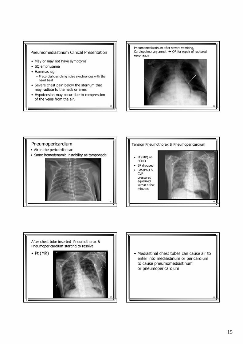
Pneumomediastinum Clinical Presentation
• May or may not have symptoms
• SQ emphysema
• Hammas sign
– Precordial crunching noise synchronous with the heart beat
• Severe chest pain below the sternum that may radiate to the neck or arms
• Hypotension may occur due to compression of the veins from the air.
85
Pneumomediastinum after severe vomiting, Cardiopulmonary arrest � OR for repair of ruptured esophagus
86
Pneumopericardium• Air in the pericardial sac
• Same hemodynamic instability as tamponade
87 88
Tension Pneumothorax & Pneumopericardium
• Pt (MR) on ECMO
• BP dropped
• PAS/PAD & CVP pressures equalized within a few minutes
89
After chest tube inserted Pneumothorax & Pneumopericardium starting to resolve
• Pt (MR) • Mediastinal chest tubes can cause air to enter into mediastinum or pericardium to cause pneumomediastinum or pneumopericardium
90

16
Case # 567 y/o female comes to ED with increasing SOB
• Bilateral pleural effusions
• Compressive atelectasis, right greater than left
• No pneumothorax seen
Thoracentesis
• Ultrasound guided thoracentesis performed
– 800 cc serosanguinous fluid removed from
left hemithorax.
– 1800cc straw color fluid removed from right hemithorax.
Following thoracentesis…
• On left, residual pleural fluid
• Right moderate pneumothorax
• Thought to represent trapped lung
A couple of days later…
• Moderate right-sided hydropneumothorax (trapped lung)
• Moderate amount of fluid in subpulmonic space
• Left base pleural effusion and/or atelectasis.
Trapped Lung
Fluid line
Post thoracotomy, debridement, chest tube placement
LG POD #4
Case # 675 y/o female Aortic Valve Replacement CXR on POD #4

17
LG POD # 6 note left hydropneumothorax LG POD # 6 CT inserted for hydropneumothorax Drained 450 ml
LG POD # 10
Pneumoperitoneum
• The presence of air within the peritoneal cavity.
• Most common cause is a perforation of the abdominal viscus — a perforated ulcer
100
In Summary• PSP
– no underlying lung disease
– Seen in young adults
• SSP
– Usually caused by underlying lung disease
– More severe due to already compromised lung state
• Both may need treatment to prevent recurrence
• Apex chest tubes for pneumos as air rises
• Keep suction on chest tubes 101
• Iatrogenic pneumothorax
– Caused by medical procedure resulting in traumatic pneumothorax
– Chest tube to aspirate air
– No need for further treatment
102

18
Other types of air leak syndromes
• Pneumopericardium
• Pneumomediastinum
• Hydropneumothorax
• Pneumoperitoneum
• Treatment for All
– aspirate air and treat underlying cause
103
Air Leak Syndromes:Be Prepared to immediately assist to insert a chest tube!
104
“I Can’t Breathe!”Rapidly Assessing & Intervening in
Air Leak Syndromes
105