Artificial Sweeteners Stimulate Adipogenesis and Suppress ...
16
Artificial Sweeteners Stimulate Adipogenesis and Suppress Lipolysis Independently of Sweet Taste Receptors * Received for publication, August 26, 2013, and in revised form, September 18, 2013 Published, JBC Papers in Press, September 24, 2013, DOI 10.1074/jbc.M113.514034 Becky R. Simon ‡ , Sebastian D. Parlee § , Brian S. Learman § , Hiroyuki Mori §1 , Erica L. Scheller § , William P. Cawthorn §¶2 , Xiaomin Ning § , Katherine Gallagher , Björn Tyrberg** ‡‡ , Fariba M. Assadi-Porter §§ , Charles R. Evans § , and Ormond A. MacDougald ‡§¶¶3 From the ‡ Program in Cellular and Molecular Biology and Departments of § Molecular and Integrative Physiology, Surgery, and ¶¶ Internal Medicine, University of Michigan, Ann Arbor, Michigan 48105, ¶ Musculoskeletal Research, Lilly Research Laboratories, Indianapolis, Indiana 46285, **Cardiovascular and Metabolic Disease, MedImmune LLC, Gaithersburg Headquarters, Gaithersburg, Maryland 20878, §§ Department of Biochemistry, University of Wisconsin, Madison, Wisconsin 53706, and ‡‡ Metabolic Signaling and Disease, Diabetes and Obesity Research Center, Sanford-Burnham Medical Research Institute, Orlando, Florida 32827 Background: Sweet taste receptors are candidate nutrient sensors in adipose tissue. Results: Sweet taste receptor ligands stimulate adipogenesis and suppress lipolysis; however, these effects do not require T1R2 and T1R3 despite their expression in adipose tissue. Conclusion: Some artificial sweeteners regulate adipocyte differentiation and metabolism through a sweet taste receptor- independent mechanism. Significance: Absorbed artificial sweeteners may regulate aspects of adipose tissue biology. G protein-coupled receptors mediate responses to a myriad of ligands, some of which regulate adipocyte differentiation and metabolism. The sweet taste receptors T1R2 and T1R3 are G protein-coupled receptors that function as carbohydrate sen- sors in taste buds, gut, and pancreas. Here we report that sweet taste receptors T1R2 and T1R3 are expressed throughout adi- pogenesis and in adipose tissues. Treatment of mouse and human precursor cells with artificial sweeteners, saccharin and acesulfame potassium, enhanced adipogenesis. Saccharin treat- ment of 3T3-L1 cells and primary mesenchymal stem cells rap- idly stimulated phosphorylation of Akt and downstream targets with functions in adipogenesis such as cAMP-response ele- ment-binding protein and FOXO1; however, increased expres- sion of peroxisome proliferator-activated receptor and CCAAT/enhancer-binding protein was not observed until rel- atively late in differentiation. Saccharin-stimulated Akt phos- phorylation at Thr-308 occurred within 5 min, was phosphati- dylinositol 3-kinase-dependent, and occurred in the presence of high concentrations of insulin and dexamethasone; phosphory- lation of Ser-473 occurred more gradually. Surprisingly, neither saccharin-stimulated adipogenesis nor Thr-308 phosphoryla- tion was dependent on expression of T1R2 and/or T1R3, although Ser-473 phosphorylation was impaired in T1R2/T1R3 double knock-out precursors. In mature adipocytes, artificial sweetener treatment suppressed lipolysis even in the presence of forskolin, and lipolytic responses were correlated with phos- phorylation of hormone-sensitive lipase. Suppression of lipoly- sis by saccharin in adipocytes was also independent of T1R2 and T1R3. These results suggest that some artificial sweeteners have previously uncharacterized metabolic effects on adipocyte dif- ferentiation and metabolism and that effects of artificial sweet- eners on adipose tissue biology may be largely independent of the classical sweet taste receptors, T1R2 and T1R3. In conditions of persistent nutrient excess, adipocytes differ- entiate from mesenchymal precursor cells to provide additional reservoirs for lipid storage. These same nutritional conditions result in reciprocal regulation of anabolic and catabolic pro- cesses in mature adipocytes to promote triacylglycerol accumu- lation. In preadipocytes, adipogenic stimulation results in the activation of transcription factors PPAR 4 and C/EBP, pri- mary drivers of the adipogenic program that stimulate expres- sion of terminal adipocyte genes such as FABP4 and GLUT4 (1). However, the upstream endogenous circulating, vascular or adipocyte-derived factors that are sensed as the key signals to indicate nutritional excess have not been fully elucidated. Nutritive signals themselves may serve this role as has been demonstrated by fatty acids acting through GPR43 and GPR120 * This work was supported, in whole or in part, by National Institutes of Health Grants R01-DK062876 and R01-DK095705 (to O. A. M.) and T32-GM007315 (to B. R. S.), a Cellular and Molecular Biology Program training grant, and T32-HD007505, a Training Program in Organogenesis grant (to B. R. S. and S. D. P.). This work was also supported by a Fulbright scholar’s award (to O. A. M.). 1 Supported by a mentor-based postdoctoral fellowship from the American Diabetes Association. 2 Supported by a Lilly Innovation Fellowship award with previous support from a postdoctoral research fellowship from the Royal Commission for the Exhibition of 1851 (United Kingdom). 3 To whom correspondence should be addressed: University of Michigan, 6313 Brehm Tower, Ann Arbor, MI 48105. E-mail: [email protected]. 4 The abbreviations used are: PPAR, peroxisome proliferator-activated recep- tor; CREB, cAMP-response element-binding protein; Sacc, saccharin; AceK, acesulfame potassium; eMSC, ear mesenchymal stem cell; HBSS, Hanks’ balanced salt solution; SVC, stromal vascular cell; Bis-Tris, 2-[bis(2-hydroxy- ethyl)amino]-2-(hydroxymethyl)propane-1,3-diol; HSL, hormone-sensi- tive lipase; p, phosphorylated; F, forward; R, reverse; DKO, double KO; DI, dexamethasone and insulin; MDI, methylisobutylxanthine, dexametha- sone, and insulin; Fsk, forskolin; pHSL, phosphorylated hormone-sensitive lipase. THE JOURNAL OF BIOLOGICAL CHEMISTRY VOL. 288, NO. 45, pp. 32475–32489, November 8, 2013 © 2013 by The American Society for Biochemistry and Molecular Biology, Inc. Published in the U.S.A. NOVEMBER 8, 2013 • VOLUME 288 • NUMBER 45 JOURNAL OF BIOLOGICAL CHEMISTRY 32475 by guest on April 7, 2018 http://www.jbc.org/ Downloaded from
-
Upload
nguyenminh -
Category
Documents
-
view
225 -
download
0
Transcript of Artificial Sweeteners Stimulate Adipogenesis and Suppress ...

Artificial Sweeteners Stimulate Adipogenesis and SuppressLipolysis Independently of Sweet Taste Receptors*
Received for publication, August 26, 2013, and in revised form, September 18, 2013 Published, JBC Papers in Press, September 24, 2013, DOI 10.1074/jbc.M113.514034
Becky R. Simon‡, Sebastian D. Parlee§, Brian S. Learman§, Hiroyuki Mori§1, Erica L. Scheller§, William P. Cawthorn§¶2,Xiaomin Ning§, Katherine Gallagher�, Björn Tyrberg**‡‡, Fariba M. Assadi-Porter§§, Charles R. Evans§,and Ormond A. MacDougald‡§¶¶3
From the ‡Program in Cellular and Molecular Biology and Departments of §Molecular and Integrative Physiology, �Surgery, and¶¶Internal Medicine, University of Michigan, Ann Arbor, Michigan 48105, ¶Musculoskeletal Research, Lilly Research Laboratories,Indianapolis, Indiana 46285, **Cardiovascular and Metabolic Disease, MedImmune LLC, Gaithersburg Headquarters,Gaithersburg, Maryland 20878, §§Department of Biochemistry, University of Wisconsin, Madison, Wisconsin 53706, and‡‡Metabolic Signaling and Disease, Diabetes and Obesity Research Center, Sanford-Burnham Medical Research Institute,Orlando, Florida 32827
Background: Sweet taste receptors are candidate nutrient sensors in adipose tissue.Results: Sweet taste receptor ligands stimulate adipogenesis and suppress lipolysis; however, these effects do not require T1R2and T1R3 despite their expression in adipose tissue.Conclusion: Some artificial sweeteners regulate adipocyte differentiation and metabolism through a sweet taste receptor-independent mechanism.Significance: Absorbed artificial sweeteners may regulate aspects of adipose tissue biology.
Gprotein-coupled receptorsmediate responses to amyriad ofligands, some of which regulate adipocyte differentiation andmetabolism. The sweet taste receptors T1R2 and T1R3 are Gprotein-coupled receptors that function as carbohydrate sen-sors in taste buds, gut, and pancreas. Here we report that sweettaste receptors T1R2 and T1R3 are expressed throughout adi-pogenesis and in adipose tissues. Treatment of mouse andhuman precursor cells with artificial sweeteners, saccharin andacesulfame potassium, enhanced adipogenesis. Saccharin treat-ment of 3T3-L1 cells and primary mesenchymal stem cells rap-idly stimulated phosphorylation of Akt and downstream targetswith functions in adipogenesis such as cAMP-response ele-ment-binding protein and FOXO1; however, increased expres-sion of peroxisome proliferator-activated receptor � andCCAAT/enhancer-binding protein�wasnot observeduntil rel-atively late in differentiation. Saccharin-stimulated Akt phos-phorylation at Thr-308 occurred within 5 min, was phosphati-dylinositol 3-kinase-dependent, and occurred in the presence ofhigh concentrations of insulin and dexamethasone; phosphory-lation of Ser-473 occurredmore gradually. Surprisingly, neithersaccharin-stimulated adipogenesis nor Thr-308 phosphoryla-tion was dependent on expression of T1R2 and/or T1R3,although Ser-473 phosphorylation was impaired in T1R2/T1R3
double knock-out precursors. In mature adipocytes, artificialsweetener treatment suppressed lipolysis even in the presenceof forskolin, and lipolytic responses were correlated with phos-phorylation of hormone-sensitive lipase. Suppression of lipoly-sis by saccharin in adipocytes was also independent of T1R2 andT1R3. These results suggest that some artificial sweeteners havepreviously uncharacterized metabolic effects on adipocyte dif-ferentiation and metabolism and that effects of artificial sweet-eners on adipose tissue biology may be largely independent ofthe classical sweet taste receptors, T1R2 and T1R3.
In conditions of persistent nutrient excess, adipocytes differ-entiate frommesenchymal precursor cells to provide additionalreservoirs for lipid storage. These same nutritional conditionsresult in reciprocal regulation of anabolic and catabolic pro-cesses inmature adipocytes to promote triacylglycerol accumu-lation. In preadipocytes, adipogenic stimulation results in theactivation of transcription factors PPAR�4 and C/EBP�, pri-mary drivers of the adipogenic program that stimulate expres-sion of terminal adipocyte genes such as FABP4 andGLUT4 (1).However, the upstream endogenous circulating, vascular oradipocyte-derived factors that are sensed as the key signals toindicate nutritional excess have not been fully elucidated.Nutritive signals themselves may serve this role as has beendemonstrated by fatty acids acting throughGPR43 andGPR120
* This work was supported, in whole or in part, by National Institutes of HealthGrants R01-DK062876 and R01-DK095705 (to O. A. M.) and T32-GM007315(to B. R. S.), a Cellular and Molecular Biology Program training grant, andT32-HD007505, a Training Program in Organogenesis grant (to B. R. S. andS. D. P.). This work was also supported by a Fulbright scholar’s award (toO. A. M.).
1 Supported by a mentor-based postdoctoral fellowship from the AmericanDiabetes Association.
2 Supported by a Lilly Innovation Fellowship award with previous supportfrom a postdoctoral research fellowship from the Royal Commission forthe Exhibition of 1851 (United Kingdom).
3 To whom correspondence should be addressed: University of Michigan,6313 Brehm Tower, Ann Arbor, MI 48105. E-mail: [email protected].
4 The abbreviations used are: PPAR, peroxisome proliferator-activated recep-tor; CREB, cAMP-response element-binding protein; Sacc, saccharin; AceK,acesulfame potassium; eMSC, ear mesenchymal stem cell; HBSS, Hanks’balanced salt solution; SVC, stromal vascular cell; Bis-Tris, 2-[bis(2-hydroxy-ethyl)amino]-2-(hydroxymethyl)propane-1,3-diol; HSL, hormone-sensi-tive lipase; p, phosphorylated; F, forward; R, reverse; DKO, double KO; DI,dexamethasone and insulin; MDI, methylisobutylxanthine, dexametha-sone, and insulin; Fsk, forskolin; pHSL, phosphorylated hormone-sensitivelipase.
THE JOURNAL OF BIOLOGICAL CHEMISTRY VOL. 288, NO. 45, pp. 32475–32489, November 8, 2013© 2013 by The American Society for Biochemistry and Molecular Biology, Inc. Published in the U.S.A.
NOVEMBER 8, 2013 • VOLUME 288 • NUMBER 45 JOURNAL OF BIOLOGICAL CHEMISTRY 32475
by guest on April 7, 2018
http://ww
w.jbc.org/
Dow
nloaded from
to promote preadipocyte differentiation in vitro (2, 3). In adi-pocytes, energy availability is communicated indirectly throughinsulin concentrations and sympathetic tone (4) but is alsosensed directly through G protein-coupled receptors. In adi-pose tissue,Gprotein-coupled receptors respond to short chainfatty acids, lactate, �-hydroxybutyrate, �-hydroxyoctanoate,and succinate tomediate effects on lipolysis (5–9). Ahypothesisexplored in this study is whether nutritive signals regulatingadipocyte differentiation and metabolism are also mediated inpart by sweet taste receptors.The sweet taste receptor is generally believed to be an obli-
gate heterodimer of the G protein-coupled receptors T1R2 andT1R3 (10, 11). Sugars and artificial sweeteners such as saccha-rin (Sacc) and acesulfame potassium (AceK) bind primarily toT1R2 (12), although direct binding to T1R3 has been described(13). Originally characterized in the tongue as a mediator ofsaccharin preference, these receptors have subsequently beendescribed in the brain, bladder, pancreas, and gut (14–17) withmetabolic roles defined in the latter two tissues. In theenteroendocrine cells of the small intestine, activation of sweettaste receptors promotes glucose uptake and release of incretinhormones such as glucagon-like peptide 1 (18, 19). T1R2/T1R3also functions in the pancreas where its activation in � cellsmediates stimulatory effects of sweeteners on glucose-inducedinsulin secretion (17, 20).Numerous reports have demonstrated sweet taste receptor
activation in response to artificial sweeteners in heterologousexpression systems (10, 12, 21, 22); however, experiments intaste receptor knock-out (KO) animals suggest that an addi-tional receptor(s) may be capable of binding and responding tosweet tastants (11, 23, 24). Additionally, binding of artificialsweeteners to the N-terminal domain of T1R2 or T1R3 in theabsence of its dimerization partner suggests that these recep-tors may be capable of functioning independently (13, 25, 26).Although the input of T1R2/T1R3 may be important in thetongue and certainmetabolic tissues, these studies indicate thatthere may be additional receptors sensitive to carbohydratesand sweeteners.Here we report that T1R2 and T1R3 were constitutively
expressed throughout adipogenesis of immortalized and pri-mary cells and within adipose tissues. Treatment with artificialsweeteners such as saccharin and AceK induced adipogenesisof mouse and human precursors. Saccharin treatment alsostimulated phosphorylation of Akt and its downstream effec-tors of preadipocyte differentiation. Surprisingly, T1R2 andT1R3 were dispensable for both saccharin-stimulated adipogene-sis and phosphorylation on Thr-308, although activation of Ser-473 was at least partially dependent on sweet taste receptors. Inmature adipocytes, exposure to artificial sweeteners suppressedboth basal and stimulated lipolysis through amechanism that wasalso independent of sweet taste receptors. Taken together, thesedata demonstrate unexpected roles for artificial sweeteners in adi-pocyte differentiation and metabolism and support the presenceof additional “sweet” taste receptors.
MATERIALS AND METHODS
Cell Culture—3T3-L1 cells were differentiated as describedpreviously (27). Briefly, cells 2 days after confluence (D0) were
treated with DMEM containing 10% fetal bovine serum, 1 �M
dexamethasone, 1 �g/ml insulin, and 0.5 mM methylisobutyl-xanthine or combinations thereof. Cells were fed every 2 dayswith insulin and fetal bovine serum (FBS) supplementation onD2 and FBS alone fromD4 to the conclusion of the experiment.In general, artificial sweeteners were added to differentiationmedium at induction and replaced with medium every 2 days.Saccharin, AceK, and sucralose were from Sigma-Aldrich. Adi-pogenesis was evaluated byOil Red-O (Sigma-Aldrich) stainingas described previously (28). LY294002, a phosphatidylinositol3-kinase (PI3K) inhibitor, andU0216, aMEK1/2 inhibitor, werepurchased from Cell Signaling Technology (Danvers, MA).U73122, a phospholipase C� inhibitor, was from CaymanChemical (Ann Arbor, MI).eMSC Isolation—eMSCs were isolated from controls, T1R2
KO, T1R3 KO, and T1R2/T1R3 double KO mice as describedpreviously (29). Briefly, mouse ears were sterilized,minced, andincubated for 1 h in collagenase to obtain a cell suspension.Preconfluent cells were supplemented with 50 �g/ml FGF dur-ing the initial growth period (30) and maintained at 5% CO2 inDMEM/F-12 supplemented with 15% FBS. For differentiation,1 �M dexamethasone, 5 �g/ml insulin, and 0.5 mM methyl-isobutylxanthine were added to maintenance medium for 2days starting at D0. Insulin remained in medium for the first 4days of adipogenesis. In general, cells were fed every 2 days untilharvest.Glucose Uptake—3T3-L1 adipocytes were serum-starved in
HBSS (Invitrogen) with 0.5% BSA for 4 h. Cells were then incu-bated with 4 nM insulin in Krebs-Ringer HEPES buffer for 10min before treating with 50 �M cytochalasin B to block back-ground translocation. After 20 min of insulin treatment, 0.1�Ci/ml 2-deoxy[14C]glucose (PerkinElmer Life Sciences) wasadded. Ten min later (30-min post-insulin), adipocytes wereplaced on ice, washed, and lysed in 0.1% SDS. Uptake of 2-de-oxyglucose was then quantified by scintillation counting.Lipolysis—Lipolysis assays were conducted in differentiated
adipocytes at least 8 days after induction of adipogenesis for3T3-L1 cells or after 12 days for eMSCs and human SVCs.Secretion of glycerol andnon-esterified fatty acid fromculturedadipocytes into HBSS was determined with assay kits from Sig-ma-Aldrich (FG0100) and Wako Diagnostics (Richmond, VA;NEFA-HR(2)). Cells were treated for 2 h or the time indicated.Lipolysis in mouse explants was performed as described under“Animals.” Saccharin was used at 4.5 mM unless indicatedotherwise.cAMP—cAMP was measured by ELISA (Cayman Chemical,
Ann Arbor, MI) according to the manufacturer’s instructions.Human SVC Isolation—SVCs were isolated from a subcuta-
neous fat depot of a 54-year-old diabetic patient in the samemanner as eMSCs (29). Human SVCs were maintained anddifferentiated in the same manner as eMSCs. Human sam-ples were obtained with the approval of the InstitutionalReview Board of the University of Michigan Medical School(HUM00060733).Immunoblot Analysis—Cell extracts were lysed in an SDS
buffer (1% SDS, 12.7 mM EDTA, 60 mM Tris-HCl, pH 6.8) andheated to 95 °C. Lysates were then centrifuged to pellet celldebris and transferred to a fresh tube, and the protein concen-
Artificial Sweeteners Regulate Adipocyte Differentiation
32476 JOURNAL OF BIOLOGICAL CHEMISTRY VOLUME 288 • NUMBER 45 • NOVEMBER 8, 2013
by guest on April 7, 2018
http://ww
w.jbc.org/
Dow
nloaded from

tration was quantified with a BCA assay (Thermo Scientific,Waltham, MA). 4� SDS loading buffer (4% SDS, 240 mM Tris-HCl, 40% glycerol, 0.05% bromphenol blue, 2.5% 2-mercapto-ethanol) was added to a constant amount of protein beforeseparation on Bis-Tris polyacrylamide gels (Invitrogen). Forevaluation of adipogenesis markers, a constant volume of lysatewas used. SDS-PAGE and immunoblotting were performed asdescribed previously (28). Membranes were immunoblottedwith antibodies from Cell Signaling Technology for C/EBP�(catalog number 2295), pAkt308 (catalog number 9271),pAkt473 (catalog number 9275 or 4060), pFOXO1 (catalognumber 9461), pCREB (catalog number 9191), phosphorylatedhormone-sensitive lipase (pHSL) (catalog number 4126), pERK(catalog number 9101), total Akt (catalog number 9272), totalERK1/2 (catalog number 4695), total FOXO1 (catalog number9462), total hormone-sensitive lipase (HSL) (catalog number4107), and total CREB (catalog number 9197). Laminin anti-body was obtained from Novus Biologicals (Littleton, CO).PPAR�1/2 antibody was obtained from Millipore (Temecula,CA). FABP4 antibody was obtained from R&D Systems (Min-neapolis, MN; MAB1143).mRNAQuantification by RT-PCR—Total RNAwas prepared
from frozen tissue or cells using RNA Stat60 according to themanufacturer’s protocol (Tel-Test, Inc., Friendsville TX). TotalRNA was quantified and reverse transcribed with random hex-amers (TaqMan Reverse Transcription kit, Applied Biosys-tems, Foster City, CA). Quantitative PCR was performed usingthe MyiQ real time PCR detection system with SYBR Greenreagents (Bio-Rad). Reverse transcription, primer design, andquantitative PCR were performed as described previously (28).Primer sequences are as follows: T1R2: F, GTCCGCTGCAC-CAAGCA; R, GTTCGTCGAAGAAGAGCTGGTT; T1R3: F,CCAGGCAACCAGGTGCCAGTC; R, CGCCTTGCAGTCC-ACGCAGT; PPAR�: F, CCAGAGCATGGTGCCTTCGC; R,TTCCGAAGTTGGTGGGCCAGA; C/EBP�: F, TGGACAA-GAACAGCAACGAG; R, TCACTGGTCAACTCCAGCAC;PREF1: F, CCTCCTGTTGCAGTATAACAGCG; R, GGTCA-TGTCAATCTTCTCGGG;WNT10b: F, ACGACATGGACT-TCGGAGAGAAGT; R, CATTCTCGCCTGGATGTCCC;C/EBP�: F, GGGACTTGATGCAATCCGG; R, AACCCCGC-AGGAACATCTTT; C/EBP�: F, CGCCGCAACCAGGAGAT;R,GCTGATGCAGCTTCTCGTTCT; FOXO1: F,GCTTTTG-TCACATGCAGGT; R, CGCACAGAGCACTCCATAAA.Animals—C57BL/6J mice were purchased from The Jackson
Laboratory (Bar Harbor, ME). Procedures for this work wereapproved by the Committee on the Use and Care of Animals atthe University of Michigan with daily care of animals overseenby the unit for laboratory animalmedicine (PRO0001369). Ani-mals were maintained on a 12-h light/dark cycle and fed stan-dard chow ad libitum (LabDiet 5L0D, Purina, St. Louis, MO).Leprdb/db mice were obtained from The Jackson Laboratory.For saccharin effects on ex vivo lipolysis, 10-week-old malemice were injected with a sodium saccharin solution at 100mg/kg. After 20 min, animals were euthanized with CO2, andepididymal adipose tissue depots were excised. Depots werethen weighed and cut into �40-mg pieces, which were thencultured at 5% CO2 in HBSS for 4 h before quantifying glycerolin medium. T1R2 and T1R3 KO animals were created and ana-
lyzed by Charles Zuker, Columbia University (11). DoubleT1R2/T1R3 KO animals were generated by initially mating T1R2KO and T1R3 KO mice to produce double heterozygote T1R2/T1R3. In turn, double heterozygote T1R2/T1R3mice werematedtoproduce littermatedoublewild-type controls anddoubleT1R2/T1R3 knock-out mice.Calcium Imaging—3T3-L1 cells were incubated with Fluo4
(Invitrogen) in glass bottom dishes (MatTek, Ashland, MA) for30 min before imaging by confocal microscopy (OlympusFV500 confocal microscope and Olympus IX-71). Sweetenerswere delivered by pipette as 10� solutions and allowed to dif-fuse through dishes maintained at 37 °C while monitoring thegreen fluorescence channel. Experiments were conducted withthe assistance of the Michigan Diabetes Research and TrainingCenter Morphology and Image Analysis Core.Unbiased Metabolomics—To prepare samples for untar-
geted metabolomics, metabolites were extracted from adi-pocytes according to the protocol of Lorenz et al. (31). Briefly,500 �l of cold 8:1:1 methanol:chloroform:water were added toeach well of a 6-well cell culture plate that had previously beenaspirated of all medium and quenched by freezing with liquidnitrogen. The plate was then scraped with a cell scraper torelease and lyse cells. The cell extract was transferred by pipetteto a microcentrifuge tube, and residual cell debris was pelletedby centrifugation at 15,000� g for 10min. The supernatant wasdirectly analyzed by LC-MS.Cell extracts were analyzed by LC-MS using an Agilent 1200
HPLC coupled to an Agilent 6210 time-of-flight mass spec-trometer. Chromatographic separation was performed viamixed mode anion exchange-hydrophilic interaction chroma-tography using a Phenomenex Luna NH2 3-�m column, 15cm � 2-mm inner diameter. Mobile phase A for the separationwas acetonitrile, and mobile phase B was 5 mM ammoniumacetate in water adjusted to pH 9.9 with ammonium hydroxide.The gradient consisted of a 20-min linear ramp from20 to 100%B followed by a 2-min hold at 100% B and a subsequent 13-minre-equilibration period at 20% B. The sample injection volumewas 20 �l, and the flow rate was 0.25 ml/min. Detection wasperformed by electrospray ionization mass spectrometry innegative ionmode.MS parameters were as follows: gas temper-ature, 350 °C; drying gas, 10 liters/min; nebulizer, 20 p.s.i.gauge; capillary voltage, 3500 V; scan range, 50–1200Da; inter-nal reference mass correction, enabled.Untargetedmetabolomics data analysis was performed using
Agilent Masshunter Qualitative Analysis and Mass ProfilerProfessional software. Features in the data were first detectedusing the Find by Feature algorithm inMasshunter QualitativeAnalysis and then aligned between samples by accurate massand retention time using Mass Profiler Professional. To mini-mize gaps in the data, recursive detection of aligned featureswas performed using the Find by Formula algorithm. Once afinal list of features was generated, compounds were assignedputative identities by searching against the online Metlin data-base. Inmany cases, theMetlin search resulted inmultiple pos-sible matches for each feature within a 10-ppmmass error win-dow. Metabolite matches were ranked in order of ascendingmass error and among matches with equivalent mass error inorder of ascending Metlin identification number. Following
Artificial Sweeteners Regulate Adipocyte Differentiation
NOVEMBER 8, 2013 • VOLUME 288 • NUMBER 45 JOURNAL OF BIOLOGICAL CHEMISTRY 32477
by guest on April 7, 2018
http://ww
w.jbc.org/
Dow
nloaded from

putative metabolite identification, statistical analysis and datareduction techniques, including principle component analysiswere used to determine statistically different features betweensample groups and to assess global differences in the metabo-lome between treatment conditions.Saturation Transfer Difference NMR—Preparation of mem-
branes and saturation transfer difference NMR was performedas described by Venkitakrishnan et al. (32) with some modifi-cations. Briefly,WT and double KO (DKO) eMSCs were grownto confluence in 500-cm2 Corning dishes (Corning, NY) andsuspended in EDTA. The cell suspension was pelleted at 800 �g and then lysed using a Polytron homogenizer (Kinematica,Lucerne, Switzerland). The resulting suspension was spun atlow speed to collect cell debris followed by ultracentrifugationat 100,000 � g to isolate cell membranes. Following membraneisolation, saturation transfer differenceNMR spectroscopy wasperformed as described previously (32).
RESULTS
SweetTasteReceptorsT1R2andT1R3AreExpressed throughoutAdipogenesis in 3T3-L1 Cells and eMSCs—To evaluate theexpression of taste receptors during adipogenesis, RNA wasisolated at the indicated time points as 3T3-L1 preadipocyteswere differentiated into mature adipocytes, and quantitativePCR analyses were performed (Fig. 1). Expression of both T1R2and T1R3 peaked at day 2 of adipogenesis, returning to nearpreadipocyte levels by day 8 (Fig. 1A).We then examined sweettaste receptor expression in primary eMSCs as an independentadipogenic model. These cells are isolated from the mouse earand are capable of chondrogenic, osteogenic, and adipogenicdifferentiation (29).We found that sweet taste receptor expres-
sion in this system was reminiscent of that in 3T3-L1 cells;T1R2 and T1R3 both peaked in expression at day 2 of adipo-genesis before returning to preadipocyte levels at day 12 (Fig.1B).To determine whether sweet taste receptors were present in
adipose tissue in vivo, we evaluated T1R3 expression in adiposetissues and liver of control and db/dbmice. Expression of T1R3was different between tissues of control mice by as much as2-fold (Fig. 1C). Based on a statistical interaction between gen-otypes, expression of T1R3 is generally reduced in obese ani-mals. However, post hoc analyses revealed only trends withinindividual tissues due to substantial variation in expression.Taken together, these data indicate that sweet taste receptorsare expressed throughout adipogenesis and in adipose tissues.Saccharin Stimulates Adipogenesis of Mouse and Human
Precursor Cells—To assess the effects of sweet taste receptoractivity on adipogenesis, we utilized the artificial sweetenersSacc and AceK as T1R2/T1R3 ligands. These agonists, asopposed to natural sugars, are useful in metabolic studiesbecause they are not metabolized and are �500-fold sweeterthan sucrose (33, 34). First, 3T3-L1 cells were treated through-out adipogenesis with a submaximal adipogenic mixture con-taining dexamethasone and insulin supplementedwith increas-ing concentrations of saccharin. The addition of saccharinrobustly stimulated adipogenesis in a concentration-dependentmanner, resulting in increased lipid accumulation and FABP4expression (Fig. 2A). FABP4 was then quantified over multipleexperiments to empirically determine minimal saccharin con-centrations necessary to enhance adipogenesis (Fig. 2B); thisdensitometric analysis indicated that 0.45 mM saccharin is the
0 2 4 6 80
2
4
6
8
10A B
0 2 4 6 8 10 120.0
0.5
1.0
1.5
2.0
2.5 T1R2T1R3
Time (Days) Time (Days)
eMSC3T3-L1 T1R2T1R3
iWAT
pWATLive
r0.0
0.5
1.0
1.5
2.0 Control
noisserpxE evit al eR
noi sser pxE evit al eR
noisserpxE 3R1T evitale
R
C
db/db
gWAT
FIGURE 1. Sweet taste receptors T1R2 and T1R3 are expressed constitutively throughout adipogenesis and in adipose tissues. A, T1R2 and T1R3expression throughout 8 days of adipogenesis in 3T3-L1 cells. B, T1R2 and T1R3 expression over 12 days of adipogenesis in eMSCs. C, the indicated tissues werecollected from control and 13–15-week-old female db/db mice maintained on a chow diet, and expression of T1R3 mRNA was evaluated by quantitative PCR.Data are expressed as mean � S.D. (error bars) with n � 4 – 6/tissue. Analysis by two-way analysis of variance indicates a significant effect of genotype (p � 0.05).gWat, gonadal white adipose tissue; iWAT, inguinal white adipose tissue; pWAT, perirenal white adipose tissue.
Artificial Sweeteners Regulate Adipocyte Differentiation
32478 JOURNAL OF BIOLOGICAL CHEMISTRY VOLUME 288 • NUMBER 45 • NOVEMBER 8, 2013
by guest on April 7, 2018
http://ww
w.jbc.org/
Dow
nloaded from

lowest concentration to significantly increase FABP4 accumu-lation in 3T3-L1 cells, whereas higher amounts resulted in anearly 10-fold increase.3T3-L1 cells induced with dexamethasone and insulin (DI)
and treated with saccharin show enhanced adipogenesis, whichcould be due to a specific synergistic interaction of the signalingpathways activated by saccharin and DI. To test this, we differ-entiated 3T3-L1 cells with various combinations of the fullmethylisobutylxanthine, dexamethasone, and insulin (MDI)mixture, including each component individually or no induc-tion at all (FBS), in the presence or absence of saccharin (Fig.2C). Using this approach, we observed that saccharin was suf-ficient to induce adipogenesis under all tested conditions with
mild effects even in the absence ofMDI. These data suggest thatsaccharin does not require permissive activation of a specificadipogenic signaling pathway to stimulate differentiation.To determine whether these effects of saccharin on adipo-
genesis are observed in other models of adipogenesis, we firstevaluated multipotent, primary eMSCs and found that saccha-rin robustly stimulated lipid accumulation and expression ofFABP4 (Fig. 2D). As observed with 3T3-L1 preadipocytes, arti-ficial sweetener effects occurred independently of differentia-tion conditions as eMSCs induced with MDI, DI, or FBS allshowed an enhancement of adipogenesis with saccharin treat-ment. We also tested the applicability of these findings tohuman precursors and observed that in SVCs isolated from
FABP4
Sacc (mM) 0 0.02 0.045 0.2 0.45 2 4.5
A
C
HumanSVCs
DIMDI FBS
B
- + - -+ +
eMSCs
3T3-L1DI
+Sacc-Sacc
FABP4ERK1/2
ERK1/2
MDI I D M DI FBSSacc - - - - - ++++++ -
FABP4Laminin
FABP4
+Sacc
-Sacc
MDI
Sacc
D
00.0
20.0
45 0.2 0.45 2 4.5
0
2
4
6
8
10
**
***
***
Saccharin concentration (mM)
E
ERK1/2
FAB
P4 E
xpre
ssio
n ) egnahC dl oF(
-Sacc
+Sacc
FIGURE 2. Saccharin stimulates adipogenesis of mouse and human precursor cells. A, 3T3-L1 cells were differentiated with DI in the presence of theindicated concentrations of saccharin. Seven days after induction, cells were stained for neutral lipid with Oil Red-O (upper panel), and lysates were evaluatedfor expression of FABP4 (lower panel). ERK1/2 was used as a loading control. B, quantification of saccharin-stimulated FABP4 expression from four independentexperiments in DI-treated 3T3-L1 cells. Data are expressed as mean � S.D. (error bars). p � 0.01 is indicated with **, and p � 0.005 is indicated with ***. C, 3T3-L1cells were differentiated with components of the MDI mixture as indicated in the presence or absence of 4.5 mM saccharin. Adipogenesis was then evaluatedafter 8 days by Western blotting for FABP4. D, eMSCs were incubated in the presence of MDI, DI, or FBS with or without 2 mM saccharin supplementation. At day16 of differentiation, the degree of differentiation was evaluated with photomicrographs (upper panels) and by expression of FABP4 (lower panels). E, humanSVCs were induced with MDI for 14 days in the absence or presence of 4.5 mM saccharin. Adipogenesis was assessed with photomicrographs and by expressionof FABP4. M, methylisobutylxanthine; D, dexamethasone; I, insulin.
Artificial Sweeteners Regulate Adipocyte Differentiation
NOVEMBER 8, 2013 • VOLUME 288 • NUMBER 45 JOURNAL OF BIOLOGICAL CHEMISTRY 32479
by guest on April 7, 2018
http://ww
w.jbc.org/
Dow
nloaded from

human white adipose tissue saccharin markedly enhanced lipidaccumulation and FABP4 expression following adipogenic induc-tionwithMDI (Fig. 2E). Taken together, these results indicate thatadipogenesisofmouseandhumanprecursors is stimulatedby sac-charin, suggesting that sweet taste receptors may sense nutritivesignals and regulate preadipocyte differentiation.AceK Stimulates Adipogenesis of Mouse and Human Precur-
sor Cells—To test whether effects on adipogenesis are specificto saccharin, we repeated the previous experiments withanother artificial sweetener and found that AceK also stimu-lated 3T3-L1 adipogenesis. Comparable with our results withsaccharin, increased adipogenesiswas observed at 4.5mMAceK(Fig. 3A). Results similar to those with saccharin were alsoobserved in eMSCs where AceK supplementation enhanced
adipogenesis regardless of the differentiation induction condi-tions (Fig. 3B). Lastly, we repeated this experiment in humanSVCs. In these primary cells, AceK stimulated lipid and FABP4accumulation (Fig. 3C). These results suggest that proadipo-genic effects may be broadly observed with different artificialsweeteners.Effects of Saccharin on Adipogenesis Are Dependent on Time
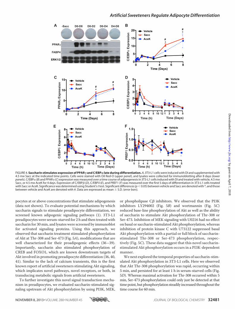
and Duration—To further characterize sweetener-stimulatedadipogenesis, we evaluated the temporal requirements of sac-charin treatment to enhance adipogenesis. To do this, wetreated differentiating 3T3-L1 cells with saccharin at varyingtime intervals (Fig. 4A). Cells treated with saccharin for the first2 or 4 days of adipogenesis had elevated expression of PPAR�and FABP4 with 4 days of treatment showing a more pro-nounced effect on Oil Red-O staining. However, maximaleffects on lipid accumulation and adipocyte gene expressionwere observedwith the full 8 days of treatment. Saccharin treat-ment must begin within an early time window because saccha-rin treatment had no effect on adipogenesis when supplemen-tation began on day 4. These observations suggest that, inaddition to concentration (Figs. 2 and 3), both time and dura-tion of saccharin treatment are important for effects onadipogenesis.Saccharin Stimulates Expression of PPAR� and C/EBP� Late
during Differentiation—To investigate mechanisms for enhance-ment of adipogenesis by artificial sweeteners, we next examinedaspects of the transcriptional profile of cells that had beentreated with DI � saccharin or AceK for the first 4 days ofadipogenesis. We observed that sweetener-stimulated PPAR�and C/EBP� expression did not differ from control cells untilD5, after the removal of saccharin from the differentiationmedium. By D8, expression of C/EBP� was increased �3-fold,and expression of PPAR�was elevated�2-fold in saccharin- orAceK-treated cells compared with DI alone (Fig. 4, B and C).These data suggest that sweeteners stimulate early events inadipogenesis (D0–D4) that are propagated following termina-tion of sweetener treatment.Because many essential elements of sweetener signaling
occur in the first 2 or 4 days of adipogenesis (Fig. 4), we profiledimportant regulators of adipogenesis active in this time period.However, after profiling mRNA expression of numerous tran-scription factors and inhibitors of differentiation, includingC/EBP� (Fig. 4D), C/EBP� (Fig. 4E), PREF1 (Fig. 4F), andFOXO1, EBF1, and EBF2 (data not shown), significant altera-tions in early events of adipogenesis were not observed. Thesedata suggest that artificial sweeteners robustly stimulate adipo-genesis in the first 4 days of adipogenesis, consistent withincreased expression of PPAR� andC/EBP� byD5 and beyond;however, the mechanistic basis appears to be independent ofthe aspects of the program of adipogenesis evaluated.Saccharin Acutely Activates Akt and ERK1/2 Signaling Path-
ways in Preadipocytes—We next examined signal transductioncascades that might mediate sweetener effects. In other con-texts (e.g. taste cells, � cells, and enteroendocrine cells), sweettaste receptor activation produces intracellular Ca2� transients(19, 20, 35). However, saccharin appears to work through anatypical signaling pathway in preadipocytes because we did notfind evidence for sweetener-stimulated calcium flux in preadi-
FIGURE 3. AceK stimulates adipogenesis of mouse and human precursorcells. A, 3T3-L1 cells were induced with DI and treated for 8 days with theindicated concentrations of AceK. After 8 days, cells were stained for neutrallipid with Oil Red-O. Lysates were evaluated for expression of FABP4. B, eMSCswere incubated in the presence of MDI, DI, or FBS with or without 2 mM AceK.After 16 days, the degree of differentiation was evaluated with photomicro-graphs (upper panels) and by expression of FABP4 (lower panels). C, humanSVCs were induced with MDI for 14 days in the absence or presence of 4.5 mM
AceK. Adipogenesis was determined with photomicrographs (upper panel)and by expression of FABP4 (lower panel).
Artificial Sweeteners Regulate Adipocyte Differentiation
32480 JOURNAL OF BIOLOGICAL CHEMISTRY VOLUME 288 • NUMBER 45 • NOVEMBER 8, 2013
by guest on April 7, 2018
http://ww
w.jbc.org/
Dow
nloaded from
pocytes at or above concentrations that stimulate adipogenesis(data not shown). To evaluate potential mechanisms by whichsaccharin signals to stimulate preadipocyte differentiation, wescreened known adipogenic signaling pathways (1). 3T3-L1preadipocytes were serum-starved for 2 h and then treatedwithsaccharin for 30min, and lysates were screened by immunoblotfor activated signaling proteins. Using this approach, weobserved that saccharin treatment stimulated phosphorylationof Akt at Thr-308 and Ser-473 (Fig. 5A), modifications that arewell characterized for their proadipogenic effects (36–39).Importantly, saccharin also stimulated phosphorylation ofCREB and FOXO1, which are known downstream targets ofAkt involved in promoting preadipocyte differentiation (36, 40,41). Similar to the lack of calcium transients, this is the firstknown report of artificial sweeteners stimulating Akt signaling,which implicates novel pathways, novel receptors, or both, intransducing metabolic signals from artificial sweeteners.To further investigate this novel signal transduction mecha-
nism in preadipocytes, we evaluated saccharin-stimulated sig-naling upstream of Akt phosphorylation by using PI3K, MEK,
or phospholipase C� inhibitors. We observed that the PI3Kinhibitors LY294002 (Fig. 5B) and wortmannin (Fig. 5C)reduced base-line phosphorylation of Akt as well as the abilityof saccharin to stimulate Akt phosphorylation of Thr-308 orSer-473. Inhibition of MEK signaling with U0216 had no effecton basal or saccharin-stimulatedAkt phosphorylation, whereasinhibition of protein kinase C with U73122 suppressed basalAkt phosphorylation with a partial or full block of saccharin-stimulated Thr-308 or Ser-473 phosphorylation, respec-tively (Fig. 5C). These data suggest that this novel saccharin-stimulated Akt phosphorylation occurs in a PI3K-dependentmanner.Wenext explored the temporal properties of saccharin-stim-
ulated Akt phosphorylation in 3T3-L1 cells. Here we observedthat Akt Thr-308 phosphorylation was rapid, occurring within5 min, and persisted for at least 1 h in serum-starved cells (Fig.5D). Whereas maximal activation for Thr-308 occurred within 5min, Ser-473 phosphorylation could only just be detected at thattimepoint, but phosphorylation steadily increased throughout thetime course for 60min.
A B-Sacc D0-D8 D0-D2 D0-D4 D4-D8
PPARγ
FABP4
Time (Days)
*
C D
E
PBE/
C δ
noi sser pxE
PBE/
Cβ
noisserpxEnoisserpxE
1FERP
PPA
Rγ
noisserpxE
PBE/
C
αnoisserpxE
F
Time (h)
Time (Days)1 2 3 4 50 2 4 6 8 10 12
Time (Days)
Time (h) Time (Days)Time (h)
1 2 3 4 50 2 4 6 8 10 12
#
#
#
#
1 2 3 4 50 2 4 6 8 10 120.0
0.5
1.0
1.5 VehicleSaccAceK
0
1
2
3
4
5
0
2
4
6
8 VehicleSaccAceK
0 2 4 6 80
5
10
15
0 2 4 6 80
5
10
15
20 VehicleSaccAceK
VehicleSaccAceK
VehicleSaccAceK
Time (Days)
*
*
*
ERK1/2
FIGURE 4. Saccharin stimulates expression of PPAR� and C/EBP� late during differentiation. A, 3T3-L1 cells were induced with DI and supplemented with4.5 mM Sacc at the indicated time points. Cells were stained with Oil Red-O (upper panel), and lysates were collected for immunoblotting after 8 days (lowerpanels). C/EBP� (B) and PPAR� (C) expression was measured over a time course of adipogenesis in 3T3-L1 cells induced with DI and treated with vehicle, 4.5 mM
Sacc, or 4.5 mM AceK for 4 days. Expression of C/EBP� (D), C/EBP� (E), and PREF1 (F) was measured over the first 5 days of differentiation in 3T3-L1 cells treatedwith Sacc or AceK. Significance was determined using Student’s t test. Significant differences (p � 0.05) between vehicle and Sacc are denoted with *, and thosebetween vehicle and AceK are denoted with #. Data are expressed as mean � S.D. (error bars).
Artificial Sweeteners Regulate Adipocyte Differentiation
NOVEMBER 8, 2013 • VOLUME 288 • NUMBER 45 JOURNAL OF BIOLOGICAL CHEMISTRY 32481
by guest on April 7, 2018
http://ww
w.jbc.org/
Dow
nloaded from
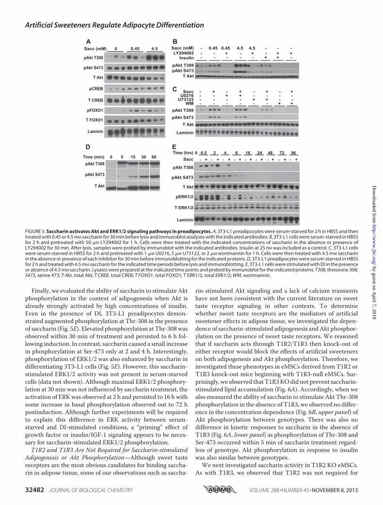
Finally, we evaluated the ability of saccharin to stimulate Aktphosphorylation in the context of adipogenesis when Akt isalready strongly activated by high concentrations of insulin.Even in the presence of DI, 3T3-L1 preadipocytes demon-strated augmented phosphorylation at Thr-308 in the presenceof saccharin (Fig. 5E). Elevated phosphorylation at Thr-308wasobserved within 30 min of treatment and persisted to 6 h fol-lowing induction. In contrast, saccharin caused a small increasein phosphorylation at Ser-473 only at 2 and 4 h. Interestingly,phosphorylation of ERK1/2 was also enhanced by saccharin indifferentiating 3T3-L1 cells (Fig. 5E). However, this saccharin-stimulated ERK1/2 activity was not present in serum-starvedcells (data not shown). Although maximal ERK1/2 phosphory-lation at 30 min was not influenced by saccharin treatment, theactivation of ERKwas observed at 2 h and persisted to 16 hwithsome increase in basal phosphorylation observed out to 72 hpostinduction. Although further experiments will be requiredto explain this difference in ERK activity between serum-starved and DI-stimulated conditions, a “priming” effect ofgrowth factor or insulin/IGF-1 signaling appears to be neces-sary for saccharin-stimulated ERK1/2 phosphorylation.T1R2 and T1R3 Are Not Required for Saccharin-stimulated
Adipogenesis or Akt Phosphorylation—Although sweet tastereceptors are the most obvious candidates for binding saccha-rin in adipose tissue, some of our observations such as saccha-
rin-stimulated Akt signaling and a lack of calcium transientshave not been consistent with the current literature on sweettaste receptor signaling in other contexts. To determinewhether sweet taste receptors are the mediators of artificialsweetener effects in adipose tissue, we investigated the depen-dence of saccharin-stimulated adipogenesis andAkt phosphor-ylation on the presence of sweet taste receptors. We reasonedthat if saccharin acts through T1R2/T1R3 then knock-out ofeither receptor would block the effects of artificial sweetenerson both adipogenesis and Akt phosphorylation. Therefore, weinvestigated these phenotypes in eMSCs derived from T1R2 orT1R3 knock-out mice beginning with T1R3-null eMSCs. Sur-prisingly, we observed thatT1R3KOdid not prevent saccharin-stimulated lipid accumulation (Fig. 6A). Accordingly, when wealsomeasured the ability of saccharin to stimulate Akt Thr-308phosphorylation in the absence of T1R3, we observed no differ-ence in the concentration dependence (Fig. 6B, upper panel) ofAkt phosphorylation between genotypes. There was also nodifference in kinetic responses to saccharin in the absence ofT1R3 (Fig. 6A, lower panel) as phosphorylation of Thr-308 andSer-473 occurred within 5 min of saccharin treatment regard-less of genotype. Akt phosphorylation in response to insulinwas also similar between genotypes.We next investigated saccharin activity in T1R2 KO eMSCs.
As with T1R3, we observed that T1R2 was not required for
pERK1/2
T Akt
Laminin
pAkt T308
0Sacc - - - - - - - - -+ + + + + + + + +
Time (hrs) 0.5 2 4 6 16 24 48 9672
A
pAkt T308
pAkt S473
T Akt
pCREB
T CREB
pFOXO1
T FOXO1
Laminin
B
C SaccU0216
WM
--
-
+-
-
-+
-
++
-
--
-
pAkt S473pAkt T308
Laminin
T Akt
+-
-
--
+
+-
+U73122 - - - - + + - -
pAkt S473pAkt T308
T Akt
Sacc (mM)LY294002
--
0.45-
4.5 4.50.45+ - +
T ERK1/2
pAkt S473
D
Insulin - - - - - -+-
++
---+
E
pAkt T308
pAkt S473
T Akt
Time (min) 0 5 15 30 60
Sacc (mM) 0.45 4.50
FIGURE 5. Saccharin activates Akt and ERK1/2 signaling pathways in preadipocytes. A, 3T3-L1 preadipocytes were serum-starved for 2 h in HBSS and thentreated with 0.45 or 4.5 mM saccharin for 30 min before lysis and immunoblot analyses with the indicated antibodies. B, 3T3-L1 cells were serum-starved in HBSSfor 2 h and pretreated with 50 �M LY294002 for 1 h. Cells were then treated with the indicated concentrations of saccharin in the absence or presence ofLY294002 for 30 min. After lysis, samples were probed by immunoblot with the indicated antibodies. Insulin at 25 nM was included as a control. C, 3T3-L1 cellswere serum-starved in HBSS for 2 h and pretreated with 1 �M U0216, 5 �M U73122, or 2 �M wortmannin for 1 h. Cells were then treated with 4.5 mM saccharinin the absence or presence of each inhibitor for 30 min before immunoblotting for the indicated proteins. D, 3T3-L1 preadipocytes were serum-starved in HBSSfor 2 h and treated with 4.5 mM saccharin for the indicated time periods before lysis and immunoblotting. E, 3T3-L1 cells were stimulated with DI in the presenceor absence of 4.5 mM saccharin. Lysates were prepared at the indicated time points and probed by immunoblot for the indicated proteins. T308, threonine 308;S473, serine 473; T Akt, total Akt; T CREB, total CREB; T FOXO1, total FOXO1; T ERK1/2, total ERK1/2; WM, wortmannin.
Artificial Sweeteners Regulate Adipocyte Differentiation
32482 JOURNAL OF BIOLOGICAL CHEMISTRY VOLUME 288 • NUMBER 45 • NOVEMBER 8, 2013
by guest on April 7, 2018
http://ww
w.jbc.org/
Dow
nloaded from

saccharin-stimulated lipid accumulation (Fig. 6C). Althoughthe T1R2 KO eMSC precursors were slightly desensitized tosaccharin in terms of Akt Thr-308 phosphorylation, T1R2was clearly not required for the effects of saccharin on Thr-308 phosphorylation (Fig. 6D, upper panel). In addition, weobserved that saccharin-stimulated Thr-308 and Ser-473 phos-phorylation in differentiating eMSCs was not affected by theabsence of T1R2 (Fig. 6D, lower panel). These data suggest thatneither T1R2 nor T1R3 is individually necessary for saccharin-mediated enhancement of adipogenesis or Akt phosphoryla-tion in preadipocytes. In this case, it is plausible that T1R2 andT1R3 might be acting as homodimers to bind saccharin andtransduce signals, or saccharin might be working throughanother receptor or mechanism.Literature suggests that T1R2 and T1R3 are capable of indi-
vidually responding to tastants rather than functioning as anobligate heterodimer (25, 42, 43). To directly address the pos-sibility of T1R2 andT1R3 homodimer activity in preadipocytes,we producedT1R2/T1R3DKOeMSCs. Using this DKOmodel,we observed that saccharin enhanced adipogenesis even in theabsence of both T1R2 and T1R3 (Fig. 6E). These data suggestthat, in this context, T1R2 and T1R3 do not function as
homodimers to mediate effects of saccharin on differentiation.Interestingly, whereas DKO had no effect on saccharin-stimu-lated Akt phosphorylation at Thr-308, phosphorylation at Ser-473 was reduced (Fig. 6F). These results suggest that althoughstimulation of Akt phosphorylation on Ser-473 is not essentialfor saccharin-induced adipogenesis it is likely that artificialsweeteners influence at least some aspects of adipose biologythrough sweet taste receptors.Artificial Sweeteners Suppress Adipocyte Lipolysis andDecrease
Hormone-sensitive Lipase Phosphorylation—Although artificialsweeteners have proadipogenic activity, this activity appears tobe independent of T1R2 and T1R3 in preadipocytes. Becausethe sweet taste receptors are expressed throughout differentia-tion and in mature adipocytes (Fig. 1), we then tested whethersaccharin regulates adipocyte metabolism in a T1R2/T1R3-de-pendent manner. Fully differentiated 3T3-L1 adipocytes weretreated with saccharin or AceK for 1 h. Cells were very rapidlywashed with ammonium acetate and frozen with liquid nitro-gen, and lysates were subjected to unbiasedmetabolomics anal-ysis. After 1 h, 81 metabolites were significantly different withsaccharin, and of these, 14 of 17 were also regulated by AceK(Fig. 7A; data not shown). All of the 14 co-regulatedmetabolites
WT T1R2 KO
Sacc - -
A
B D-
pAkt T308T Akt
Sacc
WT T1R3 KO
WT
T1R3 KO
Sacc (mM) 0 0.45
EWT
DKO
Sacc (mM) 0 0.45
WT
T1R2 KO
Sacc (mM) 0 0.45
WT WTDKO DKO
+Sacc-Sacc
pAkt T308
T AktpAkt S473
WT T1R2 KO
pAkt T308pAkt S473
Laminin
- -+ +DI DI
pAkt T308pAkt S473
T Akt
Time (min) 0 5 3015 60 InsT1R3 - - - - - -+ + + + + +
C
F
-
Sacc - -
pAkt T308T Akt
FIGURE 6. T1R3 and T1R2 are not required for saccharin-stimulated adipogenesis or Akt phosphorylation. A, WT and T1R3 KO eMSCs were differentiatedin FBS supplemented with 0.45 mM saccharin. After 12 days, lipid accumulation was evaluated with Oil Red-O. B, WT and T1R3 KO eMSCs were serum-starvedfor 2 h in HBSS and treated for 30 min with increasing concentrations of saccharin (0, 0.02, 0.045, 0.2, 0.45, 2, and 4.5 mM) before collecting lysates forimmunoblotting (upper panel). WT and T1R3 KO eMSCs were serum-starved for 2 h in HBSS before being treated with saccharin for the indicated time intervals(lower panel). C, WT and T1R2 KO eMSCs were differentiated in FBS supplemented with 0.45 mM saccharin. After 12 days, lipid accumulation was evaluated withOil Red-O. D, WT and T1R2 KO eMSCs were serum-starved for 2 h in HBSS and treated for 30 min with increasing concentrations of saccharin (0, 0.02, 0.045, 0.2,0.45, 2, and 4.5 mM) before lysis and immunoblotting (upper panel). WT and T1R2 KO eMSCs were maintained in calf serum (�) or treated with DI in the absence(�) or presence (�) of 4.5 mM saccharin for 30 min (lower panel). E, WT and T1R2/T1R3 KO (DKO) eMSCs were differentiated in FBS supplemented with 0.45 mM
saccharin. After 12 days, lipid accumulation was evaluated with Oil Red-O. F, WT and DKO eMSCs were serum-starved for 2 h in HBSS and treated for 30 min with4.5 mM saccharin before lysis and immunoblotting. T Akt, total Akt.
Artificial Sweeteners Regulate Adipocyte Differentiation
NOVEMBER 8, 2013 • VOLUME 288 • NUMBER 45 JOURNAL OF BIOLOGICAL CHEMISTRY 32483
by guest on April 7, 2018
http://ww
w.jbc.org/
Dow
nloaded from
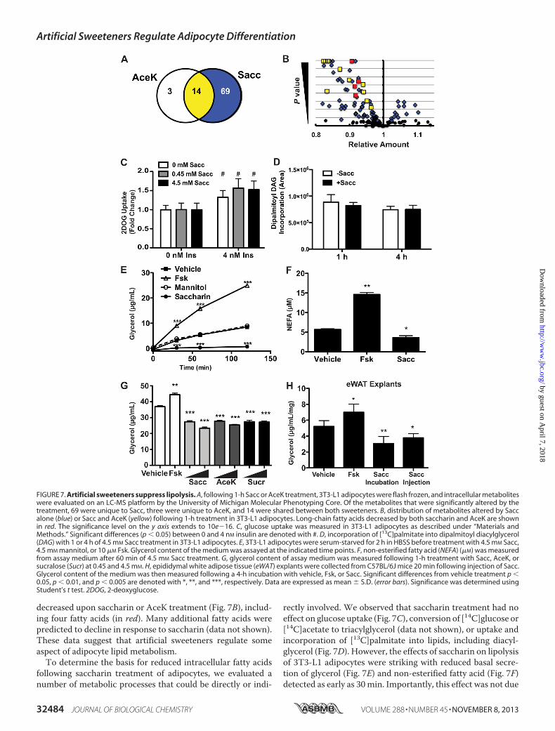
decreased upon saccharin or AceK treatment (Fig. 7B), includ-ing four fatty acids (in red). Many additional fatty acids werepredicted to decline in response to saccharin (data not shown).These data suggest that artificial sweeteners regulate someaspect of adipocyte lipid metabolism.To determine the basis for reduced intracellular fatty acids
following saccharin treatment of adipocytes, we evaluated anumber of metabolic processes that could be directly or indi-
rectly involved. We observed that saccharin treatment had noeffect on glucose uptake (Fig. 7C), conversion of [14C]glucose or[14C]acetate to triacylglycerol (data not shown), or uptake andincorporation of [13C]palmitate into lipids, including diacyl-glycerol (Fig. 7D). However, the effects of saccharin on lipolysisof 3T3-L1 adipocytes were striking with reduced basal secre-tion of glycerol (Fig. 7E) and non-esterified fatty acid (Fig. 7F)detected as early as 30min. Importantly, this effect was not due
FIGURE 7. Artificial sweeteners suppress lipolysis. A, following 1-h Sacc or AceK treatment, 3T3-L1 adipocytes were flash frozen, and intracellular metaboliteswere evaluated on an LC-MS platform by the University of Michigan Molecular Phenotyping Core. Of the metabolites that were significantly altered by thetreatment, 69 were unique to Sacc, three were unique to AceK, and 14 were shared between both sweeteners. B, distribution of metabolites altered by Saccalone (blue) or Sacc and AceK (yellow) following 1-h treatment in 3T3-L1 adipocytes. Long-chain fatty acids decreased by both saccharin and AceK are shownin red. The significance level on the y axis extends to 10e�16. C, glucose uptake was measured in 3T3-L1 adipocytes as described under “Materials andMethods.” Significant differences (p � 0.05) between 0 and 4 nM insulin are denoted with #. D, incorporation of [13C]palmitate into dipalmitoyl diacylglycerol(DAG) with 1 or 4 h of 4.5 mM Sacc treatment in 3T3-L1 adipocytes. E, 3T3-L1 adipocytes were serum-starved for 2 h in HBSS before treatment with 4.5 mM Sacc,4.5 mM mannitol, or 10 �M Fsk. Glycerol content of the medium was assayed at the indicated time points. F, non-esterified fatty acid (NEFA) (�M) was measuredfrom assay medium after 60 min of 4.5 mM Sacc treatment. G, glycerol content of assay medium was measured following 1-h treatment with Sacc, AceK, orsucralose (Sucr) at 0.45 and 4.5 mM. H, epididymal white adipose tissue (eWAT) explants were collected from C57BL/6J mice 20 min following injection of Sacc.Glycerol content of the medium was then measured following a 4-h incubation with vehicle, Fsk, or Sacc. Significant differences from vehicle treatment p �0.05, p � 0.01, and p � 0.005 are denoted with *, **, and ***, respectively. Data are expressed as mean � S.D. (error bars). Significance was determined usingStudent’s t test. 2DOG, 2-deoxyglucose.
Artificial Sweeteners Regulate Adipocyte Differentiation
32484 JOURNAL OF BIOLOGICAL CHEMISTRY VOLUME 288 • NUMBER 45 • NOVEMBER 8, 2013
by guest on April 7, 2018
http://ww
w.jbc.org/
Dow
nloaded from

to osmotic stress as lipolysis was not influenced by equimolarconcentrations of mannitol (Fig. 7E).To determine whether lipolysis was inhibited by artificial
sweeteners that are structurally distinct from saccharin, weevaluated the effects of AceK and sucralose (Fig. 7G) andobserved that, aswith saccharin, these artificial sweeteners sup-pressed basal lipolysis in 3T3-L1 cells. Lastly, we evaluated theeffects of saccharin on basal lipolysis in an ex vivo system andfound that isolated pieces of epididymal adipose tissue incu-bated with saccharin for 4 h had reduced secretion of glycerolinto culturemedium. Interestingly, an intraperitoneal injectionof saccharin was also sufficient to reduce glycerol release whenthe adipose tissue was excised 20 min postinjection and lipoly-sis was evaluated over the subsequent 4 h (Fig. 7H).We then investigated mechanisms for lipolytic regulation by
artificial sweeteners. We observed that the addition of saccha-rin to 3T3-L1 adipocytes suppressed phosphorylation of HSL(Fig. 8A). Interestingly, saccharin significantly blunted thestimulation of both glycerol release and HSL phosphorylation
by forskolin (Fsk), an adenylyl cyclase activator. This indicatesthat saccharin suppresses both basal and Fsk-stimulated lipo-lysis. We next hypothesized that saccharin might act throughAkt signaling as it does in preadipocytes (Fig. 5) to suppresslipolysis in a PI3K-dependent manner. However, treatmentwith LY294002 also failed to block saccharin-mediated sup-pression of basal lipolysis (Fig. 8B), consistent with our obser-vation that saccharin stimulated phosphorylation of Akt Thr-308 in adipocytes but only after effects on lipolysis were alreadyobserved (data not shown). As cAMP is an important regulatorof lipolytic activity upstreamofHSL,we speculated that saccha-rin treatment might reduce cAMP concentrations in adi-pocytes. However, saccharin had no effect on cAMPconcentra-tions (Fig. 8C), suggesting that artificial sweeteners may actdownstream of PKA.T1R2/T1R3AreNot Required for Suppression of Basal or Fsk-
stimulated Adipocyte Lipolysis by Saccharin—Although T1R2andT1R3 are likely binding candidates for artificial sweeteners,data in preadipocytes suggest that these receptors might not be
A B
C D
0
0
0
0
0
***** **
Vehicle Fsk Sacc Sacc + FskpHSL
Vehicle Sacc LY Sacc + LY0
20
40
60
80
100
VehicleFsk
0.45 mM
Sacc
4.5mM S
acc
0
1
2
3
4150200250 ***
E F
VehicleSacc
Fsk
Fsk + Sacc
0
1
2
3
4
5 WTT1R3 KO
*** ***
**
***
**
VehicleSacc
Fsk
Fsk + Sacc
0
1
2
3 WTT1R2 KO
* *
**
**
Gly
cero
l (µg
/mL) ) L
m/ gµ( l or ecylG
)egnahC
dl oF( lor ecylG
) egnahC
dl oF( l or ecylG
) egnahC
dl oF( l or ecylG
)Lm/lo
mp(P
MAc
VehicleSacc
Fsk
Fsk + S
acc0
1
2
3
4
5 WTDKO
** *
**
noitalyrohpsohP LSH
)egnahC dloF(
tHSL
Basa l Fsk Sacc Sacc+Fsk0
10
20
30
40****
*
10
20
30
40
FIGURE 8. T1R2/T1R3 are dispensable for saccharin-suppressed lipolysis. A, 3T3-L1 adipocytes were treated for 1 h with Sacc, Fsk, or both. Glycerolconcentration in the medium was measured before collection of protein lysates from control and treated cells. Lysates were then probed for pHSL by Westernblotting (left panel). HSL phosphorylation was then quantified by densitometry (right panel). B, 3T3-L1 adipocytes were serum-starved for 2 h and pretreatedwith LY294002 (LY) for 1 h before a 2-h Sacc treatment. Glycerol secretion was then measured in medium. C, 3T3-L1 adipocytes were serum-starved for 2 h andthen treated for 45 min with Fsk or Sacc. cAMP concentration was quantified by ELISA. T1R3 KO (D), T1R2 KO (E), or DKO (F) eMSC adipocytes were treated withSacc, Fsk, or both for 4 h before measuring glycerol accumulation in medium. Significant differences p � 0.05, p � 0.01, and p � 0.005 are denoted *, **, and***, respectively. Data are expressed as mean � S.D. (error bars). Significance was determined using Student’s t test. tHSL, total HSL.
Artificial Sweeteners Regulate Adipocyte Differentiation
NOVEMBER 8, 2013 • VOLUME 288 • NUMBER 45 JOURNAL OF BIOLOGICAL CHEMISTRY 32485
by guest on April 7, 2018
http://ww
w.jbc.org/
Dow
nloaded from

necessary. We tested for saccharin-stimulated T1R2/T1R3activity by performing lipolysis assays in T1R2 and T1R3 KOeMSCs. Similar to our observations in preadipocytes, loss ofT1R3 (Fig. 8D) or T1R2 (Fig. 8E) failed to block saccharineffects on either basal or forskolin-stimulated lipolysis, and nosignificant differences were observed between genotypes. Fur-thermore, we tested effects of saccharin in the absence of bothT1R2 and T1R3 to rule out the possibility of homodimer activ-ity. However, as observed in preadipocytes, saccharin sup-pressed basal and forskolin-induced lipolysis even in theabsence of both receptors (Fig. 8F). Taken together, theseresults suggest that saccharin reduces lipolysis independentlyof T1R2/T1R3 sweet taste receptors and through a PKA-medi-ated mechanism downstream of cAMP.Saccharin Binds to Membranes of T1R2/T1R3 Double KO
Adipocyte Progenitors—Thus far, our data have indicated thatsaccharin has effects on preadipocyte differentiation and adi-pocyte metabolism in the absence of sweet taste receptorexpression. This suggests that either additional receptors arecapable of binding and mediating effects of saccharin underthese conditions or saccharin directly initiates intracellular sig-naling downstream of receptors. To distinguish between thesetwo possibilities, we assessed saccharin binding by control andDKO preadipocyte membranes by saturation transfer differ-enceNMR.Using this approach, we observed little difference ininteractions between saccharin and either control or DKOpreadipocyte membranes (Fig. 9A). Similar results wereobserved with an independent artificial sweetener, neotame,that has been well characterized within this assay (Fig. 9B andRef. 32). Given that saccharin was capable of binding to the cellmembrane of DKO preadipocytes, this suggests that receptorsin addition to T1R2 and T1R3 mediate signal transduction ofartificial sweeteners.
DISCUSSION
One strategy currently utilized to curtail the obesity epi-demic is extensive use of non-nutritive sweeteners. Such artifi-cial sweeteners, including saccharin, AceK, aspartame, sucra-lose, and neotame, have little to no caloric value but arehundreds of times sweeter than sucrose.However, some studiessuggest that high level consumers of artificial sweeteners mayactually be at greater risk for overweight and obesity (44).Although not all studies agree with this assessment (45–47),the hypothesis that artificial sweeteners “uncouple” anticipatedcaloric density perceived by the tongue from the actual calories
ingested has gained a distinct foothold. Some argue that thisuncoupling leads to increased insulin secretion, food intake,and weight gain (48, 49), although there are little data that con-clusively implicate artificial sweeteners in either weight gain orweight loss in humans (50).Potential mechanisms for how artificial sweeteners may
influence energy balance have been complicated by reportsidentifying functional sweet taste receptors in tissues outsidethe tongue. Our results demonstrate that artificial sweetenerssuch as saccharin and AceK can regulate adipocyte differentia-tion and metabolism. The active concentrations in preadi-pocytes and adipocytes are higher than would generally beobserved in humans (51–54) as bolus oral doses of the recom-mended maximum daily intake of saccharin results in peakplasma concentrations of �75 �M (54); however, this is none-theless an important proof of principle for unexpected effects ofartificial sweetener use and raises the possibility that absorbedsweeteners may influence aspects of adipose tissue biologygiven sufficient “dose and duration” of exposure. However, fur-ther studies will be required to test this possibility.Our data indicate that saccharin increases adipogenesis and
represses adipocyte lipolysis in the absence of T1R2/T1R3, sug-gesting a novel saccharin receptor and/ormechanism of action.Some groups have hypothesized that T1R2/T1R3 does notfunction as an obligate heterodimer (42, 43), but rather eachcomponent functions independently as a homodimer, consis-tent with the N-terminal domains of both T1R2 and T1R3 con-taining ligand-binding sites and changing conformation inresponse to artificial sweeteners (13). The homodimerizationmodel has found support from Shibata and co-workers (55),who have investigated sweet taste receptor activity in cultured3T3-L1 and adipose tissue stromal cells. In their model, T1R3,but not T1R2, is required for inhibition of adipogenesis bysweet taste receptors. The authors show results conflictingwithour own in that sodium saccharin and sucralose treatmentinhibits adipogenesis and reduces expression of PPAR� andC/EBP�. Although we observed a mild inhibition of adipogen-esis with sucralose (data not shown), in our hands, saccharin,sodium saccharin, and AceK consistently stimulated adipogen-esis of mouse and human precursors. Masubuchi et al. (55) alsoshowed that inhibition of adipogenesis is partially blocked withknockdownof T1R3 using shRNA; however, we did not observeimpaired differentiation in primary eMSCs derived from micelacking T1R2, T1R3, or both receptors (Fig. 6). Although slightdifferences in experimental models might be the cause of our dis-parate results, our work suggests that receptor homodimerizationis not sufficient to mediate saccharin activity in preadipocytes.This is further supported by quantitative data demonstrating thatsaccharin and neotame bind precursormembranes in the absenceof both T1R2 and T1R3 (Fig. 9). These data support a model inwhich tastants bind to an uncharacterized “sweet” receptor.Further complexity to sweet taste receptor signaling is indi-
cated by the analysis of gustatory nerves: T1R3 KO differen-tially effects the chorda tympani, a facial nerve innervating theanterior two-thirds of the tongue, versus the glossopharyngealnerve, which innervates the posterior third of the tongue (42).Whole-nerve recordings indicate that sweet tastants are detect-able in T1R3 KO mice, and reductions in sensitivity can vary
0 10 20 30 400
1
2
3 WTDKO
1 H-1
D
0 10 20 30 400.0
0.2
0.4
0.6
0.8
1.0 WTDKO
1 H-1
D
A B
Neotame (mM)Sacc (mM)
FIGURE 9. Saccharin and neotame bind similarly to membranes of controland T1R2/T1R3 DKO eMSC progenitors. WT and DKO membranes wereincubated with the indicated concentrations of saccharin (A) or neotame (B),and binding of artificial sweeteners to membranes was evaluated by satura-tion transfer difference NMR.
Artificial Sweeteners Regulate Adipocyte Differentiation
32486 JOURNAL OF BIOLOGICAL CHEMISTRY VOLUME 288 • NUMBER 45 • NOVEMBER 8, 2013
by guest on April 7, 2018
http://ww
w.jbc.org/
Dow
nloaded from

greatly between the chorda tympani and glossopharyngealnerves. In particular, the glossopharyngeal nerve of T1R3 KOmice has only a mildly blunted response to AceK. In the case of“savory” taste mediated by T1R1/T1R3, both nerves maintainrobust responses to savory tastants in the absence of T1R3despite behavioral data showing a reduction in preference forsavory tastants in these animals.Although NMR analysis suggests that saccharin activity in
preadipocytes was not T1R2/T1R3-mediated (Fig. 9), the pos-sibility remains that artificial sweeteners have direct intracellu-lar activity. This has been suggested in rat liver where previousreports have indicated that saccharin might act directly onadenylyl cyclase (56). Additional reports in rat adipocytes haveshown saccharin-stimulated increases (57) or decreases (58) inadenylate cyclase activity and lipolysis, varying with whetherisolated adipocytes were directly treated with saccharin or ratswere previously fed a high saccharin diet. Although our data donot show any clear indications of adenylyl cyclase or cAMPregulation by saccharin, it is possible that saccharin regulateslocal or compartmentalized cAMP populations that cannot beevaluated by ELISA. Such local cAMP regulation could be amechanism for the reduction in HSL phosphorylation in boththe presence and absence of forskolin. In addition, it is possiblethat saccharin could function through regulation of the HSLphosphatase. In line with a model in which phosphatases areregulated by artificial sweeteners, a role for inhibition of PP2Aphosphatase activity in preadipocytes in response to saccharinwould be consistent with elevated phosphorylation of Akt Thr-308 phosphorylation even in the presence of DI (59). Similarly,the need for priming phosphorylation on ERK with DI andserum as well as the sustained pERK signal through severalhours (Fig. 5E) could be explained if saccharin inhibited thedual specificity ERK phosphatases (60).An additional possibility for how artificial sweeteners medi-
ate effects in preadipocytes and adipocytes is through bittertaste receptors with which saccharin and AceK interact in thetongue to cause the aversive “metallic” aftertaste at low mM
concentrations (61, 62). To date, expression of one bitter tastereceptor in an adipogenic system has been published, Tas2R46in human mesenchymal stem cells (63). However, saccharindoes not appear to activate hTas2R46 in vitro (64, 65), arguingagainst this bitter taste receptor mediating the effects observedin this study. Although to our knowledge expression has notbeen evaluated during adipogenesis, bitter taste receptorsTas2R43 andTas2R44 are activated in cultured cells by concen-trations of saccharin and AceK (62) similar to concentrationsthat regulate adipocyte differentiation and metabolism in thisstudy. Slack et al. (66) also observed that concentrations of sac-charin (ED50 of 0.61 mM) and AceK (ED50 of 0.49 mM) requiredto activate bitter taste receptors are compatible with our work.If saccharin and AceKwere working through bitter taste recep-tors, we might predict that a bitter taste receptor activatorwould mimic their effects on adipose biology. Consistent withthis notion, quinine, which activates nine bitter taste receptors(64), robustly stimulates adipocyte differentiation (data notshown) and represses adipocyte lipolysis (67). We have notdirectly tested the involvement of specificmembers of the bittertaste receptor family as these experiments are hampered by low
receptor expression and complex and overlapping pharmacol-ogy among many receptors (64).Perhaps the most intriguing question remaining from this
study is the physiological role, if any, of sweet taste receptors inadipose tissue. Although we are the second group to describethem (55), there has been no consensus on the function of thesereceptors.Wehave screened numerous pathways for sensitivityto sweetener treatment, with Akt and ERK phosphorylationincreased in preadipocytes and PKA signaling increased in adi-pocytes. However, many other candidate pathways remainuntested. In muscle, T1R1 and T1R3 regulate autophagy andmammalian target of rapamycin complex activity (68). Althoughwe have not found evidence that saccharin regulates autophagy(data not shown), the specificity of Ser-473 phosphorylation toT1R2/T1R3 DKO may represent a possible connection betweentaste receptors andTORC2activity inpreadipocytes. It is alsopos-sible that these receptors have significant functionality in vivo thatis not observed in cultured cells. Taken together, these data unveilnovel roles for artificial sweeteners in adipose biology and suggestthat sweet taste receptorsmay represent a broader andmore com-plex group of receptors than is currently appreciated.
Acknowledgments—This work utilized Animal Phenotyping, Morphol-ogy and Imaging, and Metabolomics Core Services supported byNational Institutes of Health Grants P30-DK089503, P30-DK020572,and U24 DK097153.
REFERENCES1. Rosen, E. D., and MacDougald, O. A. (2006) Adipocyte differentiation
from the inside out. Nat. Rev. Mol. Cell Biol. 7, 885–8962. Hong, Y. H., Nishimura, Y., Hishikawa, D., Tsuzuki, H., Miyahara, H.,
Gotoh, C., Choi, K. C., Feng, D. D., Chen, C., Lee, H. G., Katoh, K., Roh,S. G., and Sasaki, S. (2005) Acetate and propionate short chain fatty acidsstimulate adipogenesis via GPCR43. Endocrinology 146, 5092–5099
3. Gotoh, C., Hong, Y. H., Iga, T., Hishikawa, D., Suzuki, Y., Song, S. H., Choi,K. C., Adachi, T., Hirasawa, A., Tsujimoto, G., Sasaki, S., and Roh, S. G.(2007) The regulation of adipogenesis through GPR120. Biochem. Bio-phys. Res. Commun. 354, 591–597
4. Saltiel, A. R., andKahn, C. R. (2001) Insulin signalling and the regulation ofglucose and lipid metabolism. Nature 414, 799–806
5. Liu, C.,Wu, J., Zhu, J., Kuei, C., Yu, J., Shelton, J., Sutton, S.W., Li, X., Yun,S. J., Mirzadegan, T., Mazur, C., Kamme, F., and Lovenberg, T. W. (2009)Lactate inhibits lipolysis in fat cells through activation of an orphan G-protein-coupled receptor, GPR81. J. Biol. Chem. 284, 2811–2822
6. Taggart, A. K., Kero, J., Gan, X., Cai, T. Q., Cheng, K., Ippolito,M., Ren, N.,Kaplan, R., Wu, K., Wu, T. J., Jin, L., Liaw, C., Chen, R., Richman, J.,Connolly, D., Offermanns, S., Wright, S. D., and Waters, M. G. (2005)D-�-Hydroxybutyrate inhibits adipocyte lipolysis via the nicotinic acidreceptor PUMA-G. J. Biol. Chem. 280, 26649–26652
7. Ren, N., Kaplan, R., Hernandez, M., Cheng, K., Jin, L., Taggart, A. K., Zhu,A. Y., Gan, X.,Wright, S. D., and Cai, T. Q. (2009) Phenolic acids suppressadipocyte lipolysis via activation of the nicotinic acid receptor GPR109A(HM74a/PUMA-G). J. Lipid Res. 50, 908–914
8. Ahmed, K., Tunaru, S., and Offermanns, S. (2009) GPR109A, GPR109Band GPR81, a family of hydroxy-carboxylic acid receptors. Trends Phar-macol. Sci. 30, 557–562
9. Duncan, R. E., Sarkadi-Nagy, E., Jaworski, K., Ahmadian,M., and Sul, H. S.(2008) Identification and functional characterization of adipose-specificphospholipase A2 (AdPLA). J. Biol. Chem. 283, 25428–25436
10. Nelson, G., Hoon, M. A., Chandrashekar, J., Zhang, Y., Ryba, N. J., andZuker, C. S. (2001) Mammalian sweet taste receptors. Cell 106, 381–390
11. Zhao, G. Q., Zhang, Y., Hoon, M. A., Chandrashekar, J., Erlenbach, I.,
Artificial Sweeteners Regulate Adipocyte Differentiation
NOVEMBER 8, 2013 • VOLUME 288 • NUMBER 45 JOURNAL OF BIOLOGICAL CHEMISTRY 32487
by guest on April 7, 2018
http://ww
w.jbc.org/
Dow
nloaded from

Ryba, N. J., and Zuker, C. S. (2003) The receptors for mammalian sweetand umami taste. Cell 115, 255–266
12. Xu, H., Staszewski, L., Tang, H., Adler, E., Zoller, M., and Li, X. (2004)Different functional roles of T1R subunits in the heteromeric taste recep-tors. Proc. Natl. Acad. Sci. U.S.A. 101, 14258–14263
13. Nie, Y., Vigues, S., Hobbs, J. R., Conn, G. L., and Munger, S. D. (2005)Distinct contributions of T1R2 and T1R3 taste receptor subunits to thedetection of sweet stimuli. Curr. Biol. 15, 1948–1952
14. Elliott, R. A., Kapoor, S., and Tincello, D. G. (2011) Expression and distri-bution of the sweet taste receptor isoforms T1R2 and T1R3 in human andrat bladders. J. Urol. 186, 2455–2462
15. Ren, X., Zhou, L., Terwilliger, R., Newton, S. S., and de Araujo, I. E. (2009)Sweet taste signaling functions as a hypothalamic glucose sensor. Front.Integr. Neurosci. 3, 12
16. Dyer, J., Salmon, K. S., Zibrik, L., and Shirazi-Beechey, S. P. (2005) Expres-sion of sweet taste receptors of the T1R family in the intestinal tract andenteroendocrine cells. Biochem. Soc. Trans. 33, 302–305
17. Nakagawa, Y., Nagasawa,M., Yamada, S., Hara, A., Mogami, H., Nikolaev,V. O., Lohse, M. J., Shigemura, N., Ninomiya, Y., and Kojima, I. (2009)Sweet taste receptor expressed in pancreatic �-cells activates the calciumand cyclic AMP signaling systems and stimulates insulin secretion. PLoSOne 4, e5106
18. Jang, H. J., Kokrashvili, Z., Theodorakis, M. J., Carlson, O. D., Kim, B. J.,Zhou, J., Kim,H.H., Xu,X., Chan, S. L., Juhaszova,M., Bernier,M.,Mosinger,B., Margolskee, R. F., and Egan, J. M. (2007) Gut-expressed gustducin andtaste receptors regulate secretionof glucagon-likepeptide-1.Proc.Natl.Acad.Sci. U.S.A. 104, 15069–15074
19. Mace, O. J., Affleck, J., Patel, N., and Kellett, G. L. (2007) Sweet tastereceptors in rat small intestine stimulate glucose absorption throughapical GLUT2. J. Physiol. 582, 379–392
20. Kyriazis, G. A., Soundarapandian, M. M., and Tyrberg, B. (2012) Sweettaste receptor signaling in � cells mediates fructose-induced potentiationof glucose-stimulated insulin secretion. Proc. Natl. Acad. Sci. U.S.A. 109,E524–E532
21. Jiang, P., Cui, M., Zhao, B., Liu, Z., Snyder, L. A., Benard, L.M., Osman, R.,Margolskee, R. F., and Max, M. (2005) Lactisole interacts with the trans-membrane domains of human T1R3 to inhibit sweet taste. J. Biol. Chem.280, 15238–15246
22. Masuda, K., Koizumi, A., Nakajima, K., Tanaka, T., Abe, K., Misaka, T.,and Ishiguro,M. (2012) Characterization of themodes of binding betweenhuman sweet taste receptor and low-molecular-weight sweet compounds.PLoS One 7, e35380
23. Treesukosol, Y., Blonde, G. D., and Spector, A. C. (2009) T1R2 and T1R3subunits are individually unnecessary for normal affective licking re-sponses to Polycose: implications for saccharide taste receptors in mice.Am. J. Physiol. Regul. Integr. Comp. Physiol. 296, R855–R865
24. Zukerman, S., Glendinning, J. I., Margolskee, R. F., and Sclafani, A. (2009)T1R3 taste receptor is critical for sucrose but not Polycose taste. Am. J.Physiol. Regul. Integr. Comp. Physiol. 296, R866–R876
25. Nie, Y., Hobbs, J. R., Vigues, S., Olson,W. J., Conn,G. L., andMunger, S. D.(2006) Expression and purification of functional ligand-binding domainsof T1R3 taste receptors. Chem. Senses 31, 505–513
26. Maîtrepierre, E., Sigoillot, M., Le Pessot, L., and Briand, L. (2012) Recom-binant expression, in vitro refolding, and biophysical characterization ofthe N-terminal domain of T1R3 taste receptor. Protein Expr. Purif. 83,75–83
27. Hemati, N., Ross, S. E., Erickson, R. L., Groblewski, G. E., and MacDou-gald, O. A. (1997) Signaling pathways through which insulin regulatesCCAAT/enhancer binding protein a (C/EBP�) phosphorylation and geneexpression in 3T3-L1 adipocytes: correlation with GLUT4 gene expres-sion. J. Biol. Chem. 272, 25913–25919
28. Cawthorn, W. P., Bree, A. J., Yao, Y., Du, B., Hemati, N., Martinez-San-tibañez, G., and MacDougald, O. A. (2012) Wnt6, Wnt10a and Wnt10binhibit adipogenesis and stimulate osteoblastogenesis through a�-catenin-dependent mechanism. Bone 50, 477–489
29. Rim, J. S., Mynatt, R. L., and Gawronska-Kozak, B. (2005) Mesenchymalstem cells from the outer ear: a novel adult stem cell model system for thestudy of adipogenesis. FASEB J. 19, 1205–1207
30. Mori, H., Prestwich, T. C., Reid, M. A., Longo, K. A., Gerin, I., Cawthorn,W. P., Susulic, V. S., Krishnan, V., Greenfield, A., and Macdougald, O. A.(2012) Secreted frizzled-related protein 5 suppresses adipocytemitochon-drial metabolism through WNT inhibition. J. Clin. Investig. 122,2405–2416
31. Lorenz,M. A., Burant, C. F., and Kennedy, R. T. (2011) Reducing time andincreasing sensitivity in sample preparation for adherent mammalian cellmetabolomics. Anal. Chem. 83, 3406–3414
32. Venkitakrishnan, R. P., Benard, O., Max, M., Markley, J. L., and Assadi-Porter, F. M. (2012) Use of NMR saturation transfer difference spectros-copy to study ligand binding to membrane proteins. Methods Mol. Biol.914, 47–63
33. Sweatman, T. W., and Renwick, A. G. (1979) Saccharin metabolism andtumorigenicity. Science 205, 1019–1020
34. Renwick, A. G. (1986) Themetabolism of intense sweeteners.Xenobiotica16, 1057–1071
35. Liu, D., and Liman, E. R. (2003) Intracellular Ca2� and the phospholipidPIP2 regulate the taste transduction ion channel TRPM5.Proc.Natl. Acad.Sci. U.S.A. 100, 15160–15165
36. Nakae, J., Kitamura, T., Kitamura, Y., Biggs, W. H., 3rd, Arden, K. C., andAccili, D. (2003) The forkhead transcription factor Foxo1 regulates adi-pocyte differentiation. Dev. Cell 4, 119–129
37. Menghini, R., Marchetti, V., Cardellini, M., Hribal, M. L., Mauriello, A.,Lauro, D., Sbraccia, P., Lauro, R., and Federici, M. (2005) Phosphorylationof GATA2 by Akt increases adipose tissue differentiation and reducesadipose tissue-related inflammation: a novel pathway linking obesity toatherosclerosis. Circulation 111, 1946–1953
38. Kim, S. P., Ha, J. M., Yun, S. J., Kim, E. K., Chung, S.W., Hong, K.W., Kim,C. D., and Bae, S. S. (2010) Transcriptional activation of peroxisome pro-liferator-activated receptor-gamma requires activation of both proteinkinase A and Akt during adipocyte differentiation. Biochem. Biophys. Res.Commun. 399, 55–59
39. Magun, R., Burgering, B. M., Coffer, P. J., Pardasani, D., Lin, Y., Chabot, J.,and Sorisky, A. (1996) Expression of a constitutively activated form ofprotein kinase B (c-Akt) in 3T3-L1 preadipose cells causes spontaneousdifferentiation. Endocrinology 137, 3590–3593
40. Klemm, D. J., Leitner, J. W.,Watson, P., Nesterova, A., Reusch, J. E., Goal-stone, M. L., and Draznin, B. (2001) Insulin-induced adipocyte differenti-ation. Activation of CREB rescues adipogenesis from the arrest caused byinhibition of prenylation. J. Biol. Chem. 276, 28430–28435
41. Cypess, A. M., Zhang, H., Schulz, T. J., Huang, T. L., Espinoza, D. O.,Kristiansen, K., Unterman, T. G., and Tseng, Y. H. (2011) Insulin/IGF-Iregulation of necdin and brown adipocyte differentiation via CREB- andFoxO1-associated pathways. Endocrinology 152, 3680–3689
42. Damak, S., Rong,M., Yasumatsu, K., Kokrashvili, Z., Varadarajan, V., Zou,S., Jiang, P., Ninomiya, Y., andMargolskee, R. F. (2003) Detection of sweetand umami taste in the absence of taste receptor T1r3. Science 301,850–853
43. Geraedts,M. C., Takahashi, T., Vigues, S.,Markwardt,M. L., Nkobena, A.,Cockerham, R. E., Hajnal, A., Dotson, C. D., Rizzo, M. A., and Munger,S. D. (2012) Transformation of postingestive glucose responses after de-letion of sweet taste receptor subunits or gastric bypass surgery. Am. J.Physiol. Endocrinol. Metab. 303, E464–E474
44. Dergance, J. M., Mouton, C. P., Lichtenstein, M. J., and Hazuda, H. P.(2005) Potential mediators of ethnic differences in physical activity inolder Mexican Americans and European Americans: results from the SanAntonio Longitudinal Study of Aging. J. Am. Geriatr. Soc. 53, 1240–1247
45. Rodin, J. (1990) Comparative effects of fructose, aspartame, glucose, andwater preloads on calorie andmacronutrient intake.Am. J. Clin. Nutr. 51,428–435
46. Porikos, K. P., and Koopmans, H. S. (1988) The effect of non-nutritivesweeteners on body weight in rats. Appetite 11, Suppl. 1, 12–15
47. Blackburn, G. L., Kanders, B. S., Lavin, P. T., Keller, S. D., and Whatley, J.(1997) The effect of aspartame as part of a multidisciplinary weight-con-trol program on short- and long-term control of body weight. Am. J. Clin.Nutr. 65, 409–418
48. Swithers, S. E., Martin, A. A., and Davidson, T. L. (2010) High-intensitysweeteners and energy balance. Physiol. Behav. 100, 55–62
Artificial Sweeteners Regulate Adipocyte Differentiation
32488 JOURNAL OF BIOLOGICAL CHEMISTRY VOLUME 288 • NUMBER 45 • NOVEMBER 8, 2013
by guest on April 7, 2018
http://ww
w.jbc.org/
Dow
nloaded from
49. Swithers, S. E., and Davidson, T. L. (2008) A role for sweet taste: caloriepredictive relations in energy regulation by rats. Behav. Neurosci. 122,161–173
50. Anderson, G. H., Foreyt, J., Sigman-Grant, M., and Allison, D. B. (2012)The use of low-calorie sweeteners by adults: impact on weight manage-ment. J. Nutr. 142, 1163S–1169S
51. Sweatman, T.W., Renwick, A. G., and Burgess, C. D. (1981) The pharma-cokinetics of saccharin in man. Xenobiotica 11, 531–540
52. Colburn,W. A., Bekersky, I., and Blumenthal, H. P. (1981) Dietary saccha-rin kinetics. Clin. Pharmacol. Ther. 30, 558–563
53. Colburn, W. A., Bekersky, I., and Blumenthal, H. P. (1981) A preliminaryreport on the pharmacokinetics of saccharin in man: single oral dose ad-ministration. J. Clin. Pharmacol. 21, 147–151
54. Pantarotto, C., Salmona, M., and Garattini, S. (1981) Plasma kinetics andurinary elimination of saccharin in man. Toxicol. Lett. 9, 367–371
55. Masubuchi, Y., Nakagawa, Y., Ma, J., Sasaki, T., Kitamura, T., Yamamoto,Y., Kurose, H., Kojima, I., and Shibata, H. (2013) A novel regulatory func-tion of sweet taste-sensing receptor in adipogenic differentiation of3T3-L1 cells. PLoS One 8, e54500
56. Striem, B. J., Naim,M., Zehavi, U., and Ronen, T. (1990) Saccharin induceschanges in adenylate cyclase activity in liver and muscle membranes inrats. Life Sci. 46, 803–810
57. Dib, K., Oget, I.,Wrisez, F., El Jamali, A., Aguie-Aguie, G., Correze, C., andLambert, B. (1996) Effects of sodium saccharin diet on fat-cell lipolysis:evidence for increased function of the adenylyl cyclase catalyst. Int. J. Obes.Relat. Metab. Disord. 20, 15–20
58. Dib, K., Wrisez, F., el Jamali, A., Lambert, B., and Correze, C. (1997) So-dium saccharin inhibits adenylyl cyclase activity in non-taste cells. Cell.Signal. 9, 431–438
59. Andrabi, S., Gjoerup, O. V., Kean, J. A., Roberts, T. M., and Schaffhausen,B. (2007) Protein phosphatase 2A regulates life and death decisions viaAktin a context-dependent manner. Proc. Natl. Acad. Sci. U.S.A. 104,19011–19016
60. Huang, C. Y., and Tan, T. H. (2012) DUSPs, to MAP kinases and beyond.Cell Biosci. 2, 24
61. Bartoshuk, L. M. (1979) Bitter taste of saccharin related to the geneticability to taste the bitter substance 6-n-propylthiouracil. Science 205,934–935
62. Kuhn, C., Bufe, B., Winnig, M., Hofmann, T., Frank, O., Behrens, M.,Lewtschenko, T., Slack, J. P., Ward, C. D., andMeyerhof, W. (2004) Bittertaste receptors for saccharin and acesulfame K. J. Neurosci. 24,10260–10265
63. Lund, T. C., Kobs, A. J., Kramer, A., Nyquist, M., Kuroki, M. T., Osborn, J.,Lidke, D. S., Low-Nam, S. T., Blazar, B. R., and Tolar, J. (2013) Bonemarrow stromal and vascular smooth muscle cells have chemosensorycapacity via bitter taste receptor expression. PLoS One 8, e58945
64. Meyerhof, W., Batram, C., Kuhn, C., Brockhoff, A., Chudoba, E., Bufe, B.,Appendino, G., and Behrens,M. (2010) Themolecular receptive ranges ofhuman TAS2R bitter taste receptors. Chem. Senses 35, 157–170
65. Brockhoff, A., Behrens, M., Massarotti, A., Appendino, G., andMeyerhof,W. (2007) Broad tuning of the human bitter taste receptor hTAS2R46 tovarious sesquiterpene lactones, clerodane and labdane diterpenoids,strychnine, and denatonium. J. Agric. Food Chem. 55, 6236–6243
66. Slack, J. P., Brockhoff, A., Batram, C., Menzel, S., Sonnabend, C., Born, S.,Galindo, M. M., Kohl, S., Thalmann, S., Ostopovici-Halip, L., Simons,C. T., Ungureanu, I., Duineveld, K., Bologa, C. G., Behrens, M., Furrer, S.,Oprea, T. I., and Meyerhof, W. (2010) Modulation of bitter taste percep-tion by a small molecule hTAS2R antagonist. Curr. Biol. 20, 1104–1109
67. Markus, H. B., and Ball, E. G. (1969) Inhibition of lipolytic processes in ratadipose tissue by antimalaria drugs. Biochim. Biophys. Acta 187, 486–491
68. Wauson, E. M., Zaganjor, E., Lee, A. Y., Guerra, M. L., Ghosh, A. B.,Bookout, A. L., Chambers, C. P., Jivan, A., McGlynn, K., Hutchison, M. R.,Deberardinis, R. J., and Cobb, M. H. (2012) The G protein-coupled tastereceptor T1R1/T1R3 regulates mTORC1 and autophagy. Mol. Cell 47,851–862
Artificial Sweeteners Regulate Adipocyte Differentiation
NOVEMBER 8, 2013 • VOLUME 288 • NUMBER 45 JOURNAL OF BIOLOGICAL CHEMISTRY 32489
by guest on April 7, 2018
http://ww
w.jbc.org/
Dow
nloaded from

Fariba M. Assadi-Porter, Charles R. Evans and Ormond A. MacDougaldScheller, William P. Cawthorn, Xiaomin Ning, Katherine Gallagher, Björn Tyrberg,
Becky R. Simon, Sebastian D. Parlee, Brian S. Learman, Hiroyuki Mori, Erica L.Independently of Sweet Taste Receptors
Artificial Sweeteners Stimulate Adipogenesis and Suppress Lipolysis
doi: 10.1074/jbc.M113.514034 originally published online September 24, 20132013, 288:32475-32489.J. Biol. Chem.
10.1074/jbc.M113.514034Access the most updated version of this article at doi:
Alerts:
When a correction for this article is posted•
When this article is cited•
to choose from all of JBC's e-mail alertsClick here
http://www.jbc.org/content/288/45/32475.full.html#ref-list-1
This article cites 68 references, 20 of which can be accessed free at
by guest on April 7, 2018
http://ww
w.jbc.org/
Dow
nloaded from