ARTIFICIAL NEURAL NETWORK: AS EMERGING DIAGNOSTIC …
13
Available Online through www.ijpbs.com (or) www.ijpbsonline.com IJPBS |Volume 5| Issue 3|JUL-SEPT|2015|29-41 Review Article Biological Sciences International Journal of Pharmacy and Biological Sciences (e-ISSN: 2230-7605) S.S. Sandhu* et al Int J Pharm Bio Sci www.ijpbs.com or www.ijpbsonline.com Page29 ARTIFICIAL NEURAL NETWORK: AS EMERGING DIAGNOSTIC TOOL FOR BREAST CANCER Ishwinder K. Sandhu 1 , M. Nair 2 , H. Shukla 3 & S.S. Sandhu 3* 1 Department of Computer Science and Engineering, ITM University, Gurgaon, Haryana. 2 Center for Scientific Research & Development, Peoples University, Bhopal, M.P. 3* Department of Biological Sciences, R.D.University, Jabalpur, M.P. *Corresponding Author Email: [email protected] ABSTRACT In the present scenario, cases of breast cancer disease are on the rise as a result of which it has become one of the most deadly diseases around the world. Classical methods required cytopathologists or oncologists to examine the breast lesions for detection and classification of various stages of the cancer; which was not only labour intensive but inaccurate. Hence an efficient method using artificial neural networks as decision making tool is utilized for the diagnosis of breast cancer. Early detection and diagnosis of breast cancer is necessary for reducing the premature deaths caused by it. But the conventional biological techniques used for breast cancer diagnosis have not proved to be very efficient thereby increasing the mortality rate among cancer patients. These conventional therapies though improve the condition of patient but put them at the stake of enormous side effects. In the recent times, usage of Artificial Neural Networks (ANN) has emerged as a reliable technique for the efficient and quick diagnosis of breast cancer. Several research groups are working world wide on the development of neural networks in medical diagnosis. This review describes neural network approaches to breast cancer diagnosis including different architectures of neural networks. KEY WORDS Neural Networks; Artificial Neural Networks; UIMS, diagnosis. INTRODUCTION Breast Cancer is one of the most common diseases among women which lead to mortality. It is the most frequently encountered cancer type in women and is the second most terminal one after the lung cancer. It is also seen in men but not much frequently. In India, an average age for developing a breast cancer has undergone a shift. Preoperative or neo-adjuvant chemotherapy is an option with large operable breast cancer so to facilitate breast conservation and to downstage the disease. There has been a number of options for the prognosis and diagnosis of breast cancer. These markers provide a window of opportunity for the detection of breast cancer. Globally, the five most common cancers considered in both sexes were cancers of the lung (1,824,701; 13%), breast (1,676,633; 11.9%), colorectum (1,360,602; 9.7%), prostate (1,111,689; 7.9%), and cervix uteri (527,624; 3.7%), comprising 46.2% of the 28 cancers reported [1]. There have been various strategies utilized for the early detection of breast cancer so to reduce mortality. Newer diagnosis techniques in imaging, tissue diagnosis and cytobiological assessments have held a great promise for the
Transcript of ARTIFICIAL NEURAL NETWORK: AS EMERGING DIAGNOSTIC …

Available Online through
www.ijpbs.com (or) www.ijpbsonline.com IJPBS |Volume 5| Issue 3|JUL-SEPT|2015|29-41
Review Article
Biological Sciences
International Journal of Pharmacy and Biological Sciences (e-ISSN: 2230-7605)
S.S. Sandhu* et al Int J Pharm Bio Sci
www.ijpbs.com or www.ijpbsonline.com
Pag
e29
ARTIFICIAL NEURAL NETWORK:
AS EMERGING DIAGNOSTIC TOOL FOR BREAST CANCER
Ishwinder K. Sandhu1, M. Nair2, H. Shukla3 & S.S. Sandhu3*
1Department of Computer Science and Engineering, ITM University, Gurgaon, Haryana. 2Center for Scientific Research & Development, Peoples University, Bhopal, M.P.
3* Department of Biological Sciences, R.D.University, Jabalpur, M.P.
*Corresponding Author Email: [email protected]
ABSTRACT In the present scenario, cases of breast cancer disease are on the rise as a result of which it has become one of the
most deadly diseases around the world. Classical methods required cytopathologists or oncologists to examine the
breast lesions for detection and classification of various stages of the cancer; which was not only labour intensive
but inaccurate. Hence an efficient method using artificial neural networks as decision making tool is utilized for the
diagnosis of breast cancer. Early detection and diagnosis of breast cancer is necessary for reducing the premature
deaths caused by it. But the conventional biological techniques used for breast cancer diagnosis have not proved to
be very efficient thereby increasing the mortality rate among cancer patients. These conventional therapies
though improve the condition of patient but put them at the stake of enormous side effects. In the recent times,
usage of Artificial Neural Networks (ANN) has emerged as a reliable technique for the efficient and quick diagnosis
of breast cancer. Several research groups are working world wide on the development of neural networks in
medical diagnosis. This review describes neural network approaches to breast cancer diagnosis including different
architectures of neural networks.
KEY WORDS
Neural Networks; Artificial Neural Networks; UIMS, diagnosis.
INTRODUCTION
Breast Cancer is one of the most common
diseases among women which lead to mortality.
It is the most frequently encountered cancer
type in women and is the second most terminal
one after the lung cancer. It is also seen in men
but not much frequently. In India, an average
age for developing a breast cancer has
undergone a shift. Preoperative or neo-adjuvant
chemotherapy is an option with large operable
breast cancer so to facilitate breast
conservation and to downstage the disease.
There has been a number of options for the
prognosis and diagnosis of breast cancer. These
markers provide a window of opportunity for
the detection of breast cancer. Globally, the five
most common cancers considered in both sexes
were cancers of the lung (1,824,701; 13%),
breast (1,676,633; 11.9%), colorectum
(1,360,602; 9.7%), prostate (1,111,689; 7.9%),
and cervix uteri (527,624; 3.7%), comprising
46.2% of the 28 cancers reported [1]. There
have been various strategies utilized for the
early detection of breast cancer so to reduce
mortality. Newer diagnosis techniques in
imaging, tissue diagnosis and cytobiological
assessments have held a great promise for the

Available Online through
www.ijpbs.com (or) www.ijpbsonline.com IJPBS |Volume 5| Issue 3|JUL-SEPT|2015|29-41
International Journal of Pharmacy and Biological Sciences (e-ISSN: 2230-7605)
S.S. Sandhu* et al Int J Pharm Bio Sci
www.ijpbs.com or www.ijpbsonline.com
Pag
e30
early detection and identification of risk of the
disease. Newer and more specific screening and
diagnostic tests are required for early detection
of breast cancer, especially in high-risk
groups. Therefore, information technology has
developed different computerized methods for
easy and quick diagnosis of breast cancer, out of
which ANN is the most significant method.
CANCER
Cancer is characterized as an abnormal cell
growth with the potential to reach to other
body parts. It is divided into 100 types
depending upon the type of cell from where it
started [2]. Cancer generally accounts for 7.6
million deaths (around 13% of all deaths) in
2008, with an estimated 12 million deaths in
2030 [3]. A normal cell in a human being grows,
divides and multiplies in a controlled fashion.
Every cell in the human body follows a cell cycle
comprising of different phases i.e. G1, S, G2 and
M (fig. 1A), where “S” stands for “Synthesis”,
during this phase DNA synthesis and
chromosome duplication takes place, “M”
stands for “Mitosis”, during this phase the cell
divides and forms two daughter cells, “G1” and
“G2” stand for “gap”, during these phases there
is a regulation of cell cycle progression on the
basis of different external and internal signals
(checkpoints) [4,5,6]. In order to facilitate
smooth transition of cell from one phase to
another these checkpoints are necessary thus,
to ensure that a cell enters the next phase only
when all the tasks which were to be performed
in the prior phase have been completed [7].
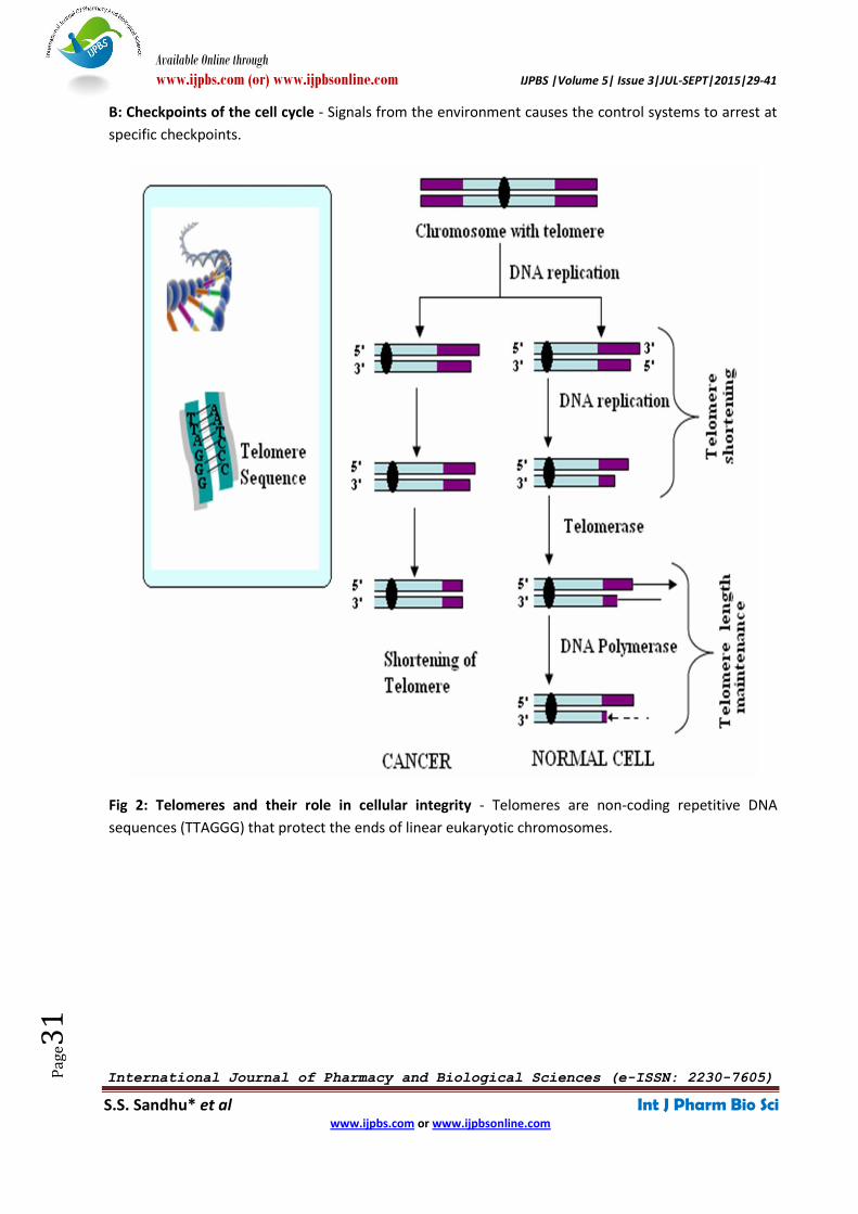
A cancerous cell disregards these checkpoints
(fig. 1B) and continues to grow in an
uncontrolled manner. Cancer cells thus have
two heritable properties: they and their
progeny (1) reproduce in defiance of the normal
restraints on cell division and (2) invade and
colonize territories normally reserved for other
cells. Apart from this a cancer cell also does not
undergo programmed cell death or apoptosis.
In Cancer cells, the length of the telomere is not
shortened over several cell divisions and hence
the cells continue to divide and bypass
senescence (fig. 2).
Fig 1 A: Cell Cycle - Cell grows in interphase, which consist of three phases: S phase; G1 phase; G2 phase
whereas in the M phase the nucleus and the cytoplasm divides.
Available Online through
www.ijpbs.com (or) www.ijpbsonline.com IJPBS |Volume 5| Issue 3|JUL-SEPT|2015|29-41
International Journal of Pharmacy and Biological Sciences (e-ISSN: 2230-7605)
S.S. Sandhu* et al Int J Pharm Bio Sci
www.ijpbs.com or www.ijpbsonline.com
Pag
e31
B: Checkpoints of the cell cycle - Signals from the environment causes the control systems to arrest at
specific checkpoints.
Fig 2: Telomeres and their role in cellular integrity - Telomeres are non-coding repetitive DNA
sequences (TTAGGG) that protect the ends of linear eukaryotic chromosomes.

Available Online through
www.ijpbs.com (or) www.ijpbsonline.com IJPBS |Volume 5| Issue 3|JUL-SEPT|2015|29-41
International Journal of Pharmacy and Biological Sciences (e-ISSN: 2230-7605)
S.S. Sandhu* et al Int J Pharm Bio Sci
www.ijpbs.com or www.ijpbsonline.com
Pag
e32
Fig 3: Schematic of ANN
Fig 4: Recurrent Network

Available Online through
www.ijpbs.com (or) www.ijpbsonline.com IJPBS |Volume 5| Issue 3|JUL-SEPT|2015|29-41
International Journal of Pharmacy and Biological Sciences (e-ISSN: 2230-7605)
S.S. Sandhu* et al Int J Pharm Bio Sci
www.ijpbs.com or www.ijpbsonline.com
Pag
e33
Fig 5: Multilayer Feedforward Network
Breast Cancer
Breast cancer is the most common disease
among the women all around the world since
last few years [8,9]. In fact, it has become one
of the leading causes of death in women [10]. In
this disease formation of malignant cells
(cancer) takes place in the tissues of the breast.
It occurs when a mutation takes place in the
cells that line the lobules (lobular carcinoma) or
the ducts (ductal carcinoma) that supply milk.
The most common type of breast
cancer is ductal carcinoma. There are many
factors which causes breast cancer these
includes certain inherited genes (BRCA1 (Breast
cancer 1), BRCA2 (Breast cancer 2), TP53 (tumor
suppressor protein p53) and ATM. Variations of
the BRCA1, BRCA2, CDH1, STK11,
and TP53 genes increase the risk of developing
breast cancer whereas AR, ATM, BARD1, BRIP1,
CHEK2, DIRAS3, ERBB2, NBN, PALB2, RAD50,
and RAD51 genes are associated with breast
cancer. These genes accounts for around 40-
50% of all cases of inherited breast cancer.
Inherited changes in several other genes,
including CDH1, STK11, and TP53, have also
been found to increase the risk of developing
breast cancer. In addition to these specific
genetic changes, researchers have also
identified many personal and environmental
factors (estrogen, radiation, electromagnetic
fields, xenoestrogens and exposure to
chemicals) that influence the risk of developing
breast cancer. These factors include gender,
age, ethnic background, a history of previous
breast cancer in closely related family members,

Available Online through
www.ijpbs.com (or) www.ijpbsonline.com IJPBS |Volume 5| Issue 3|JUL-SEPT|2015|29-41
International Journal of Pharmacy and Biological Sciences (e-ISSN: 2230-7605)
S.S. Sandhu* et al Int J Pharm Bio Sci
www.ijpbs.com or www.ijpbsonline.com
Pag
e34
certain changes in breast tissue and hormonal
factors [11, 12].
Types of Breast Cancer
There are mainly two types of breast cancer as
mentioned below [13]:
In Situ Breast Cancer: This cancer remains
within the ducts or lobules of the breasts,
where the cancer occurring within the ducts is
called as Ductal Carcinoma in situ whereas
when the cancer occurs in the lobule of the
breast it is called Lobular carcinoma in situ.
Lobular carcinoma in situ, are the most
common cancer among premenopausal women
and cannot be detected via physical
examination but also requires mammograms.
Infiltrating Breast Cancer: In this type of breast
cancer the cancer cells penetrate and cross the
membrane surrounding the milk duct or lobule,
and thereby invade the adjacent tissues.
Thereby resulting in the formation of a lump
which can be easily detected via physical
examination. Infiltrating Ductal Carcinoma
occurs across the ducts while infiltrating lobular
carcinoma occurs across the lobules.
Other types of breast cancer: Besides above
two there are other cancers (cystosarcoma
phyllodes, inflammatory cancer, breast cancer
during pregnancy, paget’s disease) which occur
comparatively less.
Clinical Stages of Breast Cancer
Cancer is divided into different stages on the
basis of three main features viz., its invasive or
non-invasive character, the widespread of
cancer in lymph nodes and size of the tumor.
Staging is used basically to describe the extent
of the cancer spread. American Joint Committee
on Cancer (AJCC) utilizes the results of physical
exam, biopsy and imaging tests (called
the clinical stage), or the results of surgery
(called the pathologic stage) for describing the
stages of the breast cancer. Pathologic staging is
more accurate than the clinical stage, as it
allows the clinicians to have an immediate idea
of the extent of the cancer.
On the basis of the TNM staging system [14]
cancers are classified according to their T, N,
and M stages. Stage T (0 to 4) depicts the size of
the tumor and its invasion in skin or wall of the
chest, Stage N (0 to 3) describes the number of
lymph nodes affected by the spreading of
cancer to lymph nodes near the breast and
Stage M (0 to 1) indicates the spread to distant
organs (bones or lungs).
Stage 0 breast cancer is also called as Ductal
carcinoma in situ, a pre-cancer of the breast
Stage I: In this stage, the size of tumor is so
small that it does not extend outside the breast
(Stage I a), small clusters of cancer cells reside
in the axillary lymph nodes, but do not invade
distant sites.
Stage II: It comprises of stage IIA and stage IIB.
In stage IIA the size of tumor is 2 cm or less and
cancer has spread to 1 to 3 axillary lymph
nodes, 1 to 3 lymph nodes under the arm or
small quantity of cancer are found in internal
mammary lymph nodes. In the stage IIB,
the cancer spreads to axillary lymph nodes,
internal mammary lymph nodes but not to
other sites and its size remains between 2 cm to
5 cm.
Stage III: It is also divided into three phases viz.,
stage IIIA, stage IIIB amd stage IIIC. In stage IIIA
the tumor size is 5 cm or less spreading upto 4-9
axillary lymph nodes, or enlarges into internal
mammary lymph nodes but does not extends to
distant sites. On the other hand in stage IIIB,
besides spreading into axillary lymph nodes and
internal mammary lymph nodes, the tumor also

Available Online through
www.ijpbs.com (or) www.ijpbsonline.com IJPBS |Volume 5| Issue 3|JUL-SEPT|2015|29-41
International Journal of Pharmacy and Biological Sciences (e-ISSN: 2230-7605)
S.S. Sandhu* et al Int J Pharm Bio Sci
www.ijpbs.com or www.ijpbsonline.com
Pag
e35
extends towards to the chest wall causing
swelling or an ulcer. In Stage IIIC, cancer
stretches from axillary lymph nodes and
mammary lymph nodes to lymph nodes above
and below the clavicle but still does not spreads
to distant sites.
Stage IV: By this stage the cancer may be of any
size and may or may not have spread to nearby
lymph nodes but it finally invades other organs
(bones, lungs, liver, or brain) or lymph nodes far
from the breast.
Diagnosis of Breast Cancer
Early diagnosis is important to reduce the
deadly impact of breast cancer. If breast cancers
are detected when they are very small, the
large majority of patients can be cured of their
disease. Various medical diagnostic methods
have been developed for diagnosis of breast
cancer. Mamogram detection is the most
commonly used method of them. It is an X-ray
of the breast that takes pictures of the fat,
fibrous tissues, ducts, lobes, and blood vessels.
But in spite of the development of medical
diagnostic techniques maximum of the breast
cancer cases (about >90%) are diagnosed in
advance stages i.e. stage II, III and stage IV [15].
This delay in diagnosis not only increases the
cost of treatment but also decreases the
chances of survival of the patients. As a result,
the problem of Breast Cancer Diagnosis (BCD)
has attracted many researchers in the area of
computational intelligence, data mining and
statistical fields.
Recently, Artificial Neural Networks (ANNs) has
emerged as an effective method for pattern
recognition, machine learning and data mining.
It is inspired from the biological neural network
of mammalian brain, capable of complex
decision making and pattern recognition.
Neural networks are used to increase the
accuracy and objectivity of medical diagnosis as
they allow physicians to distinguish benign
breast tumors from malignant ones [16].
Therefore, several research groups are working
world wide on the development of neural
networks in medical diagnosis.
Artificial Neural Networks: A New Diagnostic
Approach
Artificial Neural Network (ANN) is an intelligent
system which is inspired by the biological
nervous systems which utilizes and processes
information. The information processing system
which largely comprises of highly
interconnected processing neurons working in
unison is described as the key element of this
prototype.
Neural Network (NN), an imitator of BNN
(Biological Neural Network), is a highly
interconnected neuron system and is processed
using parallel distributed processing system.
This system therefore acquires the ability to
learn and thereby access knowledge and make
it available for use [17]. Simplified versions of
our central nervous system known as Neural
Networks are hence provoked by the functions
performed by a human brain. Neural Networks
(NN) are just simplified models of human
nervous system which performs functions such
as logical inference, cognition, pattern
recognition, etc [18]. Neurons are hence the
structural entities of a human brain. Hence this
simplified imitation of neurons is known as to
be Artificial Neural Networks (ANN). It is also
termed as Artificial Neural System (ANS).
There exists a learning process which configures
an ANN for certain applications involving
pattern recognition or classification of data. The
learning process involves adjustments to the
synaptic connections between the neurons. This
methodology is explained with the help of a
schematic model (fig. 3) showing the behavior
of a neuron with each component depicting the

Available Online through
www.ijpbs.com (or) www.ijpbsonline.com IJPBS |Volume 5| Issue 3|JUL-SEPT|2015|29-41
International Journal of Pharmacy and Biological Sciences (e-ISSN: 2230-7605)
S.S. Sandhu* et al Int J Pharm Bio Sci
www.ijpbs.com or www.ijpbsonline.com
Pag
e36
similarities to the actual elements of a biological
neuron and provides a basis to Artificial Neural
Networks.
Different Neural network architectures like Feed
forward Network, Recurrent Network, and
Multilayer Feed forward Network are broadly
specified in the literature [17]. Feed-forward
ANNs are straight forward networks that allow
signals to travel one way only. Recurrent
network consists of only single feedback loop,
such that activation can flow round the loop
(fig. 4). Multilayer Feed-forward network
consists of several layers irrespective of an
input and output layer (fig.5). In medical
research, the most commonly used ANN is the
multilayer perceptrons which use back
propagation training. This back propagation
consists of fitting the parameters (weights) of
the model by a criterion function, usually
squared error or maximum likelihood, using a
gradient optimization method. In recent times,
staging systems for cancers have evolved for
better cancer progression studies.
The TNM system is one of the most widely used
cancer staging systems based on the size and/or
extent (reach) of the primary tumor (T), the
amount of spread to nearby lymph nodes (N),
and the presence of metastasis (M) or
secondary tumors formed by the spread of
cancer cells to other parts of the body
[19].Therefore, to reduce the computation time
of diagnosing the breast cancer with reduced
death rate , this computerized breast cancer
diagnosis has been developed.
Artificial Neural Network (ANN) models are
nowadays utilized for breast cancer prognosis,
thereby predicting its prognosis and recurrence
so to aid post-operative treatments. As a
prediction model these methods have being
used in censored survival data. [20, 21, 22, 23,
24].
To assist prognosis, an ANN model learns to
predict the time to recur (recurrence time) from
previous patients and then predicts outcome
for the new patient [25]. An ANN tool reduces
the work loads on clinicians by detecting artifact
and thereby helps them in decision making.
There are various neural network algorithms
studied by various researchers to detect and
classify the Breast cancer, skin cancer etc. Table
1 provides a preview on the various ANN used
for classification, detection and of cancers.
Table 1: Existing Methods of Neural Network Algorithms for Cancer Diagnosis
S.No. Algorthims Utilized in
1. Back propagation Algorithm Classification of Breast cancer into malignant or benign with
the accuracies of 94.11% and 100%
2. Multilayer Perceptron
Training Algorithm
Classification of mammographic images of breast cancer.
Accuracy obtained is 95.49%.
3. Back propagation Algorithm Breast cancer detection and classification using ANN.
provided an accuracy of 94%
4. Hierarchical Radial Basis
Function
Breast cancer detection using hierarchical RBF with the
accuracy of 97.09%.
Over the hindsight, ANN has also been applied
in different survival analysis studies of
circulating tumor cells in metastatic breast
cancer patients [26], classification of micro-
Available Online through
www.ijpbs.com (or) www.ijpbsonline.com IJPBS |Volume 5| Issue 3|JUL-SEPT|2015|29-41
International Journal of Pharmacy and Biological Sciences (e-ISSN: 2230-7605)
S.S. Sandhu* et al Int J Pharm Bio Sci
www.ijpbs.com or www.ijpbsonline.com
Pag
e37
calcification in mammograms [27],
classification of breast cancer [28], prediction
and classification of cancer patients based
upon their gene expression profiles [29].
Previous research in this arena has been
undertaken by various researchers. Kim et al.
[30] compared the performance of logistic
regression and ANN in differentiation of benign
and malignant breast masses. In recent times,
Faradmal et al. [31] compared log-logistic
regression and artificial neural network models
in prediction of breast cancer (BC) survival. The
study demonstrated the use of ANN method
for prediction of survival in field of breast
cancer in comparison to LLM model. Bhooshan
et al. [32] classified breast lesions using
computer-aided diagnosis (CADx) of
precontrast high spectral and spatial resolution
(HiSS) MRI to that of clinical dynamic contrast-
enhanced MRI (DCE-MRI). The study utilized
Bayesian artificial neural networks and receiver
operating characteristic (ROC) analysis which
evaluated the performance with leave-one-
lesion-out validation.
Subbaiah et al. [33] proposed a complete
robust back propagation ANN model (using
Neurointelligence software)
of breast carcinomas diagnosed on fine-needle
aspiration cytology (FNAC) based on
cytomorphological, morphometric, nuclear
densitometric data and gray level co-
occurrence matrix (GLCM) of ductal carcinoma
and fibroadenomas. Wang et al. [34] utilized
ANN (STATISTICA(®) software) to predict the
five-year survivability of breast cancer patients
who were diagnosed and received
radiotherapy treatment. The accuracy rate was
found to be 85% with receiver operating
characteristic (ROC) curve of 0.79 thereby
showing ANN as a good tool to predict the five-
year survivability of breast cancer patients.
Mojarad et al. [35] corroborated the
prognostic significance of a group of well-
established prognostic markers (tumour size,
oestrogen receptor status (ER), progesterone
receptor status (PR), Ki-67 and p53 expression)
for breast cancer outcome prediction in terms
of nodal status hence obviating the necessity
to carry out axillary dissection.
Shi et al. [36] validated the use of artificial
neural network (ANN) models for predicting
quality of life (QOL) after breast cancer surgery
and compared the predictive capability of
ANNs with that of linear regression (LR)
models.
Burke et al. [37] employed an ANN composed
of three interconnected layers of nodes: an
input layer, with each input node
corresponding to a patient variable; a hidden
layer and an output layer. A transfer function
(known as activation function) to send the
information to the adjacent layer nodes. ANN
discriminates breast cancer and also helps in
the risk estimation via discriminating malignant
breast lesions from benign ones. In the past,
several Artificial Neural Network (ANN) models
have been developed for breast cancer risk
prediction.
A successful breast cancer diagnosis requires a
systematic approach utilizing image analysis,
characterization, and integration of clinical and
mammographic variables. Thus various new
prognostic factors have been identified and
novel methods have been introduced using
artificial neural network statistical models to
discriminate benign and malignant cancers. But
irrespective of the novel methods utilized, the
accuracy of predicting the probability of breast
cancer so to aid decision making is still
uncertain. Chuaqui et al. [38] describes the
measurement of gene-expression profiles
using microarray technology which is
Available Online through
www.ijpbs.com (or) www.ijpbsonline.com IJPBS |Volume 5| Issue 3|JUL-SEPT|2015|29-41
International Journal of Pharmacy and Biological Sciences (e-ISSN: 2230-7605)
S.S. Sandhu* et al Int J Pharm Bio Sci
www.ijpbs.com or www.ijpbsonline.com
Pag
e38
increasingly becoming popular among the
biomedical research community. High-end
computational analysis is also used to identify
gene expression patterns, such as clustering,
multidimensional scaling, or pattern
identification, including neural networks and
heuristic algorithms. These data pre-processing
and numerical management with a statistical
approach accurately identify the differences in
the gene expression between sample sets.
Likewise, neural networks have been used to
examine colon cancer and a range of cancers
where the model was found to be 83%
accurate at predicting which patients had
tumors, based on the blood-plasma lipid
profile, and only 8% of patients were identified
as false positives [39]. The integrative
approach between histological, biochemical
and clinical information helps in predicting and
estimating the behavior and outcome of the
disease.
Khan et al. [40] developed a method for
classifying cancers in to specific diagnostic
categories based on their gene expression
signatures using Artificial Neural Networks
(ANNs) using small, round blue-cell tumors
(SRBCTs) as a model. ANN research mainly
aims to provide a filter between the cases and
thereby distinguish the cancers hence reducing
the cost of medication and helping clinicians in
focusing on cancer prone patients. Hence, an
Artificial Neural Network (ANN) system is
utilized for diagnosis, prognosis and prediction
of cancer [41].
In ANN, the networks are firstly structured as
per the particular application. To start this
process initial weights are randomly chosen
where the ANN is trained by exposing it to a
set of existing data (based on the follow-up
history of cancer patients) and the outcomes is
known. Back-propagation algorithm, a learning
technique used in multilayer networks is
described as one of the most effective
approaches to machine learning algorithm
where the information flows from the
direction of the input layer towards the output
layer [42].
Training in ANN’s is achieved via examples
adjusting the connection weights in ANN’s
iteratively. The number of iterations of the
training algorithm and the convergence time
varies depending on the weight initialization.
After the repetition of the processes, for a
sufficiently large number of training cycles, the
network usually converges to a state where the
error in the calculations is small thus implying
the network to be learned to a certain target
function.
CONCLUSION
Over the hindsight, different methods and
softwares have been utilized to improve the
diagnosis and prognosis of breast cancer. An
integrative approach utilizing all the advanced
techniques is the best answer to the
unanswered questions. In this context,
Artificial Neural Networks certainly arose as a
valuable tool for the diagnosis of breast cancer
as they are automated with intelligent decision
making strategy which is not affected from
human error factors like emotion, lack of
attention or experience. Besides neural
networks also provides an inputs in different
layered perceptrons without maintaining any
software. Thereby, its utilization in diagnosis
diminishes the ample time which is required
for diagnosis as patients would be checked for
cancer quickly and painlessly thereby detecting
the disease at an early stage. Thus, ANN is an
effective option for cancer diagnosis so to help
clinicians and oncologists in the prediction and
prognosis of cancer. Hence, neural network
will not only assist radiologists by helping in

Available Online through
www.ijpbs.com (or) www.ijpbsonline.com IJPBS |Volume 5| Issue 3|JUL-SEPT|2015|29-41
International Journal of Pharmacy and Biological Sciences (e-ISSN: 2230-7605)
S.S. Sandhu* et al Int J Pharm Bio Sci
www.ijpbs.com or www.ijpbsonline.com
Pag
e39
diagnoses but also it will reduce the economic
and mental burden on patients caused due to
prolong treatment of breast cancer.
ACKNOWLEDGEMENTS The authors of this review would like to thank
Dr. K. N. Singh Yadav, Vice-Chancellor, R.D.
University Jabalpur for his kind support and
help. We would also like to thank the Head,
Dept. of Biological Sciences, R. D. University,
Jabalpur, India for technical and linguistic
assistance.
REFERENCES
1. Saranath D., Khanna A., Current Status of Cancer
Burden: Global and Indian Scenario. Biomed Res J,
1(1): 1-5, (2014)
2. What Is Cancer? - Medical News Today. Available at:
http://www.medicalnewstoday.com/info/cancer-
oncology. (Acccessed on: December 12, 2014).
3. World Health Organization. Cancer: Key facts about
Cancer. Available at:
http://www.who.int/cancer/about/facts/en/
index.html (Accessed on: December 9, 2014).
4. Novak B., Tyson J.J., Modeling the controls of the
eukaryotic cell cycle. Biochem Soc Trans, 31(6):
1526-9, (2003)
5. Johnson D.G., Walker C.L., Cyclins and Cell cycle.
Annu Rev Pharmacol Toxicol, 295: 312-39, (1999)
6. Ozawa K., Sato K., Oh I., Ozaki K., Uchibori R., Obara
Y., et al., Cell and gene therapy using mesenchymal
stem cells (MSCs). J Autoimmun, 30(3): 121-7, (2008)
7. Pardee A., A restriction point for control of normal
animal cell proliferation. Proc Natl Acad Sci USA,
71(4): 1286-90, (1974)
8. Kohkar A., Breast Cancer in India: Where do we
stand and where do we go? Asian Pacific Journal of
Cancer Prevention, 13(10): 4861-4866, (2012)
9. Weigelt B., Peterse J.L., Van ’t Veer L.J., Breast
Cancer Metastasis: Markers and Models. Nature
Reviews, 5: 591-602, (2005)
10. Tinoco G., Warsch S., Gluck S., Avancha K., Montero
A.J., Treating Breast Cancer in the 21st Century:
Emerging Biological Therapies. Journal of Cancer, 4:
117-132, (2013)
11. Hussein A., Assi Katia E., Khoury, Haifa D., et al.,
Epidemiology and prognosis of breast cancer in
young women. J Thorac Dis, 5: S2-S8, (2013)
12. Wang X.S., Armstrong M.E., Cairns B.J., Key T.J.,
Travis R.C. "Shift work and chronic disease: the
epidemiological evidence." Occupational medicine
(Oxford, England), 61 (2): 78–89, (2011)
13. Types of Breast Cancer. Available at:
http://www.cancer.stanford.edu/. (Accessed on:
December 12, 2015).
14. BreastCancer.Availableat:
http://www.cancer.org/Cancer/BreastCancer/Detail
edGuide/breast-cancer-staging. (Accessed on:
January 12, 2015).
15. Meshram I.I., Hiwarkar P.A., Kulkarni P.N.,
Reproductive risk factors for breast cancer: a case
control study. Online Journal of Health and Allied
Sciences, 8(3): 5, (2009)
16. Narang S., Verma H.K., Sachdev U., A review of
Breast Cancer detection using Art Model of Neural
Networks. International Journal of Advanced
Research in Computer Science and Software
Engineering, 2: 311-318, (2012)
17. A brief introduction to Neural Networks. Available
at: http://www.dkriesel.com (Accessed on January
28, 2015).
18. Rajasekaran S, Pai GAV. Neural Networks, Fuzzy
Logic, and Genetic Algorithms:Synthesis and
Aplications. PHI Learning Pvt. Ltd. 2003. Cancer
Staging Fact sheet- National Cancer Institute.
Available at:
http://www.cancer.gov/cancertopics/factsheet/dete
ction/staging (Accessed on December 26, 2014).
19. Sandhu I.K., Nair M., Aharwal R.P., Sandhu SS.
Diagnosis of Cancer Using Artificial Neural Network
and Cloud Computing Approach. World Journal of
Pharmacy and Pharmaceutical Sciences, 3(6):1533-
1548 (2014)
20. Xiang A., Lapuerta P., Ryutov A., Buckley J., Azen S.,
Comparison of the performance of neural network
methods and cox regression for censored survival
data. Comput Stat Data An, 34: 243-57, (2000)
21. Jerez J.M., Franco L., Alba E., et al,. Improvement of
breast cancer relapse prediction in high risk intervals
using artificial neural networks. Breast Cancer Res
Tr, 94: 65-72, (2005)
22. Chi C.L., Street W.N., Wolberg W.H., Application of
artificial neural network-based survival analysis on
two breast cancer datasets. AMIA Annu Symp Proc,
130-4 (2007)
23. Eleuteri A., Aung M.S., Taktak A.F., Damato B., Lisboa
P.J., Continuous and discrete time survival analysis:
neural network approaches. Conf Proc IEEE Eng Med
Biol Soc, 5420-3, (2007)
Available Online through
www.ijpbs.com (or) www.ijpbsonline.com IJPBS |Volume 5| Issue 3|JUL-SEPT|2015|29-41
International Journal of Pharmacy and Biological Sciences (e-ISSN: 2230-7605)
S.S. Sandhu* et al Int J Pharm Bio Sci
www.ijpbs.com or www.ijpbsonline.com
Pag
e40
24. Giordano A., Giuliano M., De Laurentiis M., et al.,
Artificial neural network analysis of circulating tumor
cells in metastatic breast cancer patients. Breast
Cancer Res Treat, 129: 451-8, (2011)
25. Chih-Lin C., Nick Street W., Wolbergc W.H.,
Application of Artificial Neural Network-Based
Survival Analysis on Two Breast Cancer Datasets.
Annual Proceedings/AMIA Symposium, Health
Informatics Program, University of Lowa, USA,
(2007)
26. Dheeba J., Selvi S.T., A swarm optimized neural
network system for classification of
microcalcification in mammograms. J Med Syst, 36:
3051-61, (2011)
27. Naghibi S., Teshnehlab M., Shoorehdeli M.A., Breast
cancer classification based on advanced multi
dimensional fuzzy neural network. J Med Syst, 36:
2713-20, (2011)
28. Lancashire L.J., Rees R.C., Ball G.R., Identification of
gene transcript signatures predictive for estrogen
receptor and lymph node status using a stepwise
forward selection artificial neural network modelling
approach. Artif Intell Med, 43: 99-111, (2008)
29. Kim S.M., Han H., Park J.M., Choi Y.J., Yoon
H.S., Sohn J.H., et al., A comparison of logistic
regression analysis and an artificial neural network
using the BI-RADS lexicon for ultrasonography in
conjunction with introbserver variability. J Digit
Imaging, 25(5): 599-606, (2012)
30. Faradmal J., Soltanian A.R., Roshanaei
G., Khodabakhshi R., Kasaeian A., Comparison of the
performance of log-logistic regression
and artificial neural networks for predicting breast
cancer relapse. Asian Pac J Cancer Prev, 15(14):
5883-8, (2014)
31. Bhooshan N., Giger M., Medved M., Li H., Wood
A., Yuan Y., et al., Potential of computer-aided
diagnosis of high spectral and spatial resolution
(HiSS) MRI in the classification of breast lesions. J
Magn Reson Imaging, 39(1): 59-67, (2014)
32. Subbaiah R.M., Dey P., Nijhawan R.,
Artificial neural network in breast lesions from fine-
needle aspiration cytology smears. Diagn
Cytopathol, 42(3): 218-24, (2014)
33. Wang T.N., Cheng C.H., Chiu H.W., Predicting post-
treatment survivability of patients with breast
cancer using Artificial Neural Network methods.
Conf Proc IEEE Eng Med Biol Soc, 1290-3, (2013)
34. Mojarad S., Venturini B., Fulgenzi P., Papaleo
R., Brisigotti M., Monti F., et al., Prediction of nodal
metastasis and prognosis of breast cancer by ANN-
based assessment of tumour size and p53, Ki-67 and
steroid receptor expression. Anticancer Res, 33(9):
3925-33, (2013)
35. Shi H.Y., Tsai J.T., Chen Y.M., Culbertson R., Chang
H.T., Hou M.F., Predicting two-year quality of life
after breast cancer surgery using artificial neural
network and linear regression models. Breast Cancer
Res Treat, 135(1): 221-9, (2012)
36. Burke H.B., Goodman P.H., Rosen D.B., Henson D.E.,
Weinstein J.N., Harrell F.E., et al., Artificial neural
networks improves the accuracy of cancer survival
prediction. Cancer, 79: 857–862, (1997)
37. Chuaqui R.F., Bonner R.F., Best C.J., Gillespie
J.W., Flaig M.J., Hewitt S.M., et al., Post-analysis
follow-up and validation of microarray experiments.
Nat Genet, 32: 509-14, (2002)
38. Griffin J.L., Shockcor J.P., Metabolic profiles of
Cancer cells. 4(7): 551-61, (2004)
39. Khan J., Wei J.S., Ringnér M., Saal L.H., Ladanyi
M., Westermann F., et al., Classification and
diagnostic prediction of cancers using gene
expression profiling and artificial neural networks.
Nat Med, 7(6): 673-9, (2001)
40. Janghel R.R., Shukla A., Tiwari R., Kala R., Breast
cancer diagnosis using Artificial Neural Network
models. Information Sciences and Interaction
Sciences (ICIS), 89-94, (2010)
41. Rumelhart, D.E., Hinton, G.E., Williams, R.J., in:
McClelland, J.L., Rumelhart, D.E. and the PDP
Research Group (Eds.), Parallel Distributed
Processing: Explorations in the Microstructure of
Cognition, Foundations, MIT Press, Cambridge 1986,
pp. 318–362.

Available Online through
www.ijpbs.com (or) www.ijpbsonline.com IJPBS |Volume 5| Issue 3|JUL-SEPT|2015|29-41
International Journal of Pharmacy and Biological Sciences (e-ISSN: 2230-7605)
S.S. Sandhu* et al Int J Pharm Bio Sci
www.ijpbs.com or www.ijpbsonline.com
Pag
e41
*Corresponding Author: [email protected]