Articulations - Houston Community College
79
© 2012 Pearson Education, Inc. PowerPoint ® Lecture Presentations prepared by Jason LaPres Lone Star College—North Harris 9 Articulations
Transcript of Articulations - Houston Community College

© 2012 Pearson Education, Inc.
PowerPoint® Lecture Presentations prepared by
Jason LaPres
Lone Star College—North Harris
9Articulations

© 2012 Pearson Education, Inc.
An Introduction to Articulations
• Learning Outcomes
• 9-1 Contrast the major categories of joints, and explain the relationship between structure and function for each category.
• 9-2 Describe the basic structure of a synovial joint, and describe common synovial joint accessory structures and their functions.
• 9-3 Describe how the anatomical and functional properties of synovial joints permit movements of the skeleton.
• 9-4 Describe the articulations between the vertebrae of the vertebral column.

© 2012 Pearson Education, Inc.
An Introduction to Articulations
• Learning Outcomes
• 9-5 Describe the structure and function of the
shoulder joint and the elbow joint.
• 9-6 Describe the structure and function of the hip joint
and the knee joint.
• 9-7 Describe the effects of aging on articulations, and
discuss the most common age-related clinical
problems for articulations.
• 9-8 Explain the functional relationships between the
skeletal system and other body systems.

© 2012 Pearson Education, Inc.
An Introduction to Articulations
• Articulations
• Body movement occurs at joints (articulations) where
two bones connect
• Joint Structure
• Determines direction and distance of movement
(range of motion or ROM)
• Joint strength decreases as mobility increases

© 2012 Pearson Education, Inc.
9-1 Classification of Joints
• Two Methods of Classification
1. Functional classification is based on range of motion
of the joint
2. Structural classification relies on the anatomical
organization of the joint

© 2012 Pearson Education, Inc.
9-1 Classification of Joints
• Functional Classifications
• Synarthrosis (immovable joint)
• Amphiarthrosis (slightly movable joint)
• Diarthrosis (freely movable joint)
© 2012 Pearson Education, Inc.
9-1 Classification of Joints
• Structural Classifications
• Bony
• Fibrous
• Cartilaginous
• Synovial

© 2012 Pearson Education, Inc.
Table 9-1 Functional and Structural Classifications of Articulations

© 2012 Pearson Education, Inc.
Table 9-1 Functional and Structural Classifications of Articulations

© 2012 Pearson Education, Inc.
Table 9-1 Functional and Structural Classifications of Articulations

© 2012 Pearson Education, Inc.
9-1 Classification of Joints
• Synarthroses (Immovable Joints)
• Are very strong
• Edges of bones may touch or interlock
• Four types of synarthrotic joints
1. Suture
2. Gomphosis
3. Synchondrosis
4. Synostosis
© 2012 Pearson Education, Inc.
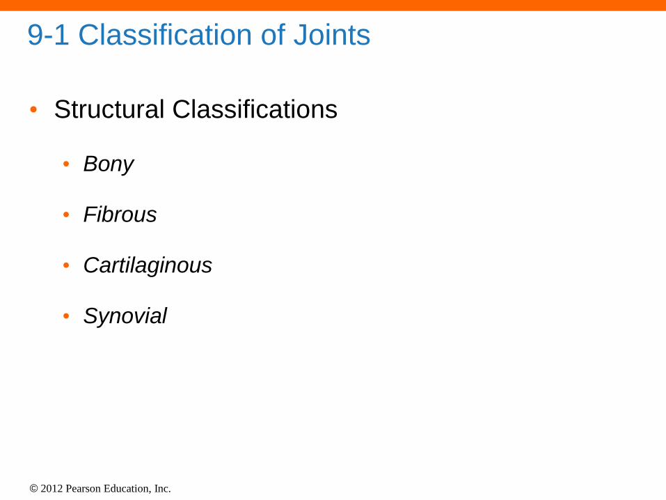
9-1 Classification of Joints
• Suture
• Bones interlocked
• Are bound by dense fibrous connective tissue
• Are found only in skull
• Gomphosis
• Fibrous connection (periodontal ligament)
• Binds teeth to sockets

© 2012 Pearson Education, Inc.
9-1 Classification of Joints
• Synchondrosis
• Is a rigid cartilaginous bridge between two bones
• Epiphyseal cartilage of long bones
• Between vertebrosternal ribs and sternum
• Synostosis
• Fused bones, immovable
• Metopic suture of skull
• Epiphyseal lines of long bones

© 2012 Pearson Education, Inc.
9-1 Classification of Joints
• Amphiarthroses
• More movable than synarthrosis
• Stronger than freely movable joint
• Two types of amphiarthroses
1. Syndesmosis
• Bones connected by ligaments
2. Symphysis
• Bones separated by fibrocartilage

© 2012 Pearson Education, Inc.
9-1 Classification of Joints
• Synovial Joints (Diarthroses)
• Also called movable joints
• At ends of long bones
• Within articular capsules
• Lined with synovial membrane

© 2012 Pearson Education, Inc.
9-2 Synovial Joints
• Articular Cartilages
• Pad articulating surfaces within articular capsules
• Prevent bones from touching
• Smooth surfaces lubricated by synovial fluid
• Reduce friction

© 2012 Pearson Education, Inc.
9-2 Synovial Joints
• Synovial Fluid
• Contains slippery proteoglycans secreted by
fibroblasts
• Functions of synovial fluid
1. Lubrication
2. Nutrient distribution
3. Shock absorption

© 2012 Pearson Education, Inc.
9-2 Synovial Joints
• Accessory Structures
• Cartilages
• Fat pads
• Ligaments
• Tendons
• Bursae

© 2012 Pearson Education, Inc.
9-2 Synovial Joints
• Cartilages
• Cushion the joint
• Fibrocartilage pad called a meniscus (or articular disc;
plural, menisci)
• Fat Pads
• Superficial to the joint capsule
• Protect articular cartilages
• Ligaments
• Support, strengthen joints
• Sprain – ligaments with torn collagen fibers

© 2012 Pearson Education, Inc.
9-2 Synovial Joints
• Tendons
• Attach to muscles around joint
• Help support joint
• Bursae
• Singular, bursa, a pouch
• Pockets of synovial fluid
• Cushion areas where tendons or ligaments rub

© 2012 Pearson Education, Inc.
9-2 Synovial Joints
• Factors That Stabilize Synovial Joints
• Prevent injury by limiting range of motion
• Collagen fibers (joint capsule, ligaments)
• Articulating surfaces and menisci
• Other bones, muscles, or fat pads
• Tendons of articulating bones

© 2012 Pearson Education, Inc.
Figure 9-1a The Structure of a Synovial Joint
Synovial joint, sagittal section
Medullary cavity
Spongy bone
Periosteum
Fibrous joint capsule
Synovial membrane
Articular cartilages
Joint cavity
(containing
synovial fluid)
Compact bone
© 2012 Pearson Education, Inc.
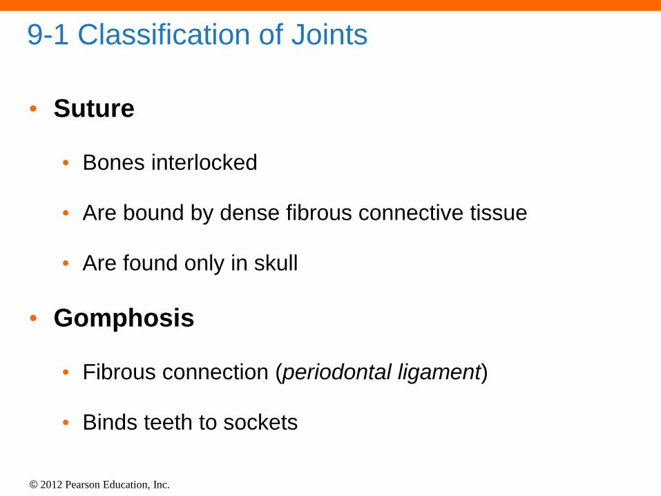
Figure 9-1b The Structure of a Synovial Joint
Knee joint, sagittal section
Synovial
membrane
Intracapsular
ligament
Joint capsule
Meniscus
Femur
Tibia
Quadriceps tendon
Patella
Articular cartilage
Fat padPatellar ligament
Joint cavityMeniscus
Bursa
© 2012 Pearson Education, Inc.
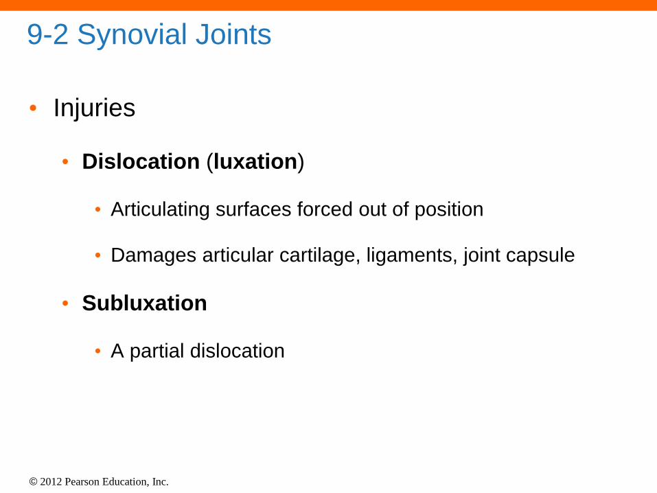
9-2 Synovial Joints
• Injuries
• Dislocation (luxation)
• Articulating surfaces forced out of position
• Damages articular cartilage, ligaments, joint capsule
• Subluxation
• A partial dislocation
© 2012 Pearson Education, Inc.
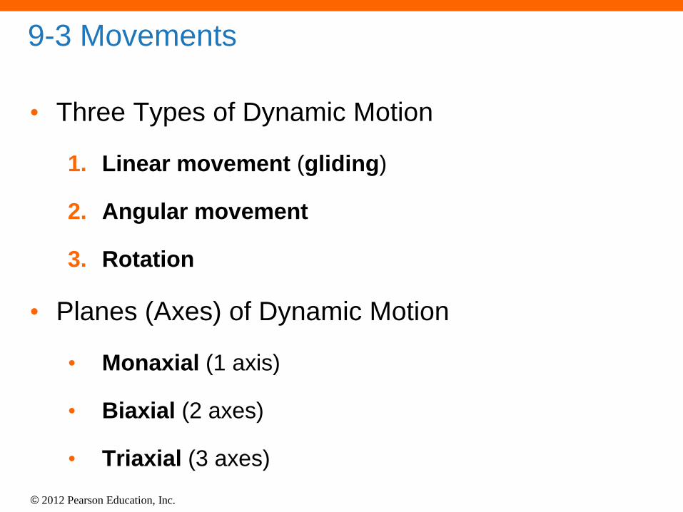
9-3 Movements
• Three Types of Dynamic Motion
1. Linear movement (gliding)
2. Angular movement
3. Rotation
• Planes (Axes) of Dynamic Motion
• Monaxial (1 axis)
• Biaxial (2 axes)
• Triaxial (3 axes)
© 2012 Pearson Education, Inc.
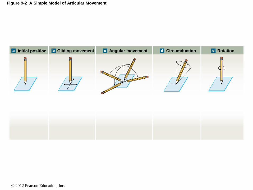
Figure 9-2 A Simple Model of Articular Movement
Initial position Gliding movement Angular movement Circumduction Rotation

© 2012 Pearson Education, Inc.
Figure 9-2a A Simple Model of Articular Movement
Initial position
Initial position of
the model. The
pencil is at right
angles to surface.

© 2012 Pearson Education, Inc.
Figure 9-2b A Simple Model of Articular Movement
Gliding movement
Possible movement 1,
showing gliding, an
example of linear
movement. The pencil
remains vertical, but
tip moves away from
point of origin.

© 2012 Pearson Education, Inc.
Figure 9-2c A Simple Model of Articular Movement
Angular movement
Possible movement 2,
showing angular movement.
The pencil tip remains
stationary, but shaft changes
angle relative to the surface.

© 2012 Pearson Education, Inc.
Figure 9-2d A Simple Model of Articular Movement
Circumduction
Possible movement 2,
showing a special type
of angular movement
called circumduction.
Pencil tip remains
stationary while the
shaft, held at an angle
less than 90º, moves in
a conical pattern to
complete a circle.

© 2012 Pearson Education, Inc.
Figure 9-2e A Simple Model of Articular Movement
Rotation
Possible movement
3, showing rotation.
With tip at same
point, the angle of
the shaft remains
unchanged as the
shaft spins around
its longitudinal axis.

© 2012 Pearson Education, Inc.
9-3 Movements
• Types of Movement at Synovial Joints
• Terms describe:
• Plane or direction of motion
• Relationship between structures

© 2012 Pearson Education, Inc.
9-3 Movements
• Types of Movement at Synovial Joints
• Gliding Movement
• Two surfaces slide past each other
• Between carpal or tarsal bones

© 2012 Pearson Education, Inc.
9-3 Movements
• Angular Movement
• Flexion
• Angular motion
• Anterior–posterior plane
• Reduces angle between elements
• Extension
• Angular motion
• Anterior–posterior plane
• Increases angle between elements

© 2012 Pearson Education, Inc.
Figure 9-3a Angular Movements
Flexion/extension
Extension
Extension
Extension
Flexion
Flexion
Flexion
Hyperextension
Hyperextension
Hyper-extension
Flexion
Extension

© 2012 Pearson Education, Inc.
9-3 Movements
• Angular Movement
• Hyperextension
• Angular motion
• Extension past anatomical position

© 2012 Pearson Education, Inc.
Figure 9-3a Angular Movements
Flexion/extension
Extension
Extension
Extension
Flexion
Flexion
Flexion
Hyperextension
Hyperextension
Hyper-extension
Flexion
Extension

© 2012 Pearson Education, Inc.
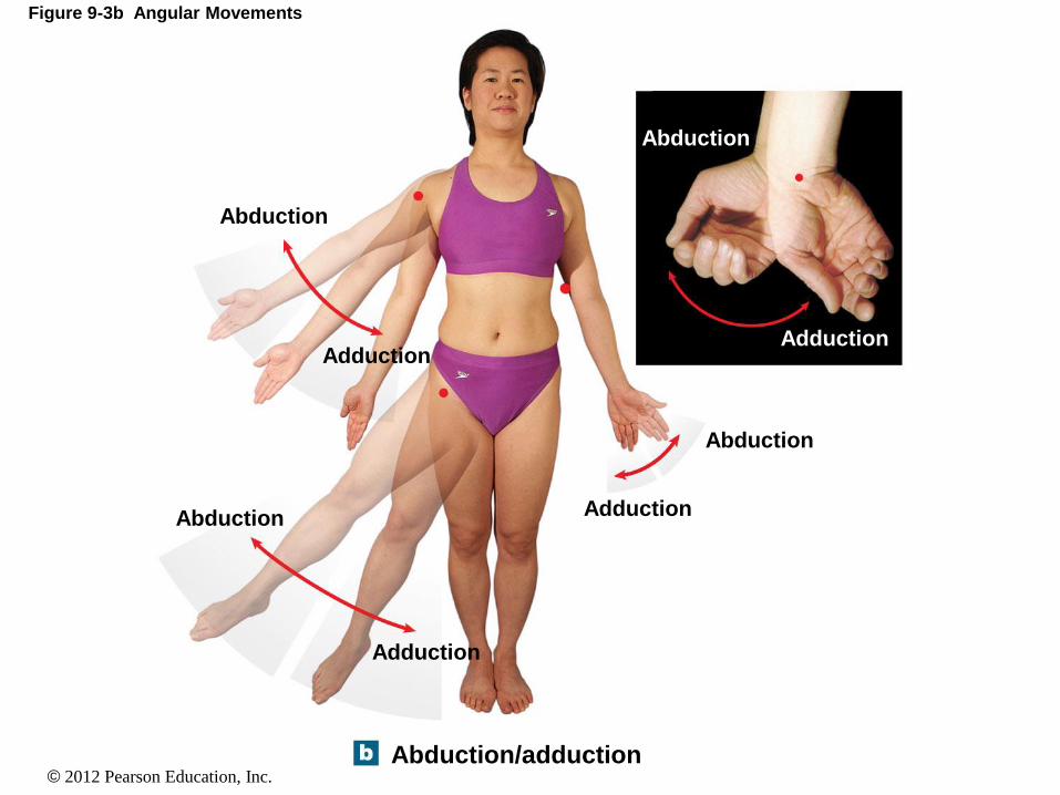
9-3 Movements
• Angular Movement
• Abduction
• Angular motion
• Frontal plane
• Moves away from longitudinal axis
• Adduction
• Angular motion
• Frontal plane
• Moves toward longitudinal axis
© 2012 Pearson Education, Inc.
Figure 9-3b Angular Movements
Abduction/adduction
Abduction
Abduction
Abduction
Adduction
Adduction
AdductionAdduction
Abduction

© 2012 Pearson Education, Inc.
Figure 9-3c Angular Movements
Adduction/abduction
AbductionAdduction

© 2012 Pearson Education, Inc.
9-3 Movements
• Angular Movement
• Circumduction
• Circular motion without rotation
• Angular motion
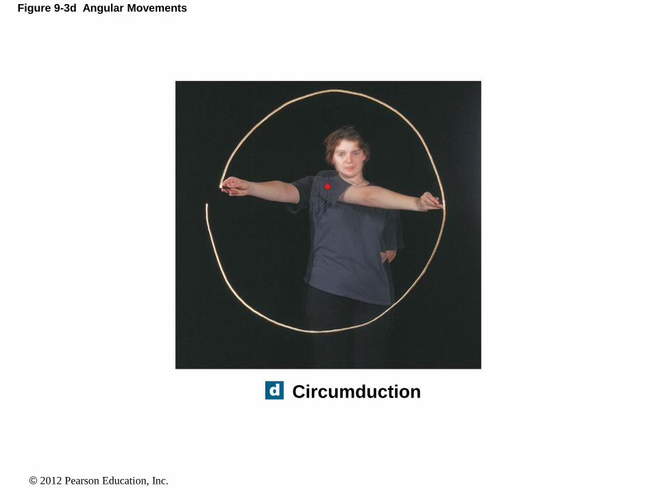
© 2012 Pearson Education, Inc.
Figure 9-3d Angular Movements
Circumduction

© 2012 Pearson Education, Inc.
9-3 Movements
• Types of Movement at Synovial Joints
• Rotation
• Direction of rotation from anatomical position
• Relative to longitudinal axis of body
• Left or right rotation
• Medial rotation (inward rotation)
• Rotates toward axis
• Lateral rotation (outward rotation)
• Rotates away from axis

© 2012 Pearson Education, Inc.
Figure 9-4a Rotational Movements
Head rotation
Rightrotation
Leftrotation
Lateral(external)rotation Medial
(internal)rotation

© 2012 Pearson Education, Inc.
9-3 Movements
• Types of Movements at Synovial Joints
• Rotation
• Pronation
• Rotates forearm, radius over ulna
• Supination
• Forearm in anatomical position

© 2012 Pearson Education, Inc.
Figure 9-4b Rotational Movements
Supination Pronation
Pronation
Supination

© 2012 Pearson Education, Inc.
9-3 Movements
• Special Movements
• Inversion
• Twists sole of foot medially
• Eversion
• Twists sole of foot laterally
• Dorsiflexion
• Flexion at ankle (lifting toes)
• Plantar flexion
• Extension at ankle (pointing toes)

© 2012 Pearson Education, Inc.
Figure 9-5 Synovial Joints
Eversion Inversion

© 2012 Pearson Education, Inc.
Figure 9-5 Synovial Joints
Plantarflexion
(ankle extension)
Dorsiflexion(ankle flexion)

© 2012 Pearson Education, Inc.
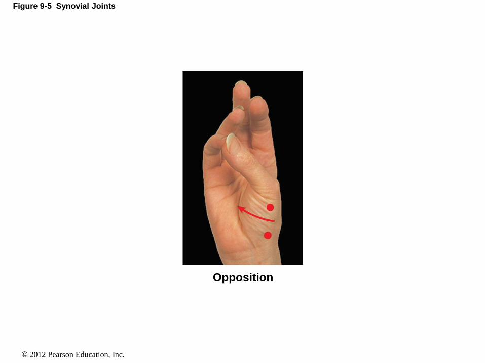
9-3 Movements
• Special Movements
• Opposition
• Thumb movement toward fingers or palm (grasping)
• Reposition
• Opposite of opposition
• Protraction
• Moves anteriorly
• In the horizontal plane (pushing forward)
• Retraction
• Opposite of protraction
• Moving anteriorly (pulling back)
© 2012 Pearson Education, Inc.
Figure 9-5 Synovial Joints
Opposition

© 2012 Pearson Education, Inc.
Figure 9-5 Synovial Joints
ProtractionRetraction

© 2012 Pearson Education, Inc.
9-3 Movements
• Special Movements
• Elevation
• Moves in superior direction (up)
• Depression
• Moves in inferior direction (down)
• Lateral flexion
• Bends vertebral column from side to side

© 2012 Pearson Education, Inc.
Figure 9-5 Synovial Joints
ElevationDepression

© 2012 Pearson Education, Inc.
Figure 9-5 Synovial Joints
Lateral flexion
© 2012 Pearson Education, Inc.
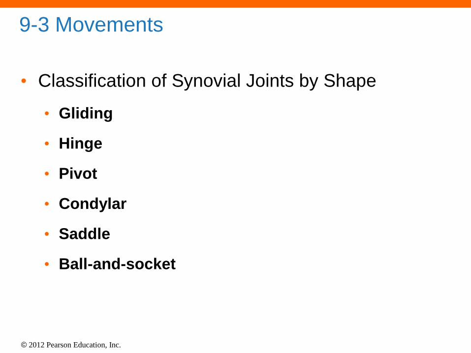
9-3 Movements
• Classification of Synovial Joints by Shape
• Gliding
• Hinge
• Pivot
• Condylar
• Saddle
• Ball-and-socket

© 2012 Pearson Education, Inc.
9-3 Movements
• Gliding Joints
• Flattened or slightly curved faces
• Limited motion (nonaxial)
• Hinge Joints
• Angular motion in a single plane (monaxial)
• Pivot Joints
• Rotation only (monaxial)

© 2012 Pearson Education, Inc.
Figure 9-6 Synovial Joints
Gliding joint
Manubrium
Movement: slight nonaxial or multiaxial
Examples:• Acromioclavicular and claviculosternal joints• Intercarpal and intertarsal joints
• Vertebrocostal joints• Sacro-iliac joints

© 2012 Pearson Education, Inc.
Figure 9-6 Synovial Joints
Hinge joint
Ulna
Humerus
Movement: monaxial
Examples:• Elbow joint• Knee joint• Ankle joint• Interphalangeal joint

© 2012 Pearson Education, Inc.
Figure 9-6 Synovial Joints
Pivot joint
Axis
Atlas
Movement: monaxial (rotation)
Examples:• Atlanto-axial joint• Proximal radio-ulnar joint

© 2012 Pearson Education, Inc.
9-3 Movements
• Condylar Joints
• Oval articular face within a depression
• Motion in two planes (biaxial)
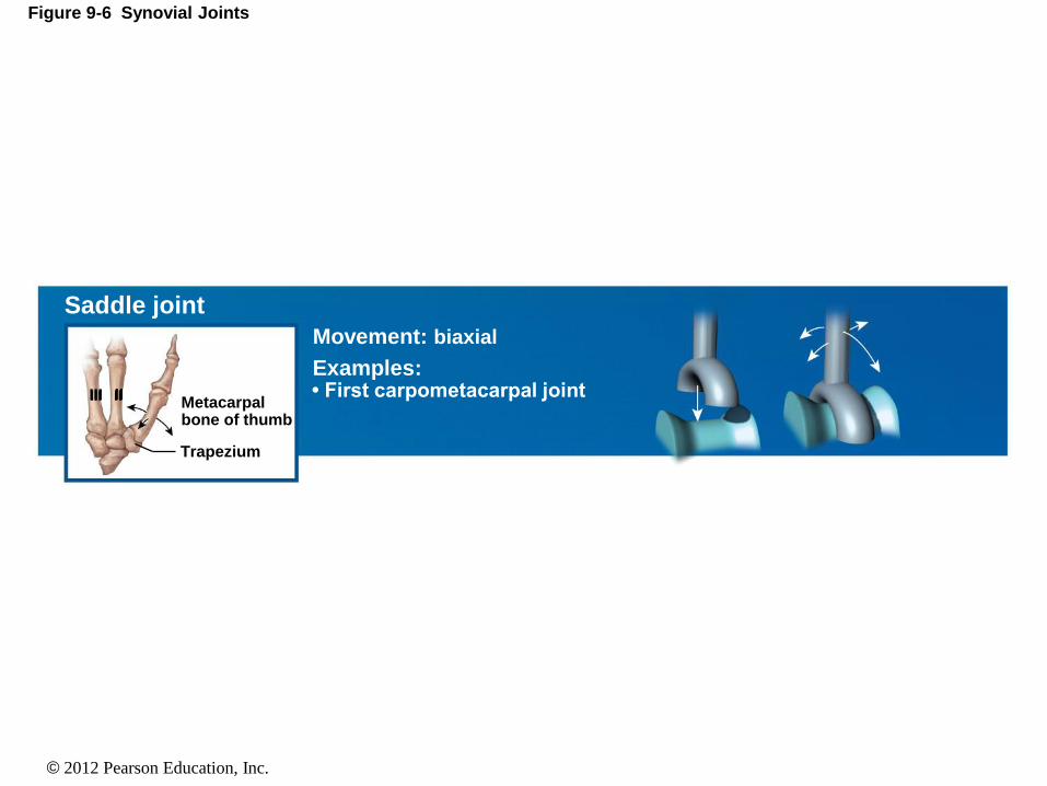
• Saddle Joints
• Two concave, straddled (biaxial)
• Ball-and-socket Joints
• Round articular face in a depression (triaxial)

© 2012 Pearson Education, Inc.
Figure 9-6 Synovial Joints
Condylar joint
Ulna
Scaphoidbone
Movement: biaxial
Examples:• Radiocarpal joint• Metacarpophalangeal joints 2–5• Metatarsophalangeal joints
© 2012 Pearson Education, Inc.
Figure 9-6 Synovial Joints
Saddle joint
Trapezium
Metacarpalbone of thumb
III II
Movement: biaxial
Examples:• First carpometacarpal joint

© 2012 Pearson Education, Inc.
Figure 9-6 Synovial Joints
Ball-and-socket joint
Humerus
Scapula
Movement: triaxial
Examples:• Shoulder joint• Hip joint

© 2012 Pearson Education, Inc.
9-3 Movements
• Joints
• A joint cannot be both mobile and strong
• The greater the mobility, the weaker the joint
• Mobile joints are supported by muscles and
ligaments, not bone-to-bone connections
ANIMATION Representative Articulations: A Functional Classification
of Synovial Joints

© 2012 Pearson Education, Inc.
9-6 The Knee Joint
• The Knee Joint
• A complicated hinge joint
• Transfers weight from femur to tibia
• Articulations of the knee joint
• Two femur–tibia articulations
• At medial and lateral condyles
• One between patella and patellar surface of femur

© 2012 Pearson Education, Inc.
9-6 The Knee Joint
• The Articular Capsule and Joint Cavity
• Medial and lateral menisci
• Fibrocartilage pads
• At femur–tibia articulations
• Cushion and stabilize joint
• Give lateral support

© 2012 Pearson Education, Inc.
9-6 The Knee Joint
• Seven Major Supporting Ligaments
1. Patellar ligament (anterior)
2. & 3. Two popliteal ligaments (posterior)
4. & 5. Anterior and posterior cruciate ligaments (inside
joint capsule)
6. Tibial collateral ligament (medial)
7. Fibular collateral ligament (lateral)

© 2012 Pearson Education, Inc.
Figure 9-12a The Right Knee Joint
Anterior view, superficial layer
Jointcapsule
Quadricepstendon
Patellarretinaculae
Fibularcollateralligament
Patella
Patellarligament
Tibia
Tibialcollateralligament

© 2012 Pearson Education, Inc.
Figure 9-12b The Right Knee Joint
Posterior view, superficial layer
Tibialcollateralligament
Jointcapsule
Bursa
Poplitealligaments
Popliteusmuscle
Femur
Tibia
Fibula
Cut tendonof bicepsfemorismuscle
Fibularcollateralligament
Gastrocnemiusmuscle,lateral head
Plantarismuscle
Gastrocnemiusmuscle,
medial head

© 2012 Pearson Education, Inc.
Figure 9-12c The Right Knee Joint
Deep anterior view, flexed
Medial condyle
Medial meniscus
Posterior cruciate
ligament
Tibial collateral
ligament
Anterior cruciate
ligament
Fibular
collateral
ligament
Lateral
meniscus
Cut
tendon
Fibula
Tibia
Lateral
condyle
Patellar
surface

© 2012 Pearson Education, Inc.
Figure 9-12d The Right Knee Joint
Deep posterior view, extended
Femur
Fibular collateral
ligament
Lateral condyle
Lateral
meniscus
Cut tendon
Fibula
Tibia
Posterior cruciate
ligament
Tibial collateral
ligament
Anterior cruciate
ligament
Medial meniscus
Medial condyle

© 2012 Pearson Education, Inc.
9-7 Effects of Aging on Articulations
• Degenerative Changes
• Rheumatism
• A pain and stiffness of skeletal and muscular systems
• Arthritis
• All forms of rheumatism that damage articular
cartilages of synovial joints
• Osteoarthritis
• Caused by wear and tear of joint surfaces, or genetic
factors affecting collagen formation
• Generally in people over age 60
© 2012 Pearson Education, Inc.
9-7 Effects of Aging on Articulations
• Rheumatoid Arthritis
• An inflammatory condition
• Caused by infection, allergy, or autoimmune disease
• Involves the immune system
• Gouty Arthritis
• Occurs when crystals (uric acid or calcium salts)
• Form within synovial fluid
• Due to metabolic disorders
© 2012 Pearson Education, Inc.
9-7 Effects of Aging on Articulations
• Joint Immobilization
• Reduces flow of synovial fluid
• Can cause arthritis symptoms
• Treated by continuous passive motion or CPM
(therapy)
• Bones and Aging
• Bone mass decreases
• Bones weaken
• Increases risk of hip fracture, hip dislocation, or pelvic
fracture

© 2012 Pearson Education, Inc.
9-8 Integration with Other Systems
• Bone Recycling
• Living bones maintain equilibrium between:
• Bone building (osteoblasts)
• And breakdown (osteoclasts)

© 2012 Pearson Education, Inc.
9-8 Integration with Other Systems
• Factors Affecting Bone Strength
1. Age
2. Physical stress
3. Hormone levels
4. Calcium and phosphorus uptake and excretion
5. Genetic and environmental factors

© 2012 Pearson Education, Inc.
9-8 Integration with Other Systems
• Bones Support Body Systems
• Support and protect other systems
• Store fat, calcium, and phosphorus
• Manufacture cells for immune system

© 2012 Pearson Education, Inc.
9-8 Integration with Other Systems
• Bones Support Body Systems
• Disorders in other body systems can cause:
• Bone tumors
• Osteoporosis
• Arthritis
• Rickets (vitamin D deficiency)