ARTICLE Live imaging of marked chromosome regions reveals ...
22
ARTICLE Live imaging of marked chromosome regions reveals their dynamic resolution and compaction in mitosis John K. Eykelenboom 1 , Marek Gierli´ nski 1,2 *, Zuojun Yue 1 *, Nadia Hegarat 3 *, Hilary Pollard 3 , Tatsuo Fukagawa 4 , Helfrid Hochegger 3 , and Tomoyuki U. Tanaka 1 When human cells enter mitosis, chromosomes undergo substantial changes in their organization to resolve sister chromatids and compact chromosomes. To comprehend the timing and coordination of these events, we need to evaluate the progression of both sister chromatid resolution and chromosome compaction in one assay. Here we achieved this by analyzing changes in configuration of marked chromosome regions over time, with high spatial and temporal resolution. This assay showed that sister chromatids cycle between nonresolved and partially resolved states with an interval of a few minutes during G2 phase before completing full resolution in prophase. Cohesins and WAPL antagonistically regulate sister chromatid resolution in late G2 and prophase while local enrichment of cohesin on chromosomes prevents precocious sister chromatid resolution. Moreover, our assay allowed quantitative evaluation of condensin II and I activities, which differentially promote sister chromatid resolution and chromosome compaction, respectively. Our assay reveals novel aspects of dynamics in mitotic chromosome resolution and compaction that were previously obscure in global chromosome assays. Introduction At the beginning of mitosis, chromosomes undergo two major structural changes in metazoan cells. First, sister chromatids are resolved from each other along chromosome arms; this process involves removal of sister chromatid cohesion and elimination of topological DNA links (Nasmyth and Haering, 2009; Pommier et al., 2016; Uhlmann, 2016). Second, each sister chromatid is compacted; as a result, they become thicker in width and shorter in length (Hirano, 2016; Uhlmann, 2016). These two changes are a prerequisite for proper chromosome segregation toward op- posite spindle poles during the subsequent anaphase. However, the precise timing and coordination of these two changes are still not fully understood. Several factors regulate sister chromatid resolution and chro- mosome compaction. Sister chromatids are held together by the cohesin complex, which forms a ring structure consisting of SMC1, SMC3, RAD21, and SA1/2 (Nasmyth and Haering, 2009). For sister chromatid resolution, the cohesin complex must be removed along chromosome arms during prophase through the destabilizing activity of the WAPL (Wings apart-like protein ho- mologue), while it is retained at the centromere to maintain sister chromatid cohesion until anaphase onset (Peters et al., 2008; Morales and Losada, 2018). In addition, topological DNA links (DNA catenation) from DNA supercoiling during DNA replication must also be removed by the de-catenation activity of topoiso- merase II (topo II; Pommier et al., 2016; Piskadlo and Oliveira, 2017). Sister chromatid resolution starts in late G2 phase (Ono et al., 2013; Stanyte et al., 2018) and continues into prophase (Nagasaka et al., 2016). However, the dynamics of sister chro- matid resolution in G2 and its regulation are not fully understood. Furthermore, the condensin complex plays important roles in both sister chromatid resolution and chromosome compaction. The condensin complex exists as two forms—condensin I and II—that consist of the common SMC2 and SMC4 subunits and distinct non-SMC subunits such as NCAPD2 and NCAPD3 (for condensin I and II, respectively) (Hirano, 2012). Condensin I and II collaboratively generate helical arrays of nested chromatin loops (Gibcus et al., 2018; Walther et al., 2018). Moreover, con- densin II operates earlier and contributes more to sister chro- matid resolution than does condensin I (Ono et al., 2003; Shintomi and Hirano, 2011; Green et al., 2012; Hirano, 2012; Nagasaka et al., 2016). The precise timing of condensin I and II activity and their relative contribution to sister chromatid res- olution and chromosome compaction remains to be fully elucidated. The analysis of chromosome reorganization in early mitosis has been advanced by several new methods, which include ............................................................................................................................................................................. 1 Centre for Gene Regulation and Expression, School of Life Sciences, University of Dundee, Dundee, UK; 2 Data Analysis Group, School of Life Sciences, University of Dundee, Dundee, UK; 3 Genome Damage and Stability Centre, University of Sussex, Brighton, UK; 4 Graduate School of Frontier Biosciences, Osaka University, Suita, Osaka, Japan. *M. Gierli´ nski, Z. Yue, and N. Hegarat contributed equally to this paper; Correspondence to Tomoyuki U. Tanaka: [email protected]. © 2019 Eykelenboom et al. This article is available under a Creative Commons License (Attribution 4.0 International, as described at https://creativecommons.org/ licenses/by/4.0/). Rockefeller University Press https://doi.org/10.1083/jcb.201807125 1531 J. Cell Biol. 2019 Vol. 218 No. 5 1531–1552 Downloaded from http://rupress.org/jcb/article-pdf/218/5/1531/1381207/jcb_201807125.pdf by guest on 22 March 2022
Transcript of ARTICLE Live imaging of marked chromosome regions reveals ...

ARTICLE
Live imaging of marked chromosome regions revealstheir dynamic resolution and compaction in mitosisJohn K. Eykelenboom1, Marek Gierlinski1,2*, Zuojun Yue1*, Nadia Hegarat3*, Hilary Pollard3, Tatsuo Fukagawa4, Helfrid Hochegger3, andTomoyuki U. Tanaka1
When human cells enter mitosis, chromosomes undergo substantial changes in their organization to resolve sister chromatidsand compact chromosomes. To comprehend the timing and coordination of these events, we need to evaluate the progressionof both sister chromatid resolution and chromosome compaction in one assay. Here we achieved this by analyzing changes inconfiguration of marked chromosome regions over time, with high spatial and temporal resolution. This assay showed thatsister chromatids cycle between nonresolved and partially resolved states with an interval of a few minutes during G2 phasebefore completing full resolution in prophase. Cohesins and WAPL antagonistically regulate sister chromatid resolution inlate G2 and prophase while local enrichment of cohesin on chromosomes prevents precocious sister chromatid resolution.Moreover, our assay allowed quantitative evaluation of condensin II and I activities, which differentially promote sisterchromatid resolution and chromosome compaction, respectively. Our assay reveals novel aspects of dynamics in mitoticchromosome resolution and compaction that were previously obscure in global chromosome assays.
IntroductionAt the beginning of mitosis, chromosomes undergo two majorstructural changes in metazoan cells. First, sister chromatids areresolved from each other along chromosome arms; this processinvolves removal of sister chromatid cohesion and eliminationof topological DNA links (Nasmyth and Haering, 2009; Pommieret al., 2016; Uhlmann, 2016). Second, each sister chromatid iscompacted; as a result, they become thicker in width and shorterin length (Hirano, 2016; Uhlmann, 2016). These two changes area prerequisite for proper chromosome segregation toward op-posite spindle poles during the subsequent anaphase. However,the precise timing and coordination of these two changes arestill not fully understood.
Several factors regulate sister chromatid resolution and chro-mosome compaction. Sister chromatids are held together by thecohesin complex, which forms a ring structure consisting ofSMC1, SMC3, RAD21, and SA1/2 (Nasmyth and Haering, 2009).For sister chromatid resolution, the cohesin complex must beremoved along chromosome arms during prophase through thedestabilizing activity of the WAPL (Wings apart-like protein ho-mologue), while it is retained at the centromere to maintain sisterchromatid cohesion until anaphase onset (Peters et al., 2008;Morales and Losada, 2018). In addition, topological DNA links(DNA catenation) from DNA supercoiling during DNA replication
must also be removed by the de-catenation activity of topoiso-merase II (topo II; Pommier et al., 2016; Piskadlo and Oliveira,2017). Sister chromatid resolution starts in late G2 phase (Onoet al., 2013; Stanyte et al., 2018) and continues into prophase(Nagasaka et al., 2016). However, the dynamics of sister chro-matid resolution in G2 and its regulation are not fully understood.
Furthermore, the condensin complex plays important roles inboth sister chromatid resolution and chromosome compaction.The condensin complex exists as two forms—condensin I andII—that consist of the common SMC2 and SMC4 subunits anddistinct non-SMC subunits such as NCAPD2 and NCAPD3 (forcondensin I and II, respectively) (Hirano, 2012). Condensin I andII collaboratively generate helical arrays of nested chromatinloops (Gibcus et al., 2018; Walther et al., 2018). Moreover, con-densin II operates earlier and contributes more to sister chro-matid resolution than does condensin I (Ono et al., 2003;Shintomi and Hirano, 2011; Green et al., 2012; Hirano, 2012;Nagasaka et al., 2016). The precise timing of condensin I and IIactivity and their relative contribution to sister chromatid res-olution and chromosome compaction remains to be fullyelucidated.
The analysis of chromosome reorganization in early mitosishas been advanced by several new methods, which include
.............................................................................................................................................................................1Centre for Gene Regulation and Expression, School of Life Sciences, University of Dundee, Dundee, UK; 2Data Analysis Group, School of Life Sciences, University ofDundee, Dundee, UK; 3Genome Damage and Stability Centre, University of Sussex, Brighton, UK; 4Graduate School of Frontier Biosciences, Osaka University, Suita, Osaka,Japan.
*M. Gierlinski, Z. Yue, and N. Hegarat contributed equally to this paper; Correspondence to Tomoyuki U. Tanaka: [email protected].
© 2019 Eykelenboom et al. This article is available under a Creative Commons License (Attribution 4.0 International, as described at https://creativecommons.org/licenses/by/4.0/).
Rockefeller University Press https://doi.org/10.1083/jcb.201807125 1531
J. Cell Biol. 2019 Vol. 218 No. 5 1531–1552
Dow
nloaded from http://rupress.org/jcb/article-pdf/218/5/1531/1381207/jcb_201807125.pdf by guest on 22 M
arch 2022

chromosome conformation capture analyses (Hi-C; Naumovaet al., 2013; Gibcus et al., 2018), differential visualization of sis-ter chromatids (Nagasaka et al., 2016), and in vitro reconstitutionof mitotic chromosomes (Shintomi et al., 2015). However, cur-rently available methods cannot attain the following two goals.First, very few methods allow quantitative evaluation of sisterchromatid resolution and chromosome compaction together. Forexample, Hi-C provides detailed information about chromosomecompaction but not about sister chromatid resolution. A simul-taneous evaluation of resolution and compaction is, however,critical since these processes might be coordinated. Second, al-though progression of global chromosome reorganization hasbeen investigated in early mitosis, few studies analyzed regionalchromosome reorganization in real time. Since global chromo-some changes are the ensemble outcome of regional changes, suchanalyses could obscure dynamic regional changes ofchromosomes—for example, any rapid or cyclical changes.
To achieve real-time measurements of regional chromosomedynamics, we investigated changes in specific chromosome re-gions over time in this study. Using bacteria-derived operatorarrays (Michaelis et al., 1997; Belmont and Straight, 1998) we havecreated a fluorescence reporter system that quantitatively eval-uates the timing of both sister chromatid resolution and chro-mosome compaction at chosen chromosome regions in humancells. This has allowed us to study dynamic chromosome reorga-nization from G2 phase to early mitosis by live cell microscopy.
ResultsVisualizing sister chromatid resolution and compaction at achosen region in live human cellsTo analyze mitotic chromosome reorganization, we developedan assay system in live HT-1080 diploid human cells. UsingCRISPR-Cas9 technology we integrated a tet operator array and alac operator array (Lau et al., 2003) with a 250-kbp interval to aregion of chromosome 5 with low gene density (Fig. 1, A–C). Thetet and lac operators (tetO and lacO) were bound by Tet-repressorfused to four monomer-Cherry fluorescent proteins (TetR-4xmCh) and by the Lac-repressor fused to GFP and a nuclearlocalization signal (EGFP-LacI-NLS), thus visualized as red andgreen fluorescent dots, respectively (Fig. 1 D). We chose a cellline where red and green fluorescent dots were found in prox-imity, reasoning that, in this cell line, tetO and lacO were inte-grated on the same copy of chromosome 5. The tetO and lacOwere stably maintained during cell proliferation since theirsignal intensity did not become weakened. As implied previ-ously (Chubb et al., 2002; Thomson et al., 2004), integration ofthese operators onto chromosome 5 did not affect cell cycleprogression or the fidelity of chromosome segregation; indeed,there was no change in DNA content of these cells as determinedby flow cytometry or no missegregation of chromosome 5 ob-served by microscopy.
We acquiredmicroscopy images of the above cells and analyzedthe z-stack images in 3D space, which revealed various config-urations of the fluorescent dots (Fig. S1 A). During interphase, weobserved one green dot and one red dot (Fig. 2 A; defined as bluestate). By contrast, in early mitosis (shortly before and after
nuclear envelope breakdown [NEBD]; see below), we often ob-served (a) one green dot and two red dots (or vice versa; defined asbrown state), (b) two green dots and two red dots without coloc-alization (defined as pink state), and (c) two green dots and two reddots with colocalization of each green and red dot (defined as redstate; Fig. 2 A). These states are likely to reflect chromosome re-organization during early mitosis, as follows (Fig. 2 B): (a) the bluestate represents “nonresolved” sister chromatids (if cells haveprogressed through S phase), (b) the brown state reflects “partiallyresolved” sister chromatids, (c) the pink state shows “resolved”(but not compacted) sister chromatids, and (d) the red state in-dicates resolved and “compacted” chromatids. Notably, the red“compacted” state was found to be the most frequent state in thefinal minutes before anaphase onset, suggesting that it reflects the“end” metaphase chromosome structure.
To analyze chromosome reorganization in early mitosis, wereleased cells with the fluorescent dots from a double thymidineblock and acquired live-cell images every minute between 8 and12 h (relative to the release). We were able to identify the timingof NEBD in individual cells, as it caused dispersion of the EGFP-LacI-NLS signal (the fraction not bound to lac operators) fromthe nucleus (Fig. S1 B). For individual cells, we aligned the se-quence of the states of fluorescent dots (as defined in Fig. 2, Aand B) relative to NEBD (defined as time zero; Fig. 2 C). Then, weplotted the proportion of cells displaying each state against time(Fig. 2 D). We noticed that as cells approached NEBD, the pink“resolved” state increased its frequency. After NEBD, there wasan increase in occurrence of the red “compacted” state. Theseobservations suggest that chromosome reorganization proceedsfrom sister chromatid resolution to chromosome compaction, asassumed in Fig. 2 B. This conclusion was also supported bymeasurement of the distances between fluorescent dots (Fig. S1,C–F): that is, the mean distances between sister tetOs and be-tween sister lacOs increased before NEBD, reflecting sisterchromatid resolution (Fig. S1 D), while the mean distance be-tween tetO and lacO gradually decreased after NEBD, reflectingchromosome compaction (Fig. S1, E and F).
To analyze the dynamics of a marked chromosome region indifferent cells, we inserted tetO and lacOwith a 100-kbp intervalon the Z chromosome of DT40 cells (Fig. S2, A and B) and vi-sualized them using the samemethod as above. In these cells, wecould identify the same four configurations of the fluorescentdots as above (Fig. S2 C). The pink “resolved” and red “com-pacted” states appeared with similar timing to that observed inhuman cells (Fig. S2 D). We conclude that amarked chromosomeregion behaves similarly during early mitosis in different ver-tebrate species and in different chromosome contexts.
Sister chromatids cycle between nonresolved and partiallyresolved states with an interval of a few minutes during lateG2 phase before attaining full resolution in prophaseFurther analysis of the HT-1080 cells revealed that the brown“partially resolved” state often (∼20% of time points) appearedup to 2 h before NEBD (Fig. 2, C and D). The brown state typicallyappeared and continued for a few minutes before returning tothe blue “nonresolved” state (Fig. 3 A). Thus, the blue and brownstates show cyclical exchanges before being converted to the
Eykelenboom et al. Journal of Cell Biology 1532
Visualizing mitotic chromosome reorganization https://doi.org/10.1083/jcb.201807125
Dow
nloaded from http://rupress.org/jcb/article-pdf/218/5/1531/1381207/jcb_201807125.pdf by guest on 22 M
arch 2022
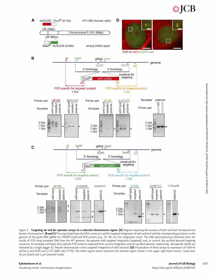
Figure 1. Targeting tet and lac operator arrays to a selected chromosome region. (A) Diagram depicting the location of tetO and lacO introduced intohuman chromosome 5. (B and C) The maps (top) show the DNA construct used for targeted integration of tetO and lacO and the corresponding positions on thegenome of the guide RNA (gRNA for CRISPR-Cas9) and PCR primers (e.g., 5F, 5R, etc.) for integration check. The DNA electrophoreses (bottom) show theresults of PCR using template DNA from the WT genome, the genome with targeted integration (targeted), and, as control, the purified plasmid targetingconstruct. Arrowheads and black dots indicate PCR products expected from correct integration and the purified plasmid, respectively. Nonspecific bands areindicated by a single dagger (†). Results demonstrate correct targeted integration of tetO and lacO. (D) Visualization of these arrays by expression of TetR-4xmCherry and EGFP-LacI in HT-1080 cells (TT75). The white square boxes represent the zoomed region shown in the upper right-hand corners. Scale bars,10 µm (main) and 1 µm (zoomed inset).
Eykelenboom et al. Journal of Cell Biology 1533
Visualizing mitotic chromosome reorganization https://doi.org/10.1083/jcb.201807125
Dow
nloaded from http://rupress.org/jcb/article-pdf/218/5/1531/1381207/jcb_201807125.pdf by guest on 22 M
arch 2022

pink “resolved” state (Fig. 3 B). To frame the timing of thisprocess in the cell cycle, we defined S, G2, and prophase in ourreal-time imaging, as follows: We identified S phase cells byvisualizing a component of the replication machinery, prolifer-ating cell nuclear antigen (PCNA) tagged with mCerulean. SinceCerulean-PCNA shows characteristic globular signals during Sphase (Fig. S2 E; Kitamura et al., 2006; Thomson et al., 2010), wedefined the end of S phase as the time its globular signals dis-appeared (Fig. S2 F, left). Our observations of PCNA and NEBDby live-cell imaging suggested that the length of G2 phase (be-tween the end of S phase and the start of prophase) was 5–7 h(Fig. S2 F, right), which is consistent with other studies (Defendiand Manson, 1963). We also defined prophase as a 20-min timewindow before NEBD, according to previous estimates (Lianget al., 2015) and based on the global change in the chromo-some volume observed in our cells (Fig. S2 G).
The brown “partially resolved” state appeared infrequentlyin late S phase (the last 30 min of S phase) and early G2 phase(first 90 min of G2 phase), but its frequency increased in late G2
(last 120 min of G2 phase; Fig. 3 C and Fig. S2 H). Subsequently,in prophase, the pink “resolved” state increased in frequency(Fig. 3 C). The brown state was also observed in ≤20% of DT40cells in late G2 phase (Fig. S2, C and D). We conclude that sisterchromatid resolution begins in late G2 phase and completesduring prophase, at least at the chromosome region of our study.Our results are consistent with other reports that sister chro-matid resolution begins before mitosis (Ono et al., 2013; Stanyteet al., 2018) and continues in prophase (Nagasaka et al., 2016). Assuggested by Stanyte et al. (2018), sister chromatids cycle be-tween nonresolved and partially resolved states in late G2 phase.We found that the two states interchange with a period of a fewminutes (i.e., more dynamically than previously thought).
Sister chromatid resolution is antagonistically regulated bycohesins and WAPL, not only during prophase but also inG2 phasePrevious studies suggested that maintenance of catenated DNArequires cohesins (Farcas et al., 2011; Sen et al., 2016) and
Figure 2. A fluorescence reporter for observing configuration of a selected chromosome region in live cells. (A) Representative images of the con-figurations of the fluorescence reporter observed in TT75 cells (see Fig. 1). Designated color codes for each configuration are indicated in the image frames.Scale bars, 1 µm. (B) Diagram shows the reporter configurations in A, with the same color codes as in A. (C) Change in the configuration of the fluorescencereporter over time (x axis) as observed in individual live cells (across the y axis). TT75 cells were synchronized by a double-thymidine block and released, imageswere taken every minute, and, at each time point, the configuration of the reporter was determined as in A and B (shownwith the same color codes). Data fromindividual cells were aligned relative to NEBD (defined as time zero). (D) The proportion of each configuration (color-coded as in A and B) was determined fromthe data in C and plotted over time with smoothing (across 9 min). The number of analyzed cells at each time point is between 15 and 55 (mean: 33).
Eykelenboom et al. Journal of Cell Biology 1534
Visualizing mitotic chromosome reorganization https://doi.org/10.1083/jcb.201807125
Dow
nloaded from http://rupress.org/jcb/article-pdf/218/5/1531/1381207/jcb_201807125.pdf by guest on 22 M
arch 2022

Figure 3. Sister chromatids cycle between nonresolved and partially resolved states during late G2 phase, antagonistically regulated by cohesinsand WAPL. (A) Time-sequence images of the fluorescence reporter showing cyclical separation of sister tetO. Times are relative to NEBD. Scale bar, 1 µm. (B)Diagram depicting cyclical partial sister resolution (brown state) in late G2 phase, leading to full resolution (pink state) in prophase. (C) The proportion of thebrown “partially resolved” or the pink “resolved” state during the indicated cell cycle phases. The cell cycle phases were determined in TT104 cells carryingCerulean-PCNA (n = 61 cells in late S and early G2 phase and 26 cells in late G2 and prophase). (D)Western blots for RAD21 (left) or WAPL (right) proteins fromcells after siRNA treatment for the indicated time. The control siRNA treatment was over 48 h. Actin is shown as a loading control. Asterisks indicate
Eykelenboom et al. Journal of Cell Biology 1535
Visualizing mitotic chromosome reorganization https://doi.org/10.1083/jcb.201807125
Dow
nloaded from http://rupress.org/jcb/article-pdf/218/5/1531/1381207/jcb_201807125.pdf by guest on 22 M
arch 2022

cohesin removal from chromosome arms by WAPL promotessister chromatid resolution in prophase (Peters et al., 2008). Wetested if these conclusions are reproduced with our assay. Wealso addressed whether the initial sister chromatid resolution inlate G2 phase, described above, depends on cohesin removal byWAPL. We used siRNA to deplete the cohesin subunit RAD21 orWAPL within 24–48 h of transfection (see Fig. 3 D and Fig. S2 I).By observing fixed metaphase chromosomes, we found thatsister chromatids were morphologically less distinct after WAPLdepletion, confirming the previously observed global defect insister chromatid resolution caused by WAPL siRNA (Gandhiet al., 2006; Fig. S2 J).
We then scored how fluorescent dots changed their config-uration from late G2 to prometaphase after 48 h of siRNAtreatment (Fig. 3 E and Fig. S2, K and L). Relative to controlsiRNA, depletion of RAD21 caused (a) an increase in the brown“partially resolved” and pink “resolved” states (Fig. 3 F) and,conversely, (b) a decrease in the blue “nonresolved” state(Fig. 3 G) in late G2 and prophase. In contrast, depletion ofWAPLled to the opposite outcomes in late G2 and prophase. Relativelymodest changes with RAD21 siRNA may be due to residualRAD21; consistently, even a small amount (∼20%) of cohesins issufficient tomaintain sister chromatid cohesion (Carvalhal et al.,2018). We conclude that cohesins inhibit precocious sisterchromatid resolution in late G2 and prophase while WAPLpromotes sister chromatid resolution during these phases. Thus,cohesins and WAPL play antagonistic roles in sister chromatidresolution not only during prophase but also during lateG2 phase.
Local cohesin enrichment regionally prevents precocious sisterchromatid resolution during prophaseIn the brown “partially resolved” state of fluorescent dots (seeFig. 2, A and B), tetO (red dot) showedmuchmore frequent sisterseparation than lacO (green dot) in late G2 and prophase (Fig. 4A). It is unlikely such sister tetO separationwas an artifact of thisarray, since the lacO also showed more frequent sister separa-tion (than in the original cell line) when it was integrated atanother chromosome region in another cell line (Fig. S3, A andB). In the original cell line, sister lacO separation may be infre-quent because of a specific chromosome feature. We inspectedthe genomic region where the operator arrays were integratedusing publicly available chromatin immunoprecipitation (ChIP;followed by DNA sequencing [ChIP-seq]) datasets (ENCODEProject Consortium, 2012). The lacO was located within 5 kbpof a cohesin-enriched region (Fig. 4 B; SMC3 and RAD21). Thisclose proximity to a cohesin-enriched site might contribute tothe reduced (or delayed) lacO separation. This cohesin peak
coincides with a CTCF (CCCTC-binding factor)-enriched region(Fig. 4 B). CTCF is a protein that binds specific DNA sequencesand acts as a barrier to cohesin movement leading to its localaccumulation (Parelho et al., 2008; Wendt et al., 2008). There-fore, deletion of the CTCF binding sequence of this region mightreduce the level of cohesins found there.
To investigate the outcome of a reduced cohesin level at thisregion, we deleted a 1277-bp DNA sequence corresponding to theCTCF enrichment site containing three CTCF-binding consensussites (Ziebarth et al., 2013) using CRISPR-Cas9 technique (Fig. 4C, left). Deletion of this region was confirmed by PCR and DNAsequencing on the two homologous chromosomes (Fig. 4 C,right; and Fig S3 C) and was designated as “CTCF-regionΔ/Δ.” Thelevel of chromosome-bound CTCF or SMC3 was examined in thevicinity of this region by ChIP followed by quantitative PCR(ChIP-qPCR; Fig. 4 C, left; and Fig. S3 D). This confirmed thatdeletion of the CTCF binding sites resulted in reduction ofchromosome-bound CTCF and SMC3 by 52% and 67%, respec-tively (Fig. 4, D and E; and Fig. S3, E and F).
In the CTCF-regionΔ/Δ strain, we scored how fluorescent dotschanged their configuration from late G2 to prometaphase(Fig. 4 F and Fig. S3, G and H). In the CTCF-regionΔ/Δ cell line, theoverall fraction of the brown “partially resolved” state wassimilar to the WT control (Fig. 4 G, top), but there was a slightincrease in sister separation of the lacO (green fluorescent dot)in late G2 phase (Fig. 4 H). Moreover, the CTCF-regionΔ/Δ cellline showed an earlier and greater increase in the pink “re-solved” state during prophase, compared with WT (Fig. 4, G[bottom] and I). We conclude that a local reduction in the co-hesin level leads to precocious sister chromatid resolution thereduring prophase, which is presumably due to weaker sisterchromatid cohesion.
Next, to study sister chromatid resolution and separation atmore chromosome sites, we used FISH. Using FISH probes set inthe region including tetO and lacO integration sites (Fig. 5 A), weinvestigated sister chromatid separation in HT-1080 cells fixedat prophase (Fig. 5 B). To exclude possible off-target signals inFISH, we used two probes (with different colors) together andanalyzed their signals only if they locate in close proximity(Fig. 5 C). Focusing on “partially resolved” states (i.e., one of thetwo probes showing separation), we first compared the fre-quency of sister separation of FISH probes in cell lines with orwithout tetO and lacO integration. Both conditions gave verysimilar results, in which Probe 1 (close to tetO integration site)showed higher sister separation than probe 3 (at lacO integrationsites; Fig. 5, A and D). The lacO integration site (probe 3) showedhigher percentage separation with FISH than with live-cellimaging (Fig. 4 A), which is probably due to (a) harsher
nonspecific binding of the anti-WAPL antibody. (E) The proportion of each configuration in cells treated with control, RAD21, and WAPL siRNA. Color codes areas in diagram (top, left). TT75 cells were arrested by a double-thymidine block and released and also treated with siRNA, as in Fig. S2 I. WAPL siRNA led tocolocalization of all four fluorescent dots (black line) after NEBD in some cells. Data from individual cells are shown in Fig. S2 K. The number of analyzed cells ateach point was between 10 and 53 (mean: 33) for control; 10 and 27 (mean: 19) for RAD21; 10 and 52 (mean: 36) for WAPL siRNA, except for the gray-shadedareas where <10 cells were analyzed. (F) The proportion of the brown “partially resolved” plus pink “resolved” state after control, RAD21 and WAPL siRNAtreatment. The data were taken from E. The gray shaded area indicates time points that include <10 cells for at least one siRNA treatment. (G) Graph shows theproportion of the blue “nonresolved” state for control, RAD21, andWAPL siRNA during late G2 phase or prophase. These data were taken from E. P values wereobtained using a chi-square test. n = 520–3,081 time points.
Eykelenboom et al. Journal of Cell Biology 1536
Visualizing mitotic chromosome reorganization https://doi.org/10.1083/jcb.201807125
Dow
nloaded from http://rupress.org/jcb/article-pdf/218/5/1531/1381207/jcb_201807125.pdf by guest on 22 M
arch 2022

Figure 4. Local reduction of cohesins at their enrichment site leads to precocious sister chromatid resolution in that region during prophase. (A)Graph shows the proportion of tetO (orange) and lacO (purple) sister separation among all “partially resolved” states in WT cells (TT75) during late G2 phase orprophase. n = 175–381 time points. (B) The ChIP-seq data show the distribution of SMC3, RAD21, and CTCF along the genomic region around integration sitesof lacO and tetO. The ChIP-seq data are taken from published data (ENCODE Project Consortium, 2012). (C) The map (left) is a zoomed view of the CTCF ChIP-seq peak at the chromosome region highlighted with # in B. The map also shows the positions on the genome to which guide RNA (gRNA; for CRISPR-Cas9) and
Eykelenboom et al. Journal of Cell Biology 1537
Visualizing mitotic chromosome reorganization https://doi.org/10.1083/jcb.201807125
Dow
nloaded from http://rupress.org/jcb/article-pdf/218/5/1531/1381207/jcb_201807125.pdf by guest on 22 M
arch 2022

treatment of cells during FISH preparation and/or (b) Probe1 being closer (than the tetO integration site) to small cohesinpeaks (∼26.05 Mbp on chromosome 5; Fig. 5 A), leading to arelatively low percentage separation of probe 1 (thus, a relativelyhigh percentage separation of probe 3). We conclude that inte-gration of tetO and lacO did not affect sister separation frequencyof their integration sites. We then compared sister separationfrequency using various FISH probes in prophase cells withouttetO and lacO arrays in “partially resolved” states (Fig. 5 E). Asshown in Fig. 5 F, larger distance from cohesin peaks (marked byarrows in Fig. 5 A) correlated well with higher sister separationfrequency. This suggests that local cohesin peaks delay sisterchromatid separation around these peaks during prophase.
Sister chromatid resolution is established in prophase andmaintained during prometaphase, relying on topo II activityUsing our live-cell assay system, we next studied how chromo-some reorganization in early mitosis is affected by the specificcatalytic inhibitor of topo II, ICRF-193 (Ishida et al., 1991). Wefirst confirmed that, following ICRF-193 treatment, chromo-somes looked tangled in the majority of metaphase cells (Fig. 6A); thus, sister chromatid resolution was indeed defective, aftertopo II inhibition, as previously reported (Ishida et al., 1991).
We then compared the change in configuration of fluorescentdots from late G2 to early mitosis in the presence and absence ofICRF-193 (Fig. 6, B and C; and Fig. S4, A–C). Since ICRF-193treatment leads to engagement of the G2/M checkpoint (Downeset al., 1994; Ishida et al., 1994), we bypassed the checkpoint byusing the WEE1 inhibitor MK-1775 (Hirai et al., 2009; Fig. S4 A).MK-1775 treatment itself (control) did not significantly affect thechange in configuration of fluorescent dots (Fig. 6 B, left; com-pare with Fig. 2 D). In contrast, treatment with both ICRF-193and MK-1775 (simply designated ICRF-193 treatment below)caused mild reduction in the pink “resolved” state during pro-phase (Fig. 6 D). ICRF-193 treatment also led to colocalization ofall four fluorescent dots after NEBD (black in Fig. 6 B, right),which we interpret as an abnormal “nonresolved and com-pacted” state (Fig. S4 C). Moreover, along the time course ofindividual ICRF-193–treated cells, the pink “resolved” state oftenreverted to the blue (and black) “nonresolved” state after NEBD(Fig. 6, C [bottom] and E). When topo II activity is reduced, DNAcatenation may still remain after overall completion of sisterchromatid resolution in this region, and may subsequently
destabilize largely resolved sisters, leading to such reversion(Fig. 6 F).
This ICRF-193 phenotype was also reproduced without usingthe WEE1 inhibitor: that is, by releasing cells from G2/M arrest(which bypassed G2/M checkpoint) and adding ICRF-193 (Fig.S4, E–G). Overall, topo II activity is required to resolve sisterchromatids in prophase, as observed by others (Gimenez-Abianet al., 1995; Liang et al., 2015; Nagasaka et al., 2016). In addition,we have found a novel role of topo II in stabilizing and main-taining resolved sister chromatids during prometaphase. Thisfinding is consistent with the recent report that topo II–dependent sister chromatid resolution could be reversible(Piskadlo et al., 2017).
Distinct roles of condensin I and II in sister chromatidresolution and chromosome compactionNext, we investigated the roles of condensin I and II with ourassay. To deplete cells of either condensin I or II, we used siRNAsagainst NCAPD2 or NCAPD3, respectively (Fig. 7 A). Their de-pletion was confirmed by Western blotting (Fig. 7 B). We char-acterized the configuration of the fluorescent dots over time(Fig. S5, A and B) and plotted proportions of each configuration(Fig. 7 C; compare with control siRNA in Fig. 3 E). NCAPD2-depleted cells showed appearance of the pink “resolved” stateduring prophase, with a similar timing to cells treated with acontrol siRNA (Fig. 7 D, left). In contrast, NCAPD3-depleted cellsshowed a delay in the appearance of the pink state comparedwith control cells (Fig. 7 D, left). Intriguingly, both NCAPD2- andNCAPD3-depleted cells showed a delay in the appearance of thered “compacted” state, relative to control cells; the extent of thisdelay was similar in NCAPD2- and NCAPD3-depleted cells(Fig. 7 D, right).
Thus, NCAPD3 (condensin II)-depleted cells show a defect insister chromatid resolution. In contrast, NCAPD2 (condensin I)-depleted cells showed no delay in sister chromatid resolution butdid show a delay in chromosome compaction. Therefore, con-densin II and I play distinct roles in sister chromatid resolutionand chromosome compaction, which is consistent with previousreports (Hirano, 2012; Nagasaka et al., 2016). However, to ad-dress the exact extent of condensin II and I in promoting sisterchromatid resolution and chromosome compaction, or theirrelative contribution in facilitating these processes, we need tounderstand the kinetics of these processes more quantitatively.
PCR primers (forward and reverse, to confirm deletion) correspond, as well as the genome intervals (qPCR1 and qPCR2) for ChIP-qPCR in D and E. PCR (right)gave a band of the expected size, ∼2 kb from intact genome DNA and ∼0.7 kb from genomic DNA containing the deletion. (D and E) Graphs show results ofChIP-qPCR. An antibody against CTCF or SMC3, or a nonspecific antibody (mouse or rabbit IgG), was used for ChIP with WT (wt) or CTCF-regionΔ/Δ (Δ) cells(TT75 and TT108, respectively). The genome intervals (qPCR1 and qPCR2 in C) were amplified by PCR following ChIP. A region on chromosome 5 (112.85 Mbp),where CTCF and cohesins are enriched, was also amplified by PCR as a control (indicated as † in B and zoomed in Fig. S3 D). For each sample, the yield (IP/inputDNA) was normalized to that at the control region inWT cells. ChIP-qPCRwas repeated four and three times for CTCF and SMC3, respectively (Fig. S3, E and F),andmeans and standard errors are shown in graphs. P values were obtained by t tests. (F) The proportion of each configuration of the fluorescence reporter forCTCF-regionΔ/Δ cells (TT108) was plotted over time. TT108 cells were synchronized and analyzed as in Fig. 2, C and D. Data from individual cells are shown inFig. S3 G. The number of analyzed cells at each point was between 10 and 36 (mean: 26), except for the gray-shaded area where <10 cells were analyzed. (G)The proportion of the brown “partially resolved” (top) or pink “resolved” (bottom) state for WT or CTCF-regionΔ/Δ cells. These data were taken from Figs. 2 Dand 4 F. The gray-shaded area is as in Fig. 3 F. (H) The graph shows the proportion of tetO (orange bars) and lacO (purple bars) sister separation among all“partial resolution” states in WT and CTCF-regionΔ/Δ cells during late G2 phase or prophase. P values were obtained using the chi-square test. n = 93–381 timepoints. (I) The proportion of the pink “resolved” state for WT or CTCF-regionΔ/Δ cells during late G2 phase or prophase. These data were taken from Figs. 2 Dand 4 F. P values were obtained using the chi-square test. n = 680–2,406 time points. ctrl, control; IP, immunoprecipitated.
Eykelenboom et al. Journal of Cell Biology 1538
Visualizing mitotic chromosome reorganization https://doi.org/10.1083/jcb.201807125
Dow
nloaded from http://rupress.org/jcb/article-pdf/218/5/1531/1381207/jcb_201807125.pdf by guest on 22 M
arch 2022

Figure 5. FISH results suggest correlation between local cohesin enrichment and robust sister chromatid cohesion during prophase. (A) The ChIP-seqdata (ENCODE Project Consortium, 2012) show the distribution of RAD21 and SMC3 along the genomic region around insertion sites of lacO and tetO. Commonenrichment sites of RAD21 and SMC3 are indicated by black arrows. The regions against which FISH probes were generated are indicated as red or green bars.(B) Experimental procedure outline. Cells were arrested in S phase using thymidine, released, and subsequently arrested at the G2-M phase boundary using theCDK1 inhibitor RO-3306. Prophase cells were fixed 10 min after washout of RO-3306. (C) Fixed prophase cells were hybridized with the indicated FISH probes(their positions are shown in A). Zoomed regions for each cluster of hybridized probes, corresponding to i or ii in the panel above, are shown below with theframes color-coded according to the key on the right. Scale bars, 5 µm (top) and 1 µm (bottom; zoomed images). (D) Graph shows the proportion of sisterseparation between FISH probes 1 (orange bars) and 3 (purple bars) shown in A among all “partial resolution” states for different prophase cells either
Eykelenboom et al. Journal of Cell Biology 1539
Visualizing mitotic chromosome reorganization https://doi.org/10.1083/jcb.201807125
Dow
nloaded from http://rupress.org/jcb/article-pdf/218/5/1531/1381207/jcb_201807125.pdf by guest on 22 M
arch 2022

Quantitative analyses reveal the specific timing andprogression of chromatid resolution and chromosomecompaction promoted by condensin II and I, respectivelyTo analyze kinetics of sister chromatid resolution and chromo-some compactionmore quantitatively, we developed a stochasticmodel that describes the following two transitions in configu-ration of fluorescent dots: (a) from the blue “nonresolved” state(including the brown “partially resolved” state) to the pink“resolved” state and (b) from the pink to the red “compacted”state (Fig. 8 A). Each of the two transitions followed a similarsequence of events. First, cells became licensed for change withtiming defined by the start time (ST) for transition (a) or thetime delay (TD) for transition (b). In each case, after licensing,the relevant transition occurred stochastically with the constantrate r1 for transition (a) or r2 for transition (b). ST and TD de-fined median time of each licensing event, as shown in Fig. 8 B(top). A larger ST value meant that sister chromatid resolutionstarted earlier, while a larger TD value meant that chromosomecompaction began with a larger delay after sister chromatidresolution (Fig. 8 B). For a given set of parameter values, thesimulation generated a sequence of states over time, and from10,000 simulations, proportions of blue, pink, and red stateswere obtained (Fig. 8 B). The best-fitting model parameter val-ues were found by minimizing the mean-square difference be-tween the simulation and the microscopy data (calculatedbetween −50 and +30 min, relative to NEBD; Fig. 8 C). Un-certainties of the best-fitting parameter values were estimatedby bootstrapping microscopy data, with the median reported asthe central estimate of the parameter value (Fig. 8, D and E).
As expected, the best-fitting parameter values obtained foruntreated and control siRNA conditions were very similar (Fig. 8E). In these WT cells, values of ST and TD were 12–13 min and6–7 min, respectively, suggesting that the final stage of sisterchromatid resolution (transition to the pink “resolved” state)starts 12–13 min before NEBD and chromosome compaction(transition to the red “compacted” state) begins 6–7 min later. InNCAPD3-depleted cells, ST was reduced by 5–6 min and r1 wasreduced by ∼40% relative to the control, which suggests a delayand inefficiency in sister chromatid resolution (Fig. 8 E and Fig.S5, C and D). Nonetheless, in NCAPD3-depleted cells, TD wasalmost normal and, once chromosome compaction started, itproceeded with almost normal kinetics since r2 was almostunchanged.
In contrast, NCAPD2-depleted cells showed similar values ofST and r1 to control values, indicating largely normal sisterchromatid resolution (Fig. 8 E and Fig. S5, C and D). In NCAPD2-depleted cells, TD was also similar to control values but r2 wasreduced by ∼55%, suggesting no significant delay but
considerable inefficiency in chromosome compaction. Thus,condensin II (NCAPD3) and condensin I (NCAPD2) specificallypromote sister chromatid resolution and chromosome compac-tion, respectively, and our modeling quantified the extent ofdefects in these processes when each factor was depleted.
To validate the above mathematical model, we made thefollowing prediction: if we deplete SMC2, which is a commonsubunit of condensin I and II (Fig. 7 A), we would see the com-bined phenotypes of NCAPD2- and NCAPD3-depleted cells(i.e., the combined changes in parameter values). To test thisprediction, we depleted SMC2 using siRNA, which was con-firmed by Western blotting (Fig. 8 F). We then analyzed theconfiguration of fluorescent dots by microscopy in the SMC2-depleted cells (Fig. 8 G and Fig. S5, A–C), applied the abovemathematical model, and fitted parameter values for ST, TD, r1,and r2 (Fig. 8 E and Fig. S5 D). In SMC2-depleted cells, the valuesof ST and r1 were similar to those in NCAPD3-depleted cellswhile the value of r2 was similar to that in NCAPD2-depletedcells—in other words, when either NCAPD2- or NCAPD3-depleted cells showed changes in parameter values (fromthose in controls), SMC2-depleted cells also showed very similarchanges in these parameter values (Fig. 8 E). Thus, SMC2-depleted cells showed the combined defects of NCAPD2- andNCAPD3-depleted cells, when their defects were described bychanges in parameter values by modeling. Therefore, we caneffectively predict the defects of SMC2 depletion from the in-dividual defects of NCAPD2 and NCAPD3 depletions. The resultssuggest that our mathematical model indeed provides accurateparameter values to represent timing and efficiency of sisterchromatid resolution and chromosome compaction.
DiscussionTo analyze dynamics of sister chromatid resolution and chro-mosome compaction, we developed a novel real-time assay usingfluorescence reporters. Compared with other assays, ours hasthe following three major advantages: First, our assay allowsanalyses of chromosome configuration with higher spatial res-olution compared with bulk chromosome assays. We visualizedtwo neighboring chromosome sites (100–250-kbp interval) assmall fluorescent dots and investigated the change in their 3Dconfiguration over time. Observation with high spatial resolu-tion allows analyses of dynamic local chromosome changes.Second, our assay allows analyses with high temporal resolution;we acquired live-cell images every minute and aligned time-course data of individual cells relative to NEBD. By observingevents in individual cells with high temporal resolution, we cananalyze and quantify rapidly changing events. Third, our
containing no operator array (WT HT-1080) or containing tetO and lacO (TT75). Mean and standard error are shown from three independent experiments (n =150–249 FISH signal clusters were analyzed in each experiment). P values were obtained by chi-square test. (E) Graph shows the proportion of sister sep-aration between indicated pairs of FISH probes (shown in A) among all “partial resolution” states in prophase of WT HT-1080 cells (no tetO or lacO). Mean andstandard error are shown from four to seven independent experiments for each FISH probe pair (n = 134–416 FISH signal clusters were analyzed in eachexperiment). (F) Sister chromatid separation rates for each FISH probe, plotted against distance to the center of the nearest cohesin peak indicated in A. Meanand standard error percentage sister separation was plotted for FISH probes 1, 2, 4, 5, and 6 (as in E). These probes were used with probe 3 in E. To representseparation of probe 3 for comparisonwith the other probes, we plotted a fixed value of 50%. This is the theoretical rate of separation for any probe whenmixedwith an identical probe for this experiment. The data points were fit to an exponential one-phase decay curve, and the R2 value shows closeness of the fit.
Eykelenboom et al. Journal of Cell Biology 1540
Visualizing mitotic chromosome reorganization https://doi.org/10.1083/jcb.201807125
Dow
nloaded from http://rupress.org/jcb/article-pdf/218/5/1531/1381207/jcb_201807125.pdf by guest on 22 M
arch 2022

Figure 6. Topo II is required to stabilize resolved sister chromatids during prometaphase. (A) Spread of metaphase chromosomes from ICRF-193–treated and control cells. Representative metaphase spreads are shown on the left with the frame of the color coding. Scale bars, 5 µm. The proportion ofeach class of spread is shown on the right (n = 27 for control and 23 for ICRF-193 treatment). The P value was obtained by the chi-square test. (B) Theproportion of each configuration of the fluorescence reporter in control and ICRF-193–treated cells. TT75 cells were arrested by a double-thymidineblock, released, treated with MK1775 and ICRF-193 (or only with MK1775 for control), and observed by live-cell microscopy, as in Fig. S4 A. Data fromindividual cells are shown in Fig. S4 B. The number of analyzed cells at each time point was between 10 and 21 (mean: 16) for control; 10 and 46 (mean:30) for ICRF-193, except for the gray-shaded areas where <10 cells were analyzed. (C) Representative live-cell images of the fluorescence reporter in a
Eykelenboom et al. Journal of Cell Biology 1541
Visualizing mitotic chromosome reorganization https://doi.org/10.1083/jcb.201807125
Dow
nloaded from http://rupress.org/jcb/article-pdf/218/5/1531/1381207/jcb_201807125.pdf by guest on 22 M
arch 2022

method allows analyses of both sister chromatid resolutionand chromosome compaction in one assay. By analyzing bothtogether, we can address their relative timing and potentialcoordination. Other assays usually focus on only one of thetwo; for example, Hi-C analyses enable detailed genome-
wide study of chromosome compaction but provide littleinformation about sister chromatid resolution. Taking theseadvantages of our assay, we found novel regulation anddynamics of mitotic chromosome organization ashighlighted below.
control or ICRF-193–treated cell. The frame colors match the color coding in B. (D) The proportion of the pink “resolved” state for control or ICRF-193–treated cells plotted against time (left) or during prophase (right). These data were taken from B. The gray-shaded area is as in Fig. 3 F. P value wasobtained by chi-square test (n = 366 and 961 for control and ICRF-193–treated cells, respectively). (E) The change in the fluorescence reporter con-figuration following NEBD. Diagram (left) shows the pipeline of assessment; cells with the pink “resolved” state for four or more time points duringprophase (the fraction of such cells shown at bottom) were assessed further. In such cells, configurations during the 20 min following NEBD were scoredin control (n = 142 time points) and ICRF-193–treated (n = 254) cells (right). P value was obtained by chi-square test. (F) Diagram explaining the outcomewith the inhibited topo II activity.
Figure 7. Condensin I and II play distinct roles in sister chromatid resolution and compaction. (A) Diagram showing composition of condensin I and II.They contain common subunits SMC2 and SMC4 and unique subunits NCAPG/H/D2 (condensin I) or NCAPG2/H2/D3 (condensin II). (B) Western blotting ofNCAPD2 (condensin I; left) or NCAPD3 (condensin II; right) proteins following treatment with the indicated siRNA for 48 h. Actin is shown as a loading control.(C) The proportion of each configuration of the fluorescence reporter in cells depleted of NCAPD2 or NCAPD3. TT75 cells were treated and analyzed as in Fig.S2 I. Data from individual cells are shown in Fig. S5 A. The number of analyzed cells at each point is between 10 and 44 (mean: 26) for NCAPD2 siRNA; 10 and 36(mean: 23) for NCAPD3 siRNA. (D) The proportion of the pink “resolved” (left) or red “compacted” (right) state is compared over time between control,NCAPD2, and NCAPD3 siRNA. These data were taken from Figs. 3 E and 7 C.
Eykelenboom et al. Journal of Cell Biology 1542
Visualizing mitotic chromosome reorganization https://doi.org/10.1083/jcb.201807125
Dow
nloaded from http://rupress.org/jcb/article-pdf/218/5/1531/1381207/jcb_201807125.pdf by guest on 22 M
arch 2022

Figure 8. Mathematical modeling of sister chromosome resolution and compaction. (A) Diagram illustrates setting of four parameters: ST, TD, r1, and r2.Also refer to B (top) for how licensing 1 and 2 were defined by ST and TD. (B) Schematic shows the procedure of modeling. Top: shows how the timing oftransitions was defined by four parameters (see A). The color codes represent the chromosome configurations as in A. Note that, in a subset of cells, thetransition to the pink “resolved” state happened after licensing 2; in these cells, transition to the red “compacted” state could only occur after the transition tothe pink state. (C)Microscopy data (thin curves; from Fig. 2 C) and the best-fit model (thick curves) for untreatedWT cells. The curves represent the proportionof each configuration of the fluorescence reporter over time. The microscopy data curves were smoothed over 5 min, while model fitting was performed onoriginal nonsmoothed data (within the orange dotted box). The color codes are as in A and gray-shaded area is as in Fig. 3 E. (D) Distributions of best-fitparameter values were obtained from 300 bootstrapping repetitions for untreated WT cells. (E) Box-and-whisker plots of best-fit parameter values, obtainedfrom bootstrapping (see D), for the indicated siRNA conditions. Thick horizontal lines show medians. (F)Western blotting analysis of SMC2 protein followingtreatment with SMC2 or control siRNA for 48 h. Actin is shown as a loading control. (G) The proportion of each configuration of the fluorescence reporter incells depleted of SMC2. TT75 cells were treated and analyzed as in Fig. S2 I. Data from individual cells are shown in Fig. S5 A. The number of analyzed cells ateach time point is between 10 and 56 (mean: 38), except for the gray-shaded areas where <10 cells were analyzed.
Eykelenboom et al. Journal of Cell Biology 1543
Visualizing mitotic chromosome reorganization https://doi.org/10.1083/jcb.201807125
Dow
nloaded from http://rupress.org/jcb/article-pdf/218/5/1531/1381207/jcb_201807125.pdf by guest on 22 M
arch 2022

First, we found dynamics of sister chromatid resolution inlate G2 phase and prophase. Sister chromatid resolution beginsin late G2 phase (Ono et al., 2013; Stanyte et al., 2018) andcontinues into prophase (Nagasaka et al., 2016). Stanyte et al.(2018) reported that sister chromatids repetitively cycle throughunresolved and partially resolved states in G2 with ∼30-minintervals. We showed that this repetitive cycle occurs evenmore dynamically (i.e., with an interval of a few minutes). Thecyclical behavior we observe might reflect multiple attempts toremove the stably bound cohesin from chromosomes. It couldalso reflect the motion of a dynamic population of cohesinmoving locally along chromosomes (Ocampo-Hafalla et al., 2016;Busslinger et al., 2017). It is also possible that cohesin-dependentchromatin loops are removed in late G2, which could enhancedynamic sister chromatid resolution (Nozaki et al., 2017; Raoet al., 2017).
Second, we found that local accumulation of cohesins alongchromosome arms correlates with the robustness of sisterchromatid cohesion. Moreover, a local reduction in cohesin ac-cumulation by deletion of CTCF binding sites led to precocioussister chromatid resolution in prophase. As far as we know,these results provide the first evidence that enriched cohesins ata chromosome site in noncentromeric regions locally contributeto robust cohesion. Recently, Stanyte et al. (2018) reported thatthe extent of sister chromatid separation in G2 phase is notcorrelated with the distance from cohesin accumulation sites onseveral chromosomes but is rather correlated with other prop-erties of chromosome domains such as replication timing. Wemay have been able to detect correlation between cohesin ac-cumulation and robustness of cohesion since we focused on onechromosome region where other properties are supposedlycommon. Meanwhile, although it was known that cohesins en-riched at CTCF binding sites promote intrachromatid loopingduring interphase (Dixon et al., 2012; Rao et al., 2014), it wasunknown if they also support sister chromatid cohesion. Ourresults provide the first evidence for this notion. Cohesins atCTCF binding sites may switch their roles between intra-chromatin looping and sister chromatid cohesion. It will be in-triguing to study whether such switching indeed occurs and, ifso, how it is regulated.
Third, by analyzing both sister chromatid resolution andchromosome compaction in one assay, we successfully evaluatedrelative timing and regulation of the two events. Moreover, byfitting computer models to live-cell imaging data, we were ableto quantify timing and progression of the two events. On aver-age, resolution begins 12–13 min before NEBD and compactionstarts 6–7 min later. Condensin II and condensin I promote sisterchromatid resolution and chromosome compaction, respec-tively. Our modeling allowed us to quantify contribution ofcondensin II and I to each process. Recent computer modelingsuggests that chromatin looping could drive not only chromo-some compaction but also sister chromatid resolution(Goloborodko et al., 2016). If so, defects in the resolution withcondensin II depletion, but not with condensin I depletion (Fig. 8E), could be explained by the observation that condensin IIpromotes chromatin looping earlier than does condensin I(Gibcus et al., 2018). Meanwhile, defects in the compaction with
condensin I depletion, but not with condensin II depletion (Fig. 8E), could be explained by the result that condensin II and Ipromote large and small chromatin looping, leading to chro-mosome axial shortening and chromosome compaction, re-spectively (Gibcus et al., 2018). Overall, our results provide animportant framework for temporal and molecular regulation ofsister chromatid resolution and chromosome compaction.
To visualize selected chromosome loci, we used tetO and lacOinserted at targeted chromosome loci. Alternative methods forlabeling selected chromosome loci in live cells use the nuclease-deficient Cas9 (dCas9) and single guide RNAs (sgRNAs; Chenet al., 2013; Ma et al., 2015). By transient transfection of differ-ent sgRNAs, the dCas9/sgRNA method would allow us to rapidlyanalyze more chromosome regions. However, this method hasbeen mainly used to visualize repetitive DNA sequences thatprovide workable signal intensities (Stanyte et al., 2018). Sinceour assay relies on visualization of two neighboring chromo-somal loci (100–250 kb apart), and suitably spaced unique re-petitive sequences are not available, dCas9/sgRNA methods arenot currently suitable to carry out the experiments we de-scribed. Nonetheless, the sensitivity of dCas9/sgRNA methods isimproving rapidly (Maass et al., 2018; Neguembor et al., 2018)and may allow more routine visualization of nonrepetitive se-quences in the near future. Analyses of a larger number ofchromosome regions in various cells and in various conditionswith live-cell imaging would broaden our knowledge aboutmitotic chromosome organization.
Materials and methodsCell cultureThe human cell line HT-1080 (obtained from American TypeCulture Collection) and derivative cell lines were cultured at37°C and 5.0% CO2 under humidified conditions in DMEM(with L-glutamine), 10% FBS, 100 U/ml penicillin, and 100 µg/ml streptomycin. Media and supplements were obtained fromInvitrogen. For microscopy, the above medium was replacedwith Fluorobrite DMEM medium (Invitrogen) supplementedwith 10% FBS, 2 mM L-glutamine, 1 mM pyruvate, and 25 mMHepes.
The DT40 cell line BM-lacO-20K-19 (Hori et al., 2013) andderivative cell lines were cultured at 37°C and 5.0% CO2 underhumidified conditions in RPMI-1640 (with L-glutamine), 10%FBS, 1% chicken serum, 100 U/ml penicillin, and 100 µg/mlstreptomycin. Media and supplements were obtained from ei-ther Invitrogen or Lonza. For microscopy, the above mediumwas replaced with colorless RPMI-1640 (phenol red–free) me-dium supplemented with 10% FBS, 1% chicken serum, 2 mML-glutamine, 1 mM pyruvate, and 25 mM Hepes.
Transfection of plasmids into HT-1080 and derivative celllines was facilitated with Fugene HD according to the manu-facturer’s instructions (Promega). Cells were transfected insingle wells of a 6-well dish using 4.5 µl Fugene HD and 1.5 µgplasmid (3:1 ratio). Selection was performed 24–48 h aftertransfection using puromycin (Sigma; 0.3 µg/ml), blasticidin(Invivogen; 2 µg/ml), hygromycin (Roche; 60 µg/ml), histidinol(Sigma; 2 mg/ml), and G418 (Sigma; 300 µg/ml).
Eykelenboom et al. Journal of Cell Biology 1544
Visualizing mitotic chromosome reorganization https://doi.org/10.1083/jcb.201807125
Dow
nloaded from http://rupress.org/jcb/article-pdf/218/5/1531/1381207/jcb_201807125.pdf by guest on 22 M
arch 2022

Transfection of DT40 cells was performed by electroporationof 1.0 × 10⁷ cells with 10–15 mg linearized plasmid using 0.4 cmGene Pulsar cuvettes and Gene Pulsar electroporation apparatus(Bio-Rad) at 550 V and 25 mF. Selection was performed usingpuromycin (final concentration: 0.5 µg/ml) or histidinol (finalconcentration: 1 mg/ml).
Protein knockdown by siRNA was performed using lip-ofectamine (Invitrogen) according to the manufacturer’s in-structions. In all cases, 0.01 nmol siRNA with 6 µl lipofectamineand 200 µl Optimem (Invitrogen) were added to cells in 2 ml ofmedium in 6-well or 3-cm microscopy dishes. The mediumcontaining the siRNAwas replaced and fresh siRNA added every24 h. Cells for analysis by Western blotting or microscopy wereused between 48 and 60 h after the first addition of siRNA.siRNAs were obtained from Ambion or Eurofins. The sequenceswere as follows; WAPL (59-CGGACUACCCUUAGCACAAdTdT-39); RAD21 (59-AUACCUUCUUGCAGACUGUdTdT-39); NCAPD2(59-CGUAAGAUGCUUGACAAUUTT-39); NCAPD3 (no. 1, 59-GAUAAAUCAGAGUAUCGUATT-39, or no. 2, 59-GAACAGCGAUUCAACAUCATT-39); SMC2 (no. 1, 59-GAAUUAGACCACAACAU-CAdTdT-39, or no. 2, 59-CUAUCACUCUGGACCUGGAdTdT-39);control (nonspecific; 59-UAACGACGCGACGACGUAATT-39).
Synchronization of human cells at the G1/S phase boundarywas achieved using a double-thymidine block (Fig. S2 I). Es-sentially, 0.10 × 10⁶ cells were seeded in 2 ml of medium in 6-well dishes or 3-cm glass-bottomed microscopy dishes (WorldPrecision Instruments) 16 to 24 h before the treatment. Thy-midine was then added at a final concentration of 2.5 mM andincubated for 16 h. Thymidine was then removed and cells werewashed 3 × 2 ml with fresh medium. Cells were incubated for afurther 8 h before 2.5 mM thymidine was added. Cells were thenincubated for 12–16 h and thymidine was then removed and cellswere washed 3 × 2 ml with fresh medium and incubated for afurther 5–10 h to observe cells in late S phase or G2/M phase, asappropriate.
Synchronization of human cells at the G2/M phase boundarywas achieved using the CDK1-inhibitor RO-3306 (Millipore; Fig.S4 E). Essentially, 0.10 × 10⁶ cells were seeded in 2 ml of mediumin 6-well dishes or 3-cm glass-bottomed microscopy dishes(World Precision Instruments) 16–24 h before the treatment.RO-3306 was then added at a final concentration of 9 µM andcells were incubated for 12 h. RO-3306 was then removed andcells were washed with 4 × 2 ml PBS before 2 ml fresh mediumwas added.
The topo II inhibitor ICRF-193 (Sigma) was used at a con-centration of 2 µg/ml and the WEE1 inhibitor MK1775 (Sell-eckchem) was used at a concentration of 0.5 µM. The DNA stainSiR-DNA (Tebu-bio) was used at a concentration of 200 nM andwas added to cells ≤12 h before imaging.
PlasmidsFor integrating tet operator arrays into human chromosome 5,the plasmids pT2770 and pT2707 were used. pT2707 contains theCas9 gene and the specific guide DNA (sgDNA; sequence: 59-ACGGGTTCTTGTCCGTCCCA-39), which was cloned into pGeneArt-CRISPR-nuclease according to the manufacturer’s instructions(Invitrogen; A1175). pT2770 was created to target the tet operator
arrays to the region and contained homology to genomic regionsupstream and downstream of the selected gDNA site of pT2707.The homology arms were amplified from genomic DNA by PCRusing primer pairs Chr5-26Mb-59F (59-AAAAAACTCGAGCGATCGTGTCTTGGGGATGTTCCACGGGTAC-39) and Chr5-26Mb-59R (59-AAAAAAAGATCTTTGGATCCACGGACAAGAACCCGTCTTCAGCTG-39), and Chr5-26Mb-39F (59-AAAAAAGGATCCTTAGATCTCCCACGGCCATGAAAATGTGGGCTC-39) and Chr5-26Mb-39R (59-AAAAAAGCGGCCGCCTTTCTTGACACATTGTTGGGAACC-39) and sequentially cloned into ploxPuro (Arakawaet al., 2001). tet operator arrays (250 repeats; 9.1 kb) with non-repetitive 10-bp DNA sequences between each repeat frompLAU44 (Lau et al., 2003) and the puromycin resistance gene(from ploxPuro) were then sequentially cloned in between theregions of homology to create pT2770. At each stage, restrictiondigestion and sequencing were used to confirm cloning.
For integrating lac operator arrays into human chromosome5, 250 kb upstream of the tet operator arrays, the plasmidspT2846 and pT2837 were used. pT2837 contains the Cas9 geneand the sgDNA (sequence: 59-CATTTAGGTTTTTCACGTAC-39)was cloned into pGeneArt-CRISPR-nuclease according to themanufacturer’s instructions (Invitrogen; A1175). pT2846 wascreated to target the lac operator arrays to the region and con-tained homology to genomic regions upstream and downstreamof the selected gDNA site of pT2837. The homology arms wereamplified from genomic DNA by PCR using primer pairs 5Chr5-26Mbii-F (59-AAAAACTCGAGATGCTAAGTGTGGGAGGGCAATTTC-39) and 5Chr5-26Mbii-R (59-AAAAAGGATCCTTGTCGACGTACTGGGATAATAGGAACATTTGAAAC-39), and 3Chr5-26Mbii-F(59-AAAAAAGGATCCTTAGATCTGTGAAAAACCTAAATGACACCATCACC-39) and 3Chr5-26Mbii-R (59-AAAAAAGCGGCCGCCTGCCTCTCTCTCTCATACACATGTG-39) and sequentially clonedinto ploxBlast (Arakawa et al., 2001). lac operator arrays (250repeats; 9.6 kb) with nonrepetitive 10-bp DNA sequences be-tween each repeat from pLAU43 (Lau et al., 2003) and theblasticidin resistance gene (from ploxBlast) were then sequen-tially cloned in between the regions of homology to createpT2846. At each stage, restriction digestion and sequencingwere used to confirm cloning.
For integrating lac operator arrays into human chromosome5, 750 kb upstream of the tet operator arrays, the plasmidspT2838 and pT2847 were used. pT2838 contains the Cas9 geneand the sgDNA (sequence: 59-TAGGCTTCACCGTAGTATCT-39)was cloned into pGeneArt-CRISPR-nuclease according to themanufacturer’s instructions (Invitrogen; A1175). pT2847 wascreated to target the lac operator arrays to the region and con-tained homology to genomic regions upstream and downstreamof the selected gDNA site of pT2838. The homology arms wereamplified from genomic DNA by PCR using primer pairs 5Chr5-26Mb-Fiii (59-AAAAACTCGAGATTAACTTCCACTACTCTACTAGAGCTG-39) and 5Chr5-26Mb-Riii (59-AAAAAGGATCCTTGTCGACATCTTGGATACTACCTACGTATGTATG-39), and 3Chr5-26Mb-Fiii (59-AAAAAAGGATCCTTAGATCTACTACGGTGAAGCCTACATAGAC-39) and 3Chr5-26Mb-Riii (59-AAAAAAGCGGCCGCCACTGTATTATTTTCCTAGAGCTGCCC-39) and sequentiallycloned into ploxBlast (Arakawa et al., 2001). lac operator arrays(250 repeats; 9.6 kb) with nonrepetitive 10-bp DNA sequences
Eykelenboom et al. Journal of Cell Biology 1545
Visualizing mitotic chromosome reorganization https://doi.org/10.1083/jcb.201807125
Dow
nloaded from http://rupress.org/jcb/article-pdf/218/5/1531/1381207/jcb_201807125.pdf by guest on 22 M
arch 2022

between each repeat from pLAU43 (Lau et al., 2003) and theblasticidin resistance gene (from ploxBlast) were then sequen-tially cloned in between the regions of homology to createpT2847. At each stage, restriction digestion and sequencing wereused to confirm cloning.
For integrating tet operator arrays into the chicken (DT40) Zchromosome, the plasmid pHH100TetO was used. pHH100TetOcontains homology to genomic regions upstream and down-stream of the selected target site. The homology arms wereamplified from genomic DNA by PCR using primer pairs100TetLA5Not (59-TATAGCGGCCGCCCTCAGATTGTTCAAACATTAATGAGATGC-39) and 100TetLA3Bgl (59-ATAAGATCTGGATCCCCATATCTGAAATCCAAATGTTTACAAAAT-39), and100TetRA5Bgl (59-TATAAGATCTACAACCTATTGAGCAGTTGAAGGTGGAAGG-39) and 100TetRA3Xho (59-TATACTCGAGGCTAGTGCTGCTGGATTATCCAGAAGCTCC-39) and sequentiallycloned into pBLUESCRIPT. Next, tet operator arrays (250 re-peats; 9.1 kb) from pLAU44 (Lau et al., 2003) and the puromycinresistance gene were sequentially cloned in between the regionsof homology to create pHH100TetO.
For visualizing tet operator arrays in live human or chickencells, a plasmid expressing TetR-4mCherry under the control ofthe β-actin promoter was created. For this, tetR (Michaelis et al.,1997) and four copies of mCherry (Renshaw et al., 2010) werecloned into pExpress (Arakawa et al., 2001) along with the histi-dinol resistance gene from pJE59 (Eykelenboom et al., 2013). Re-striction digestion and sequencing were used to confirm cloning.
For visualizing lac operator arrays in live human or chickencells, the plasmid pEGFP-lacI-NLS was used (Hori et al., 2013).For visualizing replication factories in live human cells, theplasmid pmCerulean-PCNA-19-SV40NLS-4 was used and was agift from the Davidson laboratory (Addgene plasmid 55386).
For deleting the specific CTCF-binding site in human cells(detailed in the Results and Fig. 4 C), the plasmids pT3093 andp3099 were used. pT3093 and pT3099 contain the Cas9 gene andthe sgDNAs (sequences: 59-GACTTAGTCCCTACCTCACA-39 and59-AATCACTGTGAGCCTGCCTA-39, respectively), which werecloned separately into pGeneArt-CRISPR-nuclease according tothe manufacturer’s instructions (Invitrogen; A1175). pT3093 andpT3099 were designed to cleave upstream and downstream ofthe targeted CTCF-binding site, respectively.
Cell linesThe human HT-1080–derived cell line containing tet operatorand lac operator arrays, separated by 250 kbp of DNA and ex-pressing TetR-4mCherry and EGFP-LacI was designated TT75and was created as follows. HT-1080 cells were transfected withpT2707 and pT2770 and puromycin-resistant clones were ob-tained. Targeting of the tet operator arrays to the desired ge-nomic location was confirmed using the primers 5F (59-CTTGTGACATGACCTTCTAAATAGAGTGC-39), 5R (59-CACTGCATTCTAGTTGTGGTTTGTCC-39), 5ctrlF (59-AAAAAACTCGAGCGATCGTGTCTTGGGGATGTTCCACGGGTAC-39), 5ctrlR (59-AAAAAAAGATCTTTGGATCCACGGACAAGAACCCGTCTTCAGCTG-39), 3F(59-GCCCTGATCAATAACTTCGTATAATG), 3R (59-CTCAACAGAAGACCTCCTGTTGCTC-39), 3ctrlF (59-AAAAAAGGATCCTTAGATCTCCCACGGCCATGAAAATGTGGGCTC-39), and 3ctrlR (59-
AAAAAAGCGGCCGCCTTTCTTGACACATTGTTGGGAACC-39).This cell line was designated TT51. This cell line was then se-quentially transfected with pT2415 and pEGFP-LacI-NLS withselection for histidinol and hygromycin resistance, respectively.Finally, these cells were transfected with pT2837 and pT2846and blasticidin-resistant clones were obtained. Targeting of thelac operator arrays to the desired genomic location was con-firmed using the primers 5Fii (59-GCAGGTGCATGGGAATACAAGTGTTG-39), 5Rii (59-CTCATCAATGTATCTTATCATGTCTGGATC-39), 5ctrlFii (59-AAAAACTCGAGATGCTAAGTGTGGGAGGGCAATTTC-39), and 5ctrlRii (59-AAAAAGGATCCTTGTCGACGTACTGGGATAATAGGAACATTTGAAAC-39), 3Fii (59-GCCCTGATCAATAACTTCGTATAATG), 3Rii (59-AGCGGTGTTTATTTCCCCTGGAAATG-39), 3ctrlFii (59-AAAAAAGGATCCTTAGATCTGTGAAAAACCTAAATGACACCATCACC-39), and 3ctrlRii (59-AAAAAAGCGGCCGCCTGCCTCTCTCTCTCATACACATGTG-39).
The human HT-1080–derived cell line containing tet operatorand lac operator arrays, separated by 750 kbp of DNA and ex-pressing TetR-4mCherry and EGFP-LacI was designated TT68and was created as follows. TT51 (containing tet operator arraysand whose construction is described in the previous paragraph)was sequentially transfected with pT2415 and pEGFP-LacI-NLSwith selection for histidinol and hygromycin resistance, re-spectively. These cells were then transfected with pT2838 andpT2847 and blasticidin-resistant clones were obtained. Target-ing of the lac operator arrays to the desired genomic locationwasconfirmed using the primers 5Fiii (59-CTCATTATCTGTACATTTCTTTGCATCG-39), 5Riii (59-CTCATCAATGTATCTTATCATGTCTGGATC-39), 5ctrlFiii (59-AAAAACTCGAGATTAACTTCCACTACTCTACTAGAGCTG-39), 5ctrlRiii (59-AAAAAGGATCCTTGTCGACATCTTGGATACTACCTACGTATGTATG-39), 3Fiii (59-CGTATAATGTATGCTATACGAACGGTAG-39), 3Riii (59-CTATCAGAGATCAGTACAAGAGAGCAGTTG-39), 3ctrlFiii (59-AAAAAAGGATCCTTAGATCTACTACGGTGAAGCCTACATAGAC-39), and3ctrlRiii (59-AAAAAAGCGGCCGCCACTGTATTATTTTCCTAGAGCTGCCC-39) using a similar strategy to that shown in Fig. 1 Cfor integration of the lac operator array at an alternativelocation.
The TT75 derivative expressing Cerulean-PCNA was desig-nated TT104 and was created by transfecting TT75 cells withpmCerulean-PCNA-19-SV40NLS-4 with selection for G418resistance.
The TT75 derivative with a deletion of the CTCF-bindingregion close to the lac operator array was designated TT108and was created by transfecting cells with pT3093 and pT3099.Stable clones were obtained and screened for the deletion byPCR using the primer pair CTCF-F (59-TGCATTTTAAGTGCTCACTAGAGG-39) and CTCF-R (59-GTGCCATTCAGAACATTTTTAGAG-39). The region around the deletion was sequenced usingthe primer CTCF-R.
The DT40 cell line containing lac operator arrays and tetoperator arrays was designated TT56 and was a derivative ofBM-lacO-20K-19. BM-lacO-20K-19 was a DT40 cell line in whicha lac operator array (512 repeats) had been targeted to a position3.8 Mbp along the Z chromosome (Hori et al., 2013; Fig. S2 A).The TT56 cell line was created by transfecting these cells withthe plasmid pHHTetO100 and selecting for puromycin
Eykelenboom et al. Journal of Cell Biology 1546
Visualizing mitotic chromosome reorganization https://doi.org/10.1083/jcb.201807125
Dow
nloaded from http://rupress.org/jcb/article-pdf/218/5/1531/1381207/jcb_201807125.pdf by guest on 22 M
arch 2022

resistance. Stable clones were obtained and screened bySouthern blotting; genomic DNA was digested with EcoRI andthe probe was generated using the primer pair 100South-F (59-TTTGCAGAGGTCCATGGCTCCCCAACCCAG-39) and 100South-R(59-GTTAGCAAGCCTGCAATATCAAGAAAGGAG-39). A success-fully targeted clone was then transfected with pT2415 and stableclones obtained by selecting for histidinol resistance.
SDS PAGE and Western blottingForWestern analysis, total cell extracts were obtained from cellsgrown in 6-well dishes and lysed in 30–60 µl of lysis buffer(20mMHepes, pH 7.6; 400mMNaCl; 1 mMEDTA; 25% glycerol;0.1% NP-40) containing protease inhibitors (cOmplete EDTA-free; Roche). Lysates were quantified using Bradford reagent(Thermo Fisher Scientific; 1863028) and 30 µg of total proteinfor each sample was run on precast Bis-Tris 4–12% gradient gels(Invitrogen) and protein transferred to polyvinylidene fluoridemembrane (Amersham). Membranes were blocked in PBS con-taining 5% milk and were incubated with antibodies in PBScontaining 2% BSA and 0.05% (wt:vol) sodium azide. Primaryantibodies were used as follows: WAPL (Abcam; ab70741), 1 in5000; RAD21 (Millipore; 05–908), 1 in 2000; NCAPD2 (Sigma;HPA036947), 1 in 1500; NCAPD3 (Bethyl; A300-604A), 1 in3000; SMC2 (Abcam; ab10412), 1 in 5000; actin (Sigma; A5441),1 in 20,000. Secondary antibodies were used as follows: donkey-anti-mouse-800CW (LI-COR; 926-32212), 1 in 10,000; donkey-anti-rabbit-680RD (LI-COR; 926–68073), 1 in 10,000. Signalfrom the secondary antibody was detected using a LI-COROdyssey CLx.
Metaphase spreadsAfter appropriate treatment, ∼2 × 10⁶ cells were collected andincubated in 5 ml of hypotonic solution (75 mM KCl) for 10 minat 37°C. The cells were then incubated in 5 ml of cold fixative(methanol:acetic acid, 3:1) for 20 min at 37°C. This fixation stepwas repeated and cells finally resuspended in ∼200 µl of fixativesolution and stored at −20°C. For spreading, 10–50 µl of fixedcells were dropped onto glass slides and air dried. The spreadchromosomes were mounted in Prolong Gold Antifade con-taining DAPI (Invitrogen) before imaging. In some experiments,the slides were further processed for FISH (for more details seebelow).
Generation of FISH probesFISH probes were designed, prepared, and used essentially ac-cording to protocols received from the Bickmore laboratory(Mahy et al., 2002). Specific details are provided below.
Fosmid clones containing ≤40 kb of DNA from the relevantchromosomal regions were identified using the human genomebrowser at University of California, Santa Cruz, and obtained asEscherichia coli stab cultures from http://bacpacresources.org(clones W12-1373D12, W12-1752D18, W12-2889B21, W12-1537E22, W12-819P15, W12-3198E10). They were streaked outonto LB agar plates containing 25 µg/ml chloramphenicol, andcultures were grown from single colonies. Purified fosmid DNAswere obtained from ∼10 ml of overnight culture using a plasmidminiprep kit (Qiagen; 27106). Preparations were quantified
using a nanodrop spectrophotometer and confirmed by se-quencing at one end using the T7prom primer (59-TAATACGACTCACTATAGGG-39; fosmids W12-1537E22, W12-819P15, W12-3198E10) or by PCR confirmation using primers specific for thegiven region (fosmid [primer pairs]: W12-1373D12 [5Chr5-26Mbii-F/5Chr5-26Mbii-R], W12-1752D18 [Chr5-26Mb-59F/Chr5-26Mb-59R], W12-2889B21 [5Chr5-26Mbiii-F/5Chr5-26Mbiii-R]).
Fluorescently labeled FISH probes were generated from 1 µgof fosmid DNA using a nick translation kit (ENZO; ENZ-GEN111-0050) and the 5-fluorescein (ENZO; ENZ-42831; for probe 3;green) or ATTO-647 (JENA Biosciences; NU-803-647N-S; forprobes 1, 2, 4, 5 and 6; red)–labeled dUTPs according to themanufacturer’s instructions. After labeling, 100 ng of probe wasmixed with 6 µg of human Cot1 DNA (ENZO; ENZ GEN116-0500), containing enriched human repetitive genome DNA se-quences, and 5 µg of salmon sperm DNA (Sigma). The FISHprobe mixture was then ethanol precipitated, dried, and storedat −20°C ready for FISH hybridization procedure.
FISH in methanol-acetic acid–fixed cellsCells were fixed and dropped onto glass slides as though formetaphase spreads (see above procedure). However, for FISH,after dropping the cells onto slides they were left at room tem-perature for 2–3 d. Before hybridization to probes, the slideswere incubated in 2× SSC buffer containing 100 µg/ml RNase A(Invitrogen) at 37°C for 1 h. Slides were washed briefly in 2× SSCand then dehydrated through sequential 2-min incubations in70%, 90%, and 100% ethanol before air-drying the slides. De-naturation buffer (70% deionized formamide, 2× SSC, pH 7.5)was prepared fresh and warmed to 70°C, and then the fixed cellswere denatured at 70°C for 3 min. The slides were then quicklytransferred to ice-cold 70% ethanol for 2 min and then dehy-drated through sequential 2-min incubations in 90% and 100%ethanol. The slides were then dried and stored at room tem-perature while probes were prepared for hybridization.
Single aliquots of FISH probe mixture, which had been pre-pared and stored at −20°C previously (see above), were dissolvedfor 1 h at room temperature in 30 µl hybridization buffer (50%deionized formamide, 2× SSC, 10% dextran sulfate, 1%Tween20). They were then denatured at 70°C for 5 min beforeplacing at 37°C to preanneal for 15 min. A total volume of 30 µlcontaining dissolved probes (e.g., 15 µl of probe “a”mixture plus15 µl of probe “b” mixture) was then placed onto 37°C pre-warmed slides containing the cell spreads and covered andsealed under a 15-mm coverslip and incubated at 37°C overnight.
After hybridization, slides were washed four times for 3 minwith 2× SSC at 45°C and then four times for 3 min with 0.1× SSCat 60°C. The slides were briefly washed in 4× SSC containing0.1% Tween20 and then mounted using 25 µl of prolong goldantifade containing DAPI (Invitrogen; P36935), which was al-lowed to polymerize overnight at room temperature beforeimage acquisition by microscopy.
ChIPCells were plated and grown in 10-cm dishes until confluent (1–2× 10⁶ cells). For each ChIP, ∼4 × 10⁶ cells were used (2 × 10-cm
Eykelenboom et al. Journal of Cell Biology 1547
Visualizing mitotic chromosome reorganization https://doi.org/10.1083/jcb.201807125
Dow
nloaded from http://rupress.org/jcb/article-pdf/218/5/1531/1381207/jcb_201807125.pdf by guest on 22 M
arch 2022

dishes). Cells were cross-linked with 1.42% (vol:vol) formalde-hyde for 10min and quenched with 125 mM glycine for a further10 min. Cells were washed three times in 10 ml PBS and thenscraped from the dishes. Cells were resuspended in 2 × 300 µllysis buffer containing 1% SDS, 10 mM EDTA, 50 mM Tris, pH8.1, and protease inhibitors (cOmplete EDTA-free; Roche); andchromatin was fragmented (to ∼0.5 to 1.5 kb) using a Biorupter(Diagenode) at medium setting with total sonication time of10 min (20 cycles of 30 s on/off). Lysates were cleared by cen-trifugation for 10 min at high speed, and 50 µl was saved tocreate the input sample. The remaining lysates were diluted 1:10in ChIP dilution buffer containing 1% Triton X-100, 2 mM EDTA,150 mM NaCl, 20 mM Tris, pH 8.1, and 0.1% Brij-35. The lysateswere then incubated for 1 h at 4°C with 60 µl Dynabeads ProteinG beads or Dynabeads for pan IgG mouse (both Invitrogen).Beads were then removed, and these lysates were incubatedwith 2 µg of antibody (see below) overnight at 4°C. 60 µl ofProtein G Dynabeads for antibodies from rabbit or pan-IgG-mouse Dynabeads for antibodies from mouse that had been in-cubated overnight in PBS containing 0.5% (wt:vol) BSA werethen added to the lysate plus antibody mix and incubated for afurther 4 h. The supernatant was then discarded and the beadswashed twice with 1 ml of each of the following; wash buffer I(0.1% SDS, 1% Triton X-100, 2 mM EDTA, 20 mM Tris, pH 8.1,and 150 mM NaCl), wash buffer II (0.1% SDS, 1% Triton X-100,2 mM EDTA, 20 mM Tris, pH 8.1, and 500 mM NaCl), washbuffer III (0.25M LiCl, 1% NP-40, 1% sodium deoxycholate, 1 mMEDTA, and 10 mM Tris, pH 8.1), and TE buffer (10 mM Tris, pH8.1, and 1 mM EDTA). Antibody-protein-DNA complexes werethen eluted in 2 × 100 µl of elution buffer (1% SDS and 0.1 Msodium bicarbonate). Cross-links were then reversed for thesesamples and the input sample by adding 8 µl 5-M NaCl and in-cubating at 65°C overnight. Next 10 µl of RNaseA (10 mg/ml)was added and incubated at 37°C for 30 min and then 4 µl 0.5-MEDTA, 8 µl 1-M Tris, pH 8.1, and 10 µl proteinase K (15 mg/ml)were added and incubated at 45°C for 2 h. Finally, DNA waspurified using minElute DNA purification columns (Qiagen) andeluted in 20 µl of elution buffer. Antibodies used for ChIP wereanti-CTCF and mouse-IgG (both Millipore; 17-10044), anti-SMC3(Abcam; ab9263), and rabbit-IgG (Sigma). Mouse IgG and rabbitIgG were used for ChIP control.
qPCRqPCR was performed using an Eppendorf LightCycler 96 andRotorGene SYBRGreen PCR mix (Qiagen; 1054596) according tothe manufacturer’s instructions. For each experiment, technicalreplicates were prepared for each sample. Input chromatin wasdiluted 1:100. To compare the overall pull-down efficiency be-tween cell lines, the following positive-control primers wereused: CTCF-pos-F (59-CGGAGTATCAAGGCATCAGTAA-39) andCTCF-pos_R (59-GGAATCGCACAGTTGAGAATAAG-39). To as-sess pull-down of our region of interest, two sets of query pri-mers were used CTCF-qu-F1 (59-GTCATGTCTTCAGTGCATGATTT-39), CTCF-qu-R1 (59-GGTAGGGACTAAGTCTGTTTCG-39) andCTCF-qu-F2 (59-AGTGTCATTAGTGCTTCCTTCT-39), CTCF-qu-R2 (59-GAGAATGCTCTGGCCTCTTT-39). After PCR, ΔCq valuesfor each sample were obtained by Cqinput – Cqpull-down; from this,
we calculated each as a fraction of input (2−ΔCq) to measure pull-down efficiency. Finally, each value was normalized as a fractionof the pull-down achieved for the positive control primers onchromatin from the WT cells.
Live-cell microscopy and image analysisTime-lapse images were collected at 37°C with 5% CO2 using aDeltaVision ELITE microscope (Applied Precision). We used anapochromatic 100× objective lens (Olympus; numerical aper-ture: 1.40) to minimize longitudinal chromatic aberration. Wealso regularly checked lateral and longitudinal chromatic aber-ration using 100-nm multi-color beads. We did not detect anychromatic aberration between the colors observed in thecurrent study.
For signal detection we used either a CoolSNAP HQ orEMCCD Cascade II camera (both Photometrics). For HT-1080derivative cells, we acquired 25 z-sections 0.75 µm apart. ForDT40 cells, we acquired 25 z-sections 0.5 µm apart. During live-cell imaging, Cerulean, EGFP, and mCherry signals were dis-criminated using the dichroic CFP/YFP/mCherry (52-850470-000 from API). For imaging chromosomes in live cells, SiR-DNA(Tebu-bio; emission 674 nm) and EGFP signals were discrimi-nated using the dichroic DAPI/FITC/TRITC/Cy5 (52-852111-001from API).
After acquisition, images were deconvolved using softWoRxsoftware with enhanced ratio and 10 iterations. Analysis of in-dividual cells was performed using Imaris software (Bitplane).Statistical analyses were performed using Graphpad Prism 6.0software.
The configuration of the fluorescent tet and lac operator ar-rays in live cells was determined through time and in three di-mensions using Imaris imaging software (Bitplane). In our time-lapse imaging, we use an oil-based objective while our cells weregrown in a water-based medium. To correct for the refractiveindex mismatch, we multiplied the assigned z distance of0.75 µm by a scaling factor of 0.85, which gives ∼0.64 µm per zstep (Besseling et al., 2015). For tetO-tetO, lacO-lacO, and tetO-lacO distance measurement in Fig. S1, D and E, Imaris was usedto automatically assign xyz coordinates for the center of mass ofthe fluorescent spots (the centroid) and these were used to cal-culate the distance between them. When Imaris could not rea-sonably identify centroids (∼26% of cases) when backgroundsignal was high, we manually obtained coordinates for thecenter of mass using the image inspector tool. Our assignment ofdot configurations into categories (or states) is based on meas-urements in 3D using the following rules: For both blue “non-resolved” and brown “partially resolved” states, at least oneoperator (tetO or lacO) was observed as a single fluorescent ob-ject. To distinguish blue “nonresolved” and brown “partiallyresolved” states, we looked at the second operator region suchthat (a) if it was observed as a single fluorescent object or ap-peared as two fluorescent objects whose centers were separatedby <0.85 µm, we classified it as the blue “nonresolved” state, or(b) if it was observed as two fluorescent objects whose centerswere separated by >0.85 µm, we classified it as the brown“partially resolved” state. For both pink “resolved” and red“nonresolved” states, both of the operator regions appeared as
Eykelenboom et al. Journal of Cell Biology 1548
Visualizing mitotic chromosome reorganization https://doi.org/10.1083/jcb.201807125
Dow
nloaded from http://rupress.org/jcb/article-pdf/218/5/1531/1381207/jcb_201807125.pdf by guest on 22 M
arch 2022

two separate fluorescent objects (four objects in total; two forlacO and two for tetO). To distinguish pink “resolved” and red“nonresolved” states, we looked at the colocalization of the lacO/tetO paired fluorescent objects, such that (a) if one pair (or both)showed <50% colocalization, we classified them as the pink“partially resolved” state, or (b) if both pairs showed >50% co-localization, we classified them as the red “compacted” state. Insome cases, following NEBD, all four fluorescent dots showed>50% colocalization over ≥5 consecutive time points and weclassified this as a black “nonresolved and compacted” state.White spaces in Fig. 2 B, Fig. S2 K, Fig. S3 G, Fig. S4 B, and Fig. S5A represent time points where configuration could not be de-termined. To plot the proportions of color-coded configurationsof the fluorescence reporter over time (Figs. 2 D, 3 E, 4 F, 6 B, 7 C,8 G, S2 H, and S4 C), smoothing was applied by calculating therolling mean proportion across 9 min.
Using Imaris imaging software (Bitplane) we determined thetime of transition between the end of S phase and the start of G2by observing the behavior of Cerulean-tagged PCNA—a com-ponent of the replication machinery. For the first time point ofeach movie we used a semiautomatic approach to define thenuclear PCNA dots that correspond to replication factories(Imaris detects spots according to a user-defined threshold).Imaris then automatically tracked the fate of these dots throughspace and time (allowing maximummovement of 5 µm betweentime points and only connecting those dots that have no morethan two time-point gaps). Fluorescent dots that did not fall inthese tracks, or tracks of ≥30 min, were then excluded as ran-dom background signal. We then defined the end of S phase forindividual cells as the time when all the Imaris-detectedCerulean-PCNA dots had disappeared.
Using Imaris imaging software (Bitplane) we determined theaverage length of prophase by automatically measuring thechanges in chromosome volume, as a measure of compactionstatus, over time by visualization of the DNA using the live-DNAstain SiR-DNA. We aligned these movies relative to NEBD thatwas defined as the time at which EGFP-LacI-NLS redistributesfrom the nucleus to the whole cell. For the first time point of eachmovie, we used a semiautomatic approach to accurately define thechromosomal DNA of the nucleus (Imaris defines a surface cor-responding to the far-red fluorescence of the SiR-DNA signal us-ing background subtraction according to a user-definedthreshold). Imaris then automatically detected and tracked thechromosomes, across subsequent time points during which mi-totic chromosome organization, NEBD, and anaphase occurred. Atanaphase, only one set of the divided chromosomes was trackedfurther. For individual cells, the volume of the chromosomes andtheir average mean intensity at each time point were normalizedto those at NEBD. A mean volume and mean intensity for all thecells were then plotted, and the time when DNA volume began todecrease was defined as the start of prophase.
Fixed-cell microscopy and image analysisImages of fixed cell were collected using the DeltaVision ELITEmicroscope setup mentioned above. For metaphase spreads, 15sections (0.2-µm interval) were acquired for simple DAPIstaining or 10 sections (0.5-µm interval) for three-color FISH
analysis. DAPI, ATTO-647, and 5-fluorescein signals were de-tected using the dichroic DAPI/FITC/TRITC/Cy5 (52-852111-001from API) and an EMCCD Cascade II camera.
After acquisition, images were deconvolved using softWoRxsoftware with enhanced ratio and 10 iterations. Images wereanalyzed using Imaris software (Bitplane).
For the FISH experiments in prophase, in a similar way to thelive-cell imaging we defined the configuration of individualpairs/sets of fluorescent dots (hybridized FISH probes) by as-signing them into nonresolved (blue), partially resolved(brown), or resolved (pink) states. Since preparation of cells forFISH made them flat, we considered the configuration of fluor-escent dots in two dimensions in projected images. We excludedmetaphase chromosomes (those containing individualized andhighly condensed chromosomes) from this analysis since ourfocus was prophase. In some images, punctate background sig-nals (green or far-red) were observed. To avoid such backgroundsignals, we only considered clusters of signals that showed bothgreen and far-red signals in close proximity (<1.5 µm); since allpairs of FISH probes locate within 1 Mbp on genome, their sig-nals should be in close proximity if they are properly hybridizedon genome DNA.
ENCODE datasets and analysesENCODE datasets (ENCODE Project Consortium, 2012) weresearched and displayed using the genome browser at Universityof California, Santa Cruz (http://genome.ucsc.edu/) with theHuman Feb. 2009 (GRCh37/hg19) Assembly (Kent et al., 2002).The ENCODE Genome Expression Omnibus (GEO) accessionnumbers were as follows: GSM935542 (SMC3; Bernstein labo-ratory, Harvard University, Cambridge, MA), GSM935647(RAD21; Snyder laboratory, Stanford University, Stanford, CA),and GSM733645 (CTCF; Bernstein laboratory).
To identify sites of cohesin enrichment in ChIP-seq datasets,we analyzed ENCODE datasets using the Galaxy Web platformon the public server at usegalaxy.org (Afgan et al., 2018). Weselected data obtained from HEP-G2 cells since they are near-diploid (modal chromosome number 55) and therefore havesimilar karyotype to diploid HT-1080 cells. The ENCODE GEOaccession numbers of raw ChIP DNA sequence data used are asfollows: ENCFF000XXY/ENCFF000XYC (SMC3; Snyder labo-ratory, Stanford); ENCFF000XXK/ENCFF000XXL (RAD21;Snyder laboratory; ENCODE Project Consortium, 2012). Thedatasets were mapped to the human genome (hg19), using theBowtie2 function, and filtered using Samtools. Subsequently,significant peaks of enrichment were identified using theMACS2 algorithm (Galaxy version 2.1.1.20160309.4; Zhang et al.,2008; Feng et al., 2012) with m-fold limits of 5–50, bandwidth of300, and a minimum FDR (q-value) cutoff of 0.01.
Mathematical modelingWe developed a computer model that describes both the tran-sition of a marked chromosomal region to the resolved config-uration and the subsequent transition to a compacted state(Fig. 8, A and B). The model is calculated over multiple cells.
A cell i is a sequence of states Si(tk) on a discrete time grid tkbuilt to match the extent of time points of microscopy data. We
Eykelenboom et al. Journal of Cell Biology 1549
Visualizing mitotic chromosome reorganization https://doi.org/10.1083/jcb.201807125
Dow
nloaded from http://rupress.org/jcb/article-pdf/218/5/1531/1381207/jcb_201807125.pdf by guest on 22 M
arch 2022

use tk from −140 to 90min, with a constant time step of 1 min tk =0 corresponds to NEBD. The states Si(tk) are assigned in an it-erative process over k = 1, . . . , n (here n = 231), based on thecurrent state and certain state transition rules. The transitionfrom “unresolved” to “resolved” state can happen only after alicensing time t1, with a certain probability. The transition from“resolved” to “compacted” state can occur only after anotherlicensing time t2, with a certain probability. The licensing timesand probability rules for state switching are as follows.
The model is based on Poisson processes. For the given cell,the licensing times are randomly drawn from exponential dis-tributions. Specifically, t1 takes place before NEBD, t1 � tNEBD −Re(τ1) and t2 happens after t1, and t2 � t1 + Re(τ2), where Re(τ) is arandom variable with cumulative distribution 1 − e−t/τ. In thetime coordinates defined here, tNEBD = 0.
Once the licensing times are generated, the states Si(tk) can becomputed. The simulation iterates over k = 1, . . . , n, filling Si(tk)with appropriate states. All Si(tk) for tk < t1 are filled with “un-resolved” state. After t1, the transition from the “unresolved” tothe “resolved” state is allowed and occurs at a constant rate r1.The probability of this transition per unit time Δt isp1 � 1 − e−r1Δt. At every iteration step, a uniform random variableRu is compared with p1. A condition Ru < p1 indicates a statetransition and the following elements of Si(tk) are filled with“resolved” state.
After the transition, the iteration continues until tk ≥ t2, afterwhich the transition to the “compact” state is allowed and occursat a rate r2 with probability p2 � 1 − e−r2Δt per time step. Thisstate transition is generated in the sameway as the “unresolved”to “resolved” one using a condition Ru < p2, and the subsequentelements of Si are filled with “compacted” state. At the end of theiteration, all elements of Si have an assigned state.
The calculation was repeated over m = 10,000 cells andproportions of each state at each time point are found,P(x; tk) � |{i : Si(tk) � x}|/ m, where x denotes a state. Themodel is controlled by four parameters, time scales τ1 and τ2 andtransition rates r1 and r2. The algorithm for the model is pre-sented as pseudocode in Fig. S5 E.
The model was fitted to microscopy data using theBroyden–Fletcher–Goldfarb–Shanno algorithm implemented inoptim function in R package (version 3.4.3). Using this algorithm,we obtained best-fitting parameter values that minimize thesum of squares of differences between the model and micros-copy data in the time window from −50 to +30 min relative totNEDB.
To assess the uncertainty in fitting parameter values, boot-strapping was performed. From a given microscopy dataset, thedata points were randomly sampled (the same number as in theoriginal dataset) with replacement within the above time win-dow (−50 to +30 min); this generated a dataset where somepoints were missing and some duplicated with respect to theoriginal microscopy dataset. Our model was fitted to the boot-strap dataset and best-fitting parameter values were found. Thisprocedure was repeated 300 times, giving a distribution ofvalues for each parameter (Fig. 8, D and E; and Fig. S5 D). Themedian of this distribution represents the central value of theparameter (Fig. 8 E).
In box plots (Fig. 8 E), the box indicates the value from thefirst to third quartile (interquartile range [IQR]) and a thick linein the box shows a median. The upper whisker and lowerwhisker show the maximum and minimum values, respectively,which do not exceed 1.5 IQR beyond the box. Outliers, whichexceed the range between whiskers, are shown as individualdata points.
Parameters τ1 and τ2 can be understood as typical time scalesat which state transitions are licensed. For example, for un-treated cells we find τ1 = 18.0 min. The corresponding half-lifetime (τ1 ln2 = 12.5 min) tells us that half of the cells are licensedfor transition to the “resolved” state (licensing 1) 12.5 min beforeNEBD. Similarly, for untreated cells we find τ2 = 9.3 min. Thecorresponding half-life time (τ2 ln2 = 6.5 min) shows that half ofthe cells are licensed for transition to the “compacted” state(licensing 2) 6.5 min after licensing 1. For simplicity of inter-pretation, we converted τ1 and τ2 values to half-life values in ourfigures and report them as ST and TD, respectively. Similarly,the rates r1 and r2 can be interpreted in terms of half-lives
�1rln2
�
of decay into a transformed state or as constant probabilities oftransition in a single time step, Pr(r;Δt) � 1 − e−r/Δt. For example,r1 = 0.065 min−1 for untreated cells. This corresponds to a decayhalf-life of 10.7 min or the probability of transition in a single 1-min time step of 0.063.
It should be noted that ST and TD represent typical time(median time) of licensing for transition to the pink “resolved”state (licensing 1) and to the red “compacted” state (licensing 2),respectively, but they do not directly define transition times.Times of transition to the pink “resolved” state and to the red“compacted” state are directly defined by the rates r1 and r2,respectively, in cells licensed for each transition (Fig. 8 B). Alsonote that the transition to the pink “resolved” state can happenafter licensing 2 for a fraction of cells; in these cells, transition tothe red “compacted” state can only occur after the transition tothe pink “resolved” state.
The model was implemented in R. The software with docu-mentation (R markdown document) is available from GitHub athttps://github.com/bartongroup/MG_ChromCom.
Online supplemental materialFig. S1 shows observation of fluorescent dots in 3D (A), evalua-tion of the timing of NEBD (B), and measurement of distancesbetween fluorescent dots (C–F). Fig. S2 shows use of the fluo-rescence reporter system in DT40 cells (A–D), evaluation oftiming of the S phase/G2 phase transition and the G2 phase/prophase transition (E–H), the procedure of siRNA treatmentand release from a double thymidine block (I), and supplementalresults of control, RAD21, and WAPL siRNA treatment (J–L). Fig.S3 shows comparison of two cell lines carrying lacO at differentchromosome sites (A and B) and supplemental results of deletionof CTCF-binding sites (C–H). Fig. S4 shows the procedure ofICRF-193 treatment (A), supplemental results of ICRF-193treatment (B–D), the procedure of cell synchronization usingRO-3306 treatment and subsequent washout (E), and results of
Eykelenboom et al. Journal of Cell Biology 1550
Visualizing mitotic chromosome reorganization https://doi.org/10.1083/jcb.201807125
Dow
nloaded from http://rupress.org/jcb/article-pdf/218/5/1531/1381207/jcb_201807125.pdf by guest on 22 M
arch 2022

ICRF-193 treatment after this procedure (F and G). Fig. S5 showssupplemental microscopy results of control, NCAPD2, NCAPD3,and SMC2 siRNA treatments (A and B); analyses of these mi-croscopy results usingmathematical modeling (C and D); and thepseudocode used in the mathematical modeling (E).
AcknowledgmentsWe thank J. Swedlow, T. Owen-Hughes, W.C. Earnshaw, T. Hir-ota, W. Bickmore, J. Chubb, T. Newman, and members of Tanaka,Hochegger, and Fukagawa laboratories for discussion; T. Hori, D.Sherratt, K. Nasmyth, R.Y. Tsien, andM. Davidson for reagents; S.Boyle for FISH protocols and advice; G. Ball, P. Appleton, S. Swift,M. Tsenkov, D. Alexander, and S. Ejidiran for technical help; andG. Barton for supervising the Data Analysis Group.
This work was supported by a European Research Counciladvanced grant (322682), the Wellcome Trust (grants 096535and 097945), Cancer Research UK (grant C28206/A14499), andthe Medical Research Council (grant K015869). T.U. Tanaka is aWellcome Trust Principal Research Fellow. H. Hochegger is aCancer Research UK senior research fellow.
The authors declare no competing financial interests.Author contributions: conceptualization: T.U. Tanaka, J.K.
Eykelenboom, and H. Hochegger; methodology: J.K. Eyke-lenboom, M. Gierlinski, and H. Hochegger; investigation; J.K.Eykelenboom, M. Gierlinski, Z. Yue, N. Hegarat, and H. Pollard;formal analysis: J.K. Eykelenboom, M. Gierlinski, and Z. Yue;software: M. Gierlinski; resources: T. Fukagawa; visualization: J.K. Eykelenboom, M. Gierlinski, and T.U. Tanaka; writing(original draft): J.K. Eykelenboom and M. Gierlinski; writing(review and editing): T.U. Tanaka and H. Hochegger; supervi-sion; T.U. Tanaka, H. Hochegger, and J.K. Eykelenboom; projectadministration: T.U. Tanaka and H. Hochegger; funding acqui-sition: T.U. Tanaka and H. Hochegger.
Submitted: 17 July 2018Revised: 19 December 2018Accepted: 5 February 2019
ReferencesAfgan, E., D. Baker, B. Batut, M. van den Beek, D. Bouvier, M. Cech, J. Chilton,
D. Clements, N. Coraor, B.A. Grüning, et al. 2018. The Galaxy platformfor accessible, reproducible and collaborative biomedical analyses: 2018update. Nucleic Acids Res. 46(W1):W537–W544. https://doi.org/10.1093/nar/gky379
Arakawa, H., D. Lodygin, and J.M. Buerstedde. 2001. Mutant loxP vectors forselectablemarker recycle and conditional knock-outs. BMC Biotechnol. 1:7. https://doi.org/10.1186/1472-6750-1-7
Belmont, A.S., and A.F. Straight. 1998. In vivo visualization of chromosomesusing lac operator-repressor binding. Trends Cell Biol. 8:121–124. https://doi.org/10.1016/S0962-8924(97)01211-7
Besseling, T.H., J. Jose, and A. Van Blaaderen. 2015. Methods to calibrate andscale axial distances in confocal microscopy as a function of refractiveindex. J. Microsc. 257:142–150. https://doi.org/10.1111/jmi.12194
Busslinger, G.A., R.R. Stocsits, P. van der Lelij, E. Axelsson, A. Tedeschi, N.Galjart, and J.M. Peters. 2017. Cohesin is positioned in mammalian ge-nomes by transcription, CTCF and Wapl. Nature. 544:503–507. https://doi.org/10.1038/nature22063
Carvalhal, S., A. Tavares, M.B. Santos,M.Mirkovic, and R.A. Oliveira. 2018. Aquantitative analysis of cohesin decay in mitotic fidelity. J. Cell Biol. 217:3343–3353. https://doi.org/10.1083/jcb.201801111
Chen, B., L.A. Gilbert, B.A. Cimini, J. Schnitzbauer, W. Zhang, G.W. Li, J. Park,E.H. Blackburn, J.S. Weissman, L.S. Qi, and B. Huang. 2013. Dynamicimaging of genomic loci in living human cells by an optimized CRISPR/Cas system. Cell. 155:1479–1491. https://doi.org/10.1016/j.cell.2013.12.001
Chubb, J.R., S. Boyle, P. Perry, and W.A. Bickmore. 2002. Chromatin motionis constrained by association with nuclear compartments in humancells. Curr. Biol. 12:439–445. https://doi.org/10.1016/S0960-9822(02)00695-4
Defendi, V., and L.A. Manson. 1963. Analysis of Life-Cycle in MammalianCells. Nature. 198:359–361.
Dixon, J.R., S. Selvaraj, F. Yue, A. Kim, Y. Li, Y. Shen, M. Hu, J.S. Liu, and B.Ren. 2012. Topological domains in mammalian genomes identified byanalysis of chromatin interactions. Nature. 485:376–380. https://doi.org/10.1038/nature11082
Downes, C.S., D.J. Clarke, A.M. Mullinger, J.F. Gimenez-Abian, A.M.Creighton, and R.T. Johnson. 1994. A topoisomerase II-dependent G2cycle checkpoint in mammalian cells/. Nature. 372:467–470. https://doi.org/10.1038/372467a0
ENCODE Project Consortium. 2012. An integrated encyclopedia of DNA ele-ments in the human genome. Nature. 489:57–74. https://doi.org/10.1038/nature11247
Eykelenboom, J.K., E.C. Harte, L. Canavan, A. Pastor-Peidro, I. Calvo-Asensio,M. Llorens-Agost, and N.F. Lowndes. 2013. ATR activates the S-Mcheckpoint during unperturbed growth to ensure sufficient replicationprior tomitotic onset. Cell Reports. 5:1095–1107. https://doi.org/10.1016/j.celrep.2013.10.027
Farcas, A.M., P. Uluocak, W. Helmhart, and K. Nasmyth. 2011. Cohesin’sconcatenation of sister DNAsmaintains their intertwining.Mol. Cell. 44:97–107. https://doi.org/10.1016/j.molcel.2011.07.034
Feng, J., T. Liu, B. Qin, Y. Zhang, and X.S. Liu. 2012. Identifying ChIP-seqenrichment using MACS. Nat. Protoc. 7:1728–1740. https://doi.org/10.1038/nprot.2012.101
Gandhi, R., P.J. Gillespie, and T. Hirano. 2006. HumanWapl is a cohesin-bindingprotein that promotes sister-chromatid resolution in mitotic prophase.Curr. Biol. 16:2406–2417. https://doi.org/10.1016/j.cub.2006.10.061
Gibcus, J.H., K. Samejima, A. Goloborodko, I. Samejima, N. Naumova, J.Nuebler, M.T. Kanemaki, L. Xie, J.R. Paulson, W.C. Earnshaw, et al.2018. A pathway for mitotic chromosome formation. Science. 359:eaao6135. https://doi.org/10.1126/science.aao6135
Gimenez-Abian, J.F., D.J. Clarke, A.M. Mullinger, C.S. Downes, and R.T.Johnson. 1995. A postprophase topoisomerase II-dependent chromatidcore separation step in the formation of metaphase chromosomes. J. CellBiol. 131:7–17. https://doi.org/10.1083/jcb.131.1.7
Goloborodko, A., M.V. Imakaev, J.F. Marko, and L. Mirny. 2016. Compactionand segregation of sister chromatids via active loop extrusion. eLife. 5:e14864. https://doi.org/10.7554/eLife.14864
Green, L.C., P. Kalitsis, T.M. Chang, M. Cipetic, J.H. Kim, O. Marshall, L.Turnbull, C.B. Whitchurch, P. Vagnarelli, K. Samejima, et al. 2012.Contrasting roles of condensin I and condensin II in mitotic chromo-some formation. J. Cell Sci. 125:1591–1604. https://doi.org/10.1242/jcs.097790
Hirai, H., Y. Iwasawa, M. Okada, T. Arai, T. Nishibata, M. Kobayashi, T. Ki-mura, N. Kaneko, J. Ohtani, K. Yamanaka, et al. 2009. Small-moleculeinhibition of Wee1 kinase by MK-1775 selectively sensitizes p53-deficient tumor cells to DNA-damaging agents. Mol. Cancer Ther. 8:2992–3000. https://doi.org/10.1158/1535-7163.MCT-09-0463
Hirano, T. 2012. Condensins: universal organizers of chromosomes with di-verse functions. Genes Dev. 26:1659–1678. https://doi.org/10.1101/gad.194746.112
Hirano, T. 2016. Condensin-based chromosome organization from bacteriato vertebrates. Cell. 164:847–857. https://doi.org/10.1016/j.cell.2016.01.033
Hori, T., W.H. Shang, K. Takeuchi, and T. Fukagawa. 2013. The CCAN recruitsCENP-A to the centromere and forms the structural core for kineto-chore assembly. J. Cell Biol. 200:45–60. https://doi.org/10.1083/jcb.201210106
Ishida, R., T. Miki, T. Narita, R. Yui, M. Sato, K.R. Utsumi, K. Tanabe, and T.Andoh. 1991. Inhibition of intracellular topoisomerase II by antitumorbis(2,6-dioxopiperazine) derivatives: mode of cell growth inhibitiondistinct from that of cleavable complex-forming type inhibitors. CancerRes. 51:4909–4916.
Ishida, R., M. Sato, T. Narita, K.R. Utsumi, T. Nishimoto, T. Morita, H. Nagata,and T. Andoh. 1994. Inhibition of DNA topoisomerase II by ICRF-193induces polyploidization by uncoupling chromosome dynamics from
Eykelenboom et al. Journal of Cell Biology 1551
Visualizing mitotic chromosome reorganization https://doi.org/10.1083/jcb.201807125
Dow
nloaded from http://rupress.org/jcb/article-pdf/218/5/1531/1381207/jcb_201807125.pdf by guest on 22 M
arch 2022

other cell cycle events. J. Cell Biol. 126:1341–1351. https://doi.org/10.1083/jcb.126.6.1341
Kent, W.J., C.W. Sugnet, T.S. Furey, K.M. Roskin, T.H. Pringle, A.M. Zahler,and D. Haussler. 2002. The human genome browser at UCSC. GenomeRes. 12:996–1006. https://doi.org/10.1101/gr.229102
Kitamura, E., J.J. Blow, and T.U. Tanaka. 2006. Live-cell imaging revealsreplication of individual replicons in eukaryotic replication factories.Cell. 125:1297–1308. https://doi.org/10.1016/j.cell.2006.04.041
Lau, I.F., S.R. Filipe, B. Søballe, O.A. Økstad, F.X. Barre, and D.J. Sherratt.2003. Spatial and temporal organization of replicating Escherichia colichromosomes.Mol. Microbiol. 49:731–743. https://doi.org/10.1046/j.1365-2958.2003.03640.x
Liang, Z., D. Zickler, M. Prentiss, F.S. Chang, G. Witz, K. Maeshima, and N.Kleckner. 2015. Chromosomes progress to metaphase in multiple dis-crete steps via global compaction/expansion cycles. Cell. 161:1124–1137.https://doi.org/10.1016/j.cell.2015.04.030
Ma, H., A. Naseri, P. Reyes-Gutierrez, S.A. Wolfe, S. Zhang, and T. Pederson.2015. Multicolor CRISPR labeling of chromosomal loci in human cells.Proc. Natl. Acad. Sci. USA. 112:3002–3007. https://doi.org/10.1073/pnas.1420024112
Maass, P.G., A.R. Barutcu, D.M. Shechner, C.L. Weiner, M. Mele, and J.L.Rinn. 2018. Spatiotemporal allele organization by allele-specific CRISPRlive-cell imaging (SNP-CLING). Nat. Struct. Mol. Biol. 25:176–184.https://doi.org/10.1038/s41594-017-0015-3
Mahy, N.L., P.E. Perry, S. Gilchrist, R.A. Baldock, and W.A. Bickmore. 2002.Spatial organization of active and inactive genes and noncoding DNAwithin chromosome territories. J. Cell Biol. 157:579–589. https://doi.org/10.1083/jcb.200111071
Michaelis, C., R. Ciosk, and K. Nasmyth. 1997. Cohesins: chromosomal pro-teins that prevent premature separation of sister chromatids. Cell. 91:35–45. https://doi.org/10.1016/S0092-8674(01)80007-6
Morales, C., and A. Losada. 2018. Establishing and dissolving cohesion duringthe vertebrate cell cycle. Curr. Opin. Cell Biol. 52:51–57. https://doi.org/10.1016/j.ceb.2018.01.010
Nagasaka, K., M.J. Hossain, M.J. Roberti, J. Ellenberg, and T. Hirota. 2016.Sister chromatid resolution is an intrinsic part of chromosome orga-nization in prophase. Nat. Cell Biol. 18:692–699. https://doi.org/10.1038/ncb3353
Nasmyth, K., and C.H. Haering. 2009. Cohesin: its roles and mechanisms.Annu. Rev. Genet. 43:525–558. https://doi.org/10.1146/annurev-genet-102108-134233
Naumova, N., M. Imakaev, G. Fudenberg, Y. Zhan, B.R. Lajoie, L.A. Mirny,and J. Dekker. 2013. Organization of the mitotic chromosome. Science.342:948–953. https://doi.org/10.1126/science.1236083
Neguembor, M.V., R. Sebastian-Perez, F. Aulicino, P.A. Gomez-Garcia, M.P.Cosma, and M. Lakadamyali. 2018. (Po)STAC (Polycistronic SunTAgmodified CRISPR) enables live-cell and fixed-cell super-resolutionimaging of multiple genes. Nucleic Acids Res. 46:e30. https://doi.org/10.1093/nar/gkx1271
Nozaki, T., R. Imai, M. Tanbo, R. Nagashima, S. Tamura, T. Tani, Y. Joti, M.Tomita, K. Hibino, M.T. Kanemaki, et al. 2017. Dynamic organization ofchromatin domains revealed by super-resolution live-cell imaging.Mol.Cell. 67:282–293.e7. https://doi.org/10.1016/j.molcel.2017.06.018
Ocampo-Hafalla, M., S. Muñoz, C.P. Samora, and F. Uhlmann. 2016. Evidencefor cohesin sliding along budding yeast chromosomes. Open Biol. 6:150178. https://doi.org/10.1098/rsob.150178
Ono, T., A. Losada, M. Hirano, M.P. Myers, A.F. Neuwald, and T. Hirano.2003. Differential contributions of condensin I and condensin II tomitotic chromosome architecture in vertebrate cells. Cell. 115:109–121.https://doi.org/10.1016/S0092-8674(03)00724-4
Ono, T., D. Yamashita, and T. Hirano. 2013. Condensin II initiates sisterchromatid resolution during S phase. J. Cell Biol. 200:429–441. https://doi.org/10.1083/jcb.201208008
Parelho, V., S. Hadjur, M. Spivakov, M. Leleu, S. Sauer, H.C. Gregson, A.Jarmuz, C. Canzonetta, Z. Webster, T. Nesterova, et al. 2008. Cohesins
functionally associate with CTCF on mammalian chromosome arms.Cell. 132:422–433. https://doi.org/10.1016/j.cell.2008.01.011
Peters, J.M., A. Tedeschi, and J. Schmitz. 2008. The cohesin complex and itsroles in chromosome biology. Genes Dev. 22:3089–3114. https://doi.org/10.1101/gad.1724308
Piskadlo, E., and R.A. Oliveira. 2017. A topology-centric view on mitoticchromosome architecture. Int. J. Mol. Sci. 18:18.
Piskadlo, E., A. Tavares, and R.A. Oliveira. 2017. Metaphase chromosomestructure is dynamically maintained by condensin I-directed DNA (de)catenation. eLife. 6:e26120. https://doi.org/10.7554/eLife.26120
Pommier, Y., Y. Sun, S.N. Huang, and J.L. Nitiss. 2016. Roles of eukaryotictopoisomerases in transcription, replication and genomic stability. Nat.Rev. Mol. Cell Biol. 17:703–721. https://doi.org/10.1038/nrm.2016.111
Rao, S.S., M.H. Huntley, N.C. Durand, E.K. Stamenova, I.D. Bochkov, J.T.Robinson, A.L. Sanborn, I. Machol, A.D. Omer, E.S. Lander, and E.L.Aiden. 2014. A 3D map of the human genome at kilobase resolutionreveals principles of chromatin looping. Cell. 159:1665–1680. https://doi.org/10.1016/j.cell.2014.11.021
Rao, S.S.P., S.C. Huang, B. Glenn St Hilaire, J.M. Engreitz, E.M. Perez, K.R.Kieffer-Kwon, A.L. Sanborn, S.E. Johnstone, G.D. Bascom, I.D. Bochkov,et al. 2017. Cohesin loss eliminates all loop domains. Cell. 171:305–320.e24. https://doi.org/10.1016/j.cell.2017.09.026
Renshaw, M.J., J.J. Ward, M. Kanemaki, K. Natsume, F.J. Nedelec, and T.U.Tanaka. 2010. Condensins promote chromosome recoiling during earlyanaphase to complete sister chromatid separation. Dev. Cell. 19:232–244.https://doi.org/10.1016/j.devcel.2010.07.013
Sen, N., J. Leonard, R. Torres, J. Garcia-Luis, G. Palou-Marin, and L. Aragón.2016. Physical proximity of sister chromatids promotes Top2-dependent intertwining. Mol. Cell. 64:134–147. https://doi.org/10.1016/j.molcel.2016.09.007
Shintomi, K., and T. Hirano. 2011. The relative ratio of condensin I to II de-termines chromosome shapes. Genes Dev. 25:1464–1469. https://doi.org/10.1101/gad.2060311
Shintomi, K., T.S. Takahashi, and T. Hirano. 2015. Reconstitution of mitoticchromatids with a minimum set of purified factors. Nat. Cell Biol. 17:1014–1023. https://doi.org/10.1038/ncb3187
Stanyte, R., J. Nuebler, C. Blaukopf, R. Hoefler, R. Stocsits, J.M. Peters, and D.W. Gerlich. 2018. Dynamics of sister chromatid resolution during cellcycle progression. J. Cell Biol. 217:1985–2004. https://doi.org/10.1083/jcb.201801157
Thomson, A.M., P.J. Gillespie, and J.J. Blow. 2010. Replication factory acti-vation can be decoupled from the replication timing program bymodulating Cdk levels. J. Cell Biol. 188:209–221. https://doi.org/10.1083/jcb.200911037
Thomson, I., S. Gilchrist, W.A. Bickmore, and J.R. Chubb. 2004. The radialpositioning of chromatin is not inherited through mitosis but is es-tablished de novo in early G1. Curr. Biol. 14:166–172. https://doi.org/10.1016/j.cub.2003.12.024
Uhlmann, F. 2016. SMC complexes: from DNA to chromosomes.Nat. Rev. Mol.Cell Biol. 17:399–412. https://doi.org/10.1038/nrm.2016.30
Walther, N., M.J. Hossain, A.Z. Politi, B. Koch, M. Kueblbeck, Ø. Ødegard-Fougner, M. Lampe, and J. Ellenberg. 2018. A quantitative map of humancondensins provides new insights into mitotic chromosome architecture.J. Cell Biol. 217:2309–2328. https://doi.org/10.1083/jcb.201801048
Wendt, K.S., K. Yoshida, T. Itoh, M. Bando, B. Koch, E. Schirghuber, S.Tsutsumi, G. Nagae, K. Ishihara, T. Mishiro, et al. 2008. Cohesin me-diates transcriptional insulation by CCCTC-binding factor. Nature. 451:796–801. https://doi.org/10.1038/nature06634
Zhang, Y., T. Liu, C.A. Meyer, J. Eeckhoute, D.S. Johnson, B.E. Bernstein, C.Nusbaum, R.M. Myers, M. Brown, W. Li, and X.S. Liu. 2008. Model-based analysis of ChIP-Seq (MACS). Genome Biol. 9:R137. https://doi.org/10.1186/gb-2008-9-9-r137
Ziebarth, J.D., A. Bhattacharya, and Y. Cui. 2013. CTCFBSDB 2.0: a databasefor CTCF-binding sites and genome organization. Nucleic Acids Res. 41:D188–D194. https://doi.org/10.1093/nar/gks1165
Eykelenboom et al. Journal of Cell Biology 1552
Visualizing mitotic chromosome reorganization https://doi.org/10.1083/jcb.201807125
Dow
nloaded from http://rupress.org/jcb/article-pdf/218/5/1531/1381207/jcb_201807125.pdf by guest on 22 M
arch 2022