Article - core.ac.uk · border of the cell cycle after oogenesis (Yamashita et al., 2000). Before...
13
The Journal of Cell Biology © The Rockefeller University Press, 0021-9525/2004/04/63/13 $8.00 The Journal of Cell Biology, Volume 165, Number 1, April 12, 2004 63–75 http://www.jcb.org/cgi/doi/10.1083/jcb.200309138 JCB Article 63 Ca 2 cyt negatively regulates the initiation of oocyte maturation Lu Sun and Khaled Machaca Department of Physiology and Biophysics, University of Arkansas for Medical Sciences, Little Rock, AR 72205 a 2 is a ubiquitous intracellular messenger that is important for cell cycle progression. Genetic and biochemical evidence support a role for Ca 2 in mitosis. In contrast, there has been a long-standing debate as to whether Ca 2 signals are required for oocyte meiosis. Here, we show that cytoplasmic Ca 2 (Ca 2 cyt ) plays a dual role during Xenopus oocyte maturation. Ca 2 signals are dispensable for meiosis entry (germinal vesicle breakdown and chromosome condensation), but are required for the completion of meiosis I. Interestingly, in the absence of C Ca 2 cyt signals oocytes enter meiosis more rapidly due to faster activation of the MAPK-maturation promoting factor (MPF) kinase cascade. This Ca 2 -dependent negative regulation of the cell cycle machinery (MAPK-MPF cascade) is due to Ca 2 cyt acting downstream of protein kinase A but upstream of Mos (a MAPK kinase kinase). Therefore, high Ca 2 cyt delays meiosis entry by negatively regulating the initiation of the MAPK-MPF cascade. These results show that Ca 2 modulates both the cell cycle machinery and nuclear maturation during meiosis. Introduction Mammalian and amphibian oocytes arrest at the G2/M border of the cell cycle after oogenesis (Yamashita et al., 2000). Before these oocytes become fertilization competent, they undergo a so-called “oocyte maturation” period during which they acquire the ability to activate in response to sperm entry, and to support the early stages of embryonic development (Yamashita et al., 2000). During maturation, oocytes progress through meiosis and arrest at metaphase of meiosis II until fertilization. Xenopus oocyte maturation provides a valuable model to elucidate the signal transduction cascade mediating meiosis entry and progression. In Xenopus, oocyte maturation is triggered by the hormone progesterone, which binds to a cell surface receptor and not the classical nuclear receptor/ transcription factor. Progesterone leads to inhibition of cAMP-dependent PKA and translation of the proto-oncogene c-Mos, which induces the MAPK cascade culminating in the activation of maturation promoting factor (MPF; for review see Nebreda and Ferby, 2000). MPF is the central kinase that regulates meiotic progression, and consists of a catalytic p34 cdc2 serine/threonine kinase subunit (Cdk 1), and a regulatory cyclin B subunit (Coleman and Dunphy, 1994). MPF is also activated by the removal of inhibitory phosphorylations by the Cdc25C phosphatase, which is induced by the polo-like kinase cascade (Nebreda and Ferby, 2000). A variety of genetic and biochemical evidence support a role for Ca 2 , and its downstream effectors CaM and Ca 2 -CaM–dependent protein kinase II, in mitosis initia- tion and progression (Means, 1994; Whitaker, 1995). Ca 2 signals are required for nuclear envelope breakdown (NEBD), and chromosome condensation during mitosis (Steinhardt and Alderton, 1988; Twigg et al., 1988; Kao et al., 1990; Tombes et al., 1992; Ciapa et al., 1994). Furthermore, the cytoplasmic Ca 2 (Ca 2 cyt ) rise observed at fertilization is the universal signal for egg activation in all species investigated (Stricker, 2000). Ca 2 signals at fertiliza- tion release the metaphase II arrest by activating proteolytic degradation of cytostatic factor, thus inducing completion of meiosis II and entry into the mitotic cell cycle (Tunquist and Maller, 2003). In addition, Ca 2 release at fertilization induces both the fast and slow blocks to polyspermy in Xenopus eggs (Machaca et al., 2001). In contrast to the well-defined roles for Ca 2 signals in mitosis and after Address correspondence to Khaled Machaca, Dept. of Physiology and Biophysics, University of Arkansas for Medical Sciences, 4301 West Markham St., Slot 505, Little Rock, AR 72205. Tel.: (501) 603-1596. Fax: (501) 686-8167. email: [email protected] Key words: calcium; meiosis; cell cycle; meiotic maturation; Xenopus Abbreviations used in this paper: BAPTA, 1,2-bis(2-aminophenoxy)ethane- N,N,N’,N’-tetraacetic acid; CA 2 cyt , cytoplasmic Ca 2 ; F-Ca, Ca 2 -free Ringer; GVBD, germinal vesicle breakdown; H-Ca, high Ca 2 ; I Ca,Cl , Ca 2 -activated Cl currents; L-Ca, low Ca 2 ; MPF, maturation promoting factor; N-Ca, normal Ca 2 ; NEBD, nuclear envelope breakdown; PKI, PKA inhibitor; SOCE, store-operated Ca 2 entry.
Transcript of Article - core.ac.uk · border of the cell cycle after oogenesis (Yamashita et al., 2000). Before...

The
Jour
nal o
f Cel
l Bio
logy
©
The Rockefeller University Press, 0021-9525/2004/04/63/13 $8.00The Journal of Cell Biology, Volume 165, Number 1, April 12, 2004 63–75http://www.jcb.org/cgi/doi/10.1083/jcb.200309138
JCB
Article
63
Ca
2
�
cyt
negatively regulates the initiation of oocyte maturation
Lu Sun and Khaled Machaca
Department of Physiology and Biophysics, University of Arkansas for Medical Sciences, Little Rock, AR 72205
a
2
�
is a ubiquitous intracellular messenger that isimportant for cell cycle progression. Genetic andbiochemical evidence support a role for Ca
2
�
inmitosis. In contrast, there has been a long-standing debateas to whether Ca
2
�
signals are required for oocyte meiosis.Here, we show that cytoplasmic Ca
2
�
(Ca
2
�
cyt
) plays a dualrole during
Xenopus
oocyte maturation. Ca
2
�
signals aredispensable for meiosis entry (germinal vesicle breakdownand chromosome condensation), but are required for thecompletion of meiosis I. Interestingly, in the absence of
C
Ca
2
�
cyt
signals oocytes enter meiosis more rapidly due tofaster activation of the MAPK-maturation promoting factor(MPF) kinase cascade. This Ca
2
�
-dependent negativeregulation of the cell cycle machinery (MAPK-MPF cascade)is due to Ca
2
�
cyt
acting downstream of protein kinase A butupstream of Mos (a MAPK kinase kinase). Therefore, highCa
2
�
cyt
delays meiosis entry by negatively regulating theinitiation of the MAPK-MPF cascade. These results showthat Ca
2
�
modulates both the cell cycle machinery andnuclear maturation during meiosis.
Introduction
Mammalian and amphibian oocytes arrest at the G2/Mborder of the cell cycle after oogenesis (Yamashita et al.,2000). Before these oocytes become fertilization competent,they undergo a so-called “oocyte maturation” period duringwhich they acquire the ability to activate in response tosperm entry, and to support the early stages of embryonicdevelopment (Yamashita et al., 2000). During maturation,oocytes progress through meiosis and arrest at metaphase ofmeiosis II until fertilization.
Xenopus
oocyte maturation provides a valuable model toelucidate the signal transduction cascade mediating meiosisentry and progression. In
Xenopus
, oocyte maturation istriggered by the hormone progesterone, which binds to acell surface receptor and not the classical nuclear receptor/transcription factor. Progesterone leads to inhibition ofcAMP-dependent PKA and translation of the proto-oncogenec-Mos, which induces the MAPK cascade culminating inthe activation of maturation promoting factor (MPF; forreview see Nebreda and Ferby, 2000). MPF is the centralkinase that regulates meiotic progression, and consists of acatalytic p34
cdc2
serine/threonine kinase subunit (Cdk 1),and a regulatory cyclin B subunit (Coleman and Dunphy,
1994). MPF is also activated by the removal of inhibitoryphosphorylations by the Cdc25C phosphatase, which isinduced by the polo-like kinase cascade (Nebreda andFerby, 2000).
A variety of genetic and biochemical evidence support arole for Ca
2
�
, and its downstream effectors CaM andCa
2
�
-CaM–dependent protein kinase II, in mitosis initia-tion and progression (Means, 1994; Whitaker, 1995).Ca
2
�
signals are required for nuclear envelope breakdown(NEBD), and chromosome condensation during mitosis(Steinhardt and Alderton, 1988; Twigg et al., 1988; Kaoet al., 1990; Tombes et al., 1992; Ciapa et al., 1994).Furthermore, the cytoplasmic Ca
2
�
(Ca
2
�
cyt
) rise observed atfertilization is the universal signal for egg activation in allspecies investigated (Stricker, 2000). Ca
2
�
signals at fertiliza-tion release the metaphase II arrest by activating proteolyticdegradation of cytostatic factor, thus inducing completionof meiosis II and entry into the mitotic cell cycle (Tunquistand Maller, 2003). In addition, Ca
2
�
release at fertilizationinduces both the fast and slow blocks to polyspermy in
Xenopus
eggs (Machaca et al., 2001). In contrast to thewell-defined roles for Ca
2
�
signals in mitosis and after
Address correspondence to Khaled Machaca, Dept. of Physiology andBiophysics, University of Arkansas for Medical Sciences, 4301 WestMarkham St., Slot 505, Little Rock, AR 72205. Tel.: (501) 603-1596.Fax: (501) 686-8167. email: [email protected]
Key words: calcium; meiosis; cell cycle; meiotic maturation;
Xenopus
Abbreviations used in this paper: BAPTA, 1,2-bis(2-aminophenoxy)ethane-
N,N,N’,N’
-tetraacetic acid; CA
2
�
cyt
, cytoplasmic Ca
2
�
; F-Ca, Ca
2
�
-freeRinger; GVBD, germinal vesicle breakdown; H-Ca, high Ca
2
�
; I
Ca,Cl
,Ca
2
�
-activated Cl
�
currents;
L-Ca, low Ca
2
�
; MPF, maturation promotingfactor; N-Ca, normal Ca
2
�
; NEBD, nuclear envelope breakdown; PKI,PKA inhibitor; SOCE, store-operated Ca
2
�
entry.

64 The Journal of Cell Biology
|
Volume 165, Number 1, 2004
fertilization, the role of Ca
2
�
signals during oocyte matura-tion remains contentious.
There has been a long-standing debate in the literature as towhether Ca
2
�
signals are required for
Xenopus
oocyte matura-tion/meiosis (Duesbery and Masui, 1996). Early reports ar-gued that a Ca
2
�
cyt
rise is sufficient to induce oocyte matura-tion (Wasserman and Masui, 1975; Moreau et al., 1976;Schorderet-Slatkine et al., 1976). Furthermore, oocytes in-jected with high concentrations of Ca
2
�
buffers were unableto mature (Moreau et al., 1976; Duesbery and Masui, 1996).However, injection of IP
3
, which induces Ca
2
�
release, didnot stimulate meiotic maturation (Picard et al., 1985). Addi-tional support for a Ca
2
�
role in oocyte maturation comesfrom reports that measured a Ca
2
�
cyt
rise after progesteroneaddition using
45
Ca
2
�
as a tracer, Ca
2
�
imaging, or Ca
2
�
-sen-sitive electrodes (O’Connor et al., 1977; Moreau et al., 1980;Wasserman et al., 1980). In contrast, others could not detectCa
2
�
cyt
changes downstream of progesterone addition usingsimilar techniques (Robinson, 1985; Cork et al., 1987). Arole for CaM in
Xenopus
oocyte maturation has also been pos-tulated (Wasserman and Smith, 1981) and challenged (Cici-relli and Smith, 1987). These conflicting reports argue that therelationship between Ca
2
�
and oocyte maturation is complex.We decided to revisit the role of Ca
2
�
during oocyte mat-uration/meiosis by framing the problem in terms of the spa-tially distinct sources of Ca
2
�
signals. Ca
2
�
cyt
signals can begenerated either due to Ca
2
�
release from intracellular Ca
2
�
stores (ER) or Ca
2
�
influx from the extracellular space. In
fact, these two Ca
2
�
sources are mechanistically linkedthrough the store-operated Ca
2
�
entry (SOCE) pathway,which is activated in response to intracellular Ca
2
�
stores de-pletion. Therefore, Ca
2
�
cyt
is regulated by the balance be-tween Ca
2
�
release and Ca
2
�
influx. By manipulating Ca
2
�
store load and the extent of Ca
2
�
influx through SOCE, weshow here that Ca
2
�
signals are not required for meiosis en-try. On the contrary, high Ca
2
�
cyt
delays meiosis entry.However, in the absence of Ca
2
�
cyt
signals oocytes arrest inmeiosis I, form abnormal spindles, and do not extrude a po-lar body. Surprisingly, MAPK and MPF kinetics in oocytesdeprived of Ca
2
�
signals are normal. We further mapped thesite of action of Ca
2
�
cyt
on meiosis entry and show thatCa
2
�
cyt
negatively regulates the cell cycle machinery by act-ing downstream of PKA and upstream of Mos. These dataargue that Ca
2
�
signals regulate the timing of meiosis entry,and that they are required for the completion of meiosis I.The dual role of Ca
2
�
cyt
revealed by these studies help ex-plain some of the controversy surrounding the role of Ca
2
�
in oocyte maturation, and provides a framework to explorethe role of Ca
2
�
-dependent signaling cascades in meiosis.
Results
Depleting intracellular Ca
2
�
stores accelerates entry into meiosis
Maturing oocytes in media with different Ca
2
�
concentra-tions ([Ca
2
�
]) does not affect the time course or extent of
Figure 1. Role of extracellular and store Ca2� load in GVBD. (A) Oocytes were incubated in L-15 con-taining the indicated Ca2� concentrations, stimulated with progesterone, and GVBD scored every half hour. (B) Cells were incubated in media with different calcium concentrations: low Ca2� (L-Ca) 50 �M Ca2�; normal Ca2� (N-Ca) 0.6 mM Ca2�; and high Ca2� (H-Ca) 5 mM Ca2�, and treated with either 2 �M thapsigargin for 3 h (Thap) or 10 �M ionomycin for 5 min (Ion) before progesterone addition. Control (Con) refers to cells incubated N-Ca and stimulated with progesterone. For the treatments described no GVBD was observed in the absence of progesterone, indicating that the different manipulation are not sufficient to induce maturation. (C) The rate of oocyte maturation, quantified as the time at which 50% of the oocytes have undergone GVBD (GVBD50). The GVBD50 for each indicated treatment were normalized to GVBD50 in the N-Ca control. (D) Maximal percent-age of cells that undergo GVBD for the different treatments. (C and D) Asterisks indicate significantly different means (P � 0.0146, n � 6). (E and F) Prolonged Ca2� store depletion does not inhibit GVBD. Oocytes were incubated with or without 2 �M thapsigargin for the times indicated (from 3 to 48 h) before progesterone addition. The rate of maturation (GVBD50) and maximal GVBD are shown. For all experiments GVBD was directly confirmed by fixing and bisecting oocytes. Error bars are SEM.

Ca2� role in meiosis | Sun and Machaca 65
germinal vesicle (nucleus) breakdown (GVBD; Fig. 1, A andC), which is indicative of meiosis entry. The rate of matura-tion in the population was quantified as the time requiredfor 50% of the oocytes to undergo GVBD (GVBD50). Be-cause the rate and extent of GVBD were unaffected in lowCa2� (L-Ca) medium, this shows that Ca2� influx is not re-quired for entry into meiosis (Fig. 1, C and D).
To test whether intracellular Ca2� levels affect oocyte mat-uration, we emptied Ca2� stores either by treating cells withthapsigargin, an inhibitor of the ER Ca2� ATPase (SERCA),or with the Ca2� ionophore ionomycin. Thapsigargin leadsto Ca2� store depletion because of a poorly defined Ca2� leakpathway from the ER (Camello et al., 2002). Emptying Ca2�
stores activates Ca2� influx through SOCE (Parekh and Pen-ner, 1997). Because the extent of Ca2� influx through SOCEdepends on [Ca2�] in the medium, oocytes incubated in highCa2� (H-Ca) medium will have more Ca2� influx than thosein normal Ca2� (N-Ca) medium, and no Ca2� influx in ex-pected in L-Ca medium (see Fig. 4).
Emptying Ca2� stores with either thapsigargin or ionomy-cin in N-Ca does not affect the time to GVBD50 (Fig. 1, Band C, Thap-N-Ca and Ion-N-Ca), but decreases maximallevels of GVBD (Fig. 1 D, Thap-N-Ca and Ion-N-Ca). InH-Ca, emptying Ca2� stores results in high percentage ofcellular degeneration due to excessive Ca2� influx, thusprohibiting analysis of the rate of meiosis entry becauseGVBD50 is rarely reached under these conditions (Fig. 1 B,Thap-H-Ca and Ion-H-Ca). In contrast, emptying Ca2�
stores in L-Ca medium accelerates the rate of maturation(Fig. 1, B and C, Thap-L-Ca and Ion-L-Ca), without affect-ing maximal maturation levels (Fig. 1 D, Thap-L-Ca andIon-L-Ca). In L-Ca medium with Ca2� stores depleted, oo-cytes are unable to generate Ca2� signals after progesteroneaddition because Ca2� stores are depleted and Ca2� influx isprevented in L-Ca medium (see Fig. 4); nonetheless they en-ter meiosis at an accelerated rate. These data show that Ca2�
signals are not required for GVBD and argue that Ca2�cyt
negatively regulates meiosis entry.Although Ca2� signals after progesterone addition are dis-
pensable for GVBD, it is conceivable that Ca2�cyt signals
generated before progesterone addition are required for mei-osis entry. To determine whether this is the case, we de-pleted Ca2� stores with thapsigargin and waited for ex-tended periods of time before inducing maturation withprogesterone. We reasoned that if Ca2� signals are requiredfor GVBD, the longer we wait after depriving oocytes ofCa2� signals the less effective progesterone will be in induc-ing maturation. Depleting stores for as long as 48 h does notaffect the extent of oocyte maturation (Fig. 1 F), but still en-hances oocyte maturation rate (Fig. 1 E). These results sup-port the conclusion that Ca2� signals are not required forentry into meiosis.
Lowering Ca2�cyt levels accelerates meiosis entry
The more rapid maturation observed in L-Ca medium whenCa2� stores are depleted, argues that Ca2�
cyt negatively regu-lates meiosis entry. It follows then that buffering Ca2�
cyt atlow levels should also accelerate meiosis entry. This is in-deed the case as injection of 500 �M 1,2-bis(2-aminophe-noxy)ethane-N,N,N’,N’-tetraacetic acid (BAPTA) alone or
in combination with thapsigargin in varying orders, resultsin a more rapid maturation (Fig. 2, A and B). BAPTA andthapsigargin treatments accelerate maturation to a similarextent with no significant additive effect. However, thelonger the interval between BAPTA injection and progester-one addition the more rapid the maturation rate (Fig. 2 B).In all treatments the extent of maturation was comparable(Fig. 2 C). Similar results were obtained when BAPTA wasinjected at 1 mM (unpublished data). These data argue thatat resting Ca2�
cyt levels some Ca2�-dependent pathways areactive and negatively regulate meiosis entry.
As reported by others, injecting high BAPTA concentra-tions (2.5–5 mM) blocks GVBD, arguing that Ca2� is re-quired for GVBD (Moreau et al., 1976; Duesbery and Ma-sui, 1996). However, at such high BAPTA concentrations weobserve significant levels of oocyte degeneration, bringinginto question the specificity of this treatment. As shown be-low, injecting BAPTA at 500 �M effectively buffers Ca2�
cyt
transients (Fig. 4). In addition, depleting Ca2� stores in theabsence of extracellular Ca2�, thus depriving oocytes of Ca2�
signals, does not block GVBD. Furthermore, treating oocytes
Figure 2. Buffering Ca2�cyt with BAPTA accelerates meiosis entry.
(A) GVBD time course of BAPTA-injected cells. Oocytes were incu-bated in L-Ca medium and treated in one of the following ways: injected with 500 �M BAPTA (BAPTA); incubated in 2 �M thapsi-gargin for 3 h (Thaps); or treated with both BAPTA and thapsigargin (Thaps-BAPTA) in the order and for the times indicated. The control group (Con) refers to 5 �g/ml of progesterone alone. (B and C) The rate of maturation (B, GVBD50), and maximal GVBD (C) are shown for the different treatments. Letter designations refer to significantly different means (P � 0.018, n � 5).

66 The Journal of Cell Biology | Volume 165, Number 1, 2004
with the heavy metal chelator TPEN blocks oocyte matura-tion (unpublished data). Based on these findings, it is possi-ble that high BAPTA concentrations block GVBD in a Ca2�-independent manner, either due to a nonspecific effect ofBAPTA, or chelation of other metal ions because BAPTA is apotent chelator of transition metals (Arslan et al., 1985).
High Ca2�cyt delays meiosis entry
If Ca2�cyt negatively regulates meiosis entry it is expected
that raising Ca2�cyt levels would lead to a slower rate of oo-
cyte maturation. To test whether this is the case we induceddifferent levels of Ca2� influx through SOCE by depletingCa2� stores with thapsigargin and incubating oocytes withsolutions containing 5 mM, 3 mM, 1.5 mM, 0.6 mM, and50 �M Ca2� (H-5, H-3, H-1.5, N-Ca, and L-Ca, respec-tively). Inducing maturation in solutions with different[Ca2�] has no effect on the rate of oocyte maturation, exceptfor L-Ca medium where maturation rate was more rapid(Fig. 3, A and C). Although this enhancement was morepronounced in this set of experiments, Fig. 1 shows a similartendency toward a more rapid maturation in L-Ca medium.More importantly, after store depletion with thapsigargin,the higher the concentration of extracellular Ca2� the slowerthe rate of maturation (Fig. 3, B and C). Furthermore, theextent of maturation was reduced in H-Ca containing solu-tions (H-5, H-3, and H1.5). At both 3 and 5 mM of extra-cellular Ca2� (H-5 and H-3) some cellular degeneration wasobserved, but at 1.5 mM of extracellular Ca2� the oocyteswere healthy, but the rate of oocyte maturation was slower.These data show that the higher the level of Ca2� influx theslower the rate of maturation, supporting the conclusionthat high Ca2�
cyt levels negatively regulate meiosis entry.
Ca2�-activated Cl� currents (ICa,Cl) as markers for Ca2�
cyt levelsTo confirm that the different treatments are modulatingCa2�
cyt levels as predicted, we used endogenous Ca2�-acti-
vated Cl� current (ICl,Ca), as an in situ marker of Ca2�cyt lev-
els (Fig. 4). We have shown previously that ICl,Ca provides anaccurate measure of both Ca2� release and influx (Machacaand Hartzell, 1999). During Ca2� release ICl,Ca is activated asa sustained current (ICl1) at depolarized voltages (�40 mV;Fig. 4 A, left, trace t). ICl1 is sustained because during Ca2�
release Ca2�cyt levels remain high for the duration of the
voltage pulse. In contrast, during Ca2� influx ICl,Ca is acti-vated as a transient current (IClT) only when the �40 mVpulse is preceded by a hyperpolarization step (�140 mV) toinduce Ca2� influx (Fig. 4 A, right, traces w–z). IClT is tran-sient because Ca2� flows into the cell during the preceding�140 mV pulse, and then dissipates rapidly resulting in cur-rent inactivation (Machaca and Hartzell, 1999). We haveshown using simultaneous electrical recording and Ca2� im-aging that ICl,Ca faithfully reports the levels and kinetics ofCa2� release and influx (Machaca and Hartzell, 1999).However, it is important to note that although ICl,Ca pro-vides an accurate measure of Ca2� release and Ca2� influx, itdoes not directly reflect Ca2� levels deep in the cytosol asthese channels localize to the plasma membrane.
To determine store Ca2� load in the different treatments,we incubated oocytes in Ca2�-free Ringer (F-Ca), and de-pleted Ca2� stores with ionomycin. Because no Ca2� influxis possible in Ca2�-free solution, the level of ICl1 activated inresponse to ionomycin provides a measure of the extent ofstore Ca2� load (Fig. 4, A and B). After the dissipation of theCa2� release transient indicated by the return of ICl1 to base-line (Fig. 4 B, squares), oocytes were sequentially exposed toL-Ca, N-Ca, H1.5-Ca, H3-Ca, and H5-Ca to determine theextent of Ca2� influx (Fig. 4, B, D, and F). This protocolwas applied to control untreated oocytes (Fig. 4 B) or to oo-cytes incubated in thapsigargin to fully deplete Ca2� stores(Fig. 4 D), or injected with 500 �M BAPTA (Fig. 4 F). Thelevels of Ca2� release as indicated by ICl1 and the levels ofCa2� influx as indicated by IClT were quantified in the differ-ent treatments (Fig. 4, C, E, and G). In control oocytes ion-
Figure 3. Effects of high Ca2�cyt on meiosis entry.
(A and B) GVBD time course of oocytes incubated in media containing increasing Ca2� concentrations: 50 �M, 0.6 mM, 1.5 mM, 3 mM, and 5 mM for L-Ca, N-Ca, H1.5-Ca, H3-Ca, and H5-Ca, respec-tively, with (B) or without (A) thapsigargin (T) treatment (2 �M, 3 h). Oocytes with high Ca2� (3 and 5 mM) had a diffuse white spot on the animal pole, but when fixed and cut the nucleus was present although flattened and close to the mem-brane. In addition, oocyte degeneration was observed in these treatments, which partly contrib-utes to lower GVBD levels. No degeneration was seen in 1.5 mM Ca2� (H1.5-Ca). (C and D) Relative GVBD50 and maximal GVBD for the different treatments. For GVBD50 (C) the letters above the bars indicate significantly different means (P � 0.05, n � 3). Oocytes in H3-Ca2� and H5-Ca2� are excluded from this analysis because they rarely reach GVBD50. For maximal GVBD (D) the asterisk indicates significantly different means (P � 0.0155, n � 3).

Ca2� role in meiosis | Sun and Machaca 67
omycin activates a large ICl1, indicating that Ca2� stores arefully loaded (Fig. 4 B, squares; Fig. 4 C, Ca Rel.). Ca2� re-lease leads to store depletion which activates SOCE. As ex-pected, no IClT is detected in either Ca2�-free solution(F-Ca) or in L-Ca medium (L-Ca) confirming our predictionthat at 50 �M of extracellular Ca2� no Ca2� influx occurs(Fig. 4 B, circles; Fig. 4 C). Increasing levels of Ca2� influx(indicated by IClT) are observed in media with increasing[Ca2�] (Fig. 4 B, circles; Fig. 4 C; Fig. 4 A, right).
Oocytes treated with thapsigargin did not release Ca2� inresponse to ionomycin as no ICl1 was activated (Fig. 4 D,squares; Fig. 4 E, Ca Rel.), showing that Ca2� stores weredepleted. Because thapsigargin depletes Ca2� stores, it acti-vates Ca2� influx through SOCE. As for control oocytes no
Ca2� influx is observed in F-Ca or L-Ca solutions, andhigher levels of Ca2� influx, as indicated by IClT, are detectedin solutions containing increasing Ca2� (Fig. 4 D, circles;Fig. 4 E). Ca2� influx levels in thapsigargin-treated cellswere similar to those in control cells (Fig. 4, C and E).
BAPTA injection dramatically reduces both Ca2� release(ICl1) and Ca2� influx (IClT) transients (Fig. 4, F and G).Small levels of Ca2� release are observed in BAPTA-injectedcells (Fig. 4 F, squares; Fig. 4 G, Ca Rel.), indicating thatCa2� stores still contain Ca2�, but that as Ca2� is released itis chelated by BAPTA, thus drastically reducing the levels offree Ca2� available to activate ICa,Cl. The same is true duringthe Ca2� influx phase in different [Ca2�] (Fig. 4, F and G).No Ca2� influx can be detected in L-Ca solution, and small
Figure 4. ICa,Cl as marker for Ca2�cyt levels. ICa,Cl
was recorded from control cells (untreated), cells treated with thapsigargin (2 �M, 3 h), or injected with 500 �M BAPTA to estimate store Ca2� load and the levels of Ca2� influx. ICa,Cl provide endoge-nous reporters of Ca2� release from stores (ICl1) and Ca2� influx from the extracellular space (IClT) as described in the text. (A) Voltage protocol and representative current traces of the Ica,Cl. ICl1 is a sustained current recorded upon depolarization to �40 mV (trace t), whereas IClT is a transient current detected only when the �40 mV pulse is preceded by a hyperpolarization step to �140 mV (tracesx–z). Note that at high 5 mM Ca2� levels, Ca2� influx at �140 mV activates an inward Cl� current (trace z). The current traces shown are from the control oocyte in B. The time at which each trace was obtained is indicated in B. (B–G) Oocytes were incubated in Ca2�-free Ringer (F-Ca) and treated with ionomycin to release store Ca2�. The levels of ICl1 induced in response to ionomycin provide a measure of store Ca2� load. ICl1 (squares) is plotted as the maximal current at the end of the �40 mV pulse as indicated by the arrow in A (left). After store depletion oocytes were sequentially exposed to solutions containing the indicated Ca2� concen-tration: L (50 �M Ca2�), N (0.6 mM Ca2�), H1.5 (1.5 mM Ca2�), H3 (3 mM Ca2�), and H5 (5 mM Ca2�). Store depletion activates Ca2� influx through the SOCE pathway, which activates IClT. IClT (circles) is plotted as the maximal current during the second �40 mV pulse as indicated by the arrow in A (right). B, D, and F show the time course of ICl1 and IClT in control, thapsigargin, and BAPTA-treated cells, respectively. The time of solution changes and ionomycin (Ion.) addition are indicated above each panel. C, E, and G show statistical analysis of ICl1 and IClT. ICl1 levels were significantly different in the three treatments (P � 0.0041, n � 5–7). No ICl1 was detected in the thapsigargin treatment indicating complete Ca2� store depletion. For IClT in each panel the asterisks indicate significantly different means: (C) Con, P � 0.015, n � 5; (E) Thaps, P � 0.00012, n � 7; (G) BAPTA, P � 0.00022, n � 6.
68 The Journal of Cell Biology | Volume 165, Number 1, 2004
levels of IClT are observed in N-Ca through H3-Ca. OnlyH5-Ca produced evident, but small IClT consistently (Fig. 4G). This indicates that the primary effect of BAPTA injec-tion is to buffer Ca2�
cyt at low levels. The fact that BAPTAinjection enhances the rate of meiosis entry in a similar fash-ion to store depletion argues that this enhancement is due toa reduction of Ca2�
cyt levels. It is noteworthy that Ca2� storedepletion has been shown to alter ER protein expression(Soboloff and Berger, 2002), however, based on the BAPTAdata and the delayed maturation rate with high Ca2�
cyt, it isunlikely that this is affecting meiosis entry.
Kinetics of MAPK and MPF activationWe assayed the rate and extent of oocyte maturation abovebased on the GVBD time course. GVBD marks entry intomeiosis but does not provide any information about meiosisprogression. Oocyte maturation is considered complete onceoocytes reach metaphase of meiosis II. Although, the datapresented so far show that Ca2� signals are not required forentry into meiosis, they do not address whether meiosis/oo-cyte maturation can progress normally in the absence ofCa2� signals. To determine whether interfering with Ca2�
signaling pathways affects meiosis progression, we tested theactivation kinetics of the MAPK-MPF kinase cascade, whichregulates meiosis transitions (Nebreda and Ferby, 2000). Asdescribed above (Figs. 1 and 2), treating cells with either
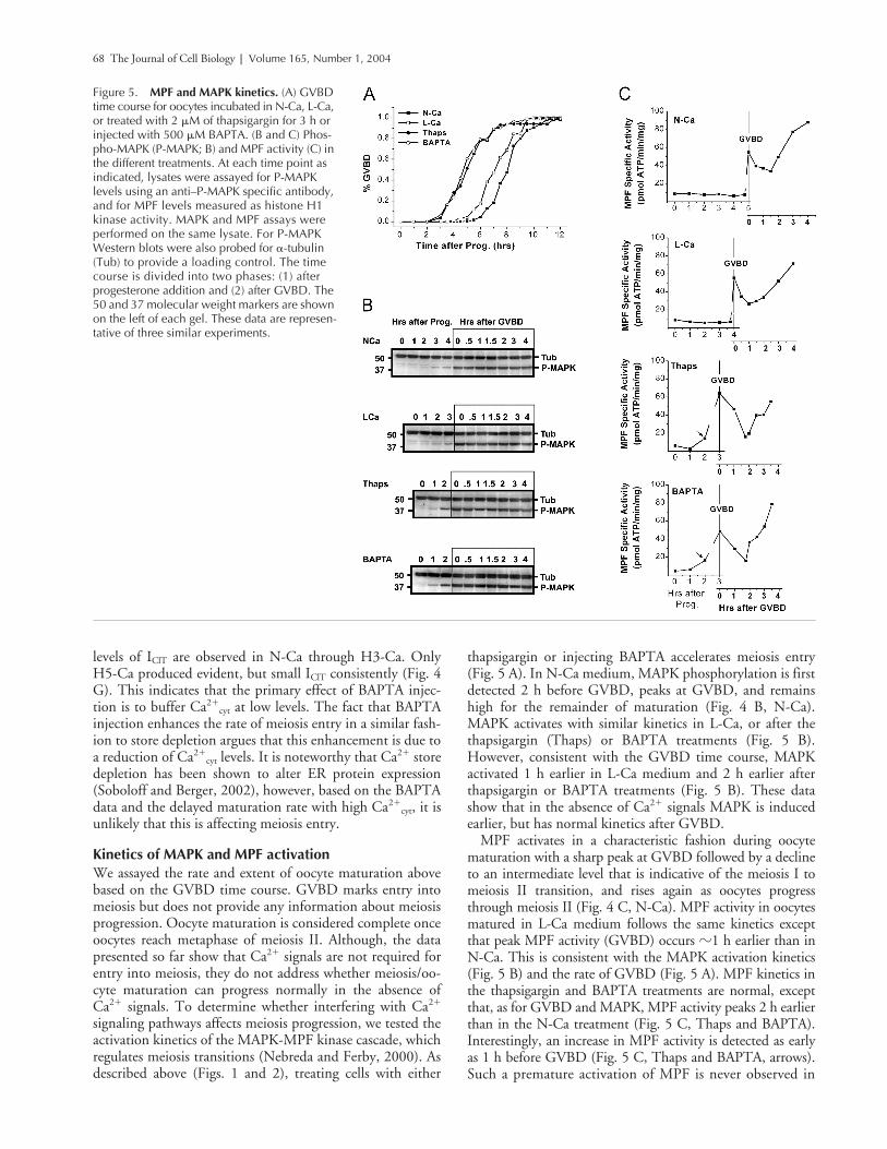
thapsigargin or injecting BAPTA accelerates meiosis entry(Fig. 5 A). In N-Ca medium, MAPK phosphorylation is firstdetected 2 h before GVBD, peaks at GVBD, and remainshigh for the remainder of maturation (Fig. 4 B, N-Ca).MAPK activates with similar kinetics in L-Ca, or after thethapsigargin (Thaps) or BAPTA treatments (Fig. 5 B).However, consistent with the GVBD time course, MAPKactivated 1 h earlier in L-Ca medium and 2 h earlier afterthapsigargin or BAPTA treatments (Fig. 5 B). These datashow that in the absence of Ca2� signals MAPK is inducedearlier, but has normal kinetics after GVBD.
MPF activates in a characteristic fashion during oocytematuration with a sharp peak at GVBD followed by a declineto an intermediate level that is indicative of the meiosis I tomeiosis II transition, and rises again as oocytes progressthrough meiosis II (Fig. 4 C, N-Ca). MPF activity in oocytesmatured in L-Ca medium follows the same kinetics exceptthat peak MPF activity (GVBD) occurs �1 h earlier than inN-Ca. This is consistent with the MAPK activation kinetics(Fig. 5 B) and the rate of GVBD (Fig. 5 A). MPF kinetics inthe thapsigargin and BAPTA treatments are normal, exceptthat, as for GVBD and MAPK, MPF activity peaks 2 h earlierthan in the N-Ca treatment (Fig. 5 C, Thaps and BAPTA).Interestingly, an increase in MPF activity is detected as earlyas 1 h before GVBD (Fig. 5 C, Thaps and BAPTA, arrows).Such a premature activation of MPF is never observed in
Figure 5. MPF and MAPK kinetics. (A) GVBD time course for oocytes incubated in N-Ca, L-Ca, or treated with 2 �M of thapsigargin for 3 h or injected with 500 �M BAPTA. (B and C) Phos-pho-MAPK (P-MAPK; B) and MPF activity (C) in the different treatments. At each time point as indicated, lysates were assayed for P-MAPK levels using an anti–P-MAPK specific antibody, and for MPF levels measured as histone H1 kinase activity. MAPK and MPF assays were performed on the same lysate. For P-MAPK Western blots were also probed for �-tubulin (Tub) to provide a loading control. The time course is divided into two phases: (1) after progesterone addition and (2) after GVBD. The 50 and 37 molecular weight markers are shown on the left of each gel. These data are represen-tative of three similar experiments.

Ca2� role in meiosis | Sun and Machaca 69
N-Ca or L-Ca. Therefore, MAPK and MPF activation kinet-ics (Fig. 5, B and C) correlate well with each other and withthe rate of GVBD (Fig. 5 A), and show that reducing Ca2�
cyt
transients leads to premature activation of the MAPK-MPFkinase cascade. This premature activation explains the acceler-ated maturation rate in treatments that reduce Ca2�
cyt tran-sients. Therefore, Ca2�
cyt modulates meiosis entry by nega-tively regulating the MAPK-MPF cascade.
Spindle formation and nuclear maturationKinase data in oocytes deprived of Ca2� signals (Thaps andBAPTA) suggest that in the absence of Ca2�
cyt signals meio-sis proceeds normally after GVBD. To determine whetherthis is the case we imaged meiotic spindle structure andchromosome dynamics in oocytes matured in N-Ca, L-Ca,and oocytes treated with thapsigargin or BAPTA (Fig. 6).This allowed us to assess the progression of nuclear matura-tion, and directly compare it to MPF, MAPK, and GVBDkinetics because all three experiments were performed on thesame batch of oocytes.
Control oocytes matured in N-Ca medium progress nor-mally through meiosis (Fig. 6 A, Table I, N-Ca). At GVBDoocytes were at the late prophase I stage (Fig. 6 A, N-Ca, P),which refers to oocytes that have undergone GVBD, havecondensed chromosomes, and organized microtubules aroundthe chromosomes, but have not yet formed a bipolar spindle(Fig. 6 A, N-Ca, P). At 0.5 h after GVBD prometaphase Istructures (30%; Table I) are observed with a typical bipolarspindle and associated chromosomes (Fig. 6 A, N-Ca, PM I).
This is followed by metaphase I with chromosome lined up atthe metaphase plate (Fig. 6 A, N-Ca, M I) at �1 h afterGVBD (Table I). Between 2–4 h after GVBD oocytesprogress from M I to metaphase II (Table I) at which stagethey arrest. Examples of anaphase I (A), prometaphase II (PMII), and metaphase II spindles are shown in Fig. 6 A (N-Ca).
Surprisingly, oocytes matured in L-Ca medium alone ortreated with thapsigargin or BAPTA formed abnormal spin-dles. The progression through meiosis was not significantlydifferent between the three treatments which will be discuss asa group. At GVBD a large percentage of these oocytes(�57%; Table I) had condensed chromosomes, but the mi-crotubule were still dispersed over a large area. We refer to thisstage as early prophase (EP; Fig. 6 A), because eventually theseoocytes do progress to the late prophase stage (P) as describedfor the control group (N-Ca) above (Table I). However, oo-cytes matured in L-Ca medium or treated with BAPTA orthapsigargin rarely progress to prometaphase I and never reachmetaphase I (Fig. 6 A; Table I). Instead, they form abnormalstructures at different rates depending on the treatment as de-tailed in Table I. Based on the severity of defects we dividedabnormal spindles into three groups: (1) We refer to smalland/or slightly disorganized spindles as prometaphase like(PM-L; Fig. 6 A). These spindles are the least disorganizedand are observed throughout the time period studied (TableI). (2) The second group represents completely disruptedspindles (Ab) with no clear structure and with the microtu-bules highly condensed and/or spread over a large area. Inmost but not all instances condensed chromosomes were still
Figure 6. Spindle structure. Oocytes were stained for tubulin and DNA with Sytox orange to visualize spindle structure. (A) The treatment designation for N-Ca, L-Ca, Thaps, and BAPTA is as indicated in Fig. 5. P refers to cells in late prophase with clustering of the microtubules and chromosome condensation. PMI, prometa-phase I; MI, metaphase I; A, Anaphase I; PMII, prometaphase II; MII, metaphase II; EP, early prophase with chromosome condensation but microtubules still spread over a large area; Ab, abnormal spindle structure; PM-L, prometaphase-like spindle with short or slightly disorganized structure; DS, double spindle with two overlapping spindles. (B and C) Polar body extrusion. (B) Representative images showing an oocyte with a polar body (PB) matured in N-Ca medium 3 h after GVBD (top); and an oocyte matured in L-Ca medium 3 h after GVBD, with no polar body (bottom). (C) Normalized polar body emission levels from 2–4 h after GVBD in the different treatments: N-Ca (0.6 mM); L-Ca (50 �M); thapsigar-gin (Thaps, 2 �M for 3 h); injected with 500 �M BAPTA (BAPTA); incubated in N-Ca until GVBD and then switched to L-Ca medium (N-L); or incubated in L-Ca until GVBD and then switched to N-Ca (L-N). Asterisks above the bar indicate significantly different means from N-Ca and N-L (P � 0.002; n � 3). Bars, 10 �m.
70 The Journal of Cell Biology | Volume 165, Number 1, 2004
associated with the disrupted spindle (Fig. 6 A, Ab). (3) Thelast and most interesting group we refer to as the double spin-dle (DS) group (see Fig. 6 A). These double spindles weremost common in the L-Ca group (Table I), but were also ob-served in the thapsigargin treatment (Fig. 6 A, Thaps, DS).
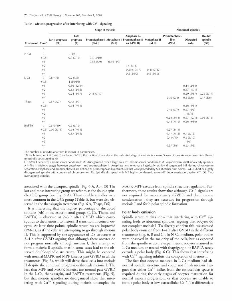
It is interesting that the highest percentage of disruptedspindles (Ab) in the experimental groups (L-Ca, Thaps, andBAPTA) is observed at 2–3 h after GVBD which corre-sponds to the meiosis I to meiosis II transition in control oo-cytes. At later time points, spindle structures are improved(PM-L), as if the cells are attempting to go through meiosisII. This is supported by the appearance of DS structures at3–4 h after GVBD arguing that although these oocytes donot progress normally through meiosis I, they attempt toform a meiosis II spindle, that in some cases lead to the ob-served double-spindle structures. This might be expectedwith normal MAPK and MPF kinetics past GVBD in all thetreatments (Fig. 5), which will drive these cells into meiosisII despite the abnormal progression through meiosis I. Thefact that MPF and MAPK kinetics are normal past GVBDin the L-Ca, thapsigargin, and BAPTA treatments (Fig. 5),but that meiotic spindles are disorganized show that inter-fering with Ca2� signaling during meiosis uncouples the
MAPK-MPF cascade from spindle structure regulation. Fur-thermore, these results show that although Ca2� signals arenot required for meiosis entry (GVBD and chromosomecondensation), they are necessary for progression throughmeiosis I and for bipolar spindle formation.
Polar body emissionSpindle structure data show that interfering with Ca2� sig-naling leads to abnormal spindles, arguing that oocytes donot complete meiosis I. To directly confirm this, we assessedpolar body emission from 1–4 h after GVBD in the differenttreatments (Fig. 6, B and C). In N-Ca medium, polar bodieswere observed in the majority of the cells, but as expectedfrom the spindle structure experiments, oocytes matured inL-Ca medium or treated with thapsigargin or BAPTA rarelyextrude a polar body (Fig. 6 C). This shows that interferingwith Ca2� signaling inhibits the completion of meiosis I.
The fact that oocytes matured in L-Ca medium had ab-normal spindle structure and could not finish meiosis I ar-gues that either Ca2� influx from the extracellular space isrequired during the early stages of oocytes maturation fornormal meiosis progression, or that oocytes are unable toform a polar body at low extracellular Ca2�. To differentiate
Table I. Meiosis progression after interfering with Ca2� signaling
Stage of meiosis Abnormal spindles
Treatment TimeaEarly prophase
(EP)
Late prophase
(P)Prometaphase I
(PM I)Metaphase I
(M I)
Anaphase I–Prometaphase II
(A I–PM II)Metaphase II
(M II)
Prometaphase-like
(PM-L)Disrupted
(Ab)
Doublespindle
(DS)
h
N-Ca 0 1 (5/5)�0.5 0.7 (7/10) 0.3 (3/10)�1 0.55 (5/9) 0.44 (4/9)�2 1 (13/13)�3 0.59 (10/17) 0.41 (7/17)�4 0.5 (5/10) 0.5 (5/10)
L-Ca 0 0.8 (4/5) 0.2 (1/5)�0.5 1 (10/10)�1 0.86 (12/14) 0.14 (2/14)�2 0.13 (2/15) 0.87 (13/15)�3 0.24 (4/17) 0.18 (3/17) 0.29 (5/17) 0.29 (5/17)�4 0.33 (2/6) 0.5 (3/6) 0.17 (1/6)
Thaps 0 0.57 (4/7) 0.43 (3/7)�0.5 0.64 (7/11) 0.36 (4/11)�1 0.43 (3/7) 0.67 (6/9)�2 1 (15/15)�3 0.28 (5/18) 0.67 (12/18) 0.05 (1/18)�4 0.44 (7/16) 0.56 (9/16)
BAPTA 0 0.5 (5/10) 0.5 (5/10)�0.5 0.09 (1/11) 0.64 (7/11) 0.27 (3/11)�1 0.13 (2/15) 0.47 (7/15) 0.4 (6/15)�2 0.4 (4/10) 0.6 (6/10)�3 1 (6/6)�4 0.37 (3/8) 0.63 (5/8)
The number of oocytes analyzed is shown in parentheses.aAt each time point at (time 0) and after GVBD, the fraction of oocytes at the indicated stage of meiosis is shown. Stages of meiosis were determined basedon spindle structure (Fig. 6).EP: GVBD occurred; chromosomes condensed; MT disorganized over a large area. P: Chromosomes condensed; MT organized in small area (early spindle).A I–PM II: Meiotic stages between anaphase I and prometaphase II. Anaphase and telophase I typically exhibit disorganized MT during chromosomeseparation. Prophase and prometaphase II are defined as prometaphase-like structures that were preceded by M I at earlier time points. PM-L: Short or slightlydisorganized spindle with condensed chromosomes. Ab: Spindle disrupted with MT highly condensed; some MT depolimerization; spiky MT. DS: Twooverlapping spindles.

Ca2� role in meiosis | Sun and Machaca 71
between these possibilities we assessed polar body emissionin oocytes incubated in N-Ca solution until GVBD andthen switched to L-Ca solution (N-L); or in oocytes incu-bated in L-Ca solution until GVBD and then switched toN-Ca medium (L-N). Oocytes in the N-L group, but notthe L-N group, emitted polar bodies to the same extent asthe N-Ca control group (Fig. 6, B and C), arguing that Ca2�
influx in the early stages of oocyte maturation is required formeiosis I progression.
Ca2�cyt acts between PKA and Mos to negatively
regulate entry into meiosisTo better define the mechanism by which Ca2�
cyt negativelyregulates meiosis entry, we mapped the site of action ofCa2�
cyt on the cell cycle machinery using an epistatic ap-
proach. For these experiments thapsigargin-treated oocytesrepresented the experimental group deprived of Ca2� signals.Control oocytes were activated in N-Ca solution. Our ap-proach was to activate the cell cycle machinery at differentpoints along the MAPK-MPF signal transduction cascade,and determine whether the enhancing effect of Ca2� depra-vation on the rate of maturation is still observed. Oocyteswere activated with progesterone, or by injection of an in-hibitor of PKA inhibitor (PKI), Mos RNA (Mos), cyclinB1 RNA (Cy) and 87cyclin B1 protein (CyP; Fig. 7 A).Thapsigargin treatment enhanced the rate of oocyte matura-tion to a similar extent in oocytes activated with progesteroneor PKI (Fig. 7 A, PKI). Thapsigargin-treated oocytes activatefaster than controls after Mos RNA injection, but the effecton Ca2� deprivation on the rate of maturation is smaller than
Figure 7. Mapping the site of action of Ca2�
cyt on meiosis entry. (A) GVBD time course for oocytes incubated in N-Ca solution (N), or L-Ca, and treated with thapsigargin (T). Oocyte maturation was induced with progesterone (N and T), PKI (PKI), Mos RNA 10 ng (Mos), cyclin B1 RNA 10 ng (Cy), or 87cyclin B1 protein �200 pg (CyP). (B) Relative time to GVBD50 normalized to GVBD50 in the N-Ca for each activator. P values are shown above the graph, n � 4–7. (C) Biochemical analysis of the cell cycle machinery. Maturation was induced as described in A and lysates were collected from immature oocytes (ooc), at hourly intervals after progesterone addition as indicated, at first GVBD (GVBD) and at GVBD50 (G50). For the GVBD50 time point we collected lysates form oocytes that have undergone GVBD (white spot, w) and those that have not (no white spot, nw). (D) Working model for the role of Ca2�
cyt in oocyte maturation/meiosis (see text for details). Dashed arrows indicated yet unknown steps in the cascade. The change in arrow shape and the bold font indicate transitions between the different stages of meiosis. M I, meiosis I; PB, polar body emission; M II, meiosis II.

72 The Journal of Cell Biology | Volume 165, Number 1, 2004
in the case of progesterone (Fig. 7 A, Mos). Because the ki-netics of the kinase cascade downstream of Mos are similar incontrol and thapsigargin-treated oocytes (Fig. 7 C; Fig. 5),we wondered whether the faster maturation rate in thapsigar-gin-treated Mos-injected oocytes is due to an effect of Ca2�
deprivation on RNA translation. To determine whether thisis the case we induced meiosis by directly activating MPFthrough the expression of cyclin B1 to activate the free cdc2pool in the oocyte. Similar to Mos RNA injection, thapsigar-gin-treated oocytes activated faster than controls after cyclinB1 RNA injection (Fig. 7 A, Cy). In contrast, injecting oo-cytes with cyclin B1 protein induces GVBD with a similartime course in both thapsigargin-treated and control oocytes(Fig. 7 A, CyP). The rate of oocyte maturation with the dif-ferent activators is summarized in Fig. 7 B. Because the rateof maturation varies between activators we normalized matu-ration rate for each activator to the rate of activation in N-Ca(Fig. 7 B) to allow a better visualization of the relative effectof Ca2� deprivation on maturation. For example, althoughcyclin B activates maturation faster than Mos (Fig. 7 A), therelative enhancement in the rate of maturation (�20%faster) is similar between the two activators in the absence ofCa2� signals (Fig. 7 B). This argues that the more rapid mat-uration in oocytes deprived of Ca2� signals after both Mosand cyclin RNA injections is due to an effect of Ca2�
cyt ontranslation of the injected RNAs. This conclusion is sup-ported by the fact that control and thapsigargin-treated oo-cytes mature at similar rates when activated with 87cyclinB1 protein. However, the different responses after cyclinRNA or protein injections could be due to an effect ofCa2�
cyt on protein turnover because we injected full-lengthcyclin B1 RNA and 87cyclin B1 protein, which is missingthe first 87 aa and is thus nondegradable because it lacks thedestruction box (Kumagai and Dunphy, 1995). Nonetheless,we favor an effect of Ca2�
cyt on RNA translation becausematuration is enhanced to a similar level when oocytes are ac-tivated with Mos or Cyclin B1, two activators that induce thecell cycle kinase cascade at different points.
These data show that Ca2�cyt negatively regulates meiosis
entry by acting on at least two sites between PKA inhibitionand Mos activation. One site is downstream of PKA inhibi-tion and the other site appears to be mRNA translation,which is required for the induction of the cell cycle machin-ery (Fig. 7 D). Furthermore, the fact that the rate of matura-tion is enhanced to a similar extent in oocytes activated withprogesterone and PKI (Fig. 7, A and B, PKI) argues thatCa2�
cyt acts downstream of PKI (Fig. 7 D).
Ca2�cyt negatively regulates the initiation of the
MAPK-MPF cascadeTo confirm that Ca2�
cyt acts upstream of Mos we analyzedin more details the steps of the cell cycle machinery down-stream of Mos in both control and thapsigargin-treated oo-cytes (Fig. 7 C). As described above MAPK activates signifi-cantly earlier in thapsigargin-treated oocytes consistent withthe GVBD time course (Fig. 7 C). p90RSK, the down-stream substrate of MAPK is activated with a similar timecourse to MAPK. For these experiments we analyzed lysatesform oocytes at GVBD and at GVBD50. For the latter timepoint lysates from oocytes that have undergone GVBD (w)
or not (nw) were collected. Interestingly, p90RSK was phos-phorylated to higher levels in the GVBD50-nw group inthapsigargin-treated oocytes as compared with controls, thusconfirming the earlier activation of the MAPK cascade inthapsigargin-treated oocytes.
Xenopus oocytes contain two pools of cdc2, the catalyticsubunit of MPF: the preMPF and the free cdc2 pool. ThepreMPF pool which is activated at GVBD, contains cdc2 as-sociated with cyclin B. Pre-MPF is kept inactive by phos-phorylation on Tyr15 of cdc2 (Nebreda and Ferby, 2000).The free cdc2 pool is activated after association with B-typecyclins synthesized during meiosis I (Hochegger et al., 2001).To determine which pool of cdc2 is activated in thapsigargin-treated oocytes we probed Western blots with a phosphospe-cific antibody against Tyr15 of cdc2 (Fig. 7 C, P-Y15-cdc2),and determined MPF activity as the H1-kinase activity fromp13suc1 pulldowns (Fig. 7 C, MPF). In both control andthapsigargin-treated oocytes the disappearance of P-Y15 im-munoreactivity coincides with an increase in MPF activity in-dicating that as in control oocytes the preMPF pool is acti-vated in thapsigargin-treated oocytes. Interestingly, a smalllevel of MPF activity is detected at the GVBD50 time point inthapsigargin-treated oocytes that have not undergone GVBD(Fig. 7, Thaps, G50 nw), confirming the early activation ofMPF before GVBD observed in Fig. 5.
These data show that in the absence of Ca2�cyt signals the
cell cycle kinase cascade downstream of Mos activatesnormally. Therefore, the more rapid entry into meiosis(GVBD) observed in oocytes deprived of Ca2� signals is dueto a negative regulation of Ca2�
cyt on the initiation of this ki-nase cascade upstream of Mos. Blocking Ca2�
cyt signals re-lieves this negative regulation thus allowing more rapid in-duction of the MAPK-MPF cascade and GVBD.
DiscussionIn contrast to the established role of Ca2� signaling in mito-sis (Whitaker and Larman, 2001), the requirement for Ca2�
in both Xenopus and mammalian oocyte meiotic maturationhas been difficult to define (Homa et al., 1993; Duesberyand Masui, 1996). To delineate the function of Ca2� duringXenopus oocyte meiosis we manipulated Ca2�
cyt, extracellularCa2�, and store Ca2� load and tested the effect on nuclearmaturation and the cell cycle machinery. Our data show thatCa2� has two opposing roles during Xenopus oocyte matura-tion: It negatively regulates meiosis entry by delaying the ac-tivation of the cell cycle machinery, and it is required forcompletion of meiosis I (Fig. 7 D).
Ca2�cyt negatively regulates the activation of
the cell cycle kinase cascadeProgesterone leads to lower cAMP levels and PKA inhibitionwithin 10 min (Sadler and Maller, 1981), but the next knownstep in the pathway, that is polyadenylation of maternalRNAs to induce their translation, does not occur until muchlater (Sheets et al., 1995). The molecular steps during thistime lag are not known. Our data show that Ca2�
cyt is an im-portant regulator of the transition between PKA inhibitionand mRNA translation. Ca2�
cyt negatively regulates the acti-vation of the cell cycle machinery by acting on at least two
Ca2� role in meiosis | Sun and Machaca 73
sites between PKA and Mos (Fig. 7 D). One site of action ap-pears to be mRNA translation. Therefore, the level of Ca2�
cyt
provides a timing mechanism for entry into meiosis by regu-lating the initiation of the MAPK-MPF cascade downstreamof PKA inhibition (Fig. 7 D). It is tempting to propose thatby acting in this capacity Ca2�
cyt could synchronize morpho-logical and biochemical changes during oocyte maturation.Under such a scenario, which is completely speculative at thispoint, Ca2�
cyt levels could signal the physiological prepared-ness of the oocyte to begin maturation. Relatively low Ca2�
cyt
levels would be indicative of proper functioning of the Ca2�
signaling machinery, and thus a healthy oocyte that is readyto mature. In contrast, relatively high Ca2�
cyt levels would in-dicate a compromised oocyte where Ca2�
cyt would negativelyregulate initiation of maturation. It is interesting in that con-text that Ca2�
cyt acts upstream of Mos, that is before theoocyte activates the cell cycle machinery and commits tomaturation. The proposed role of Ca2�
cyt in synchronizingmorphological and biochemical changes in the oocyte duringmaturation is further supported by the fact that disruptingCa2�
cyt signaling uncouples the nuclear cell cycle from theMAPK-MPF kinase cascade (Fig. 6).
Ca2�cyt is required for completion of meiosis I
Oocytes deprived of Ca2� signals do not complete meiosis Ias they do not extrude a polar body. Rather, they form ab-normal spindles early in meiosis I despite normal MAPK andMPF kinetics. This shows that progression through meiosis Irequires Ca2�, possibly Ca2� influx before GVBD becauseoocytes are dependent on extracellular Ca2� only beforeGVBD (Fig. 6). A Ca2� influx requirement before GVBDfits nicely with the regulation of SOCE during oocyte matu-ration because SOCE inactivates at the GVBD stage due toMPF activation (Machaca and Haun, 2000, 2002).
Interestingly, others have shown that inhibition of theMAPK cascade (Gross et al., 2000), down-regulation ofMPF (Nakajo et al., 2000), or inhibition of protein synthe-sis (Kanki and Donoghue, 1991) block completion of mei-osis I. These treatments lead to a decrease in MPF activity,and induce an interphase-like state that is usually absentbetween meiosis I and II. In contrast, interfering with Ca2�
signaling blocks meiosis I completion, but is not associatedwith an interphase-like state. Rather, spindle structure isdisrupted and polar body formation inhibited, but thechromosomes remain condensed. Furthermore, MPF ac-tivity cycles normally with a dip in activity between 1–2 hafter GVBD, the expected time for meiosis I to meiosis IItransition. These data show that disruption of Ca2� signal-ing uncouples the cell cycle machinery (MAPK-MPF)from nuclear maturation (i.e., bipolar spindle formationand completion of meiosis I).
It is interesting that the disrupted meiosis I spindle doesnot activate a spindle checkpoint to arrest the cell cycle.However, there is good evidence against the existence of ameiosis I spindle checkpoint in Xenopus oocytes. Blockingthe activity of the APC/C or the checkpoint protein Mad2does not affect progression through meiosis I (Peter et al.,2001; Taieb et al., 2001). This is consistent with our obser-vation of a lack of cell cycle arrest in the absence of Ca2� sig-nals despite disrupted meiosis I spindles.
Recently, Castro et al. (2003) described a similar block ofmeiosis I after inhibition of Aurora A kinase or its substrateEg5 (a kinesin-like protein) in Xenopus oocytes. Unfortu-nately, these authors did not assess spindle morphology, butthey show that blocking Aurora A inhibits polar body for-mation with chromosomes maintaining their condensedstate long after GVBD (Castro et al., 2003). Members of theAurora kinase family associate with the spindle and havebeen shown to be important for both meiosis and mitosistransitions. Therefore, it is possible that Ca2�-dependentpathways somehow modulate Aurora A kinase activitywhich in turn regulates spindle structure. This possibility re-mains to be explored.
Role of Ca2� signaling in GVBDDuring mitosis NEBD has been shown to be dependent onCa2� (Poenie et al., 1985; Steinhardt and Alderton, 1988;Twigg et al., 1988; Kao et al., 1990; Wilding et al., 1996),and studies in sea urchin embryos suggest that Ca2� exertsits effect through CaMKII activation (Baitinger et al.,1990). In contrast, during meiosis GVBD is Ca2�-indepen-dent as shown here for Xenopus oocytes, and in both mouse(Carroll and Swann, 1992; Tombes et al., 1992) and star-fish oocytes (Witchel and Steinhardt, 1990). One exceptionto this rule are some bivalve molluscs where GVBD hasbeen shown to require Ca2� (Deguchi and Osanai, 1994),but unlike amphibian and mammalian oocytes, in thiscase oocyte maturation and GVBD occur after fertilizationwhich invariably induces a Ca2�
cyt rise. Nonetheless, thedifferential requirement of NEBD on Ca2� signals duringmeiosis and mitosis is surprising because both NEBD andGVBD require the activation of MPF (Lenart and Ellen-berg, 2003), and it is reasonable to assume that the basicstructural properties of the nuclear envelope are similar inmitotic and meiotic cells. It has been argued that a Ca2�
signal is still required for GVBD but occurs very early oreven before the initiation of oocyte maturation in some spe-cies (Tombes et al., 1992; Homa et al., 1993). This doesnot seem to be the case for Xenopus, because as shown inFig. 1 F, eliminating Ca2� signals for as long as 48 h be-fore inducing oocyte maturation has no effect on GVBD,strongly arguing that GVBD is Ca2� independent. There-fore, the differential requirement for Ca2� during thebreakdown of the nuclear envelope suggests that NEBDand GVBD are mechanistically distinct.
In conclusion, our results show that Ca2� signals are dis-pensable for GVBD and chromosome condensation, thatCa2�
cyt controls the timing of meiosis entry by negatively reg-ulating the initiation of cell cycle machinery, and that N-Ca2�
homeostasis is important for bipolar spindle formation andcompletion of meiosis I. These results provide a framework tofurther explore and better define the role of Ca2�-dependentsignaling pathways in meiosis and oocyte maturation.
Materials and methodsOocyte maturationXenopus oocytes were obtained as described previously (Machaca andHaun, 2002). The control L-15 solution contains 0.63 mM Ca2�. Ca2� wasbuffered at 50 �M in the low solution as calculated using the MaxChelatorprogram (http://www.stanford.edu/~cpatton/maxc.html) by the addition of
74 The Journal of Cell Biology | Volume 165, Number 1, 2004
0.58 mM EGTA. For the H-Ca L-15 solutions Ca2� was added to the indi-cated concentration as CaCl2. In all experiments, GVBD was visually con-firmed by fixing oocytes in methanol and bisecting them in half.
Electrophysiological methodsRecording of the ICa,Cl was performed as described previously (Machacaand Haun, 2000). ICa,Cl were recorded in the following solutions: F-Ca con-tains in micromolars: 96 NaCl, 2.5 KCl, 5 MgCl2.6H2O, 0.1 EGTA, 10Hepes, pH 7.4. Low Ca2� Ringer solution (50 �M free Ca2�) contains inmicromolars: 96 NaCl, 2.5 KCl, 4.37 MgCl2.6H2O, 0.63 CaCl2.2H2O, 0.58EGTA, 10 Hepes, pH 7.4. Normal Ringer (N-Ca, 0.63 mM Ca2�) and H-Casolutions (1.5, 3, and 5 mM Ca2�) had the following composition: 96NaCl, 2.5 KCl, 10 Hepes, pH 7.4; with Ca2� and Mg2� concentrations add-ing up to 5 mM.
Western blots and MPF kinase assaysMPF kinase activity assay and phospho-MAPK Western blots were pre-pared as described previously (Machaca and Haun, 2002), except that�-tubulin was used as the loading control for MAPK Western blots. The ac-tivation of p90RSK and preMPF was assessed using phosphospecific anti-bodies against phospho-Thr573 of p90RSK and phospho-Tyr15 of cdc2(Cell Signaling). MPF activity was also assayed from lysates affinity purifiedon p13suc1 beads using histone-H1 kinase as a substrate essentially as de-scribed previously (Howard et al., 1999).
Plasmids and reagentsHeat stable PKI was purchased from Calbiochem. Cyclin B1 and MosRNAs were synthesized from a pXen-GST-Mos and pSP64-cyclinB1xen
plasmids provided by A. Macnicol (University of Arkansas for Medical Sci-ences; Freeman et al., 1991; Howard et al., 1999) using the mMessageMmachine transcription kit (Ambion). The His6-tagged 87cyclin B1 pro-tein was used as described previously (Machaca and Haun, 2002).
Spindle and polar body staining and image acquisitionOocytes were fixed in 100% methanol, bisected in half, and incubated inDM1A an antitubulin mAb (Sigma-Aldrich) in TBS containing 2% BSA, fol-lowed by a Cy2-conjugated donkey anti–mouse secondary (Jackson Immu-noResearch Laboratory) for 24 h each. The oocytes were washed, dehy-drated, stained in 1 �M Sytox® orange (Molecular Probes), and cleared inbenzyl alcohol/benzyl benzoate (1:2). Images were collected using a Fluo-view confocal (Olympus) coupled to a microscope (model IX70; Olympus)at RT, using a UPlanApo 40 oil objective with an NA of 1.00. The acqui-sition software was Fluoview 2.1 and figures were compiled using AdobePhotoshop 7.0. For each spindle a z section was obtained and projectedonto a single plane to visualize the entire spindle. For polar body emissionstudies oocytes were fixed in methanol, stained with Sytox® orange, andvisualized by confocal microscopy as described for the spindle staining.
We thank members of the Machaca Lab for critical reading of the manu-script, A. Macnicol for providing plasmid constructs, and A. Charlesworthfor help with the p13suc1 MPF assay.
This work was supported by grant GM-61829 from the National Insti-tutes of Health.
Submitted: 23 September 2003Accepted: 26 February 2004
ReferencesArslan, P., F. Di Virgilio, M. Beltrame, R.Y. Tsien, and T. Pozzan. 1985. Cytosolic
Ca2� homeostasis in Ehrlich and Yoshida carcinomas. J. Biol. Chem. 260:2719–2727.
Baitinger, C., J. Alderton, M. Poenie, H. Schulman, and R.A. Steinhardt. 1990.Multifunctional Ca2�/calmodulin-dependent protein kinase is necessary fornuclear envelope breakdown. J. Cell Biol. 111:1763–1773.
Camello, C., R. Lomax, O.H. Petersen, and A.V. Tepikin. 2002. Calcium leakfrom intracellular stores–the enigma of calcium signalling. Cell Calcium. 32:355–361.
Carroll, J., and K. Swann. 1992. Spontaneous cytosolic calcium oscillations drivenby inositol trisphosphate occur during in vitro maturation of mouse oocytes.J. Biol. Chem. 267:11196–11201.
Castro, A., E. Mandart, T. Lorca, and S. Galas. 2003. Involvement of Aurora A ki-nase during meiosis I-II transition in Xenopus oocytes. J. Biol. Chem. 278:2236–2241.
Ciapa, B., D. Pesando, M. Wilding, and M. Whitaker. 1994. Cell-cycle calciumtransients driven by cyclic changes in inositol trisphosphate levels. Nature.368:875–878.
Cicirelli, M.F., and L.D. Smith. 1987. Do calcium and calmodulin trigger matura-tion in amphibian oocytes? Dev. Biol. 121:48–57.
Coleman, T.R., and W.G. Dunphy. 1994. Cdc2 regulatory factors. Curr. Opin.Cell Biol. 6:877–882.
Cork, R.J., M.F. Cicirelli, and K.R. Robinson. 1987. A rise in cytosolic calcium isnot necessary for maturation of Xenopus laevis oocytes. Dev. Biol. 121:41–47.
Deguchi, R., and K. Osanai. 1994. Meiosis reinitiation from the first prophase isdependent on the levels of intracellular Ca2� and pH in oocytes of the bi-valves Mactra chinensis and Limaria hakodatensis. Dev. Biol. 166:587–599.
Duesbery, N.S., and Y. Masui. 1996. The role of Ca2� in progesterone-inducedgerminal vesicle breakdown of Xenopus laevis oocytes: the synergic effects ofmicrotubule depolymerization and Ca2�. Dev. Genes Evol. 206:110–124.
Freeman, R.S., S.M. Ballantyne, and D.J. Donoghue. 1991. Meiotic induction byXenopus cyclin B is accelerated by coexpression with mosXe. Mol. Cell. Biol.11:1713–1717.
Gross, S.D., M.S. Schwab, F.E. Taieb, A.L. Lewellyn, Y.W. Qian, and J.L. Maller.2000. The critical role of the MAP kinase pathway in meiosis II in Xenopusoocytes is mediated by p90(Rsk). Curr. Biol. 10:430–438.
Hochegger, H., A. Klotzbucher, J. Kirk, M. Howell, K. le Guellec, K. Fletcher, T.Duncan, M. Sohail, and T. Hunt. 2001. New B-type cyclin synthesis is re-quired between meiosis I and II during Xenopus oocyte maturation. Develop-ment. 128:3795–3807.
Homa, S.T., J. Carroll, and K. Swann. 1993. The role of calcium in mammalianoocyte maturation and egg activation. Hum. Reprod. 8:1274–1281.
Howard, E.L., A. Charlesworth, J. Welk, and A.M. MacNicol. 1999. The mito-gen-activated protein kinase signaling pathway stimulates mos mRNA cyto-plasmic polyadenylation during Xenopus oocyte maturation. Mol. Cell. Biol.19:1990–1999.
Kanki, J.P., and D.J. Donoghue. 1991. Progression from meiosis I to meiosis II inXenopus oocytes requires de novo translation of the mosxe protooncogene.Proc. Natl. Acad. Sci. USA. 88:5794–5798.
Kao, J.P., J.M. Alderton, R.Y. Tsien, and R.A. Steinhardt. 1990. Active involve-ment of Ca2� in mitotic progression of Swiss 3T3 fibroblasts. J. Cell Biol.111:183–196.
Kumagai, A., and W.G. Dunphy. 1995. Control of the Cdc2/cyclin B complex inXenopus egg extracts arrested at a G2/M checkpoint with DNA synthesis in-hibitors. Mol. Biol. Cell. 6:199–213.
Lenart, P., and J. Ellenberg. 2003. Nuclear envelope dynamics in oocytes: fromgerminal vesicle breakdown to mitosis. Curr. Opin. Cell Biol. 15:88–95.
Machaca, K., and H.C. Hartzell. 1999. Reversible Ca gradients between the sub-plasmalemma and cytosol differentially activate Ca-dependent Cl currents. J.Gen. Physiol. 113:249–266.
Machaca, K., and S. Haun. 2000. Store-operated calcium entry inactivates at thegerminal vesicle breakdown stage of Xenopus meiosis. J. Biol. Chem. 275:38710–38715.
Machaca, K., and S. Haun. 2002. Induction of maturation-promoting factor dur-ing Xenopus oocyte maturation uncouples Ca2� store depletion from store-operated Ca2� entry. J. Cell Biol. 156:75–86.
Machaca, K., Z. Qu, A. Kuruma, H.C. Hartzell, and N. McCarty. 2001. The en-dogenous calcium-activated Cl channel in Xenopus oocytes: a physiologicallyand biophysically rich model system. In Calcium Activates Chloride Chan-nels. C.M. Fuller, editor. Academic Press, San Diego. 3–39.
Means, A.R. 1994. Calcium, calmodulin and cell cycle regulation. FEBS Lett. 347:1–4.
Moreau, M., M. Doree, and P. Guerrier. 1976. Electrophoretic introduction ofcalcium into the cortex of Xenopus laevis oocytes triggers meiosis reinitiation.J. Exp. Zool. 197:443–449.
Moreau, M., J.P. Vilain, and P. Guerrier. 1980. Free calcium changes associatedwith hormone action in amphibian oocytes. Dev. Biol. 78:201–214.
Nakajo, N., S. Yoshitome, J. Iwashita, M. Iida, K. Uto, S. Ueno, K. Okamoto, andN. Sagata. 2000. Absence of Wee1 ensures the meiotic cell cycle in Xenopusoocytes. Genes Dev. 14:328–338.
Nebreda, A.R., and I. Ferby. 2000. Regulation of the meiotic cell cycle in oocytes.Curr. Opin. Cell Biol. 12:666–675.
O’Connor, C.M., K.R. Robinson, and L.D. Smith. 1977. Calcium, potassium,and sodium exchange by full-grown and maturing Xenopus laevis oocytes.Dev. Biol. 61:28–40.
Parekh, A.B., and R. Penner. 1997. Store depletion and calcium influx. Physiol.Rev. 77:901–930.
Ca2� role in meiosis | Sun and Machaca 75
Peter, M., A. Castro, T. Lorca, C. Le Peuch, L. Magnaghi-Jaulin, M. Doree, andJ.C. Labbe. 2001. The APC is dispensable for first meiotic anaphase in Xeno-pus oocytes. Nat. Cell Biol. 3:83–87.
Picard, A., F. Giraud, F. Le Bouffant, F. Sladeczek, C. Le Peuch, and M. Doree.1985. Inositol 1,4,5-trisphosphate microinjection triggers activation, butnot meiotic maturation in amphibian and starfish oocytes. FEBS Lett. 182:446–450.
Poenie, M., J.M. Alderton, R.Y. Tsien, and R.A. Steinhardt. 1985. Changes of freecalcium levels with stages of the cell division cycle. Nature. 315:147–149.
Robinson, K.R. 1985. Maturation of Xenopus oocytes is not accompanied by elec-trode-detectable calcium changes. Dev. Biol. 109:504–508.
Sadler, S.E., and J.L. Maller. 1981. Progesterone inhibits adenylate cyclase in Xeno-pus oocytes. Action on the guanine nucleotide regulatory protein. J. Biol.Chem. 256:6368–6373.
Schorderet-Slatkine, S., M. Schorderet, and E.E. Baulieu. 1976. Initiation of meioticmaturation in Xenopus laevis oocytes by lanthanum. Nature. 262:289–290.
Sheets, M.D., M. Wu, and M. Wickens. 1995. Polyadenylation of c-mos mRNAas a control point in Xenopus meiotic maturation. Nature. 374:511–516.
Soboloff, J., and S.A. Berger. 2002. Sustained ER Ca2� depletion suppresses pro-tein synthesis and induces activation-enhanced cell death in mast cells. J.Biol. Chem. 277:13812–13820.
Steinhardt, R.A., and J.M. Alderton. 1988. Intracellular free calcium rise triggersnuclear envelope breakdown in the sea urchin embryo. Nature. 332:364–366.
Stricker, S.A. 2000. Comparative biology of calcium signaling during fertilizationand egg activation in animals. Dev. Biol. 211:157–176.
Taieb, F.E., S.D. Gross, A.L. Lewellyn, and J.L. Maller. 2001. Activation of theanaphase-promoting complex and degradation of cyclin B is not required forprogression from Meiosis I to II in Xenopus oocytes. Curr. Biol. 11:508–513.
Tombes, R.M., C. Simerly, G.G. Borisy, and G. Schatten. 1992. Meiosis, egg acti-vation, and nuclear envelope breakdown are differentially reliant on Ca2�,
whereas germinal vesicle breakdown is Ca2� independent in the mouse oo-cyte. J. Cell Biol. 117:799–811.
Tunquist, B.J., and J.L. Maller. 2003. Under arrest: cytostatic factor (CSF)-medi-ated metaphase arrest in vertebrate eggs. Genes Dev. 17:683–710.
Twigg, J., R. Patel, and M. Whitaker. 1988. Translational control of InsP3-induced chromatin condensation during the early cell cycles of sea urchinembryos. Nature. 332:366–369.
Wasserman, W.J., and Y. Masui. 1975. Initiation of meiotic maturation in Xenopuslaevis oocytes by the combination of divalent cations and ionophoreA23187. J. Exp. Zool. 193:369–375.
Wasserman, W.J., and L.D. Smith. 1981. Calmodulin triggers the resumption ofmeiosis in amphibian oocytes. J. Cell Biol. 89:389–394.
Wasserman, W.J., L.H. Pinto, C.M. O’Connor, and L.D. Smith. 1980. Progester-one induces a rapid increase in [Ca2�]in of Xenopus laevis oocytes. Proc. Natl.Acad. Sci. USA. 77:1534–1536.
Whitaker, M. 1995. Regulation of the cell division cycle by inositol trisphosphateand the calcium signaling pathway. Adv. Second Messenger Phosphoprotein.Res. 30:299–310.
Whitaker, M., and M.G. Larman. 2001. Calcium and mitosis. Semin. Cell Dev.Biol. 12:53–58.
Wilding, M., E.M. Wright, R. Patel, G. Ellis-Davies, and M. Whitaker. 1996. Lo-cal perinuclear calcium signals associated with mitosis-entry in early sea ur-chin embryos. J. Cell Biol. 135:191–199.
Witchel, H.J., and R.A. Steinhardt. 1990. 1-Methyladenine can consistently in-duce a fura-detectable transient calcium increase which is neither necessarynor sufficient for maturation in oocytes of the starfish Asterina miniata. Dev.Biol. 141:393–398.
Yamashita, M., K. Mita, N. Yoshida, and T. Kondo. 2000. Molecular mechanismsof initiation of oocyte maturation: general and species-specific aspects. Prog.Cell Cycle Res. 4:115–129.