
Article Accelerated In Vitro Degradation of Optically...
11
DOI: 10.1167/tvst.2.3.2 Article Accelerated In Vitro Degradation of Optically Clear Low b-Sheet Silk Films by Enzyme-Mediated Pretreatment Ke Shang 1 $ , Jelena Rnjak-Kovacina 1 $ , Yinan Lin 1 $ , Rebecca S. Hayden 1 $ , Hu Tao 1 $ , and David L. Kaplan 1 $ 1 Department of Biomedical Engineering, Tufts University, Medford, MA Correspondence: David L. Kaplan, Department of Biomedical Engi- neering, Tufts University, 4 Colby Street, Medford, MA, USA. e-mail: [email protected]. Received: 2 August 2012 Accepted: 5 February 2013 Published: 25 June 2013 Keywords: corneal tissue engi- neering; silk; films; degradation; b- sheet content; protease pretreat- ment Citation: Shang K, Rnjak-Kovacina J, Lin Y, Hayden RS, Tao H, Kaplan DL. Accelerated in vitro degradation of optically clear low b-sheet silk films by enzyme-mediated pretreatment. Trans Vis Sci Tech. 2013;2(3):2, http://tvstjournal.org/doi/full/10. 1167/tvst.2.3.2, doi:10.1167/tvst.2. 3.2 Purpose: To design patterned, transparent silk films with fast degradation rates for the purpose of tissue engineering corneal stroma. Methods: b-sheet (crystalline) content of silk films was decreased significantly by using a short water annealing time. Additionally, a protocol combining short water annealing time with enzymatic pretreatment of silk films with protease XIV was developed. Results: Low b-sheet content (17%–18%) and enzymatic pretreatment provided film stability in aqueous environments and accelerated degradation of the silk films in the presence of human corneal fibroblasts in vitro. The results demonstrate a direct relationship between reduced b-sheet content and enzymatic pretreatment, and overall degradation rate of the protein films. Conclusions: The novel protocol developed here provides new approaches to modulate the regeneration rate of silk biomaterials for corneal tissue regeneration needs. Translational Relevance: Patterned silk protein films possess desirable characteristics for corneal tissue engineering, including optical transparency, biocompatibility, cell alignment, and tunable mechanical properties, but current fabrication protocols do not provide adequate degradation rates to match the regeneration properties of the human cornea. This novel processing protocol makes silk films more suitable for the construction of human corneal stroma tissue and a promising way to tune silk film degradation properties to match corneal tissue regeneration. Introduction A transparent, avascular, elastic cornea protects the eye from the exterior environment, providing 75% of refractive power by forming a sharp image at the retinal receptors. 1–3 As the most commonly trans- planted tissue in the United States, the supply of healthy corneas does not meet current clinical demand 2,4–5 and, therefore, successful tissue engineer- ing of human cornea would address a major need. Tissue engineered epithelium and endothelium have been effectively studied and tested in animal models; however, the cornea stroma, which accounts for 90% of corneal thickness, remains challenging to recon- struct due to its complex structural hierarchy. 6 Human central corneal stroma consists of stacked collagen fibril lamellae populated by quiescent kerato- cyte cells. In each lamella, type I and type V collagen fiber bundles run parallel to each other with regular interfibrillar spacing. Collagen fibrils in the stroma show preferred alignment in horizontal and vertical directions 3,7–9 arranged in a pseudo crystalline lattice orientation. 2,10 The specific collagen orientation con- tributes to the mechanical strength and the optical clarity of the cornea. 3,11 The special arrangement of the parallel lamellae with spacing less than one light wavelength and the uniform spacing of the fibers cause ‘‘destructive interference’’ of incoming light rays. The destructive interference reduces light scatter and leads to corneal transparency. 3,11 Isotropically organized collagen fibrils balance intraocular pressure while fibrils aligned along the superior–inferior and nasal– temporal meridians withstand the tensile stress from the orbicularis and rectus muscles. 12 Recent interest in corneal tissue engineering has prompted the development of a range of biomaterials with specific features aimed at mimicking the complex http://tvstjournal.org/doi/full/10.1167/tvst.2.3.2 TVST j 2013 j Vol. 2 j No. 3 j Article 2 1
-
Upload
phungkhanh -
Category
Documents
-
view
217 -
download
3
Transcript of Article Accelerated In Vitro Degradation of Optically...

DOI: 10.1167/tvst.2.3.2
Article
Accelerated In Vitro Degradation of Optically Clear Lowb-Sheet Silk Films by Enzyme-Mediated Pretreatment
Ke Shang1 $ , Jelena Rnjak-Kovacina1 $ , Yinan Lin1 $ , Rebecca S. Hayden1 $ ,Hu Tao1 $ , and David L. Kaplan1 $
1 Department of Biomedical Engineering, Tufts University, Medford, MA
Correspondence: David L. Kaplan,
Department of Biomedical Engi-
neering, Tufts University, 4 Colby
Street, Medford, MA, USA. e-mail:
Received: 2 August 2012
Accepted: 5 February 2013
Published: 25 June 2013
Keywords: corneal tissue engi-
neering; silk; films; degradation; b-
sheet content; protease pretreat-
ment
Citation: Shang K, Rnjak-Kovacina J,
Lin Y, Hayden RS, Tao H, Kaplan DL.
Accelerated in vitro degradation of
optically clear low b-sheet silk films
by enzyme-mediated pretreatment.
Trans Vis Sci Tech. 2013;2(3):2,
http://tvstjournal.org/doi/full/10.
1167/tvst.2.3.2, doi:10.1167/tvst.2.
3.2
Purpose: To design patterned, transparent silk films with fast degradation rates forthe purpose of tissue engineering corneal stroma.
Methods: b-sheet (crystalline) content of silk films was decreased significantly by usinga short water annealing time. Additionally, a protocol combining short water annealingtime with enzymatic pretreatment of silk films with protease XIV was developed.
Results: Low b-sheet content (17%–18%) and enzymatic pretreatment provided filmstability in aqueous environments and accelerated degradation of the silk films in thepresence of human corneal fibroblasts in vitro. The results demonstrate a directrelationship between reduced b-sheet content and enzymatic pretreatment, andoverall degradation rate of the protein films.
Conclusions: The novel protocol developed here provides new approaches tomodulate the regeneration rate of silk biomaterials for corneal tissue regenerationneeds.
Translational Relevance: Patterned silk protein films possess desirable characteristicsfor corneal tissue engineering, including optical transparency, biocompatibility, cellalignment, and tunable mechanical properties, but current fabrication protocols donot provide adequate degradation rates to match the regeneration properties of thehuman cornea. This novel processing protocol makes silk films more suitable for theconstruction of human corneal stroma tissue and a promising way to tune silk filmdegradation properties to match corneal tissue regeneration.
Introduction
A transparent, avascular, elastic cornea protectsthe eye from the exterior environment, providing 75%of refractive power by forming a sharp image at theretinal receptors.1–3 As the most commonly trans-planted tissue in the United States, the supply ofhealthy corneas does not meet current clinicaldemand2,4–5 and, therefore, successful tissue engineer-ing of human cornea would address a major need.Tissue engineered epithelium and endothelium havebeen effectively studied and tested in animal models;however, the cornea stroma, which accounts for 90%of corneal thickness, remains challenging to recon-struct due to its complex structural hierarchy.6
Human central corneal stroma consists of stackedcollagen fibril lamellae populated by quiescent kerato-cyte cells. In each lamella, type I and type V collagen
fiber bundles run parallel to each other with regularinterfibrillar spacing. Collagen fibrils in the stromashow preferred alignment in horizontal and verticaldirections3,7–9 arranged in a pseudo crystalline latticeorientation.2,10 The specific collagen orientation con-tributes to the mechanical strength and the opticalclarity of the cornea.3,11 The special arrangement of theparallel lamellae with spacing less than one lightwavelength and the uniform spacing of the fibers cause‘‘destructive interference’’ of incoming light rays. Thedestructive interference reduces light scatter and leadsto corneal transparency.3,11 Isotropically organizedcollagen fibrils balance intraocular pressure whilefibrils aligned along the superior–inferior and nasal–temporal meridians withstand the tensile stress fromthe orbicularis and rectus muscles.12
Recent interest in corneal tissue engineering hasprompted the development of a range of biomaterialswith specific features aimed at mimicking the complex
http://tvstjournal.org/doi/full/10.1167/tvst.2.3.2 TVST j 2013 j Vol. 2 j No. 3 j Article 21
stromal structure and function. However, due to thecomplexity of the structural hierarchy and require-ments for mechanical strength and optical transpar-ency, the development of a corneal equivalent remainschallenging. Silk fibroin is a promising biomaterial fora range of tissue engineering and regenerativemedicine purposes due to its abundance, biocompat-ibility, mechanical robustness, and tunable degrada-tion properties. Silk also possesses a unique set offeatures that make it a promising candidate forsuccessful corneal tissue engineering. Silk can be castinto thin (3–4 lm), optically transparent films withsurface features that have been optimized for cornealfibroblast alignment.6 Over 14 days in culture onthese films, corneal fibroblasts express importantmarkers of corneal stroma, including collagen V,decorin, and biglycans. These extracellular matrix(ECM) components, as well as the cellular cytoskel-etal filaments were oriented along the aligned,patterned grooves in silk films.6 The advantage ofthis system over many other technologies developedas corneal stroma equivalents is that the complexmultilamellar arrangement of the stromal cells andtheir associated ECM can be recapitulated. Bystacking multiple silk films (seeded with cornealfibroblasts) in helicoidal, multilamellar arrangement,a single, cellular, optically transparent stromalequivalent can be generated.7,13 Furthermore, therelative ease of silk surface modification allows silkfilms functionalization with the arginine-glycine-aspartic acid (RGD) cell adhesion peptide, thereby,significantly improving corneal fibroblast attachmentand proliferation on silk matrices.7
In order for these constructs to serve as clinically-relevant corneal equivalents, their degradation rateneeds to match the rate of corneal regeneration. Silkdegradation correlates with its b-sheet structure,consisting of highly repetitive alanine–glycine oralanine repeats. Increased ratio of b-sheet structure insilk leads to lower rates of enzymatic degradation byprotease XIV and a-chymotrypsin.14 The b-sheetstructure determines the stabilization of silk in aqueousenvironments.15 For this reason, the b-sheet structureneeds to be induced into regenerated silk to ensurewater resistance of silk films.15 The b-sheet–inducingprocess includes treatment with methanol, ethanol, orphysical methods such as water vapor annealing andslow drying16–18 to generate the physical crosslinks inthe crystals. Among the above methods, water vaporannealing is the most controllable.18 The current waterannealing protocol for silk films used in corneal tissueengineering (2 hour water annealing at 258C) generates
films that do not degrade after 3 months whenimplanted in rabbit corneal pockets (unpublished data)and, therefore, warrants further investigation towardmore rapidly degrading films that maintain structuralintegrity, allow surface patterning, and remain opti-cally transparent. Reduced b-sheet content will resultin a less compacted structure for easier access byproteolytic enzymes and, thus, faster hydrolysis,making it possible to adjust the degradation propertiesto match tissue regeneration and ensure optimummechanical and physiologic integration.17
In the present study, short water annealing (low b-sheet content) times and an enzymatic pretreatmentwere combined to increase the degradation rate of silkfilms for corneal tissue engineering. Silk film degra-dation by bacterial protease XIV and human pancre-atic a-chymotrypsin was used to confirm the b-sheetcontent related to degradation rate. Human cornealfibroblasts (hCFs) produce metalloproteinases capa-ble of degrading basement membrane components.19
Among matrix metalloproteinases (MMPs) producedby human corneal cells, MMP-1, MMP-2, MMP-3,and MMP-4 have recognition sites that are present insilk fibroin.20,21 Therefore, hCFs were utilized in vitroto biologically assess the degradation of these silkfilms with different b-sheet contents and with enzymepretreatment.
Methods
Preparation of Regenerated Silk FibroinSolutions
All procedures followed our prior published meth-ods.22 Bombyx mori silk cocoons from Tajima ShojiCo., LTD (Sumiyoshicho, Naka-ku, Yokohama,Japan) were cut into fourths. For every 5 g of cocoondegumming proceeded for 20 minutes in boiling 0.02M sodium carbonate solution to remove the glue-likesericin proteins from the silk fibroin proteins. Thefibroin fiber extract was rinsed 6 to 7 times in distilledwater and air dried overnight. The dried degummedsilk fibers were dissolved in 9.3 M lithium bromidesolution at a ratio of 1 g of silk fibers per 4 mL of LiBrsolution in 608C oven for 4 hours. The solution wasplaced in a cellulose membrane based Slide-A-Lyzerdialysis cassette (MWCO3500; Pierce Biotechnology,Rockford, IL) and dialyzed against distilled water for48 hours to remove lithium bromide salt. The dialyzedsilk solution was then centrifuged twice at 8800 rpm for20 minutes. The supernatant was collected and storedat 48C. The final concentration of aqueous silk solution
http://tvstjournal.org/doi/full/10.1167/tvst.2.3.2 TVST j 2013 j Vol. 2 j No. 3 j Article 22
Shang et al.
was between 5 and 7 wt/vol percent, determined bygravimetric analysis.13
Preparation of Polydimethylsiloxane (PDMS)Molds for Silk Casting
Patterned PDMS (Sylgard 184 Silicone ElastomerKit; Dow Corning, Midland, MI) substrates of 1.5- to3.0-mm thickness were generated by casting on apatterned optical mirror surface with grooves of 3.5-lm width and 500-nm depth (Edmund Optics, Inc,Barrington, NJ). The PDMS substrates were placedcast side up and prepared for silk solution casting.13,23
Preparation of Silk Films from RegeneratedSilk Fibroin Solutions
A 1.1 mL aliquot of 1 wt/vol percent silk solutionwas cast upon grooved PDMS molds resulting in 3- to4-lm thick films after drying. The films were coveredwith a venting lid and allowed to dry overnight in achemical hood. Once dry, the films were waterannealed in a vacuum oven with a 200 mL water trayat the bottom at 258C, 20 mm Hg vacuum for 30 to 60minutes to obtain silk films with different levels of b-sheet. After annealing and drying, the silk filmmargins were sealed with Scotch Tape (Hutchinson,MN) to form a frame to prevent the films fromcollapsing when wet.
Fourier Transform Infrared SpectroscopyAnalysis
To assess the secondary structure of the silk films,Fourier transform infrared spectroscopy (FTIR) anal-ysis was performed by a multiple reflection, horizontalMIRacle attenuated total reflectance (ATR) (Gecrystal; Pike Tech., Madison, WI) attached Jasco FT/IR-6200 spectrometer (JASCO, Tokyo, Japan), usingour previously published procedures.18 For each mid-infrared spectrum (400 ~ 4000 cm�1), 128 scans werecollected in reflection mode at 4 cm�1 resolution. TheAmide I bands (1595 ~ 1705 cm�1) were resolved byFourier self-deconvolution (FSD) in the Opus 5.0software package (Bruker Optics, Billerica, MA) usinga Lorentzian line shape and parameters equivalent to20 cm�1 bandwidth at half height and a noisesuppression factor of 0.3. Gaussian curve-fitting wasconducted on the amide I spectra after resolutionenhancement FSD. The b-sheet content of silk filmswas calculated from the areas of the individualassigned bands and their fraction of the total area inthe amide I region. The peak absorption bands at 1610to 1625 and 1696 to 1704 cm�1 were assigned as b-sheet
structure; the peak from 1640 to 1650 cm�1 wasrandom coil structure; 1650 to 1660 cm�1 was alpha-helical bands position; and 1660 to approximately 1695cm�1 was peak position for b turns. For each type ofsilk film, the conformational characterization wascarried out on at least three samples preparedindependently. Further comparison among the threetypes of silk films was made based on the averagepercentage of secondary structure.18
Preparation and Screening of Silk Films withDifferent b-Sheet Contents
To prepare silk films with different b-sheetcontent, water annealing, and methanol treatmentwere used. Low b-sheet content silk film wasgenerated by room temperature water vapor anneal-ing for 30 to 45 minutes. Silk films with this b-sheetlevel were just water stable with a b-sheet content of17% to 18%. Medium b-sheet content silk films wereprepared by room temperature water vapor annealingfor 45 to 60 minutes (b-sheet content 23%–24%).High b-sheet silk films were produced by treating thefilms with methanol for 5 minutes to achieve around55% b-sheet content. High b-sheet content films forcell culture studies were achieved by water vaporannealing using boiling water for 15 minutes,generating 43% to 45% b-sheet content.
Transmission Measurements
Transmission spectra of silk films with differentbeta sheet contents were measured by placing thesamples in close proximity between two optical fiberprobes (distance ~5 mm) that couple to a portablespectrometer (time constant: 5 ms; average number:10; wavelength: 350 nm–1000 nm, USB2000; OceanOptics, Dunedin, FL).
Enzymatic Pretreatment
Some of low b-sheet silk films were pretreated with0.0125 U/mL protease XIV for 24 hours to furtheraccelerate silk film degradation.
RGD Surface Modification
RGD functionalization followed our previouslypublished procedures.24 The silk films were pre-soaked in BupH MES buffered saline, pH 6.0(Thermo Scientific, Waltham, MA) solution for 30minutes. The –COOH groups from the silk fibroinwere activated with 1-ethyl-3-(dimethylaminoprop-yl) carbodiimide hydrochloride (EDC–HCl)/N-hy-
http://tvstjournal.org/doi/full/10.1167/tvst.2.3.2 TVST j 2013 j Vol. 2 j No. 3 j Article 23
Shang et al.

droxysuccinimide (NHS) solution for 30 minutes atroom temperature, generating amine-reactive NHS-esters on the silk film surface. The films werewashed with 2-(N-morpholino)ethanesulfonic acid(MES) buffer three times and subsequently treatedin 1 mg/mL glycine-arginine-glycine-aspartic acid-serine (GRGDS) peptide (Bachem, Torrance, CA)in MES buffer, pH 6.0 at room temperature for 2hours.5 After the GRGDS coupling reaction, thesurface modified silk films were washed twice inMES buffer and five times in double distilled water.Silk films were sterilized by ultraviolet (UV) light inbiosafety hoods on both sides of silk film over-night.
Enzyme Degradation
Silk film samples of 15 mg (61 mg) with low,medium, and high b-sheet content were incubated at378C in phosphate-buffered saline (PBS) solutioncontaining protease XIV from Streptomyces griseus(EC 3.4.24.31) or a-chymotrypsin from humanpancreas (EC3.4.21.1) in 1.5 mL Eppendorf tubes(Eppendorf, Hauppauge, NY). Serial enzyme concen-trations were applied to identify the degradationdifferences of silk films with different amounts of b-sheet. Degradation studies were performed with 0.1,0.05, and 0.025 U/mL Protease XIV and 1, 0.5, and0.25 U/mL a-chymotrypsin in PBS.25 At daily timepoints, silk film residues were centrifuged at 10,000rpm for 10 minutes and wash twice with Milli-Q water(Life Technologies, Grand Island, NY). After rinsing,silk films residues were centrifuged again at 10,000rpm for 10 minutes to collect the silk residues. Thesilk film residues were dried overnight in a chemicalhood and the mass determined using an analyticalbalance. Fresh enzyme solution was added to eachtube and the samples were incubated at 378C. Sixsamples from each group were taken at each timepoint to get statistically significant data (N ¼ 6).Samples incubated in PBS at 378C without enzymesserved as controls.18
Human Corneal Fibroblast Cell Culture andSilk Film Seeding
Primary hCFs isolated from donor corneas (pro-vided by May Griffith, University of Ottawa EyeInstitute) were cultured in Dulbecco’s Modified EagleMedium (DMEM; Life Technologies, Grand Island,NY) with high glucose and GlutaMAX (Invitrogen,Grand Island, NY) containing 10% fetal bovine serum(FBS) and 1% antibiotic-antimycotic (Invitrogen).
Cells were maintained at 378C in a 5% CO2 incubator.The cultures were detached from their substrates using0.25% Trypsin (Invitrogen). The hCF cells were seededon sterile square silk films (1.53 1.5 cm2) with varyingb-sheet contents at a cell density of 20,000 cells/cm2 for8 weeks.5 Nonseeded silk films with different b-sheetcontents served as controls. Cell proliferation rates onsilk films were analyzed by Alamar blue (LifeTechnologies, Grand Island, NY) at day 1, 7, and 14according to manufacturer’s protocol. Cell number ateach time point was normalized to seeding number atday 0. Silk film degradation by hCFs was examined byscanning electron microscopy (SEM) at week 2, 4, 6,and 8 post seeding following cell detachment asdescribed below.
Scanning Electron Microscopy
Morphological analysis of degraded silk films wasperformed by SEM (Zeiss UltraPlus SEM or ZeissSupra 55 VP SEM; Carl Zeiss SMT Inc., Peabody,MA) at a voltage of 2 to 3 kV. During hCF celldegradation, samples were taken at 2 weeks intervalsuntil week 8. After trypsinizing the hCF cells, the silkfilms were punched into round biopsies of 10-mmdiameter. Silk film biopsies were progressively dehy-drated twice in a graded series of ethanols (50%,70%, 80%, 90%, 95%, and in 100%, 10 minutes ateach concentration). The samples were subsequentlydried by hexamethyldisilazane (HMDS; ElectronMicroscopy Science, Hatfield, PA) overnight. Afterdrying samples were coated with a thin (10 nm) layerof platinum/palladium using a sputter coater (208HR;Cressington Scientific Instruments Inc., CranberryTwp, PA) prior to imaging. The roughness increase ofdegraded silk films was quantified by MeX software(Alicona, Bartlett, IL) on three samples. AverageRoughness (Ra) was calculated based on the formula:
Ra¼ 1/nPn
i¼1 jyij. Root mean square roughness (Rq)
was calculated based on the formula: Rq ¼ffiffiffiffiffiffiffiffiffiffiffiffiffiffiffiffiffiffiffi1n
Pni¼1 y
2i
q. To compare the dimensions of the
degradation features, the length, width, and area of10 irregular degradation pits was measured (N ¼ 10)using ImageJ image processing and analysis software(National Institutes of Health, Bethesda, MD) acrossdiametrically opposite ends of the pits. The pitdistribution density was counted based on sevenviews. The numbers of degradation holes and crackswere determined based on measurements from threeviews.
http://tvstjournal.org/doi/full/10.1167/tvst.2.3.2 TVST j 2013 j Vol. 2 j No. 3 j Article 24
Shang et al.
Statistical Analyses
All data are presented as mean 6 SD. Statisticalanalyses were performed by using one-way or two-way analysis of variance (ANOVA) to calculatedifferences. P less than 0.05 were regarded asstatistically significant. Statistically significant differ-ences are indicated as * (P , 0.05), ** (P , 0.01), and*** (P , 0.001).
Results
Water Resistant Silk Films Produced by
Water Vapor Annealing-Induced Physical b-Sheet Cross-Links
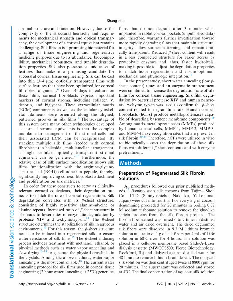
Silk films with various b-sheet contents wereproduced by water vapor annealing or methanolimmersion and were studied by FTIR spectroscopy.Deconvolution of the amide I region was performedfor secondary structure quantification. Methanolimmersed silk films (top curve in Fig. 1A) showed astrong absorption peak centered at 1610 to 1625cm�1, which was assigned to the b-sheets of silk.18
These films had 54% to 55% b-sheet content andwere classified as ‘high b-sheet films’ Silk films watervapor annealed for 30 to 45 minutes had b-sheet
Figure 1. (A) FTIR spectra of the Amide I region of the silk films with various b-sheet contents: low (bottom), medium (middle) and high(top). The percentages of b-sheets were determined by Fourier self-deconvolution. (B) Photographs of hydrated silk films with different b-sheet contents (low, medium, high) placed over a piece of paper displaying the word ‘silk’. (C) Transmittance measurements of silk filmswith different b-sheet contents (low, medium, high) in the visible light range.
http://tvstjournal.org/doi/full/10.1167/tvst.2.3.2 TVST j 2013 j Vol. 2 j No. 3 j Article 25
Shang et al.

content of 17% to 18% and were deemed ‘low b-sheetfilms’ as shorter treatments resulted in films with nostructural integrity that dissolved in aqueous envi-ronments. The secondary structure of ‘low b-sheetfilms’ (bottom curve in Fig. 1A), was dominated byrandom coil conformations (1640–1650 cm�1). ‘Me-dium b-sheet films’ were produced by water vaporannealing at 258C for 45 to 60 minutes and showedincreased b-sheet content compared with the low b-sheet silk films as demonstrated by the small shoulderpeak (middle curve in Fig. 1A) and b-sheet content of23% to 24%. Low, medium and high b-sheet filmswere structurally stable, possessed clear groovedpatterns necessary for cell alignment and wereoptically transparent (Fig. 1B, 1C).
Degradation of Silk Film in Vitro by HumanPancreatic a-Chymotrypsin
The a-chymotrypsin from human pancreas wasused to degrade silk films at concentrations of 1, 0.5,and 0.25 U/ml. No degradation occurred on thecontrol silk films incubated in PBS without enzyme.Bulk degradation was observed in the opticalmicrographs of all the silk films incubated with a-chymotrypsin at day 4 (Fig. 2). The films broke intopieces with the nanometer scale film pattern remain-ing mostly intact. The remaining mass of silk filmsover time was quantified by weighing the silk filmresidues after drying (Fig. 3A). Silk degradation level
correlated with b-sheet content, where high b-sheetfilms showed no significant mass loss over 4 days witha-chymotrypsin concentrations up to 1 U/mL, whilelow b-sheet showed significantly more mass losscompared with medium b-sheet films at all enzymeconcentrations (Fig. 3A).
Degradation of Silk Films In Vitro byProtease
Unlike a-chymotryspin, high b-sheet films showed3% and 12% mass loss at 0.05 U/mL and 0.1 U/mLof enzyme. Mass differences between low andmedium, as well as low and high b-sheet films weresignificant at day 4 at all enzyme concentrations.
Human Corneal Fibroblast Cells Proliferationon Silk Films with Various b-Sheet Levels
hCFs were seeded on silk films with different b-sheet contents (low, medium, and high) to assess theeffect of b-sheet content on cell proliferation andtherefore ensure that film degradation in the presenceof cells correlates with b-sheet content and is notaffected by differences in cell number on differenttreatments. No significant difference was observed incell numbers on different silk films over a 15 dayperiod, assessed by Alamar blue metabolic assay (Fig.4). At week 2 post seeding, the cells were aligned andcompletely confluent on all the films.
Figure 2. Optical micrographs of bulk degraded silk films in a-chymotrypsin at various concentrations after 4 days of degradation.
http://tvstjournal.org/doi/full/10.1167/tvst.2.3.2 TVST j 2013 j Vol. 2 j No. 3 j Article 26
Shang et al.

Cell Degradation of Silk Films with Differentb-Sheet Levels
Since there was no proliferation difference amonghCFs on silk films with different b-sheet content,hCFs were seeded on the different films at the samedensity and cultivated to compare degradationdifferences of silk films. In order to assist cell
degradation in addition to the lower b-sheet contentfilms, protease XIV enzyme pretreated films were alsoassessed. Silk film degradation by hCFs was assessedby analyzing surface roughness and quantifyingdegradation features on the film surface followingcell removal from the silk films at 2,4, 6, and 8 weekspost seeding. In order to quantify the process at theearly stages when no obvious degradation features
Figure 3. Time-dependent enzyme degradation profiles of silk films with various b-sheet contents with (A) human pancreatic a-chymotrypsin and (B) bacterial protease XIV. Film degradation is expressed as percent remaining mass, such that lower remaining massindicates greater level of film degradation.
http://tvstjournal.org/doi/full/10.1167/tvst.2.3.2 TVST j 2013 j Vol. 2 j No. 3 j Article 27
Shang et al.

were observed, roughness changes on the film surfaceswere monitored. The most commonly assessedroughness parameter, average roughness (Ra) androot mean square roughness (Rq), a parameter thatmeasures the variance of the amplitude distributionfunction were monitored on low, medium, and high b-sheet films and on enzyme pretreated films followingincubation with hCFs (Fig. 5). Enzyme pretreatmentcaused a 23% increase in the film surface roughness(Ra and Rq) prior to incubation with hCFs, but didnot affect the structural integrity or optical transpar-ency of the silk films. Films pretreated with enzymealso showed the greatest overall increase in Ra andRq in the presence of hCFs, with Ra increasing 1.6-fold and Rq increasing 1.4-fold by week 8 postseeding. At week 8 post seeding, Ra and Rq ofenzyme pretreated silk films were significantly higher
compared with all nontreated films. The Ra of low b-sheet films increased 1.3-fold by week 8 post seeding,compared with 0.2-fold increase in medium and 0-foldincrease in high b-sheet films (Fig. 5A). Similarly, Rqof low b-sheet films increased by 1.2-fold, comparedwith 0.3-fold and 0-fold increase in medium and highb-sheet films, respectively.
By week 8 post seeding, hCF cells formeddegradation features on the enzyme pretreated films,but not on the other films. The degradation featuresincluded degradation pits, cracks, and holes (Fig. 6).Degradation pits were shallow pits where surfacepattern grooves of silk films were eroded due to hCFdegradation (Fig. 6B). Small degradation holesformed along the base of the grooves in the patternedsilk films and a number of small degradation holesconnected together to form degradation cracks.Unlike the degradation pits which crossed thegrooves, the degradation cracks formed along thegrooves of the patterned silk films, where the filmswere eroded and split (Fig. 6C). The dimensions andnumbers of the above degradation features werequantified (Tables 1, 2). Degradation pits for enzymepretreated low b-sheet silk films presented at 150 6 80/mm2, were around 3 6 0.8 lm in length, 2 6 1 lm inwidth and 10 6 7 lm2 in area. There were around 300smaller degradation holes and 36 cracks formed permillimeter groove. Since smaller holes linked togetherto form cracks, the ratio of small holes and cracks wasalso counted at 10:1.
Discussion
Patterned silk films are promising biomaterialcandidates for engineering the complex hierarchicalstructure of the human cornea, but do not degrade
Figure 4. Equivalent hCF cell proliferation behavior on silk filmswith various b-sheet levels. The cell number was normalized toinitial cell number seeded on each silk film.
Figure 5. Increase in surface roughness features induced by hCF degradation over an 8 week culture period quantified from SEMimages of decellularized samples using MeX software. (A) Ra quantification (left) and three-dimensional reconstruction of the controlsurfaces (0 weeks) and following 4 and 6 weeks of cell culture. (B) Rq quantification.
http://tvstjournal.org/doi/full/10.1167/tvst.2.3.2 TVST j 2013 j Vol. 2 j No. 3 j Article 28
Shang et al.
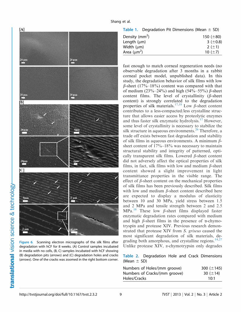
fast enough to match corneal regeneration needs (noobservable degradation after 3 months in a rabbitcorneal pocket model, unpublished data). In thisstudy, the degradation behavior of silk films with lowb-sheet (17%–18%) content was compared with thatof medium (23%–24%) and high (54%–55%) b-sheetcontent films. The level of crystallinity (b-sheetcontent) is strongly correlated to the degradationproperties of silk materials.17,18 Low b-sheet contentcontributes to a less-compacted/less crystalline struc-ture that allows easier access by proteolytic enzymesand thus faster silk enzymatic hydrolysis.17 However,some level of crystallinity is necessary to stabilize thesilk structure in aqueous environments.26 Therefore, atrade off exists between fast degradation and stabilityof silk films in aqueous environments. A minimum b-sheet content of 17%–18% was necessary to maintainstructural stability and integrity of patterned, opti-cally transparent silk films. Lowered b-sheet contentdid not adversely affect the optical properties of silkfilms, in fact, silk films with low and medium b-sheetcontent showed a slight improvement in lighttransmittance properties in the visible range. Theeffect of b-sheet content on the mechanical propertiesof silk films has been previously described. Silk filmswith low and medium b-sheet content described hereare expected to display a modulus of elasticitybetween 10 and 30 MPa, yield stress between 1.5and 2 MPa and tensile strength between 2 and 2.5MPa.18 These low b-sheet films displayed fasterenzymatic degradation rates compared with mediumand high b-sheet films in the presence of a-chymo-tryspin and protease XIV. Previous research demon-strated that protease XIV from S. griseus caused themost significant degradation of silk materials, de-grading both amorphous, and crystalline regions.14,27
Unlike protease XIV, a-chymotrypsin only degradesFigure 6. Scanning electron micrographs of the silk films afterdegradation with hCF for 8 weeks. (A) Control samples incubatedin media with no cells, (B, C) samples incubated with hCF showing(B) degradation pits (arrows) and (C) degradation holes and cracks(arrows). One of the cracks was zoomed in the right bottom corner.
Table 1. Degradation Pit Dimensions (Mean 6 SD)
Density (mm2) 150 (680)Length (lm) 3 (60.8)Width (lm) 2 (61)Area (lm2) 10 (67)
Table 2. Degradation Hole and Crack Dimensions(Mean 6 SD)
Numbers of Holes/(mm groove) 300 (6145)Numbers of Cracks/(mm groove) 30 (614)Holes/Cracks 10:1
http://tvstjournal.org/doi/full/10.1167/tvst.2.3.2 TVST j 2013 j Vol. 2 j No. 3 j Article 29
Shang et al.

the less crystalline domains in the silk28 withoutactivity toward the b-sheet crystals.14 While neither ofthese enzymes are expressed in the human cornea,they are invaluable tools in studying the effects ofmaterial properties on silk construct degradationprofiles in vitro. Both enzymes have been usedextensively to study silk degradation, allowing newfindings to be easily correlated with previouslypublished findings. Degradation of silk materials hasbeen demonstrated in numerous animal models,17 aswell with collagenases, enzymes known to beexpressed in injured corneal tissue.25
The surface roughness properties (Ra and Rq) oflow b-sheet silk films also increased to a greater extentcompared with medium and high b-sheet films in thepresence of hCFs in vitro. hCFs produce MMP-1,MMP-2, MMP-3, and MMP-9, which have recogni-tion sites on silk fibroin.19,21,29,30 However, other thanan increase in surface roughness, hCFs did notgenerate other discernible degradation features onthe silk films over the 8 week culture period. Othercell types, including osteoblasts, osteoclasts, andhuman mesenchymal stem cells have previously beenshown to generate degradation features, includinglarge 3-lm pits on the patterned silk surfaces.31
To further enhance the degradation behavior oflow b-sheet films, the films were pretreated with a lowconcentration (0.0125 U/mL) of protease XIV priorto cell culture. This treatment initiated the degrada-tion of silk films without compromising the structuralintegrity. It is likely that protease XIV loosened thecompact secondary structure of low b-sheet silk films,leading to the increased initial surface roughness andfaster degradation. Enzyme pretreated films displayedthe greatest increase in surface roughness (Ra and Rq)and higher overall surface roughness at 8 weeks postseeding. These films were also the only condition thatdisplayed discernible degradation features on the filmsurface, including degradation pits, holes, and cracks.Most degradation features, especially degradationholes and cracks were observed along the base ofthe patterned grooves on the film surface. The widthand the depth of these grooves were nanotopographyparameters to align the cells.32 As hCF cells alignedalong the grooves, the edges of grooves were likelyinitially exposed to MMPs secreted by aligned cells,resulting in the degradation features observed. First,small degradation holes appeared along the groovesand this feature increased as the degradation holesconnected into degradation cracks.
This study provided a protocol to significantlyaccelerate degradation of silk films starting with short
time water vapor annealing to generate low b-sheet(17%–18%) films, followed by enzymatic pretreat-ment with protease XIV. Lowering b-sheet content tothe limit of water stability balanced the tradeoffbetween silk film structural integrity in aqueousenvironments and faster degradation rates, whileenzyme pretreatment further enhanced degradationproperties allowing initiation of film degradation byhCFs within an 8 week period. This novel processingprotocol makes silk films more suitable for theconstruction of human corneal stroma tissue and apromising way to tune silk film degradation proper-ties to match corneal tissue regeneration. Successfulfilm degradation by hCFs in vitro warrants furtherinvestigation of these films in vivo. This protocol isalso of potential utility in a broader range ofbiomaterial needs where more rapid degradation orremodeling rates would be advantageous in tissuerepairs and regenerative medicine needs.
Acknowledgments
The authors thank Xiao Hu for his help during theinitial exploration of the b-sheet structure and watervapor annealing method, and also Biman B. Mandalfor his contribution to the corneal tissue engineeringproject and his help during the early stages of thisproject.
This work was supported by the National Insti-tutes of Health (NIH; EY020856) and the NIH P41Tissue Engineering Resource Center (EB002520).This work was performed in part at the Center forNanoscale Systems (CNS), a member of the NationalNanotechnology Infrastructure Network (NNIN).CNS is part of the Faculty of Arts and Sciences atHarvard University.
Disclosure: K. Shang, None; J. Rnjak-Kovacina,None; Y. Lin, None; R.S. Hayden, None; H. Tao,None; D.L. Kaplan, None
References
1. Sugioka K, Yoshida K, Kodama A, et al.Connective tissue growth factor cooperates withfibronectin in enhancing attachment and migra-tion of corneal epithelial cells. Tohoku J ExpMed. 2010;222:45–50.
2. Shah A, Brugnano J, Sun S, Vase A, Orwin E.The development of a tissue-engineered cornea:
http://tvstjournal.org/doi/full/10.1167/tvst.2.3.2 TVST j 2013 j Vol. 2 j No. 3 j Article 210
Shang et al.

biomaterials and culture methods. Pediatr Res.2008;63:535–544.
3. West-Mays JA, Dwivedi DJ. The keratocyte:corneal stromal cell with variable repair pheno-types. Int J Biochem Cell Biol. 2006;38:1625–1631.
4. Crabb RAB, Chau EP, Evans MC, Barogas VH,Hubel A. Biomechanical and microstructuralcharacteristics of a collagen film-based cornealstroma equivalent. Tissue Eng. 2006;12:1565–1575.
5. Gil ES, Mandal BB, Park SH, Marchant J,Omenetto FG, Kaplan DL. Helicoidal multi-lamellar features of RGD-functionalized silkbiomaterials. Biomaterials. 2010b;31:8953–8963.
6. Gil ES, Park SH, Marchant J, Omenetto FG,Kaplan DL. Response of human corneal fibro-blast on silk film surface patterns. MacromolBiosci. 2010a;10:664–673.
7. Abahussin M, Hayes S, Cartwright NEK, et al.3D collagen orientation study of the humancornea using x-ray diffraction and femtosecondlaser technology. Invest Ophthalmol Vis Sci. 2009;50:5159–5164.
8. Daxer A, Fratzl P. Collagen fibril orientation inthe human corneal stroma and its implication inkeratoconus. Invest Ophthalmol Vis Sci. 1997;38:121–129.
9. Komai Y, Ushiki T. The three-dimensionalorganization of collagen fibrils in the humancornea and sclera. Invest Ophthalmol Vis Sci.1991;32:2244–2258.
10. Chen L, Meng Q, Kao W, Xia Y. IjB kinase bregulates epithelium migration during cornealwound healing. PLoS ONE. 2011;6:e16132.
11. Chirila TV, Hicks CR, Dalton PD, et al. Artificialcornea. Prog Polym Sci. 1998;23:447–473.
12. Boote C, Dennis S, Huang Y, Quantock AJ,Meek KM. Lamellar orientation in human corneain relation to mechanical properties. J Struct Biol.2005;149:1–6.
13. Lawrence BD, Marchant JK, Pindrus MA,Omenetto FG, Kaplan DL. Silk film biomaterialsfor cornea tissue engineering. Biomaterials. 2009;30:1299–1308.
14. Numata K, Cebe P, Kaplan DL. Mechanism ofenzymatic degradation of b-sheet crystals. Bio-materials. 2010:31:2926–2933.
15. Lu Q, Zhang B, Li M, et al. Degradationmechanism and control of silk fibroin. Biomacro-molecules. 2011;12:1080–1086.
16. Jin HJ, Park J, Karageorgiou V. Water-stable silkfilms with reduced b-sheet content. Adv FunctMater. 2005;15:1241–1247.
17. Cao Y, Wang B. Biodegradation of silk bioma-terials. Int J Mol Sci. 2009;10:1514–1524.
18. Hu X, Shmelev K, Sun L, et al. Regulation of silkmaterial structure by temperature-controlled wa-ter vapor annealing. Biomacromolecules. 2011;12:1686–1696.
19. Ko�zak I, Klisenbauer D, Juhas T. UV-B inducedproduction of MMP-2 and MMP-9 in humancorneal cells. Physiol Res. 2003;52:229–234.
20. Leonardi A, Cortivo R, Fregona I, Plebani M,Secchi AG, Abatangelo G. Effects of Th 2cytokines on expression of collagen, MMP-1,and TIMP-1 in conjunctival fibroblasts. InvestOphthalmol Vis Sci. 2003;44:183–189.
21. Li DQ, Lee SB, Gunja-Smith Z, et al. Overex-pression of collagenase (MMP-1) and stromelysin(MMP-3) by pterygium head fibroblasts. ArchOphthalmol. 2001;119:71–80.
22. Rockwood DN, Preda RC, Yucel T, Wang X,Lovett ML, Kaplan DL. Materials fabricationfrom Bombyx mori silk fibroin. Nat Protoc. 2011;6:1612–1631.
23. Lawrence BD, Wharram S, Kluge JA, et al. Effectof hydration on silk film material properties.Macromol Biosci. 2010;10:393–403.
24. Sofia S, McCarthy MB, Gronowicz G, KaplanDL. Functionalized silk-based biomaterials forbone formation. J Biomed Mater Res. 2001;54:139–148.
25. Li M, Ogiso M, Minoura N. Enzymatic degra-dation behavior of porous silk fibroin sheets.Biomaterials. 2003;24:357–365.
26. Vepari C, Kaplan DL. Silk as a biomaterial. ProgPolym Sci. 2007;32:991–1007.
27. Horan RL, Antle K, Collette A, et al. In vitrodegradation of silk fibroin. Biomaterials. 2005;26:3385–3393.
28. Altman GH, Diaz F, Jakuba C, et al. Silk-basedbiomaterials. Biomaterials. 2003;24:401–416.
29. Berman M, Leary R, Gage J. Collagenase fromcorneal cell cultures and its modulation byphagocytosis. Invest Ophthalmol Vis Sci. 1979;18:588–601.
30. Kimura K, Nomi N, Yan ZH, Orita T, NishidaT. Inhibition of poly(I:C)–induced matrix metal-loproteinase expression in human corneal fibro-blasts by triptolide. Mol Vis. 2011;17:526–532.
31. Sengupta S, Park SH, Gil SE, et al. Quantifyingosteogenic cell degradation of silk biomaterials.Biomacromolecules. 2010;11:3592–3599.
32. Loesberg WA, te Riet J, van Delft FC, et al. Thethreshold at which substrate nanogroove dimen-sions may influence fibroblast alignment andadhesion. Biomaterials. 2007; 28:3944–3951.
http://tvstjournal.org/doi/full/10.1167/tvst.2.3.2 TVST j 2013 j Vol. 2 j No. 3 j Article 211
Shang et al.


![Accelerated Degradation Tests Planning With Competing ... · accelerated tests. Bai and Chun [13] discussed the optimal simple step-stress ALT (SSALT) plans with independent competing](https://static.fdocuments.in/doc/165x107/5ec5928fa51e9b1376067df5/accelerated-degradation-tests-planning-with-competing-accelerated-tests-bai.jpg)

