
Arthrography
53
Arthrography Arthrography Spring 2013 Spring 2013
-
Upload
reece-molina -
Category
Documents
-
view
34 -
download
6
description
Arthrography. Spring 2013. Arthrography. Used to obtain diagnostic information regarding the: Joint space Surrounding soft tissue Cartilage Lesions of the menisci Delineates the joint space and its surrounding structures Largely replaced by MRI. Joint Overview. - PowerPoint PPT Presentation
Transcript of Arthrography
ArthrographyArthrography
Spring 2013Spring 2013

ArthrographyArthrography
Used to obtain diagnostic information regarding Used to obtain diagnostic information regarding the:the: Joint spaceJoint space Surrounding soft tissueSurrounding soft tissue CartilageCartilage Lesions of the menisciLesions of the menisci
Delineates the joint space and its surrounding Delineates the joint space and its surrounding structuresstructures
Largely replaced by MRILargely replaced by MRI
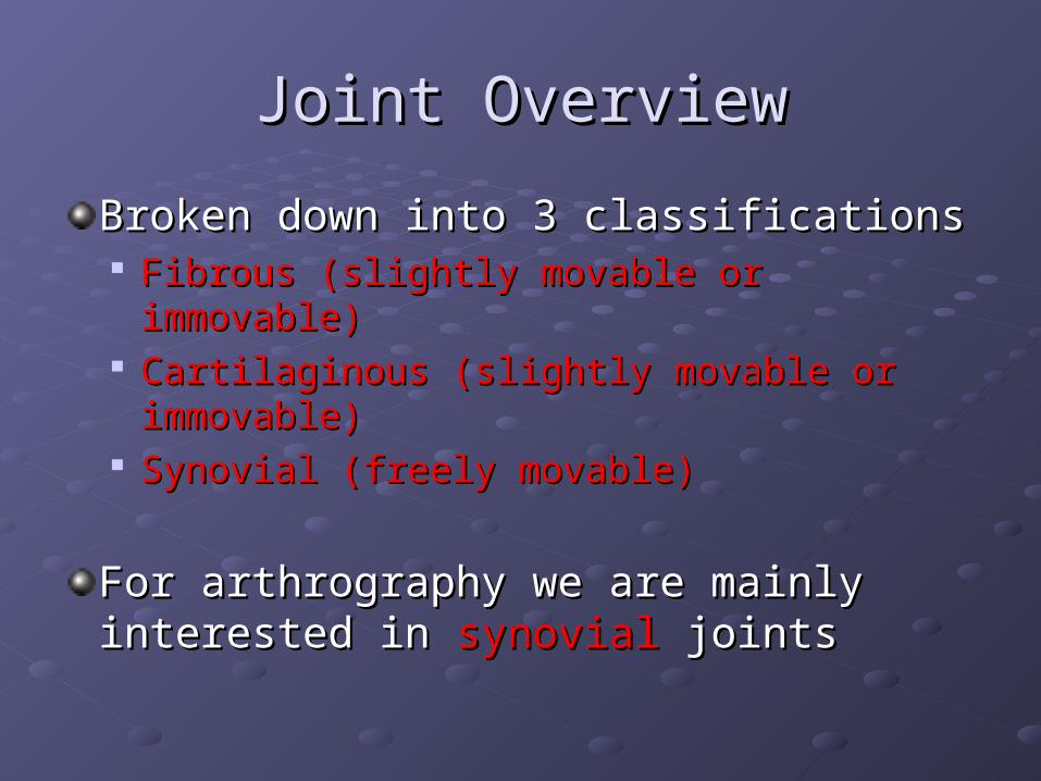
Joint OverviewJoint Overview
Broken down into 3 classificationsBroken down into 3 classifications Fibrous (slightly movable or immovable)Fibrous (slightly movable or immovable) Cartilaginous (slightly movable or immovable)Cartilaginous (slightly movable or immovable) Synovial (freely movable)Synovial (freely movable)
For arthrography we are mainly interested For arthrography we are mainly interested in in synovial synovial jointsjoints
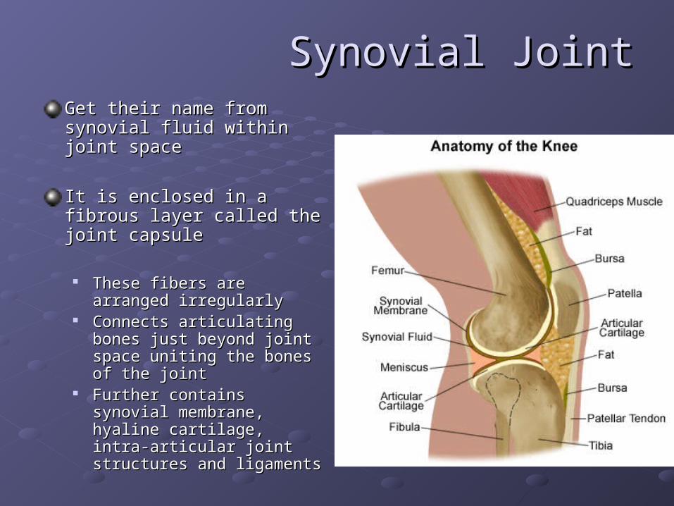
Synovial JointSynovial JointGet their name from synovial Get their name from synovial fluid within joint spacefluid within joint space
It is enclosed in a fibrous layer It is enclosed in a fibrous layer called the joint capsulecalled the joint capsule
These fibers are arranged These fibers are arranged irregularlyirregularly
Connects articulating bones Connects articulating bones just beyond joint space uniting just beyond joint space uniting the bones of the jointthe bones of the joint
Further contains synovial Further contains synovial membrane, hyaline cartilage, membrane, hyaline cartilage, intra-articular joint structures intra-articular joint structures and ligamentsand ligaments
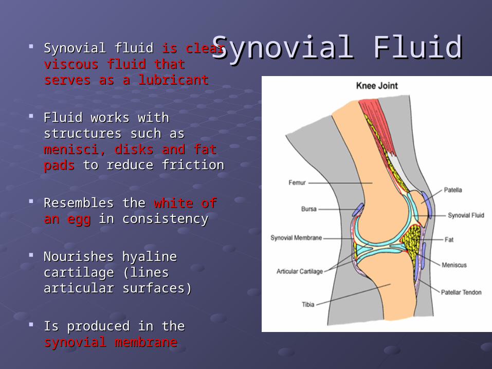
Synovial FluidSynovial Fluid Synovial fluid Synovial fluid is clear viscous is clear viscous fluid that serves as a lubricantfluid that serves as a lubricant
Fluid works with structures Fluid works with structures such as such as menisci, disks and fat menisci, disks and fat padspads to reduce friction to reduce friction
Resembles the Resembles the white of an egg white of an egg in consistencyin consistency
Nourishes hyaline cartilage Nourishes hyaline cartilage (lines articular surfaces)(lines articular surfaces)
Is produced in the Is produced in the synovial synovial membranemembrane

Anatomy of a Synovial JointAnatomy of a Synovial Joint
Synovial membraneSynovial membrane
Hyaline articular cartilageHyaline articular cartilage
Intra-articular JT Intra-articular JT structuresstructures
Menisci, fat pads, and Menisci, fat pads, and intra-articular disksintra-articular disks
LigamentsLigaments

Most Common Areas of Most Common Areas of ExaminationExamination
Arthrography can be done on any Arthrography can be done on any encapsuled JTencapsuled JT
KneeKnee is most common type of arthrogram is most common type of arthrogram performedperformed
Other joint spaces include:Other joint spaces include:WristWristShoulderShoulderTMJTMJHipHip

PneumoathrogramsPneumoathrograms
Air or gaseous medium is usedAir or gaseous medium is used
100-150100-150 ml ml
Produces Produces painful painful distention of jointdistention of joint
Possible Possible air embolismair embolism
Accuracy is considerably less than that when 2 Accuracy is considerably less than that when 2 contrast methods are usedcontrast methods are used

Positive or Opaque ArthrographyPositive or Opaque Arthrography
Water soluble iodinated Water soluble iodinated contrastcontrast
Ionic or non-ionicIonic or non-ionic30-100ml can be used30-100ml can be used
Contrast is readily Contrast is readily absorbed, tolerated and absorbed, tolerated and excretedexcreted
Produces greater Produces greater diagnostic accuracydiagnostic accuracy
Concentration should be Concentration should be no more than 30%no more than 30%

Double contrast ArthrographyDouble contrast Arthrography
Both Both gaseous and water soluble contrast gaseous and water soluble contrast employedemployed
By using both contrasts less of each can be By using both contrasts less of each can be used.used.
Reducing patient discomfortReducing patient discomfort
Decreasing chance of air embolismDecreasing chance of air embolism
Highly accurate diagnostic studyHighly accurate diagnostic study
Contrast PrecautionsContrast Precautions
Verify it is the correct contrastVerify it is the correct contrast Ionic or Non-ionic iodinated contrastIonic or Non-ionic iodinated contrast
Omnipaque or Isovue (non-ionic)Omnipaque or Isovue (non-ionic) Correct concentrationCorrect concentration
Check expiration dateCheck expiration date
Keep contrast vial in room until procedure is Keep contrast vial in room until procedure is completecomplete
Indications and Contraindications Indications and Contraindications for Arthrographyfor Arthrography
Indications:Indications: Suspected injury of meniscus (tears)Suspected injury of meniscus (tears) Suspected capsular damageSuspected capsular damage Rupture of articular ligamentsRupture of articular ligaments Cartilaginous defectsCartilaginous defects Arthritic deformities (specifically TMJ)Arthritic deformities (specifically TMJ) Congenital luxation ( dislocation) of hipCongenital luxation ( dislocation) of hip Extent of damage from traumaExtent of damage from trauma
Contraindications:Contraindications: Hypersensitivity to iodineHypersensitivity to iodine

Clinical SymptomsClinical Symptoms
Pain Pain
SwellingSwelling
Limited range of motionLimited range of motion
Recurrent instability (such as ankle)Recurrent instability (such as ankle)

RisksRisks
It is an invasive procedure therefore there It is an invasive procedure therefore there are certain risks to the patientare certain risks to the patient
Reaction to contrast mediaReaction to contrast media Vasovagal reactionVasovagal reaction
Nausea, perspiration and pallorNausea, perspiration and pallor Allergy to anesthetic agentAllergy to anesthetic agent Inflammatory synovitisInflammatory synovitis
Patient PREPPatient PREP
Get thorough pt Get thorough pt historyhistory Reason for examReason for exam AllergiesAllergies
Ease patients Ease patients anxietiesanxieties Answer questionsAnswer questions Explain procedureExplain procedure
PT comfortPT comfort Allow them to use Allow them to use
restroomrestroom Get pt into gownGet pt into gown Blankets Blankets
Obtain informed Obtain informed consentconsent Sometimes hospitals Sometimes hospitals
require doctor to do require doctor to do thisthis
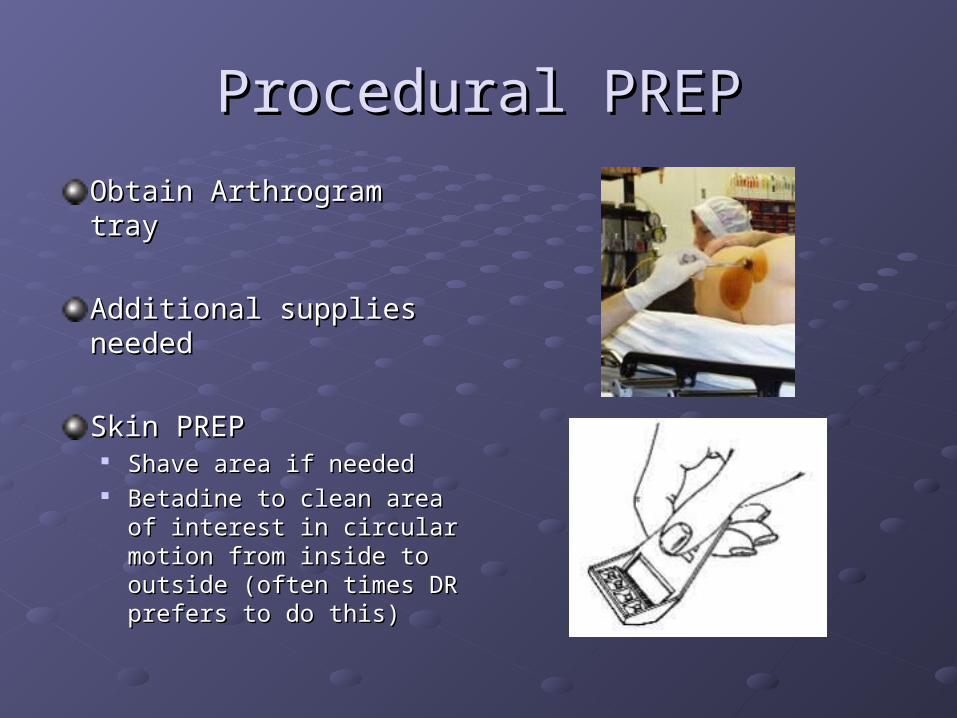
Procedural PREPProcedural PREP
Obtain Arthrogram trayObtain Arthrogram tray
Additional supplies Additional supplies neededneeded
Skin PREPSkin PREP Shave area if neededShave area if needed Betadine to clean area of Betadine to clean area of
interest in circular motion interest in circular motion from inside to outside from inside to outside (often times DR prefers to (often times DR prefers to do this)do this)
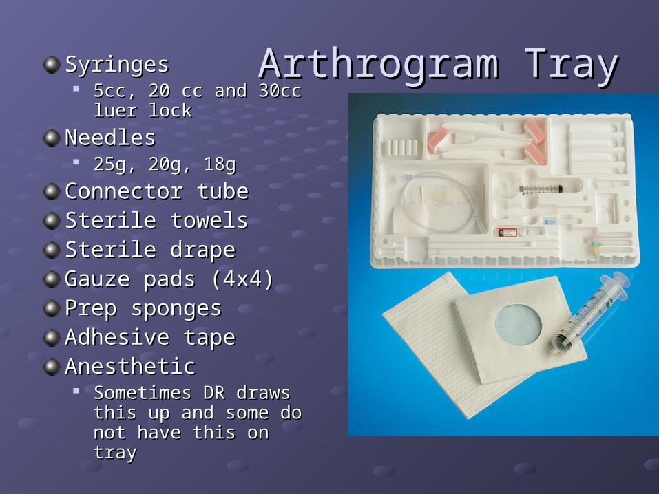
Arthrogram TrayArthrogram TraySyringesSyringes 5cc, 20 cc and 30cc luer 5cc, 20 cc and 30cc luer
locklock
NeedlesNeedles 25g, 20g, 18g25g, 20g, 18g
Connector tubeConnector tubeSterile towelsSterile towelsSterile drapeSterile drapeGauze pads (4x4)Gauze pads (4x4)Prep spongesPrep spongesAdhesive tapeAdhesive tapeAnestheticAnesthetic
Sometimes DR draws this Sometimes DR draws this up and some do not have up and some do not have this on traythis on tray
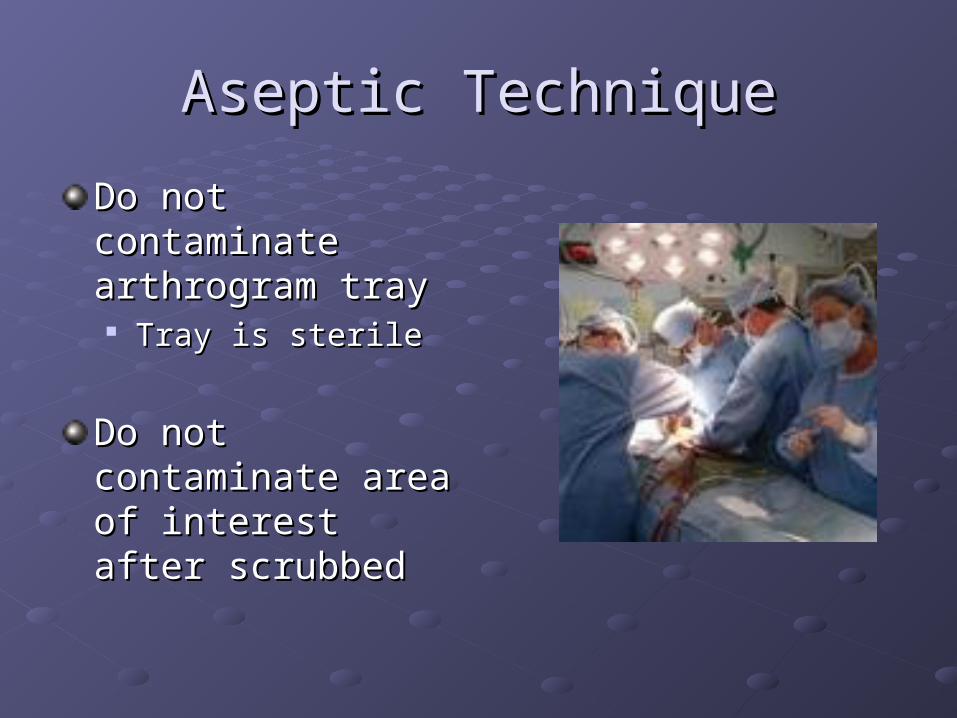
Aseptic TechniqueAseptic Technique
Do not contaminate Do not contaminate arthrogram trayarthrogram tray Tray is sterileTray is sterile
Do not contaminate Do not contaminate area of interest after area of interest after scrubbedscrubbed
Additional Equipment & SuppliesAdditional Equipment & Supplies
ShieldsShieldsTowels and blanketsTowels and blanketsContrastContrastSterile glovesSterile glovesAntiseptic solutionAntiseptic solutionGauzeGauzeAce bandages (if Ace bandages (if needed)needed)Fluoroscopy & Fluoroscopy & radiographic radiographic capabilities capabilities
GownGownExtra syringes and Extra syringes and needlesneedlesBandaidsBandaidsForceps (if part of Forceps (if part of protocol)protocol)GlovesGlovesSpecimen tubes (if Specimen tubes (if needed)needed)
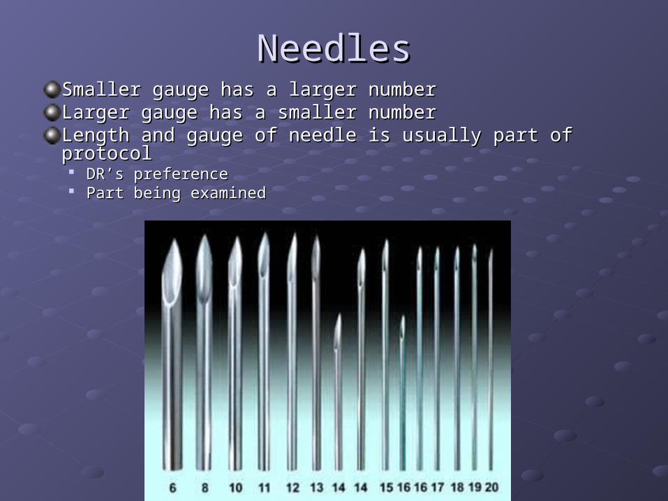
NeedlesNeedlesSmaller gauge has a larger numberSmaller gauge has a larger numberLarger gauge has a smaller numberLarger gauge has a smaller numberLength and gauge of needle is usually part of protocolLength and gauge of needle is usually part of protocol
DR’s preferenceDR’s preference Part being examinedPart being examined
Radiation SafetyRadiation Safety
Have shields for PT’s, DR and yourselfHave shields for PT’s, DR and yourself
Question LMP and the possibility of being Question LMP and the possibility of being pregnantpregnant
Use cardinal rulesUse cardinal rules TimeTime DistanceDistance ShieldingShielding
ALARAALARA Use pulse if possibleUse pulse if possible Save the last image on screen when possibleSave the last image on screen when possible

General GuidelinesGeneral Guidelines
Also refer to DEPT protocolAlso refer to DEPT protocol
Many hospitals have different protocols for Many hospitals have different protocols for different DR’sdifferent DR’s
Make sure you have everything readyMake sure you have everything ready This makes the procedure go smoothlyThis makes the procedure go smoothly

AspirationAspiration
Dr’s may aspirate Dr’s may aspirate fluids before injecting fluids before injecting contrast mediacontrast media If there is a joint If there is a joint
effusion especiallyeffusion especially
Fluid is sent to lab in Fluid is sent to lab in specimen vialsspecimen vials
Clinical Indications for Knee Clinical Indications for Knee ArthrogramsArthrograms
Pain, swelling and limited ROMPain, swelling and limited ROM
Trauma or athletic injuriesTrauma or athletic injuries
Suspected damage to menisci and Suspected damage to menisci and capsulecapsule
Rupture of articular ligamentsRupture of articular ligaments
Cartilaginous defectsCartilaginous defects
ArthritisArthritis

Knee Arthrogram: Vertical methodKnee Arthrogram: Vertical method
Apply all principles from Apply all principles from slides 15-21slides 15-21
Scout films: often AP, Scout films: often AP, Lateral and obliqueLateral and oblique
Check with DEPT protocolCheck with DEPT protocol
Anesthetic injectedAnesthetic injected
Contrast is injected Contrast is injected (single contrast study)(single contrast study)
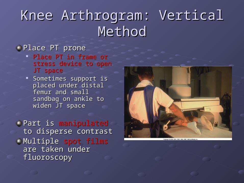
Knee Arthrogram: Vertical MethodKnee Arthrogram: Vertical Method
Place PT prone Place PT prone Place PT in frame or stress Place PT in frame or stress
device to open JT spacedevice to open JT space Sometimes support is Sometimes support is
placed under distal femur placed under distal femur and small sandbag on and small sandbag on ankle to widen JT spaceankle to widen JT space
Part is Part is manipulatedmanipulated to to disperse contrast disperse contrast Multiple Multiple spot films spot films are are taken under fluoroscopy taken under fluoroscopy
Knee Arthrogram: Vertical MethodKnee Arthrogram: Vertical Method
Overheads are doneOverheads are done AP, lateral, 20 degree right and left obliqueAP, lateral, 20 degree right and left oblique Sometimes Interconyloid fossa projections are requiredSometimes Interconyloid fossa projections are required
Single contrast study for Single contrast study for a torn meniscus a torn meniscus may fail to may fail to demonstrate the teardemonstrate the tear
Usually single contrast studies are used to demonstrate Usually single contrast studies are used to demonstrate loose particles of the JTloose particles of the JT
Post procedurePost procedure PT may feel tightness PT may feel tightness This should go away in 1-2 daysThis should go away in 1-2 days Can be treated with analgesicsCan be treated with analgesics
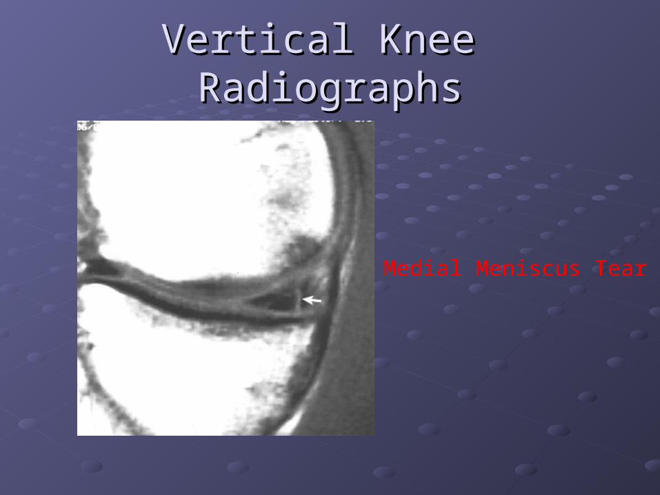
Vertical Knee RadiographsVertical Knee Radiographs
Medial Meniscus Tear

Meniscus TearsMeniscus Tears
Symptoms may include: Symptoms may include:
"Popping" sound "Popping" sound at the at the time of the injury time of the injury
Pain Pain
Tightness Tightness
Swelling within the knee, Swelling within the knee, often called "water on the often called "water on the knee" knee"
Locking up, catching, or Locking up, catching, or giving way of the knee giving way of the knee
Tenderness in the jointTenderness in the joint
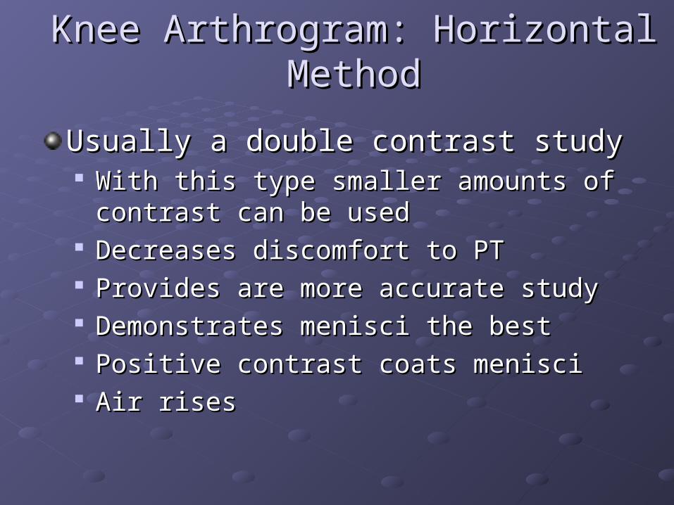
Knee Arthrogram: Horizontal MethodKnee Arthrogram: Horizontal Method
Usually a double contrast studyUsually a double contrast study With this type smaller amounts of contrast can With this type smaller amounts of contrast can
be usedbe used Decreases discomfort to PTDecreases discomfort to PT Provides are more accurate studyProvides are more accurate study Demonstrates menisci the bestDemonstrates menisci the best Positive contrast coats menisciPositive contrast coats menisci Air rises Air rises
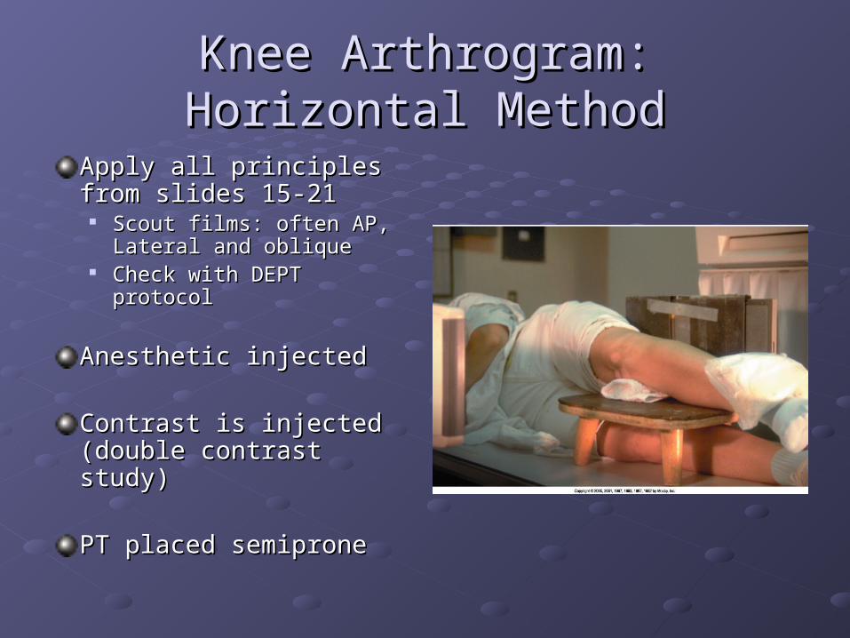
Knee Arthrogram: Horizontal Knee Arthrogram: Horizontal MethodMethod
Apply all principles from Apply all principles from slides 15-21slides 15-21
Scout films: often AP, Scout films: often AP, Lateral and obliqueLateral and oblique
Check with DEPT protocolCheck with DEPT protocol
Anesthetic injectedAnesthetic injected
Contrast is injected Contrast is injected (double contrast study)(double contrast study)
PT placed semipronePT placed semiprone
Knee Arthrogram: Horizontal MethodKnee Arthrogram: Horizontal Method
Knee is manually stressed while spot films Knee is manually stressed while spot films are taken (medial & lateral meniscus)are taken (medial & lateral meniscus)
Draw a line on medial or lateral side of Draw a line on medial or lateral side of knee and then direct CR to the meniscusknee and then direct CR to the meniscus
Rotate knee toward the supine positionRotate knee toward the supine position Turn 30 degrees for each of the projectionsTurn 30 degrees for each of the projections
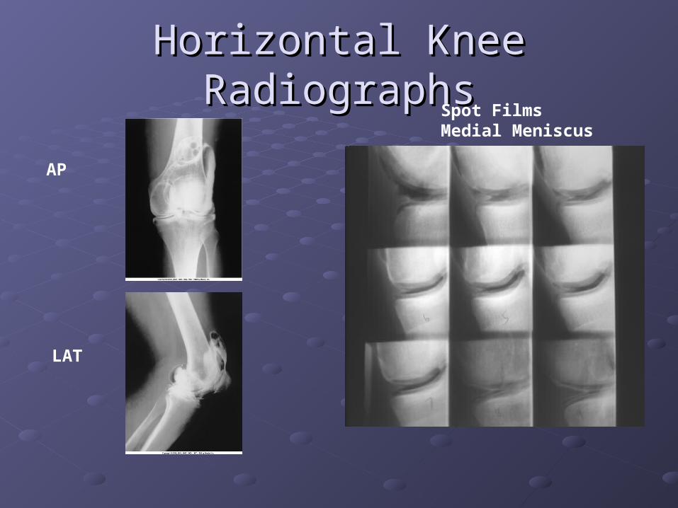
Horizontal Knee RadiographsHorizontal Knee RadiographsSpot Films Medial Meniscus
AP
LAT
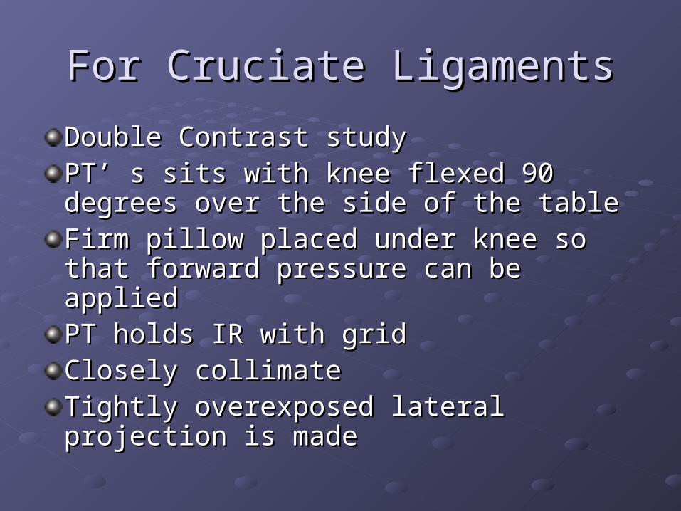
For Cruciate LigamentsFor Cruciate Ligaments
Double Contrast studyDouble Contrast studyPT’ s sits with knee flexed 90 degrees PT’ s sits with knee flexed 90 degrees over the side of the tableover the side of the tableFirm pillow placed under knee so that Firm pillow placed under knee so that forward pressure can be appliedforward pressure can be appliedPT holds IR with gridPT holds IR with gridClosely collimate Closely collimate Tightly overexposed lateral projection is Tightly overexposed lateral projection is mademade

CT Knee CT Knee ArthrographyArthrography
PT gets a regular PT gets a regular arthrogram in radiology arthrogram in radiology
Then is taken to CT for Then is taken to CT for imagingimaging
Can be single or double Can be single or double contrast (water soluble contrast (water soluble iodine)iodine)
Usually doubleUsually double
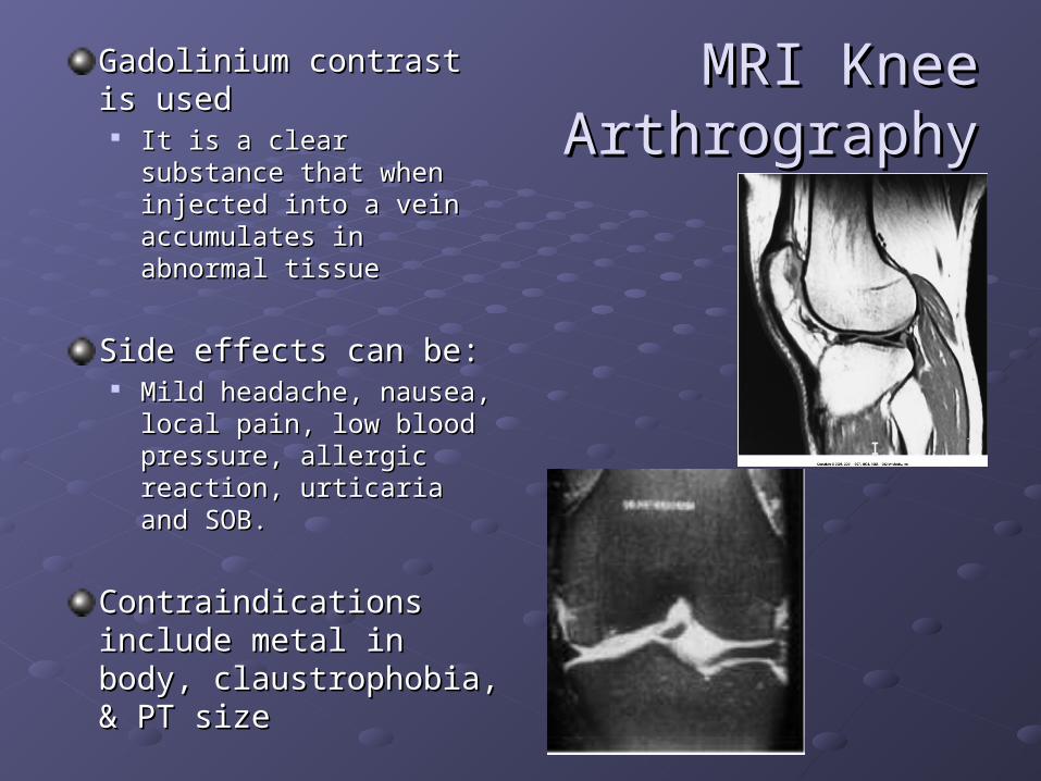
MRI KneeMRI Knee Arthrography Arthrography
Gadolinium contrast is Gadolinium contrast is usedused
It is a clear substance that It is a clear substance that when injected into a vein when injected into a vein accumulates in abnormal accumulates in abnormal tissuetissue
Side effects can be:Side effects can be: Mild headache, nausea, Mild headache, nausea,
local pain, low blood local pain, low blood pressure, allergic reaction, pressure, allergic reaction, urticaria and SOB.urticaria and SOB.
Contraindications include Contraindications include metal in body, metal in body, claustrophobia, & PT sizeclaustrophobia, & PT size

Shoulder ArthrogramShoulder Arthrogram
Indications:Indications: Partial or complete tears of rotator cuffPartial or complete tears of rotator cuff Tears of glenoid labrumTears of glenoid labrum Persistent pain or weaknessPersistent pain or weakness Frozen shoulderFrozen shoulder
Single or double contrast can be usedSingle or double contrast can be used Single 10-12 mlSingle 10-12 ml Double 3-4 positive contrast and 10-12 of airDouble 3-4 positive contrast and 10-12 of air

Shoulder Shoulder ArthrogramArthrogram
The usual objection site is The usual objection site is approx ½ inch inferior & lateral approx ½ inch inferior & lateral to the coracoid processto the coracoid process
Usually spinal needle is used Usually spinal needle is used because the joint capsule is because the joint capsule is usually deepusually deep
Scout films: AP (internal & Scout films: AP (internal & external), 30 degree oblique, external), 30 degree oblique, axillary, tangentialaxillary, tangential
See Chapter 5 for PT and part See Chapter 5 for PT and part positioningpositioning
AP scout
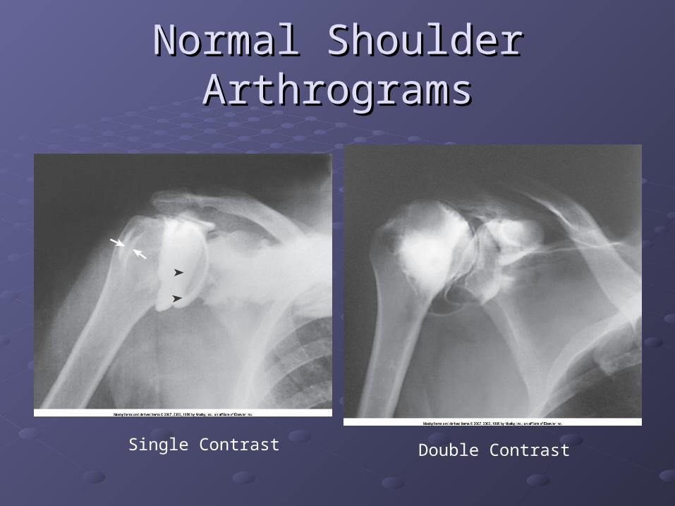
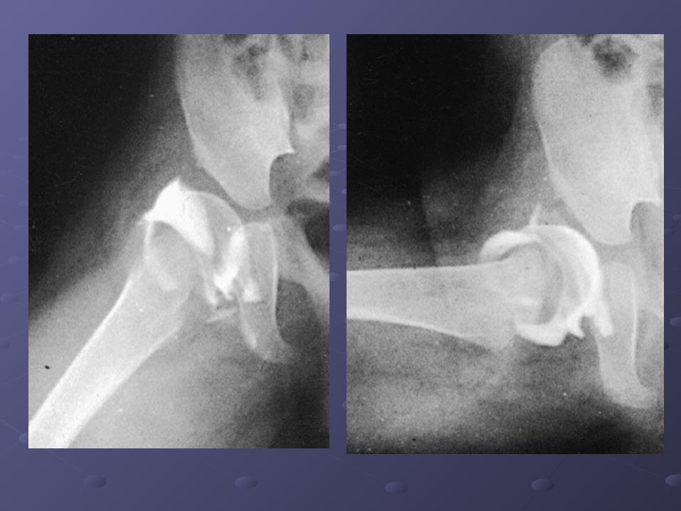
Normal Shoulder ArthrogramsNormal Shoulder Arthrograms
Single Contrast Double Contrast

Shoulder Single and Double Shoulder Single and Double contrastcontrast
Single contrastDouble contrast

Rotator Cuff TearRotator Cuff Tear
Shoulder ArthrogramShoulder Arthrogram
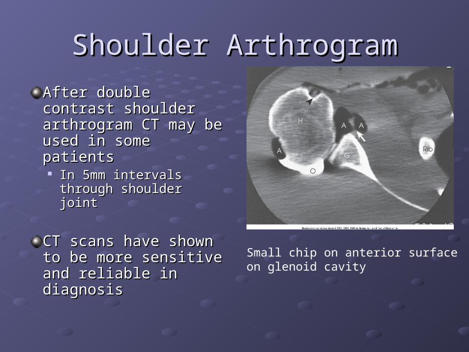
After double contrast After double contrast shoulder arthrogram shoulder arthrogram CT may be used in CT may be used in some patientssome patients In 5mm intervals In 5mm intervals
through shoulder jointthrough shoulder joint
CT scans have shown CT scans have shown to be more sensitive to be more sensitive and reliable in and reliable in diagnosisdiagnosis
Small chip on anterior surfaceon glenoid cavity
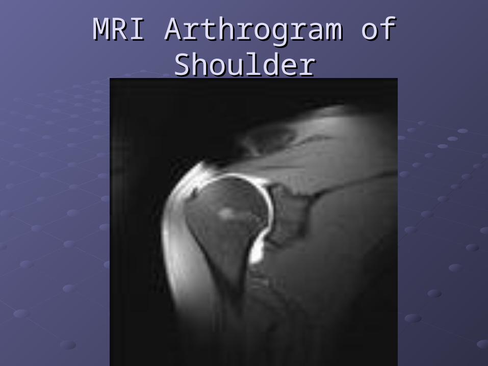
MRI Arthrogram of ShoulderMRI Arthrogram of Shoulder

Hip ArthrogramHip Arthrogram
Performed most often on children for congenital Performed most often on children for congenital dislocation pre and post treatmentdislocation pre and post treatment
Performed on adults to detect loose prosthetics Performed on adults to detect loose prosthetics or confirm presence of an infectionor confirm presence of an infection Cement & barium are added to hold prostheses and Cement & barium are added to hold prostheses and
to be able to check it radiographicallyto be able to check it radiographically BA and cement have approx same Z# making BA and cement have approx same Z# making
evaluation of JT by arthrographyevaluation of JT by arthrography Digital subtraction is used to overcome this problemDigital subtraction is used to overcome this problem

Hip Arthrogram & Hip Arthrogram & Digital SubtractionDigital Subtraction

Hip ArthrogramHip Arthrogram
Common puncture siteCommon puncture site ¾ “ distal to the inguinal crease¾ “ distal to the inguinal crease ¾” lateral to the palpated femoral pulse¾” lateral to the palpated femoral pulse
Spinal needle is used due to how deep the Spinal needle is used due to how deep the hip joint is into the body.hip joint is into the body.

Children Hip ArthrographyChildren Hip Arthrography
Wrist ArthrogramWrist ArthrogramIndications: trauma, persistent pain, limited ROM.Indications: trauma, persistent pain, limited ROM.
Contrast is injected through the dorsal wrist at the Contrast is injected through the dorsal wrist at the articulation of the radius, scaphoid and lunatearticulation of the radius, scaphoid and lunate
1.5-4ml water soluble iodinated contrast1.5-4ml water soluble iodinated contrast
After injection the wrist is carefully moved to spread After injection the wrist is carefully moved to spread contrastcontrast
Under fluoro or tape recording the wrist is rotated for Under fluoro or tape recording the wrist is rotated for exact area of leakageexact area of leakage
AP, LAT and both obliques often taken (check DEPT AP, LAT and both obliques often taken (check DEPT protocolsprotocols
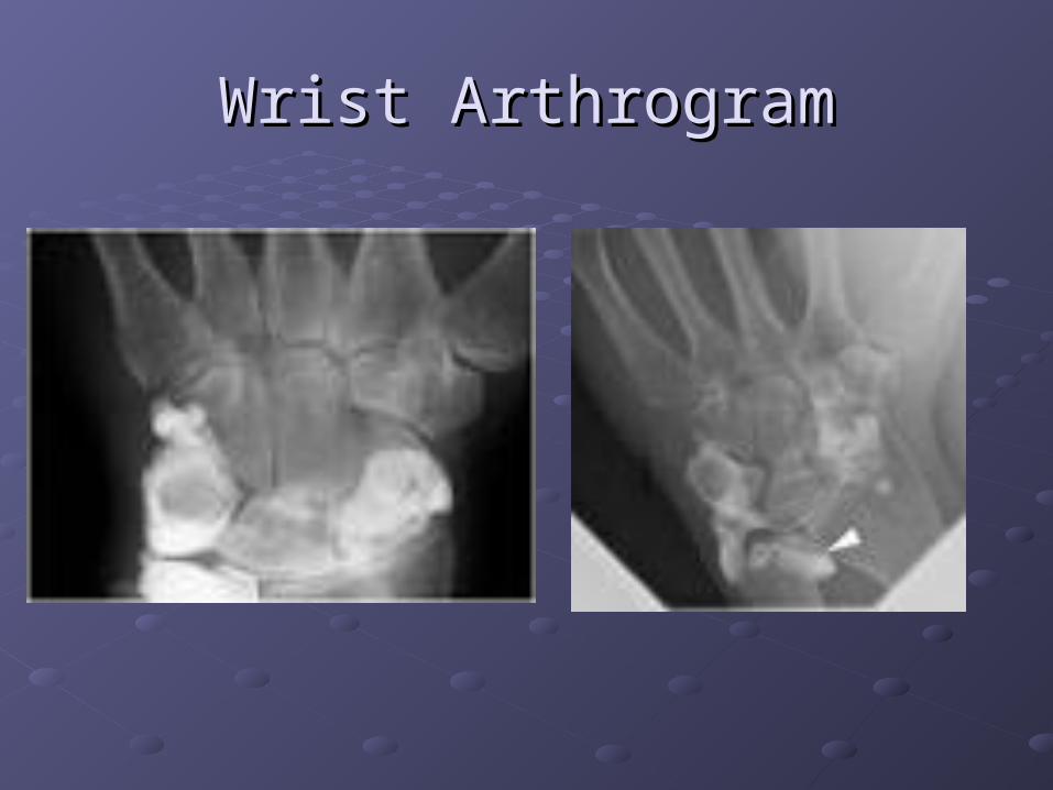
Wrist ArthrogramWrist Arthrogram

Wrist ArthrogramWrist Arthrogram
Rheumatoid Arthritis

TMJ ArthrogramTMJ Arthrogram
CT and MRI have largely replaced TMJ CT and MRI have largely replaced TMJ arthrography because they are noninvasivearthrography because they are noninvasive
Useful in diagnosingUseful in diagnosing Abnormalities of the articular diskAbnormalities of the articular disk
Indications: pain, clicking sound, lock jaw when Indications: pain, clicking sound, lock jaw when chewing sticky candychewing sticky candy StarburstStarburst TaffyTaffy

TMJ ArthrogramsTMJ Arthrograms
MRI
Tomography


![Ankle MR Arthrography: How, Why, When · of the extensor hallucis longus [1–3]. The arthrogram usually is performed under fluoroscopy control; how-ever, ultrasound, CT, or MR guidance](https://static.fdocuments.in/doc/165x107/601cf30d56915f4fae43da82/ankle-mr-arthrography-how-why-of-the-extensor-hallucis-longus-1a3-the-arthrogram.jpg)


