ARROCase Orbital MALT
24
ARROCase Orbital MALT Stephen Rosenberg, MD Faculty Advisors: Steven Howard, MD, PhD, Michael Bassetti, MD, PhD, and Kristin Bradley, MD Department of Human Oncology, University of Wisconsin
Transcript of ARROCase Orbital MALT
ARROCase
Orbital MALT
Stephen Rosenberg, MD
Faculty Advisors: Steven Howard, MD,
PhD, Michael Bassetti, MD, PhD, and
Kristin Bradley, MD
Department of Human Oncology,
University of Wisconsin
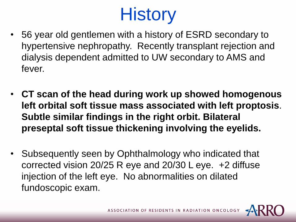
History• 56 year old gentlemen with a history of ESRD secondary to
hypertensive nephropathy. Recently transplant rejection and
dialysis dependent admitted to UW secondary to AMS and
fever.
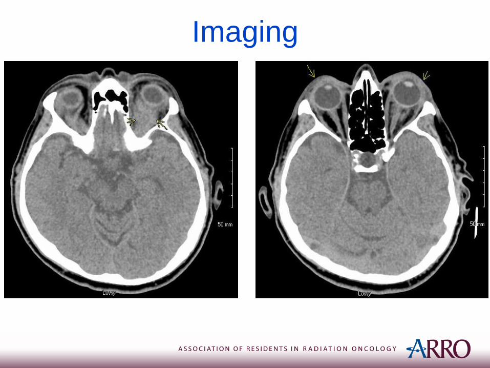
• CT scan of the head during work up showed homogenous
left orbital soft tissue mass associated with left proptosis.
Subtle similar findings in the right orbit. Bilateral
preseptal soft tissue thickening involving the eyelids.
• Subsequently seen by Ophthalmology who indicated that
corrected vision 20/25 R eye and 20/30 L eye. +2 diffuse
injection of the left eye. No abnormalities on dilated
fundoscopic exam.
Imaging
History
• Patient’s AMS improved during hospitalization secondary to adjustment of home opiate & benzodiazepines.
• Fever work up was unremarkable, including blood cultures. Antibiotics discontinued.
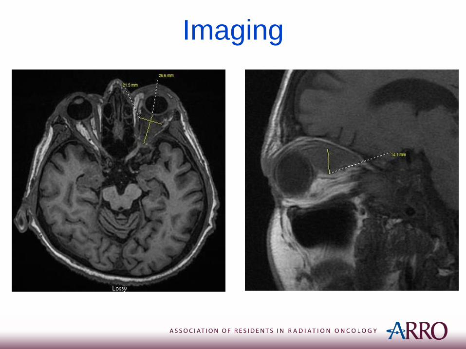
• MRI of the Orbits revealed infiltrative bilateral soft tissue masses involving the left greater than right orbits with abnormal infiltration and thickening of the preseptal soft tissues/conjunctiva.
Imaging
History
PMH: ESRD, Rheumatic Fever, Acute rejection of kidney transplant,
Hydronephrosis, Neutropenia, Pericardial effusion, Community-acquired
pneumonia, GERD, Hypothyroidism, Depression, Anxiety, Valve with AV fistula
infection, Secondary hyperparathyroidism, Hypertension
Family History: No history of malignancy
ROS: He denies any diplopia or blurry vision. He has noticed keratoconjuntivitis
sicca. Additionally, he notes pruritus in the left eye for at least 5 to 6 months. No
headaches, chest pain, or other systemic symptoms.
PSH: Renal transplant, Renal Biopsy, Knee arthroscopy, Cystoscopy,
Parathyroidectomy
Medications: Tylenol, Atenolol, Calcitriol, Calcium carbonate, Klonopin, Lunesta,
Heparin, Fosrenol, Synthroid, MTV, Zoloft
Differential Diagnosis
• Lymphoma
• Lymphoproliferative disease (including post transplant disorder in this patient)
• Idiopathic orbital inflammatory disease
• Cellulitis
• Metastatic disease
• Vascular or lymphatic malformation
• Sarcoidosis
• Granulomatosis with polyangiitis
History (continued)
• A left extensive superior and posterior orbitotomy with biopsy of the posterior orbital mass and orbital fat was performed.
• Pathology consistent with low-grade B-cell lymphoma, most consistent with a marginal zone lymphoma.
• Patient underwent complete systemic staging with a CT chest, abdomen, and pelvis which showed no metastatic disease.
• No significant elevation in Chlamydia psittaciantibodies.

Mucosa Associated Lymphoid
Tissue (MALT) Lymphoma
• MALT lymphomas are often secondary to chronic
inflammation (autoimmune or infectious etiology).
• The most common locations for MALT Lymphomas are
the GI tract (stomach>small intestine>colon). Other
locations include: lung, thyroid, salivary gland, tonsil,
breast, or orbit.
• MALT lymphomas typically arise from the marginal
zone of lymphoid follicle.
• Typically these malignancies are low grade B-cell
lymphomas that are CD20+, CD35+, CD5-, and CD10-.

Orbital/Ocular Adnexal
Lymphoma• Lymphomas are the most frequent tumor of the ocular
adnexa.
• Ocular Lymphomas:
1) Marginal Zone Lymphoma of MALT (~40-80%)
2) Follicular Lymphoma (~20%)
3) Diffuse Large B-Cell Lymphoma (~8%)
4) Mantle cell, small lymphocytic, lymphoplasmacytic (these
are less common)
• t(11;18)(q21;q21) is seen in 15-40% of patients with
Orbital MALT. Fusing API2 (apoptosis inhibitor 2) gene on
chromosome 11 and the MALT1 gene on chromosome 18.
Stefanovic A. and Lossos I. Blood 2009;114:501-510
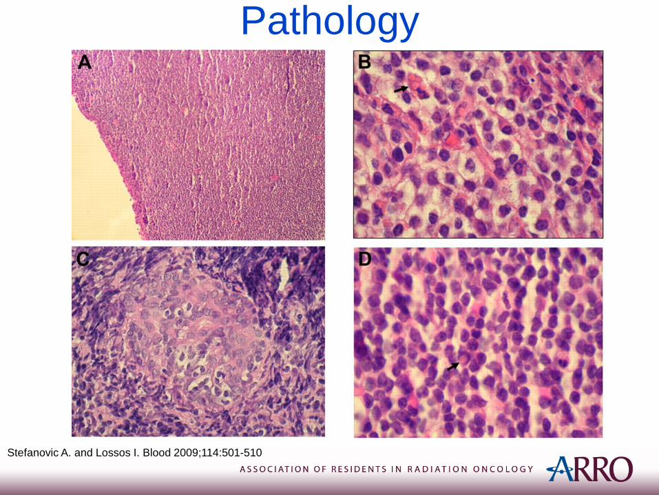
Pathology
Stefanovic A. and Lossos I. Blood 2009;114:501-510
Orbital MALT
• Patients often present between 5th – 7th decade
• Female predominance (1.5-2.0:1.0).
• 10-15% of patients present with bilateral orbital disease.
• Most frequent sites: Orbit (40%), Conjunctiva (35-40%),
Lacrimal gland (10-15%), and Eyelid.
• Chlamydia psittaci identified in ~25% of orbital MALT on
meta-analysis.
• Chlamydia psittaci association with orbital MALT lymphoma
varies by region:
– Common in Italy, Austria, Germany, and parts of the US
Stefanovic A. and Lossos I. Blood 2009;114:501-510
Ferreri., et. al. JCO 2012; 30(24):2988-2994
Husain., et. al. Cancer 2007; 110:809-815
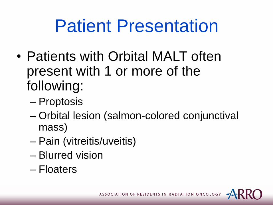
Patient Presentation
• Patients with Orbital MALT often present with 1 or more of the following:– Proptosis
– Orbital lesion (salmon-colored conjunctival mass)
– Pain (vitreitis/uveitis)
– Blurred vision
– Floaters
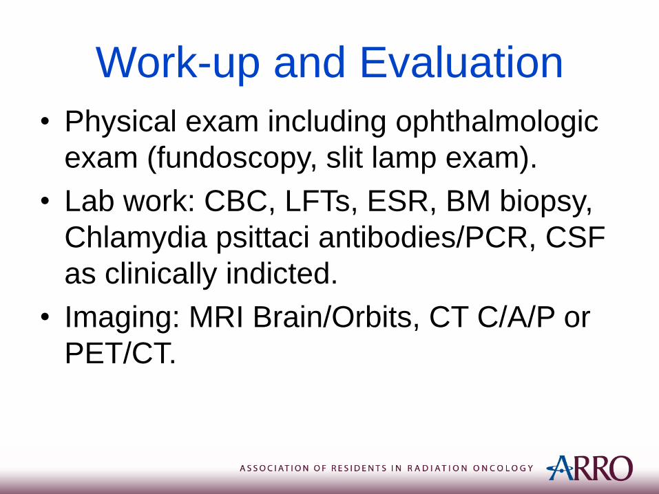
Work-up and Evaluation
• Physical exam including ophthalmologic
exam (fundoscopy, slit lamp exam).
• Lab work: CBC, LFTs, ESR, BM biopsy,
Chlamydia psittaci antibodies/PCR, CSF
as clinically indicted.
• Imaging: MRI Brain/Orbits, CT C/A/P or
PET/CT.
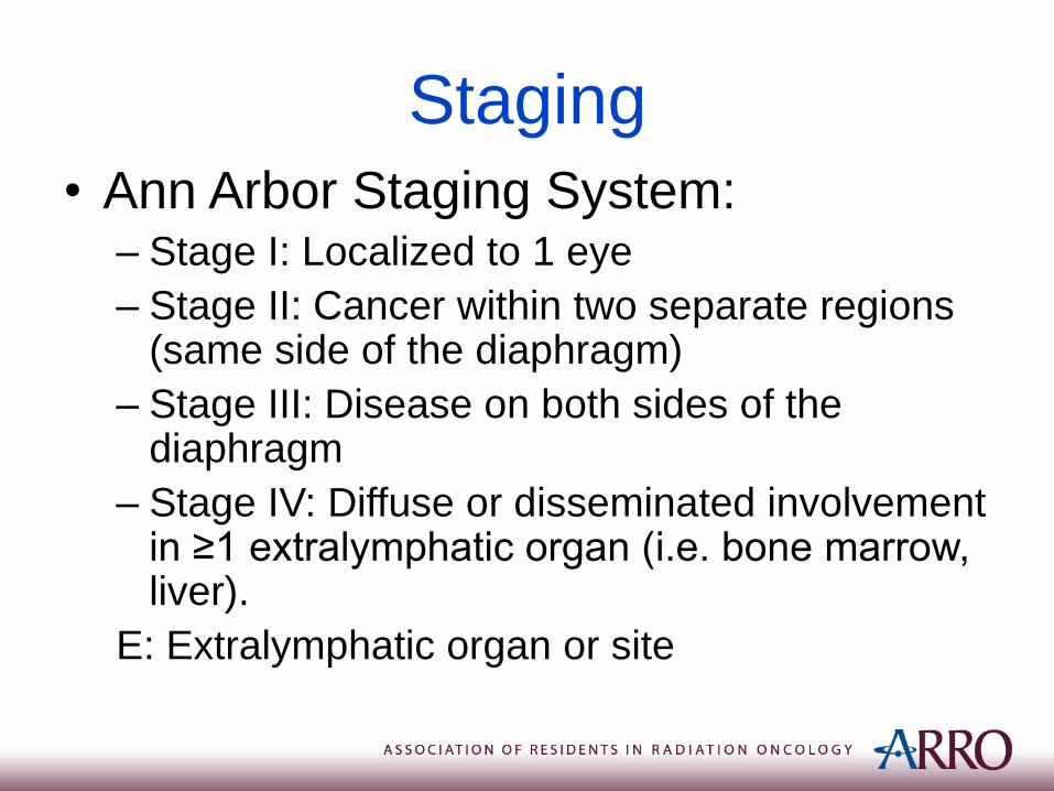
Staging
• Ann Arbor Staging System:– Stage I: Localized to 1 eye
– Stage II: Cancer within two separate regions (same side of the diaphragm)
– Stage III: Disease on both sides of the diaphragm
– Stage IV: Diffuse or disseminated involvement in ≥1 extralymphatic organ (i.e. bone marrow, liver).
E: Extralymphatic organ or site
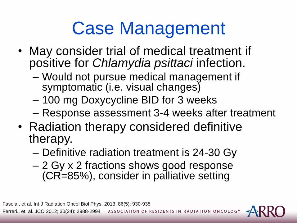
Case Management• May consider trial of medical treatment if
positive for Chlamydia psittaci infection.– Would not pursue medical management if
symptomatic (i.e. visual changes)
– 100 mg Doxycycline BID for 3 weeks
– Response assessment 3-4 weeks after treatment
• Radiation therapy considered definitive therapy.– Definitive radiation treatment is 24-30 Gy
– 2 Gy x 2 fractions shows good response (CR=85%), consider in palliative setting
Ferreri., et. al. JCO 2012; 30(24): 2988-2994
Fasola., et al. Int J Radiation Oncol Biol Phys. 2013. 86(5): 930-935
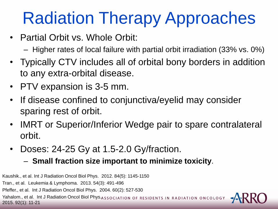
Radiation Therapy Approaches• Partial Orbit vs. Whole Orbit:
– Higher rates of local failure with partial orbit irradiation (33% vs. 0%)
• Typically CTV includes all of orbital bony borders in addition
to any extra-orbital disease.
• PTV expansion is 3-5 mm.
• If disease confined to conjunctiva/eyelid may consider
sparing rest of orbit.
• IMRT or Superior/Inferior Wedge pair to spare contralateral
orbit.
• Doses: 24-25 Gy at 1.5-2.0 Gy/fraction.
– Small fraction size important to minimize toxicity.
Yahalom., et al. Int J Radiation Oncol Biol Phys.
2015. 92(1): 11-21
Pfeffer., et al. Int J Radiation Oncol Biol Phys. 2004. 60(2): 527-530
Tran., et al. Leukemia & Lymphoma. 2013. 54(3): 491-496
Kaushik., et al. Int J Radiation Oncol Biol Phys. 2012. 84(5): 1145-1150
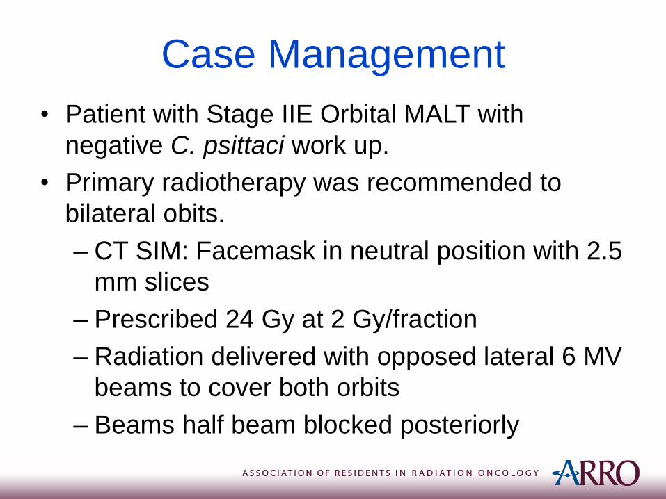
Case Management
• Patient with Stage IIE Orbital MALT with
negative C. psittaci work up.
• Primary radiotherapy was recommended to
bilateral obits.
– CT SIM: Facemask in neutral position with 2.5
mm slices
– Prescribed 24 Gy at 2 Gy/fraction
– Radiation delivered with opposed lateral 6 MV
beams to cover both orbits
– Beams half beam blocked posteriorly

Radiation Therapy: Bilateral Orbits
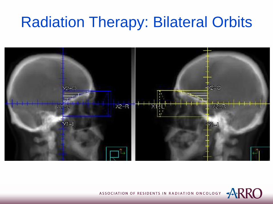
Radiation Therapy: Bilateral Orbits
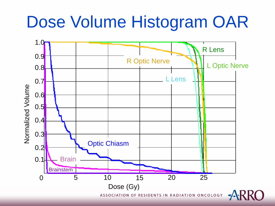
Dose Volume Histogram OAR
0.1
0.2
0.3
0.4
0.5
0.6
0.7
0.8
0.9
1.0
0 5 10 15 20 25
Dose (Gy)
Norm
aliz
ed V
olu
me
Optic Chiasm
Brain
Brainstem
R Optic NerveL Optic Nerve
R Lens
L Lens
Prognosis
Yahalom., et al. Int J Radiation Oncol Biol
Phys. 2015. 92(1): 11-21
• 85-100% local control rate with radiotherapy
• Distant failure rates vary from 20 -50%
usually involving other MALT tissues with
indolent behavior and prolonged survival
• Primary Site correlates with risk for systemic
involvement:
– Conjunctival (lowest risk)
– Eyelid (Highest risk)
Stefanovic A. and Lossos I. Blood 2009;114:501-510
Side Effects of Treatment• Acute toxicity:
– Conjunctival reactions (erythema/irritation)
• Long-term complications (~50% of patients):– Cataract formation (30-50%)
– Xerophthalmia (20-40%)
• RT doses >36 Gy associated with significant more toxicity– Ischemic retinopathy
– Corneal ulceration
– Optic atrophy
– Neovascular glaucoma
– Risk of vision loss
Yahalom., et al. Int J Radiation Oncol Biol
Phys. 2015. 92(1): 11-21
References• Fasola., et al. Low-Dose Radiation Therapy (2 Gy × 2) in the Treatment of
Orbital Lymphoma. Int J Radiation Oncol Biol Phys. 2013. 86(5): 930-935.
• Ferreri., et. al. Chlamydophila Psittaci Eradication With Doxycycline As
First-Line Targeted Therapy for Ocular Adnexae Lymphoma: Final Results
of an International Phase II Trial. JCO 2012; 30(24): 2988-2994.
• Husain., et. al. Meta–analyses of the association between Chlamydia
psittaci and ocular adnexal lymphoma and the response of ocular adnexal
lymphoma to antibiotics. Cancer 2007; 110:809-815.
• Kaushik., et al. Risk of radiation retinopathy in patients with orbital and
ocular lymphoma. Int J Radiation Oncol Biol Phys. 2012. 84(5): 1145-1150.
• Pfeffer., et al. Orbital lymphoma: Is it necessary to treat the entire orbit? Int
J Radiation Oncol Biol Phys. 2004. 60(2): 527-530.
• Stefanovic A. and Lossos I. Extranodal marginal zone lymphoma of the
ocular adnexa. Blood 2009;114:501-510.
• Tran., et al. Efficacy of low dose radiotherapy for primary orbital marginal
zone lymphoma. Leukemia & Lymphoma. 2013. 54(3): 491-496.
• Yahalom., et al. Modern Radiation Therapy for Extranodal Lymphomas:
Field and Dose Guidelines From the International Lymphoma Radiation
Oncology Group. Int J Radiation Oncol Biol Phys. 2015. 92(1): 11-21.