Arrhythmias Complicating Acute Myocardial Infarction.
27
Arrhythmias Complicating Acute Myocardial Infarction
-
Upload
patience-warner -
Category
Documents
-
view
224 -
download
0
Transcript of Arrhythmias Complicating Acute Myocardial Infarction.
Arrhythmias Complicating Acute Myocardial
Infarction

Sinus Bradycardia Most common arrhythmia occurring during the early
hours after MI and may occur in up to 40% of inferior and posterior infarcts.
May be related to autonomic imbalance or to atrial and sinus node ischemia or both.
Profound bradycardia may predispose the patient to ventricular ectopy.
Usually resolves spontaneously, treatment is reserved for hemodynamically symptomatic arrhythmias and those with bradycardia dependent vent. Arrhythmias.
Atropine usually successful for symptomatic bradycardia. Temporary pacing is rarely required.
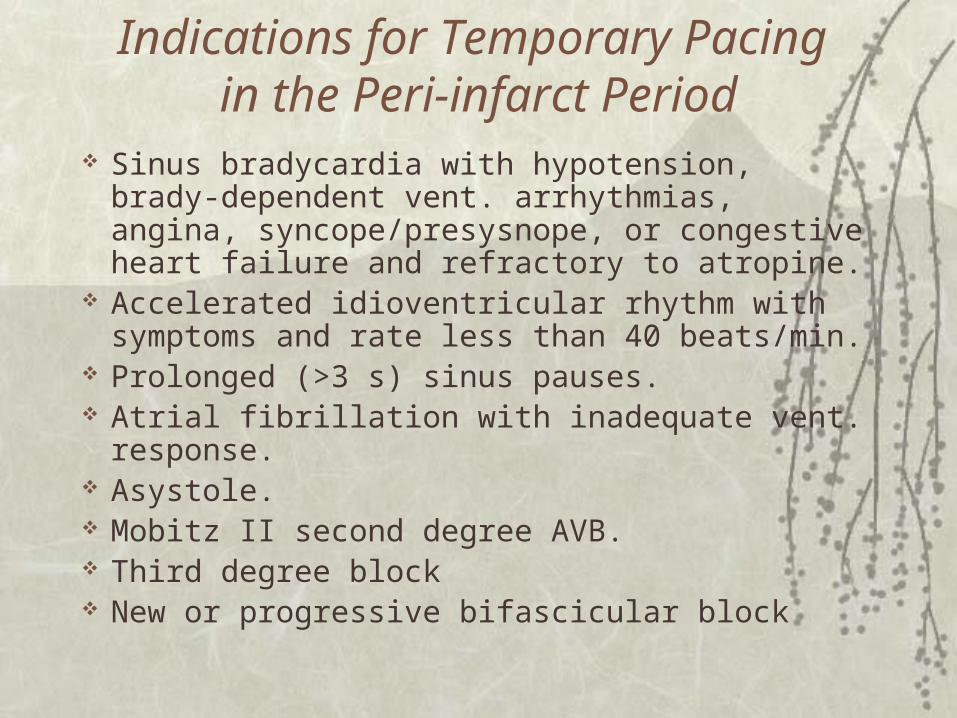
Indications for Temporary Pacing in the Peri-infarct Period
Sinus bradycardia with hypotension, brady-dependent vent. arrhythmias, angina, syncope/presysnope, or congestive heart failure and refractory to atropine.
Accelerated idioventricular rhythm with symptoms and rate less than 40 beats/min.
Prolonged (>3 s) sinus pauses. Atrial fibrillation with inadequate vent. response. Asystole. Mobitz II second degree AVB. Third degree block New or progressive bifascicular block
Sinus Tachycardia May occur in up to 1/3 of patients in the peri-infarct period,
esp. with ant. MI. The ischemic left ventricle may have a relatively fixed
stroke volume; thus augmenting CO by increasing HR. It may occur as a result of sympathetic stimulation from
locally released and circulating catecholamines, concurrent anemia, hypo or hypervolemia, hypoxia, pericarditis, inotropic drugs, pain, fever.
Treatment includes optimizing hemodynamics, oxygenation, correction of anemia, electrolyte and acid base abnormalities, pain control, and anxiolytic agents.
Beta blockers are indicated for patients without evidence of significant LV dysfunction or hypovolemia.
Persistent sinus tachycardia as an early manifestation of heart failure is an indicator of poor prognosis.
Premature atrial Contractions Up to one half of patients with MI May be due to atrial or sinus node ischemia,
atrial fibrillation, pericarditis, anxiety or pain. The combination of atrial asystole and rapid
ventricular rate markedly decreases cardiac output and increase oxygen demands.
Attempts should be made to restore sinus rhythm; if not, rate should be controlled aggressively to minimize oxygen demand.
May have no prognostic significance after MI.
First degree AVB
5 % to 10 % patients with MI at some times during peri-infarct period.
Almost all have supra-Hisian conduction abnormalities.
Rare cases of infranodal block are seen with anterior MI and associated fascicular block; these patients are at risk for progressive block, including third degree block with ventricular asystole.
May be associated with drugs that prolong AV conduction.

2nd degree Mobitz Type I Block May be seen with up to 10% cases of MI,
typically inferior infarcts, and is due to increased vagal tone and ischemia.
Conduction defect is usually in the AV node. When seen early after MI, usually responds to
atropine and resolves within 48-72 hours. Late occurring Wenckebach is less sensitive to
atropine and may be due to recurrent ischemia. Very rarely it may progress to higher grades of
block that require permanent pacing. It has no impact on long-term prognosis.

Second-degree Mobitz Type II Block
1% of cases of MI and more common with anterior MI.
High risk of progression to higher degrees of block, including sudden complete heart block with ventricular asystole.
Should have temporary pacing wire placed prophylactically.
Conduction defect is most likely infranodal. Most patients need permanent pacing and if it is
uncertain, EP evaluation should be performed before discharge to assess integrity of the infranodal conduction system.
Long term prognosis is related to size of infarct rather than conduction abnormality.
Complete Heart Block With either anterior or inferior infarct. With inferior infarcts , the defect is likely to be in the
AV node, with escape rhythms exceeding 40 beats/min and exhibiting a narrow QRS complex.
With anterior infarct, the conduction defect is infranodal and the escape rhythm (if present) is usually less than 40 beats/min with a wide QRS complex.
Typically CHB seen with ant. MI is preceded by progressive fascicular, bundle, or mobitz type II block.
Temporary pacing may be required with inferior MI if the patient is hemodynamically unstable. It should always be used with anterior MI if progressive or CHB is present.
Complete Heart Block - Continued
Permanent pacing is almost always required for high grade block in the setting of anterior MI.
The prognosis is poor for these patients because of the large amount of myocardium involved.
EP evaluation should be considered for patients with Anterior MI and transient CHB to assess the integrity of the infranodal conduction system.
Transient CHB with inferior MI rarely requires permanent pacing and usually resolves spontaneously.
BBB New BBB has been reported in about 15% of
cases of MI and is associated with an increased risk of CHB, CHF, cardiogenic shock, vent. arrhythmias and sudden death.
Most commonly seen is RBBB; LBBB or alternating BBB being less common. This may be related to discrete anatomical location of RB compared to broad, fan shaped LB.
The highest incidence of BBB occurs with LAD.
BBB Progressive infra-Hisian block indicates a
significant risk of sudden CHB and asystole, and patients demonstrating progression should have temporary pacing wires placed.
Persistent BBB confers a significantly higher mortality, because of the large amount of myocardium that must be involved to include the BB.
Thrombolytic therapy and catheter-based early reperfusion appear to decrease the incidence of BBB in the peri-infarct period.
Intraventricular Block New isolated LAFB occurs in 3% to 5% of
patients with MI; New isolated LPFB occurs in 1% to 2% of patients with acute MI.
Anatomically, LPFB is larger; hence a large infarct is required to produce block. Mortality is greater among those patients.
LAFB with new RBBB is also indicative of a larger infarct and higher subsequent mortality.

Ventricular arrhythmias - Factors Damaged myocardium: A substrate for development of
reentrant circuits. Large MI, Early LV dilatation and remodeling, perhaps due to ventricular stretching and electromechanical feedback.
Arrhythmia trigger: spontaneous ventricular arrhythmias, variation in cycle length and HR.
Electrolyte Imbalance, autonomic nervous system dysfunction/imbalance, increased catecholamines, continued ischemia, impaired LV function.
Mechanism Changes in the resting membrane potential and the
inward and outward ionic fluxes during action potential lead to alterations in the conduction, refractoriness, and automaticity of cardiac muscle cells.
Increase in QT dispersion due to inhomogeneity in myocardial cells may be a contributing factor. The central ischemic zone has decreased impulse velocity and increased refractoriness; the normal zone has increased or normal impulse velocity with decreased refractoriness; and the area in between has intermediate properties.
Mechanism Less negative resting membrane potential due to loss of
energy dependent ATP-ase pump and consequently decrease K influx and increased K loss.
The ventricular arrhythmias after MI can be divided into early ( upto 30 minutes after total occlusion of artery) and delayed phase (3 to 6 hours and as long as 3 days). Early phase is further divided into 1a (2-10 minutes after total occlusion of artery) or 1b (10 to 30 minutes). According to animal studies arrhythmias occurring in 1a phase are mainly due to re-entry. Occasionally it may be due to nonreentry initiated PVBs which may precipitate eventually reentry arrhythmias.
Mechanism
1b phase has no clear mechanism established but it is thought to be due to abnormal automaticity.
Arrhythmias occurring in late phase are thought to be due to surviving purkinje tissue located within the subendocardium which displays abnormal automaticity. It also shows increase sensitivity to catecholamines.
Arrhythmias occurring in chronic phase (after 3 days) - reentry.

Ventricular Fibrillation
Incidence of primary VF in MI is about 5% in patients in whom a documented rhythm is obtained. It occurs without antecedent warning arrhythmias in over half.
The true incidence of primary VF is probably higher because it has been estimated that one-half of all patients with coronary artery disease die of sudden death presumably VF.
Factors associated with an increased incidence of VF include current smoking, LBBB and hypokalemia.
Ventricular Fibrillation Patients with Ant. MI and VF have worse long-
term prognosis than those with inferior MI. VF may occur with reperfusion after
thrombolytics or catheter-based therapy. Treatment with prompt defibrillation. Beta
blockers appear to decrease the incidence of lethal vent arrhythmias including VF, in the peri-infarct period
Ventricular Tachycardia VT occurs in 10% to 40% of cases of MI. Early VT
during the first 24 hours is usually transient and benign.
Late occurring VT is associated with transmural infarction, LV dysfunction, hemodynamic deterioration, and a markedly higher mortality, both in-hospital and long-term.
If the rate is slow and hemodynamically tolerated, cardioversion with drugs may be attempted. Rapid VT (>150 beats/min) or VT with hemodynamic deterioration should be treated with prompt DC cardioversion.
Accelerated Idioventricular rhythm Rate faster than the normal ventricular escape rhythm
but slower than the VT. Onset and offset is usually gradual, and isorhythmic
dissociation is often present. Has been reported in 10% to 40% of cases of MI,
especially but not necessarily with early reperfusion. The incidence is equal in anterior and inferior infarcts
and not related to infarct size. Not related to increased mortality or VF. May also be seen with dig toxicity, myocarditis, and
cocaine use.. Symptoms are usually related to loss of AV synchrony
or slow ventricular rates or both.

Premature ventricular Complexes
Occur frequently during MI. Their significance preceding VT and VF is unclear. Treatment of PVCs in the peri-infarct period has not
been shown conclusively to decrease incidence of malignant vent. Arrhythmias or to improve mortality.
The pooled data of randomized trials in which PVCs were treated prophylactically in the peri-infarct period with lidocaine demonstrated an increased mortality.
Beta blockers may be the best option for treating PVCs and preventing malignant arrhythmias
Reperfusion Arrhythmias Typically accelerated idioventricular rhythm has
been credited with being a marker of reperfusion, However, any arrhythmia or no arrhythmia may be seen with reperfusion; conversely, AIVR may occur without reperfusion. Other clinical factors should be considered when deciding whether reperfusion has occurred.
The appearance of reperfusion arrhythmias is related to size of infarct, length and severity of ischemia, rate of reperfusion, HR, extracellular K concentration, and the presence of congestive heart failure or LVH or both.
Asystole and EMD
Occur in a small fraction of patients with MI and are usually associated with large infarcts.
The prognosis is extremely poor even with aggressive therapy.
Defibrillation should be attempted in patients with apparent systole, because the rhythm may be actually fine VF
Other Miscellaneous
T-Wave alternans: Transient finding usually seen with ischemia and most pronounced in leads overlying the affected myocardium.
Other Miscellaneous
Regional pericarditis sometimes seen after Q-wave MI may present with PR depression, but more commonly with atypical ST-segment and T-wave changes. These changes typically consist of gradual premature reversal of initially inverted T waves or persistent/recurrent ST-segment elevation or both.
Persistent ST-segment elevation after infarct may be due to continuing ischemia or aneurysm formation or may herald free wall rupture
Other Miscellaneous LV free wall rupture occurs in approximately 10%
cases of fatal transmural MI. EKG finding include failure of the characteristic
evolution of the ST segment, T wave or both. Persistent, progressive, or recurrent ST-segment
elevation in the absence of recurrent ischemia may be seen.
Failure of T waves to invert or initial inversion followed by reversion may be seen.
Abrupt bradycardia responsive to atropine may occur and is believed to mark the time or rupture