Arrhythmias 101 Fundamentals and what you should know for the big, bad BOARDS!
59
Arrhythmias 101 Arrhythmias 101 Fundamentals and what you Fundamentals and what you should know for the big, should know for the big, bad bad BOARDS! BOARDS!
-
date post
20-Dec-2015 -
Category
Documents
-
view
216 -
download
0
Transcript of Arrhythmias 101 Fundamentals and what you should know for the big, bad BOARDS!
Arrhythmias 101Arrhythmias 101Arrhythmias 101Arrhythmias 101
Fundamentals and what you Fundamentals and what you should know for the big, bad should know for the big, bad
BOARDS!BOARDS!
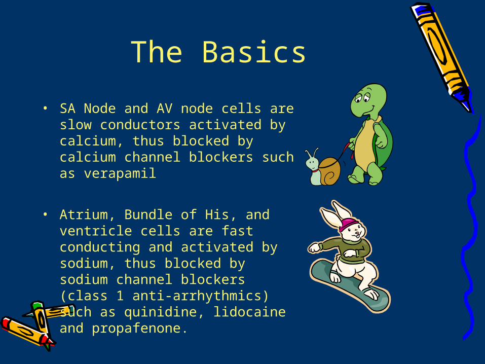
The Basics
• SA Node and AV node cells are slow conductors activated by calcium, thus blocked by calcium channel blockers such as verapamil
• Atrium, Bundle of His, and ventricle cells are fast conducting and activated by sodium, thus blocked by sodium channel blockers (class 1 anti-arrhythmics) such as quinidine, lidocaine and propafenone.
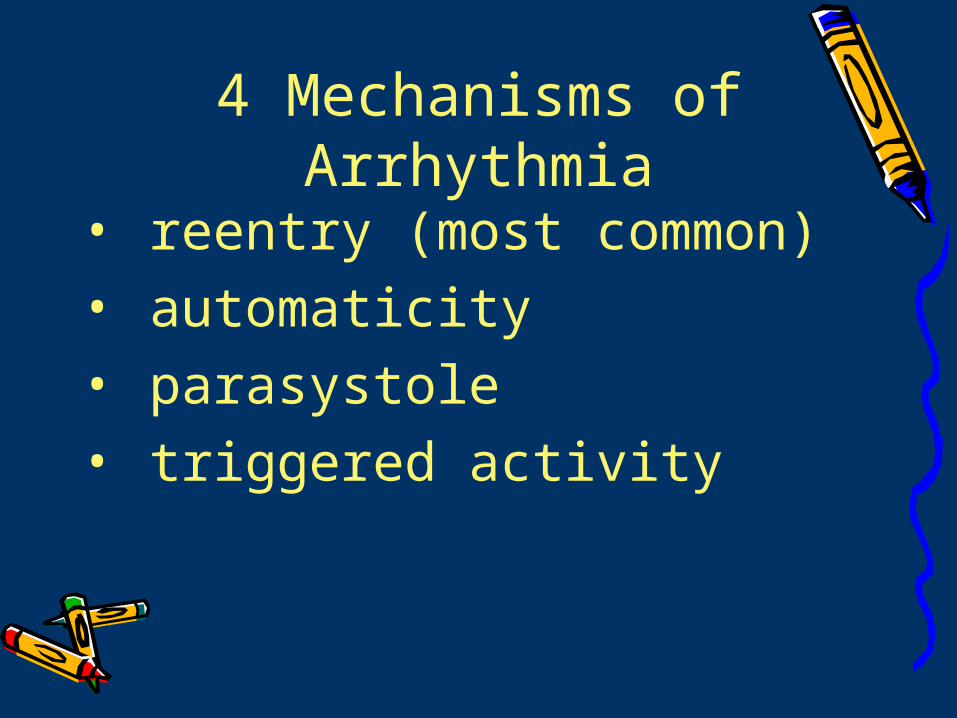
4 Mechanisms of Arrhythmia
• reentry (most common)• automaticity• parasystole • triggered activity
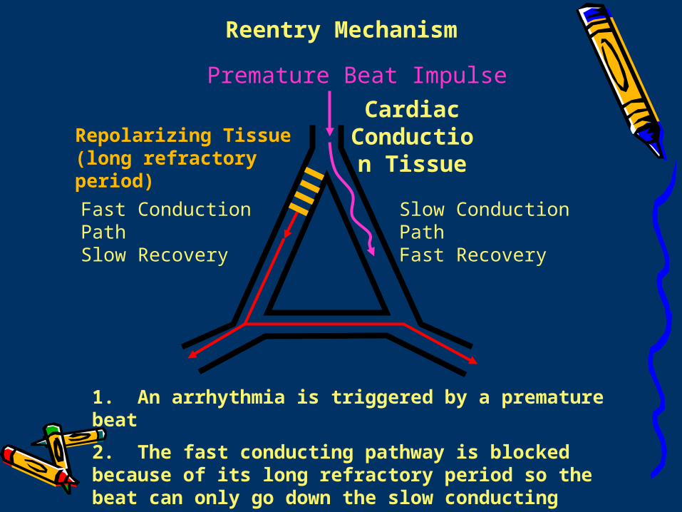
Fast Conduction PathSlow Recovery
Slow Conduction PathFast Recovery
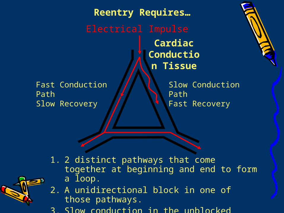
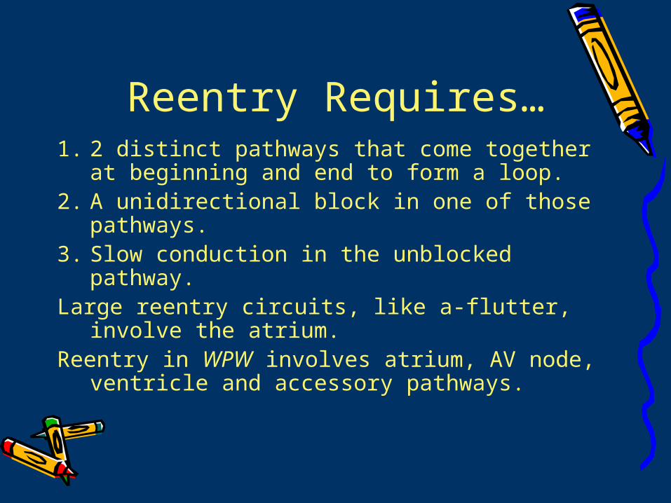
Reentry Requires…
Electrical Impulse
Cardiac Conduction
Tissue
1. 2 distinct pathways that come together at beginning and end to form a loop.
2. A unidirectional block in one of those pathways.
3. Slow conduction in the unblocked pathway.
Fast Conduction PathSlow Recovery
Slow Conduction PathFast Recovery
Premature Beat Impulse
Cardiac Conduction
Tissue
1. An arrhythmia is triggered by a premature beat
2. The fast conducting pathway is blocked because of its long refractory period so the beat can only go down the slow conducting pathway
Repolarizing Tissue (long refractory period)
Reentry Mechanism
3. The wave of excitation from the premature beat arrives at the distal end of the fast conducting pathway, which has now recovered and therefore travels retrogradely (backwards) up the fast pathway
Fast Conduction PathSlow Recovery
Slow Conduction PathFast Recovery
Cardiac Conduction
Tissue
Reentry Mechanism
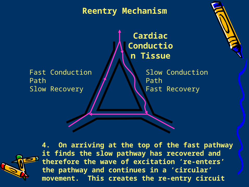
4. On arriving at the top of the fast pathway it finds the slow pathway has recovered and therefore the wave of excitation ‘re-enters’ the pathway and continues in a ‘circular’ movement. This creates the re-entry circuit
Fast Conduction PathSlow Recovery
Slow Conduction PathFast Recovery
Cardiac Conduction
Tissue
Reentry Mechanism

Atrial Reentry• atrial tachycardia• atrial fibrillation• atrial flutter
Atrio-Ventricular Reentry• WPW• SVT
Ventricular Re-entry• ventricular tachycardia
AV Nodal Reentry•SVT
Reentry Circuits
SA Node
Reentry Requires…1. 2 distinct pathways that come together at
beginning and end to form a loop. 2. A unidirectional block in one of those
pathways. 3. Slow conduction in the unblocked pathway. Large reentry circuits, like a-flutter, involve the
atrium. Reentry in WPW involves atrium, AV node,
ventricle and accessory pathways.
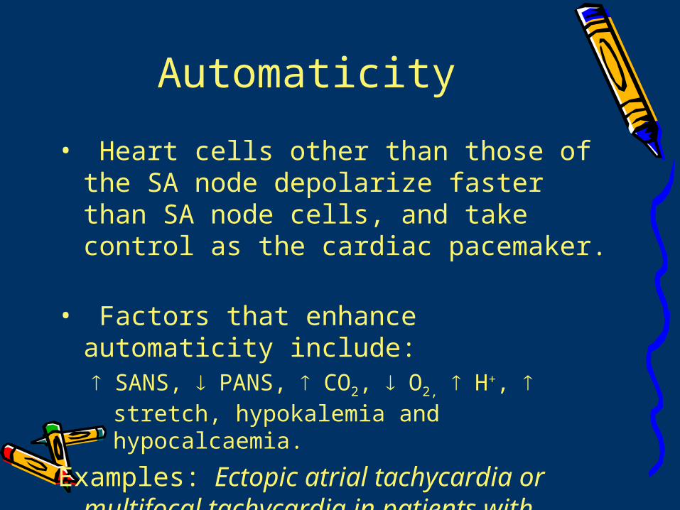
Automaticity
• Heart cells other than those of the SA node depolarize faster than SA node cells, and take control as the cardiac pacemaker.
• Factors that enhance automaticity include: SANS, PANS, CO2, O2, H+, stretch,
hypokalemia and hypocalcaemia.
Examples: Ectopic atrial tachycardia or multifocal tachycardia in patients with chronic lung disease OR ventricular ectopy after MI
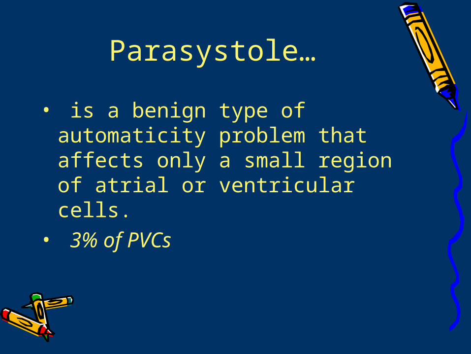
Parasystole…
• is a benign type of automaticity problem that affects only a small region of atrial or ventricular cells.
• 3% of PVCs
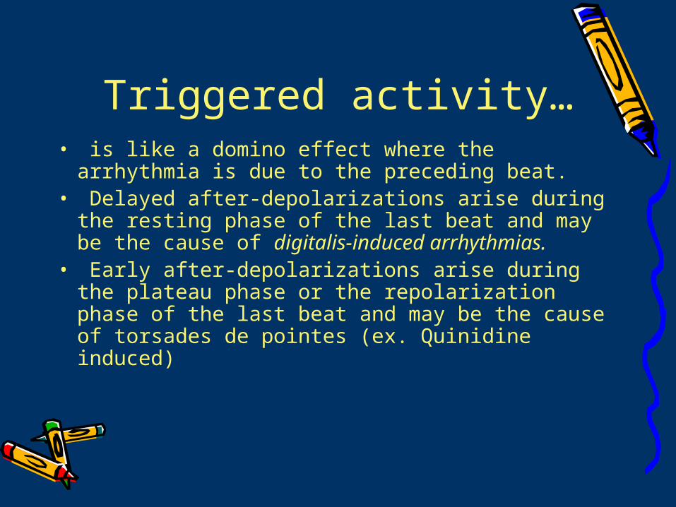
Triggered activity…• is like a domino effect where the
arrhythmia is due to the preceding beat. • Delayed after-depolarizations arise during
the resting phase of the last beat and may be the cause of digitalis-induced arrhythmias.
• Early after-depolarizations arise during the plateau phase or the repolarization phase of the last beat and may be the cause of torsades de pointes (ex. Quinidine induced)
Diagnosis…Diagnosis…Diagnosis…Diagnosis…
What tools to use and when to What tools to use and when to use it…use it…
Event Monitors• Holter monitoring: Document
symptomatic and asymptomatic arrhythmias over 24-48 hours. Can also evaluate treatment effectiveness in a-fib, pacemaker effectiveness and identify silent MIs.
• Trans-telephonic event recording: patient either wears monitor for several days or attaches it during symptomatic events and an ECG is recorded and transmitted for evaluation via telephone. Only 20% are positive, but still helpful.
Exercise testing• Symptoms only appear or worsen with
exercise. • Also used to evaluate medication
effectiveness (esp. flecanide & propafenone)
You can assess SA node function with exercise testing.
Mobitz 1 (Wenkebach) is blockage at the AV node, so catecholamines from exercise actually help!
Mobitz 2 is blockage at bundle of His, so it worsens as catecholamines from exercise increase AV node conduction, thus prognosis is worse.
*PVCs occur in 10% without and 60% of patients with CAD. *PVCs DO NOT predict severity of CAD (neither for nor against)!
Signal Averaged ECG
• Used only in people post MI to evaluate risk for v-fib or v-tach.
• Damage around the infarct is variable, so this measures late potentials (low-signal, delayed action potentials) as they pass through damaged areas.
• Positive predictive value is 25%-50% but negative predictive value is 90%-95%, thus if test is negative, patient is at low risk.
Electrophysiologic Testing…
• Catheters are placed in RA, AV node, Bundle of HIS, right ventricle, and coronary sinus (to monitor LA and LV).
• Used to evaluate cardiogenic syncope of unknown origin, symptomatic SVT, symptomatic WPW, and sustained v-tach.
*Ablative therapy is beneficial in AV node reentry, WPW, atrial tachycardia, a-flutter, and some v-tach. Complication is 1%
BradyarrhythmiasBradyarrhythmiasBradyarrhythmiasBradyarrhythmias
The slow pokes (HR<60)…The slow pokes (HR<60)…
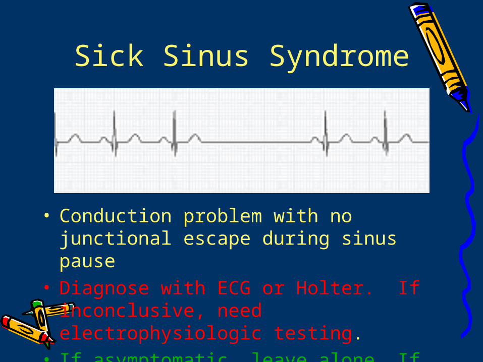
Sick Sinus Syndrome
• Conduction problem with no junctional escape during sinus pause
• Diagnose with ECG or Holter. If inconclusive, need electrophysiologic testing.
• If asymptomatic, leave alone. If symptomatic, needs pacemaker.
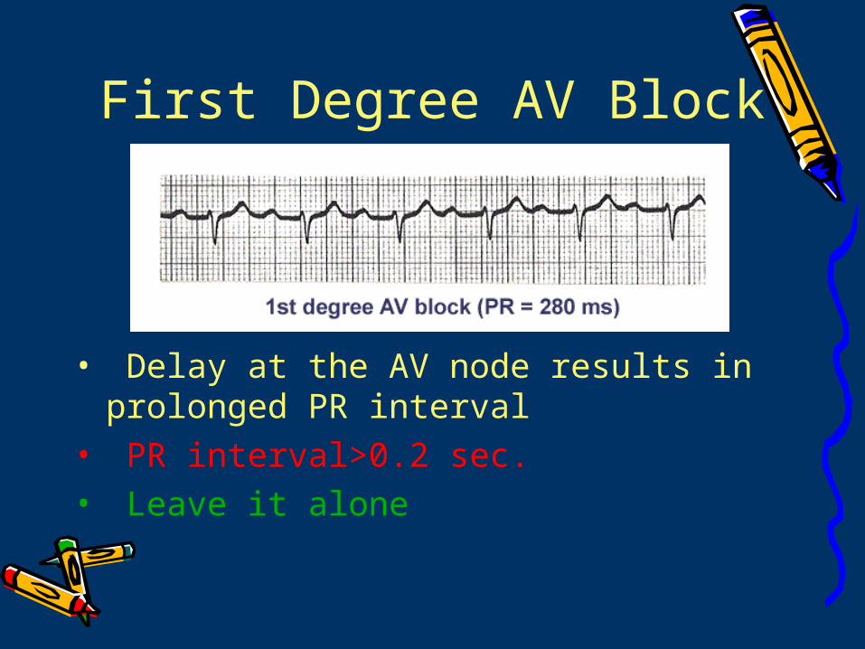
First Degree AV Block
• Delay at the AV node results in prolonged PR interval
• PR interval>0.2 sec.• Leave it alone
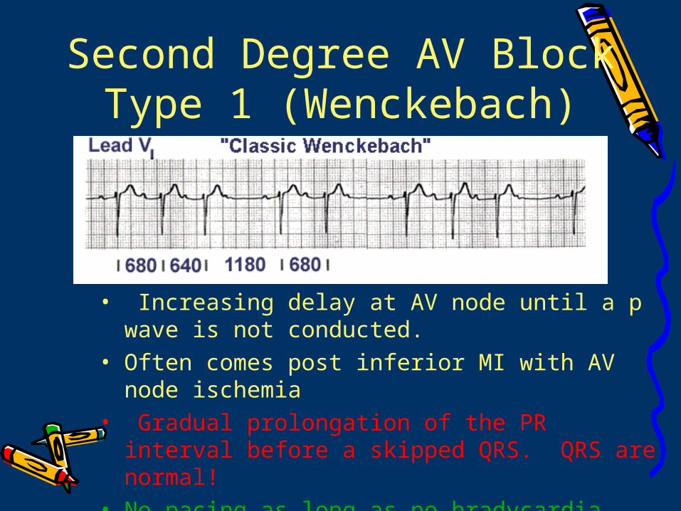
Second Degree AV Block Type 1 (Wenckebach)
• Increasing delay at AV node until a p wave is not conducted.
• Often comes post inferior MI with AV node ischemia
• Gradual prolongation of the PR interval before a skipped QRS. QRS are normal!
• No pacing as long as no bradycardia.
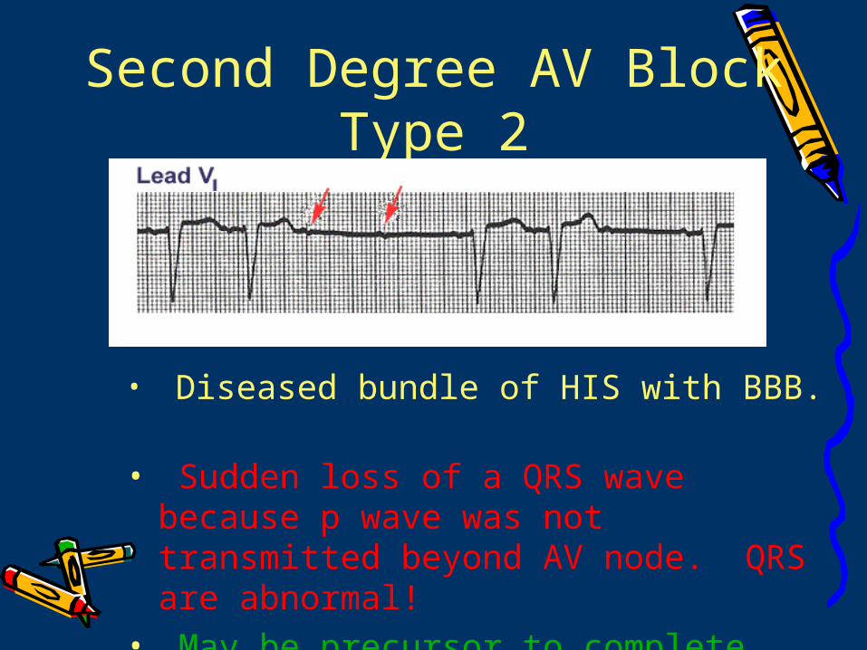
Second Degree AV Block Type 2
• Diseased bundle of HIS with BBB. • Sudden loss of a QRS wave because p
wave was not transmitted beyond AV node. QRS are abnormal!
• May be precursor to complete heart block and needs pacing.
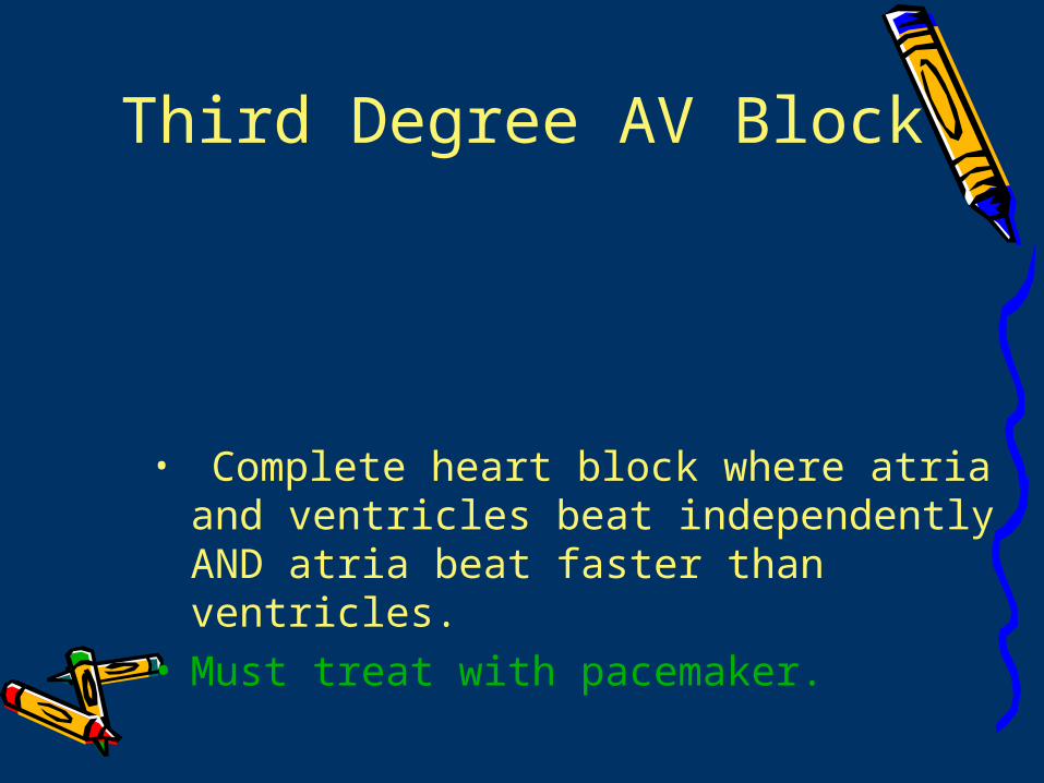
Third Degree AV Block
• Complete heart block where atria and ventricles beat independently AND atria beat faster than ventricles.
• Must treat with pacemaker.
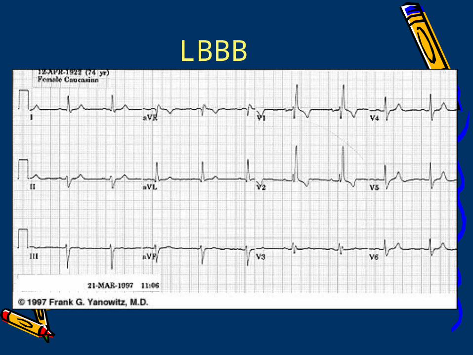
LBBB
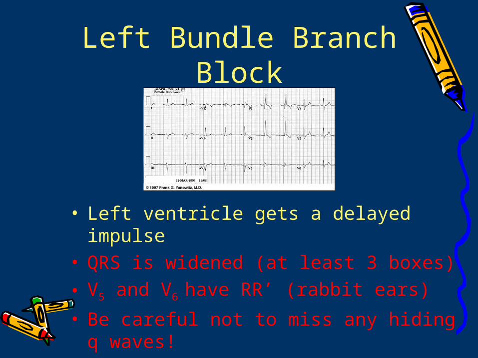
Left Bundle Branch Block
• Left ventricle gets a delayed impulse• QRS is widened (at least 3 boxes)
• V5 and V6 have RR’ (rabbit ears)
• Be careful not to miss any hiding q waves!
• Pacemaker if syncope occurs
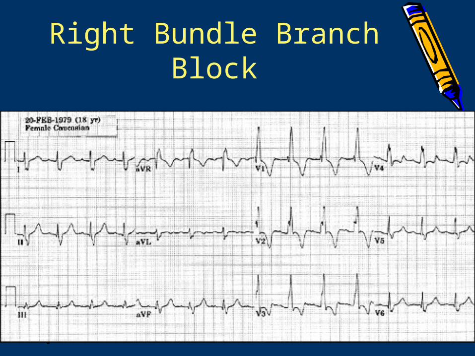
Right Bundle Branch Block
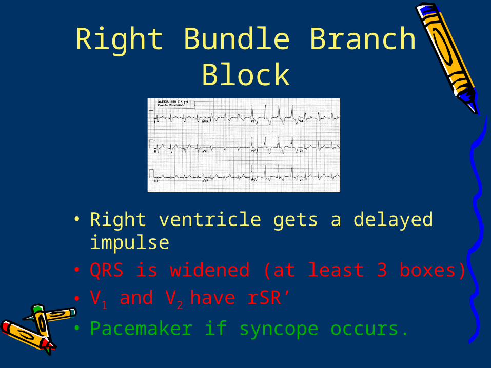
Right Bundle Branch Block
• Right ventricle gets a delayed impulse• QRS is widened (at least 3 boxes)
• V1 and V2 have rSR’
• Pacemaker if syncope occurs.
Bifascicular Block
• RBBB plus LABB OR RBBB plus LPBB
• QRS is widened (at least 3 boxes)
• V5 and V6 have RR’ (rabbit ears)
• V1 and V2 have rSR’
• Pacemaker if syncope occurs
TachyarrhythmiasTachyarrhythmiasTachyarrhythmiasTachyarrhythmias
The speed demons…(HR >100)The speed demons…(HR >100)
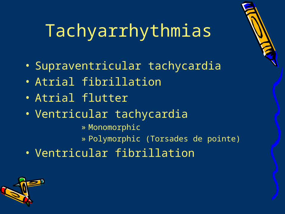
Tachyarrhythmias
• Supraventricular tachycardia• Atrial fibrillation• Atrial flutter• Ventricular tachycardia
» Monomorphic» Polymorphic (Torsades de pointe)
• Ventricular fibrillation
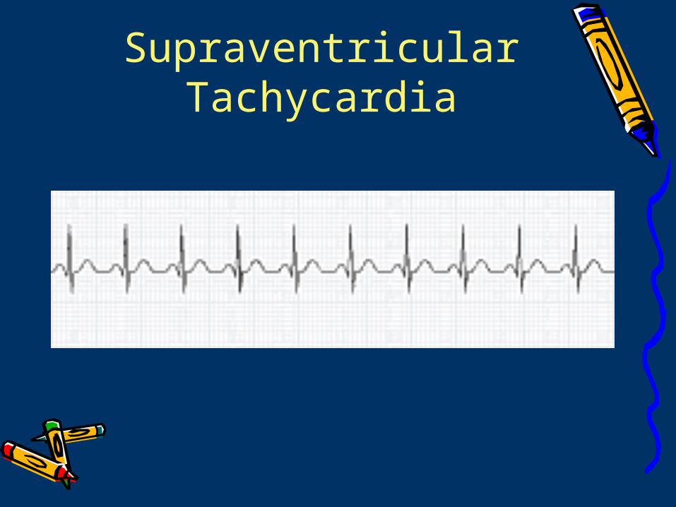
Supraventricular Tachycardia
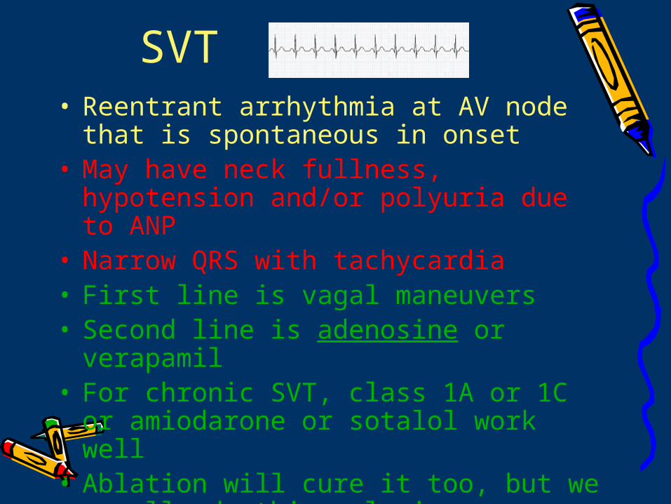
SVT• Reentrant arrhythmia at AV node that is
spontaneous in onset • May have neck fullness, hypotension
and/or polyuria due to ANP• Narrow QRS with tachycardia• First line is vagal maneuvers • Second line is adenosine or verapamil• For chronic SVT, class 1A or 1C or
amiodarone or sotalol work well• Ablation will cure it too, but we usually
do this only in young patients
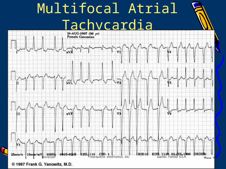
Multifocal Atrial Tachycardia
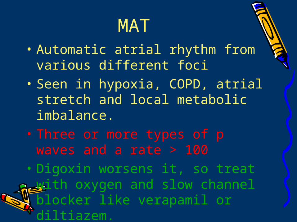
MAT• Automatic atrial rhythm from
various different foci • Seen in hypoxia, COPD, atrial
stretch and local metabolic imbalance.
• Three or more types of p waves and a rate > 100
• Digoxin worsens it, so treat with oxygen and slow channel blocker like verapamil or diltiazem.
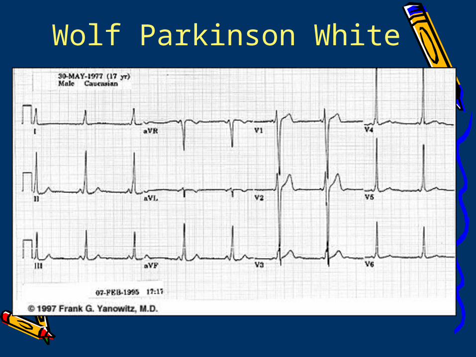
Wolf Parkinson White
WPW
• Ventricles receive partial signal normally and partially through accessory pathway
• Symptomatic tachycardia, short PR interval (<0.12), a delta wave and prolonged QRS (>0.12)
• Electrophysiologic testing helps to identify the reentry pathway and location of the accessory pathway
WPW Because WPW has both normal conduction
through the AV node and accessory pathway conduction that bypasses the AV node, a-fib can happen via the accessory pathway
Inhibition of the AV node will end up in worsening the a-fib because none of the signals are slowed down by the AV node before hitting the ventricle.
* Do not use any meds that will slow AV node conduction, ie digoxin, beta-blockers, adenosine or calcium channel blockers.
* The best choice is procainamide as it slows the accessory pathway. *If patient becomes hypotensive, cardiovert immediately!
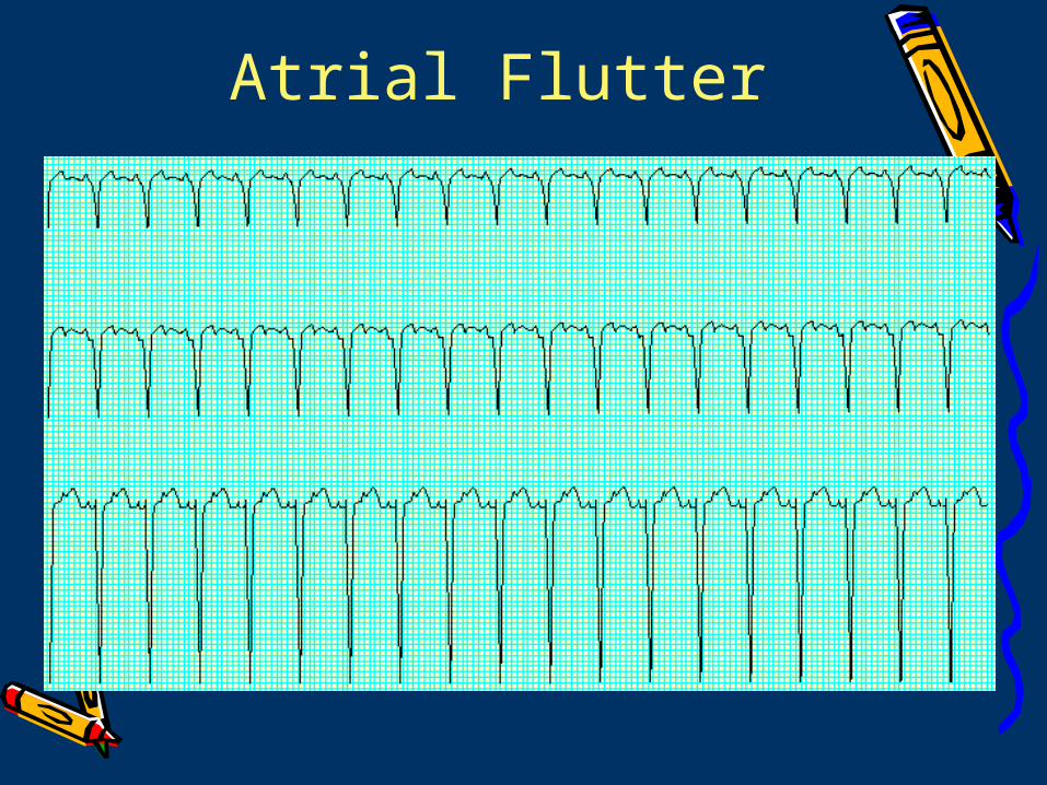
Atrial Flutter
Atrial Flutter• Atrial activity of 240-320 with sawtooth
pattern. Usually a 2:1 conduction pattern; if it is 3:1 or higher, there is AV node damage
• Treatment is to slow AV node conduction with amiodarone, propafenone or sotalol
• DC cardiovert if <48 hours or unstable• You can also ablate the reentry
pathway within the atrium between the tricuspid and the IVC.
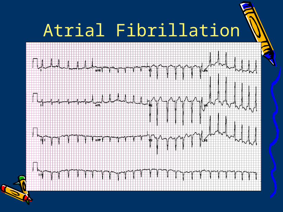
Atrial Fibrillation
A-Fib
• Can be due to HTN, cardiomyopathy, valvular heart desease, sick sinus, WPW, thyrotoxicosis or ETOH
• Therapy is either rate control via slowing AV node conduction with stroke prophylaxis or rhythm control
Rate control Beta-blockers
Continuation after CABG may prevent a-fib Good for hyperthyroid or post-MI patients with
a-fibCarvedilol decreases mortality in patients with
CHFEsmolol is good for acute management
Digoxin actually increases vagal tone, thus indirectly slowing AV node conduction. But it is used essentially only in patients with LV dysfunction because it’s inotropic.
Rate control
Calcium Channel Blockers Nondihydropyridines (verapamil or
dilitiazem) block AV node conduction but also have negative inotropy, so don’t use in CHF.
Dihydropyridines (nifedipine, amlodipine, felodipine) have no effect on AV node conduction
Adenosine is too short acting to be of any use in a-fib
Last choice is AV node ablation and permanent pacing
Rhythm control
Rhythm control does not decrease thromboembolic risk and may be proarrhythmic
Class 1A (quinidine, procainamide, disopyramide) slows conduction through HIS can cause torsades de pointes during conversion. They also enhance AV node conduction, so they should be used only after rate is controlled
Class 1B (lidocaine, meilitine, tocainide) are useless for a-fib
Class 1C (propafenone, and flecainide) slow conduction through HIS are good first choice.
• Amiodarone is good if patient is post-MI or has systolic dysfunction.
Cardioversion for A-Fib
• Cardiovert if symptomatic• Patients with a-fib for more than 2
days should be receive 3 weeks of anticoagulation before electrical cardioversion.
• Give coumadin for 4 weeks after cardioversion
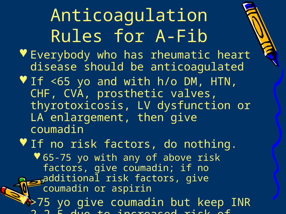
Anticoagulation Rules for A-Fib
Everybody who has rheumatic heart disease should be anticoagulated
If <65 yo and with h/o DM, HTN, CHF, CVA, prosthetic valves, thyrotoxicosis, LV dysfunction or LA enlargement, then give coumadin
If no risk factors, do nothing. 65-75 yo with any of above risk factors,
give coumadin; if no additional risk factors, give coumadin or aspirin
>75 yo give coumadin but keep INR 2-2.5 due to increased risk of bleed
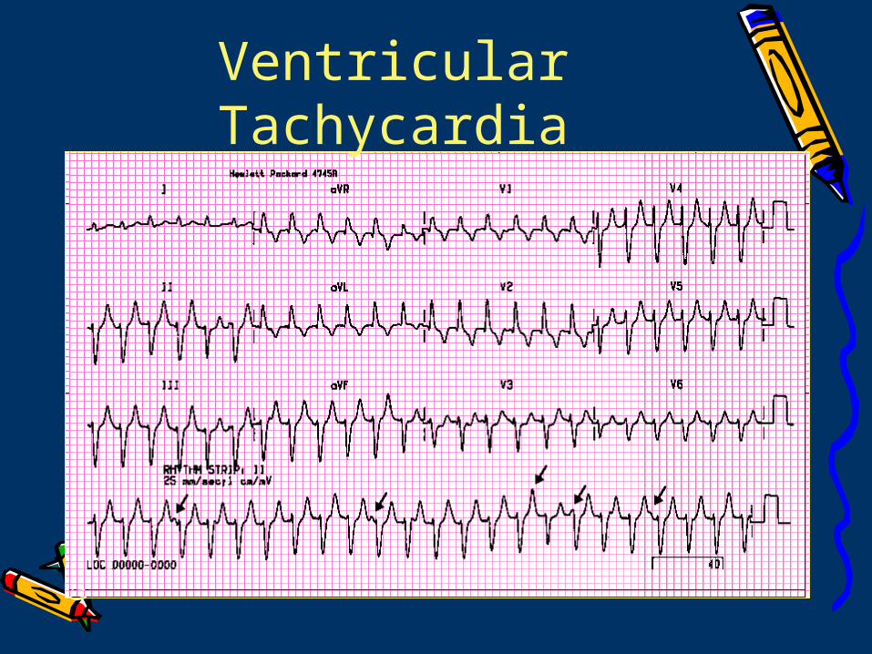
Ventricular Tachycardia
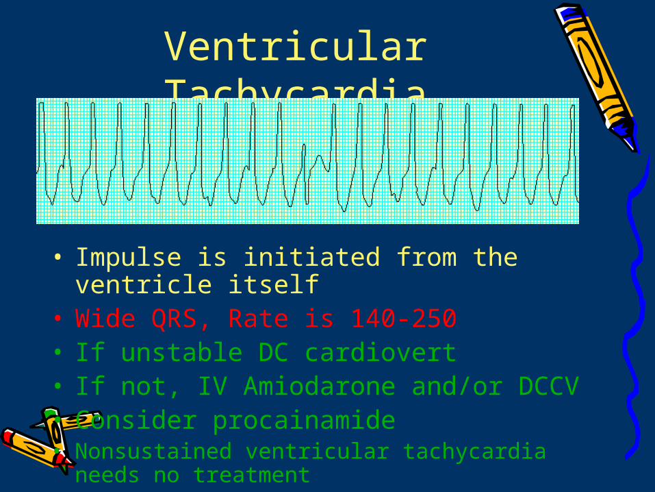
• Impulse is initiated from the ventricle itself
• Wide QRS, Rate is 140-250• If unstable DC cardiovert• If not, IV Amiodarone and/or DCCV• Consider procainamide• Nonsustained ventricular tachycardia needs
no treatment
Ventricular Tachycardia
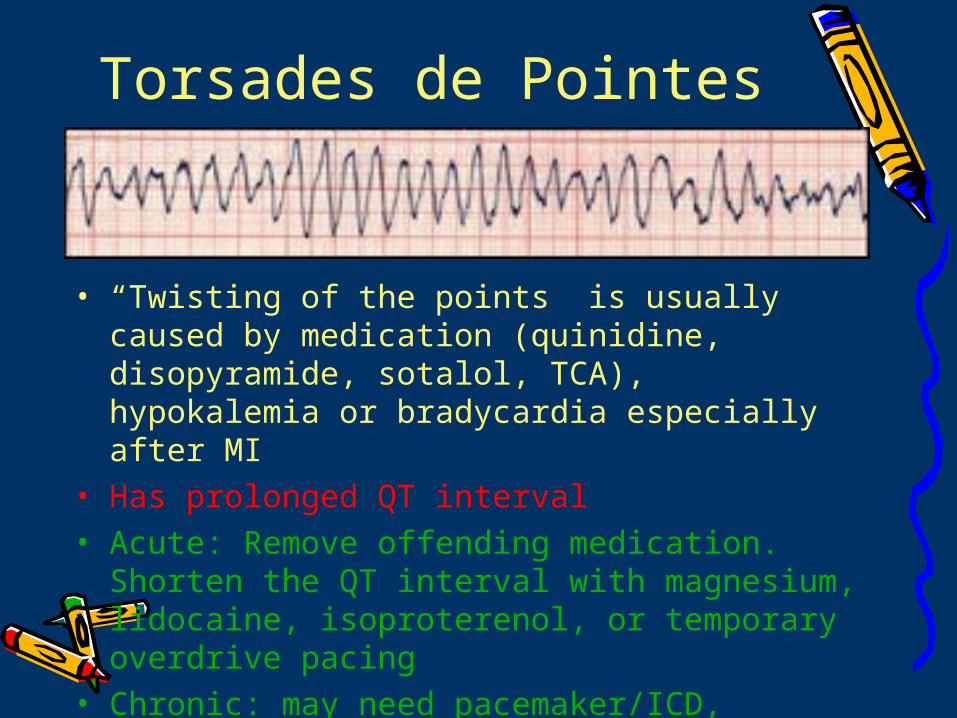
Torsades de Pointes
• “Twisting of the points” is usually caused by medication (quinidine, disopyramide, sotalol, TCA), hypokalemia or bradycardia especially after MI
• Has prolonged QT interval• Acute: Remove offending medication. Shorten
the QT interval with magnesium, lidocaine, isoproterenol, or temporary overdrive pacing
• Chronic: may need pacemaker/ICD, amiodarone, beta-blockers
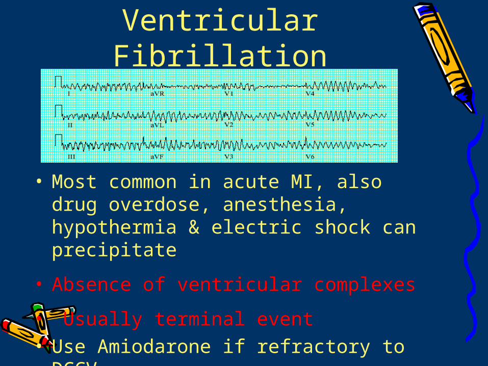
Ventricular Fibrillation
• Most common in acute MI, also drug overdose, anesthesia, hypothermia & electric shock can precipitate
• Absence of ventricular complexes
• Usually terminal event• Use Amiodarone if refractory to DCCV.

TreatmentTreatmentTreatmentTreatment
Here comes the fun part!Here comes the fun part!
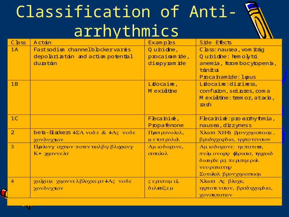
Classification of Anti-arrhythmics
Class Action Examples Side Effects1A Fast sodium channel blocker varies
depolarization and action potentialduration
Quinidine,procainamide,disopyramide
Class: nausea, vomitingQuinidine: hemolyticanemia, thrombocytopenia,tinnitusProcainamide: lupus
1B Lidocaine,Mexiletine
Lidocaine: dizziness,confusion, seizures, comaMexiletine: tremor, ataxia,rash
1C Flecainide,Propafenone
Flecainide: pro-arrhythmia,nausea, dizzyness
2 beta-blockers ↓SA nod e & ↓A V nodeconduction
Propranolo ,lmetoprolol
Class: CHF, bronchospasm,bradycardia, hypotension
3 Prolon g actio n potential b y blocking+K channels
Amiodaron ,esotalol
Amiodaron :e hepatiti ,spulmonar y fibrosi ,s thyroiddisorders, peripheralneuropathySotalol: bronchospasm
4 calciu mchannel blockers ↓A V nodeconduction
Verapamil,dilitiazem
Class: A V block,hypotension, bradycardi ,aconstipation
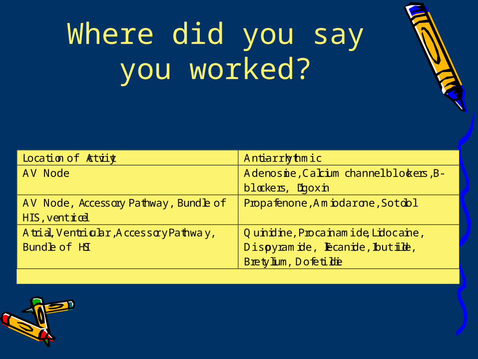
Where did you say you worked?
Location of Activity Anti-arrhythmicAV Node Adenosine, Calcium channel blockers, B-
blockers, DigoxinAV Node, Accessory Pathway, Bundle ofHIS, ventricle
Propafenone, Amiodarone, Sotolol
Atrial, Ventricular, Accessory Pathway,Bundle of HIS
Quinidine, Procainamide, Lidocaine,Disopyramide, Flecanide, Ibutilide,Bretylium, Dofetilide
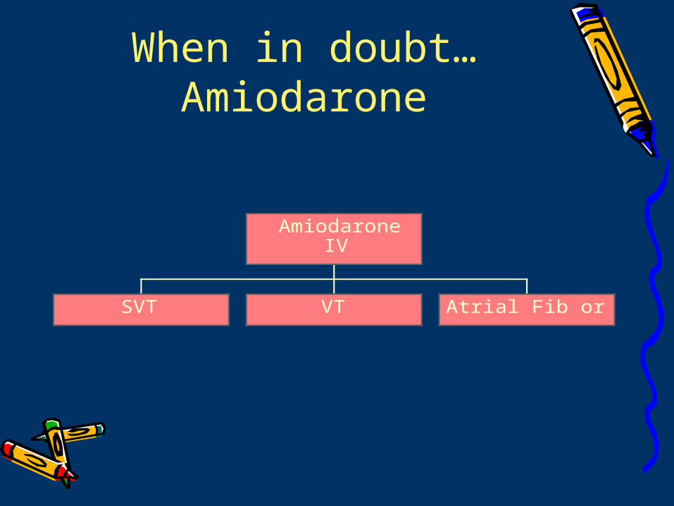
When in doubt…Amiodarone
SVT VT Atrial Fib or flutter
AmiodaroneIV
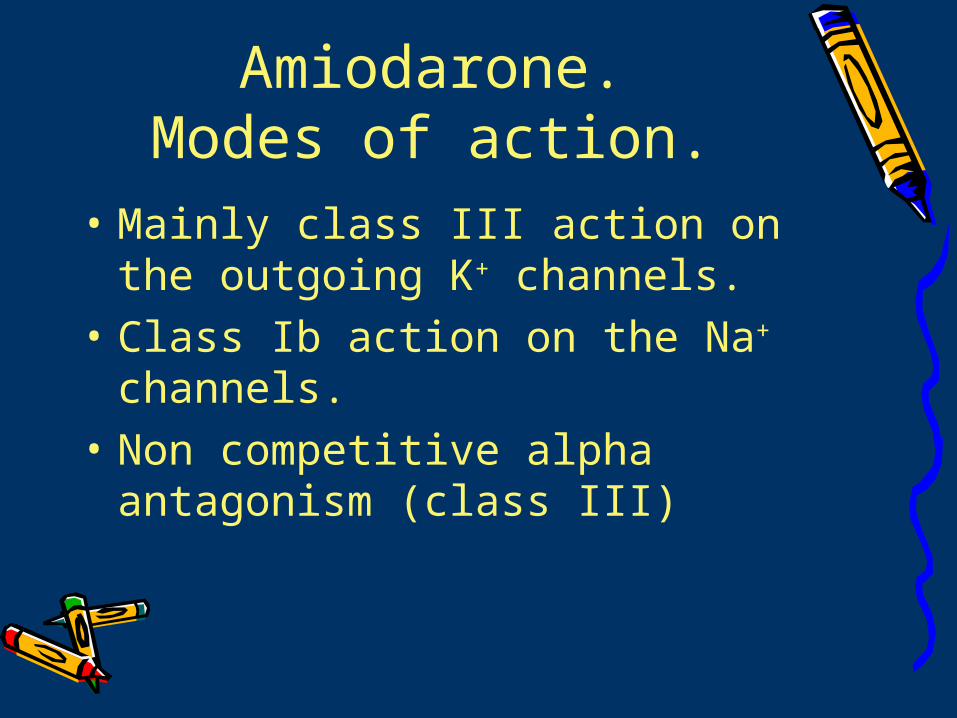
Amiodarone.Modes of action.
• Mainly class III action on the outgoing K+ channels.
• Class Ib action on the Na+ channels.
• Non competitive alpha antagonism (class III)
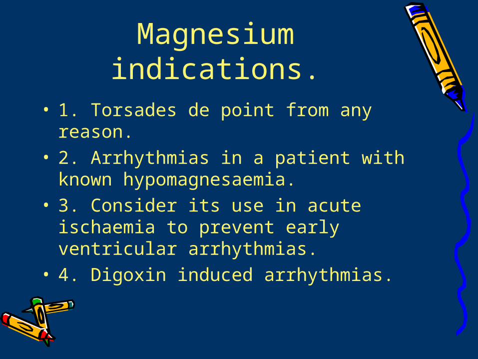
Magnesium indications.
• 1. Torsades de point from any reason.• 2. Arrhythmias in a patient with known
hypomagnesaemia.• 3. Consider its use in acute ischaemia
to prevent early ventricular arrhythmias.
• 4. Digoxin induced arrhythmias.
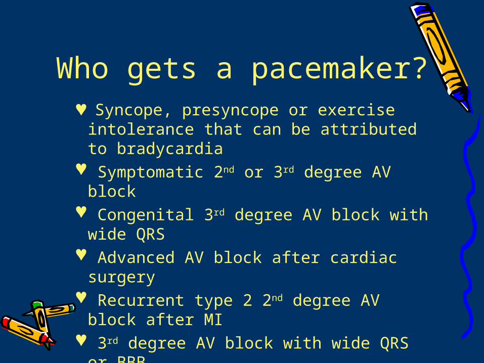
Who gets a pacemaker? Syncope, presyncope or exercise
intolerance that can be attributed to bradycardia
Symptomatic 2nd or 3rd degree AV block Congenital 3rd degree AV block with wide
QRS Advanced AV block after cardiac surgery Recurrent type 2 2nd degree AV block
after MI 3rd degree AV block with wide QRS or
BBB.
QUESTIONSQUESTIONSQUESTIONSQUESTIONS