Arranging the elements of the potassium channel: the T1 domain occludes the cytoplasmic face of the...
7
BIOPHYSICS LETTER Anurag Varshney Æ Baron Chanda Æ M. K. Mathew Arranging the elements of the potassium channel: the T1 domain occludes the cytoplasmic face of the channel Received: 10 July 2003 / Revised: 13 September 2003 / Accepted: 22 October 2003 / Published online: 11 December 2003 Ó EBSA 2003 Abstract The voltage-gated potassium channel is cur- rently one of the few membrane proteins where func- tional roles have been mapped onto specific segments of sequence. Although high-resolution structures of the transmembrane portions of three bacterial potassium channels, the tetramerization domain and the cytoplas- mic ‘‘ball’’ are available, their relative spatial arrange- ment in mammalian channels remains a matter of ongoing debate. Cryo-electron microscopic images of the six transmembrane voltage-gated Kv channel have been reconstructed at up to 18 A ˚ resolution, revealing that the T1 domain tetramerizes and is suspended below the transmembrane segments. However, the resolution of these images is insufficient to reveal the location of the third piece of the puzzle, the inactivating ball domain. We have used the aberrant interactions observed in a series of chimæric channels to establish that an assem- bled T1 domain restricts access to the cytoplasmic face of the channel, suggesting that the N-terminal ‘‘ball and chain’’ may be confined in the space between the T1 domain and the transmembrane portion of the channel. Keywords Activation Æ N-terminus Æ Potassium channels Æ Structure Æ Voltage dependence Introduction The physiology of a neuronal cell is dictated in no small part by the complement of ion channels it expresses, particularly the voltage-gated potassium channels that sculpt the falling phase of the action potential. These are tetrameric proteins, each subunit of which has six membrane-embedded helices and a re-entrant P-loop that lines the aqueous pore. The key elements whose functions have been identified are the inactivation gate, the voltage sensor and the pore lining loop. Rapidly inactivating channels have a ball-like structure at the N-terminus of their cytoplasmic domains, which physi- cally occludes open channels, causing inactivation by the ‘‘ball and chain’’ mechanism (Zagotta et al. 1990). The fourth transmembrane segment (S4) is positively charged and has been shown to move in response to changes in transmembrane electrical potential (for re- view, see Bezanilla 2000), thus serving as a voltage sen- sor. The last two helices and the included loop contribute to the lining of the aqueous pore and carry the elements of ionic selectivity (Sather et al. 1994; MacKinnon 1995). Movement of the last transmem- brane helices S5 and S6 has been associated with channel opening and closing (Yellen 1998). Between the first transmembrane helix and the N-terminal ball is a seg- ment called the T1 domain, which is responsible for restricting the promiscuity of subunit–subunit interac- tions in the formation of the functional tetramer (Shen and Pfafinger 1995; Xu et al. 1995). Functional roles have been mapped onto specific segments of the sequence of K + channels by employing a combination of molecular biology and electrophysi- ology (Yellen 1998). However, a detailed understanding of the mechanism by which these proteins function requires a knowledge of their three-dimensional archi- tecture. The structures of three major segments of the a-subunit of the K + channel have been solved at atomic resolution: the central ion-conducting pore in the form of four bacterial potassium channels (Doyle et al. 1998; Eur Biophys J (2004) 33: 370–376 DOI 10.1007/s00249-003-0372-1 A. Varshney Æ B. Chanda Æ M. K. Mathew (&) National Centre for Biological Sciences, TIFR, UAS-GKVK Campus, 560 065 Bangalore, India E-mail: [email protected] Tel.: +91-80-3636421x3270 Fax: +91-80-3636662/675 Present address: A. Varshney Department of Pharmacology, Yale University School of Medicine, New Haven, CT, USA B. Chanda Department of Physiology, David Geffen School of Medicine at UCLA, Los Angeles, CA, USA
-
Upload
anurag-varshney -
Category
Documents
-
view
212 -
download
0
Transcript of Arranging the elements of the potassium channel: the T1 domain occludes the cytoplasmic face of the...

BIOPHYSICS LETTER
Anurag Varshney Æ Baron Chanda Æ M. K. Mathew
Arranging the elements of the potassium channel:the T1 domain occludes the cytoplasmic face of the channel
Received: 10 July 2003 / Revised: 13 September 2003 / Accepted: 22 October 2003 / Published online: 11 December 2003� EBSA 2003
Abstract The voltage-gated potassium channel is cur-rently one of the few membrane proteins where func-tional roles have been mapped onto specific segments ofsequence. Although high-resolution structures of thetransmembrane portions of three bacterial potassiumchannels, the tetramerization domain and the cytoplas-mic ‘‘ball’’ are available, their relative spatial arrange-ment in mammalian channels remains a matter ofongoing debate. Cryo-electron microscopic images ofthe six transmembrane voltage-gated Kv channel havebeen reconstructed at up to 18 A resolution, revealingthat the T1 domain tetramerizes and is suspended belowthe transmembrane segments. However, the resolutionof these images is insufficient to reveal the location of thethird piece of the puzzle, the inactivating ball domain.We have used the aberrant interactions observed in aseries of chimæric channels to establish that an assem-bled T1 domain restricts access to the cytoplasmic faceof the channel, suggesting that the N-terminal ‘‘ball andchain’’ may be confined in the space between the T1domain and the transmembrane portion of the channel.
Keywords Activation Æ N-terminus Æ Potassiumchannels Æ Structure Æ Voltage dependence
Introduction
The physiology of a neuronal cell is dictated in no smallpart by the complement of ion channels it expresses,particularly the voltage-gated potassium channels thatsculpt the falling phase of the action potential. These aretetrameric proteins, each subunit of which has sixmembrane-embedded helices and a re-entrant P-loopthat lines the aqueous pore. The key elements whosefunctions have been identified are the inactivation gate,the voltage sensor and the pore lining loop. Rapidlyinactivating channels have a ball-like structure at theN-terminus of their cytoplasmic domains, which physi-cally occludes open channels, causing inactivation by the‘‘ball and chain’’ mechanism (Zagotta et al. 1990). Thefourth transmembrane segment (S4) is positivelycharged and has been shown to move in response tochanges in transmembrane electrical potential (for re-view, see Bezanilla 2000), thus serving as a voltage sen-sor. The last two helices and the included loopcontribute to the lining of the aqueous pore and carrythe elements of ionic selectivity (Sather et al. 1994;MacKinnon 1995). Movement of the last transmem-brane helices S5 and S6 has been associated with channelopening and closing (Yellen 1998). Between the firsttransmembrane helix and the N-terminal ball is a seg-ment called the T1 domain, which is responsible forrestricting the promiscuity of subunit–subunit interac-tions in the formation of the functional tetramer (Shenand Pfafinger 1995; Xu et al. 1995).
Functional roles have been mapped onto specificsegments of the sequence of K+ channels by employinga combination of molecular biology and electrophysi-ology (Yellen 1998). However, a detailed understandingof the mechanism by which these proteins functionrequires a knowledge of their three-dimensional archi-tecture. The structures of three major segments of thea-subunit of the K+ channel have been solved at atomicresolution: the central ion-conducting pore in the formof four bacterial potassium channels (Doyle et al. 1998;
Eur Biophys J (2004) 33: 370–376DOI 10.1007/s00249-003-0372-1
A. Varshney Æ B. Chanda Æ M. K. Mathew (&)National Centre for Biological Sciences, TIFR, UAS-GKVKCampus, 560 065 Bangalore, IndiaE-mail: [email protected].: +91-80-3636421x3270Fax: +91-80-3636662/675
Present address: A. VarshneyDepartment of Pharmacology,Yale University School of Medicine, New Haven, CT, USA
B. ChandaDepartment of Physiology, David Geffen Schoolof Medicine at UCLA, Los Angeles, CA, USA

Jiang et al. 2002, 2003a; Kuo et al. 2003); the cytoplas-mic tetramerization domain T1 (Kreusch et al. 1998;Bixby et al. 1999) and the N-terminal cytoplasmic ‘‘ball’’structure responsible for rapid N-type inactivation (Antzet al. 1997). The crystal structures of a bacterial voltage-gated K+ channel, KvAP, that have been solvedrecently (Jiang et al. 2003a) provide insight into thepossible architecture of the membrane-embedded por-tion of the protein (see Sigworth 2003 for comments). Inaddition, low-resolution cryo-electron microscopic(cryo-EM) structures of Shaker (25 A) and of mamma-lian Kv1.1 channels (18 A) have been reported (Sokol-ova et al. 2001; Orlova et al. 2003). The organization ofthe cytoplasmic structural elements of channel withrespect to the membrane-embedded portion is still to beelucidated.
Much of the data mapping function onto structurehas been based on domain swaps using Shaker, a Dro-sophila K+ channel where diversity is brought about byalternative splicing. A common exon in all transcriptsencodes a region stretching from the T1 cytoplasmicdomain through almost the entire transmembrane re-gion. Mammalian K+ channel subunits are encoded onseparate genes with high, but not absolute, conservationin this region (Stuhmer et al. 1989; Ramaswami et al.1990). Thus, domain swaps among mammalian K+
channels can generate novel phenotypes by aberrantinteractions not normally observed in nature [seeVarshney and Mathew (2003b) for a review on cytosoliccontrol of channel function]. We have generated achimæric channel, 4N/1, composed of the N-terminalcytoplasmic region of the rapidly inactivating humanKv1.4 and the transmembrane body of the non-inacti-vating hKv1.1. The 4N/1 chimæra mediates inwardcurrents on hyperpolarization (Chanda et al. 1999b),owing to the aberrant interaction of the two portions ofthe channel. We have used the appearance of voltage-dependent inward currents to assay the accessibility ofthe cytoplasmic face of the channel to the ball and chainsegment. We show that the presence of an intact T1domain occludes the transmembrane body of the chan-nel, preventing access to ball and chain peptides in thecytosol. The data suggest that the ball and chain canassemble within the basket formed by the T1 tetramersuspended from the transmembrane embedded portionof the channel.
Methods
Potassium channel cDNAs were cloned from human brain stemand were subcloned into pGEMA (Ramaswami et al. 1990). The4N/1 chimæra was generated by transplanting the first 210 residuesof hKv1.4 onto hKv1.1, as described earlier (Chanda et al. 1999b)and schematically indicated in Fig. 1B. A start site was introducedinto hKv1.1 near the chimæra junction to generate the truncatedconstruct D78N1 (Chanda et al. 1999b). A construct 4BC (D410C4)encoding just the ‘‘ball and chain’’ segment of hKv1.4 and con-sisting of the first 210 residues was generated and cloned down-stream of the T7 promoter in pGEMA (Chanda et al. 1999a)(Fig. 1B). Similarly, The 4N/1T1 and 4Nb/1T1 chimæric channels
were constructed by transplanting the first 183 and 39 residues ofhKv1.4, respectively, onto 480 transmembrane amino acids ofhKv1.1 as described earlier (Varshney and Mathew 2003a) andschematically indicated in insets to Fig. 3A and Fig. 3C. The T1domain of hKv1.1 starts 20 residues downstream from the junctionin these chimæras. All mutagenesis was confirmed by sequencingthe constructs on an ABI PRISM 310 automated sequencer.Methods of Xenopus oocyte retrieval and their maintenance havebeen previously described (Ramaswami et al. 1990; Varshney et al.2002). Capped polyadenylated RNA of all the constructs wasgenerated using T7 RNA polymerase (mMessage mMachine tran-scription kit, Ambion, USA). 46 nL of transcribed RNA (150–300 ng/lL) was injected per oocyte 48 h prior to recording. RNAinjection, two-electrode voltage clamp of Xenopus oocytes and dataanalysis were carried out as described earlier (Ramaswami et al.1990; Varshney et al. 2002). Voltage-dependent activation andinactivation curves are best fits to the Boltzmann functions, asdescribed previously (Ramaswami et al. 1990).
Results and discussion
A series of chimæric and truncated constructs weregenerated using parental channels hKv1.1 and hKv1.4(schematically represented in Fig. 1A). The chimæras4N/1, 4N/1T1 and 4Nb/1T1 consist of N-terminal re-gions of hKv1.4 (the ‘‘ball and chain’’) spliced onto thebody of hKv1.1. Figure 1B presents an alignment of thesequences of hKv1.1, hKv1.4, 4N/1, 4N/1T1, 4BC andD78N1 over the T1 region. The secondary structurestaken by amino acid sequence are indicated against thealignment: five stretches of b strand and four of a heli-ces. The third and fourth b strands are deleted in themaking of the 4N/1 chimæra. It is conceivable that theT1 region of 4N/1 may tetramerize. However, the do-main has 28 charged residues out of 93 and the netcharge over the T1 domain changes from +2 to +4 ingoing from hKv1.1 to the 4N/1 chimæra. This, togetherwith the 10 amino acid deletion in the middle of thechimæric T1, makes it unlikely that it will form a stabletetramer (Minor et al. 2000). Similarly, the T1 residuesremaining in 4BC could lead to an association with thetruncated T1 in D78N1, thus facilitating tetramerizationwhen the two constructs are expressed together. How-ever, given the extent of deletions in both of the con-structs, this association appears extremely unlikely.Nonetheless, the data presented here do not directlyaddress this issue.
In contrast, the transplant site in the 4N/1T1 and4Nb/1T1 chimæras is situated 20 residues upstream ofthe T1 domain (Varshney and Mathew 2003a), whichtherefore have intact T1 domains derived entirely fromhKv1.1 (Fig. 1B). The truncation construct, D78N1, isgenerated by deleting the N-terminal residues of hKv1.1at the splice site in the 4N/1 chimæra. Thus, the D78N1construct consists of the hKv1.1 portion of 4N/1 whichlacks the N-terminal chain of hKv1.1 and contains atruncated T1 domain (Fig. 1A, B). The complementarytruncation construct 4BC encodes the N-terminus regionof hKv1.4 that was transplanted onto the transmem-brane body of hKv1.1 to generate the 4N/1 chimæra(Fig. 1A, B).
371
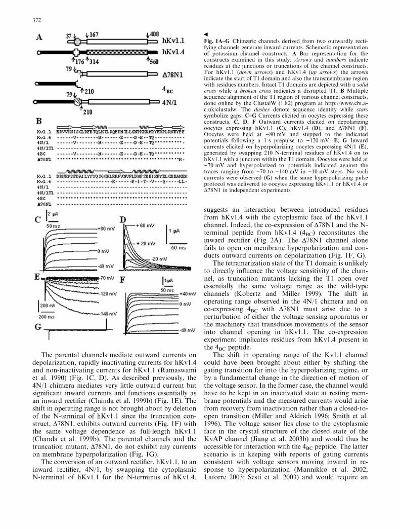
The parental channels mediate outward currents ondepolarization, rapidly inactivating currents for hKv1.4and non-inactivating currents for hKv1.1 (Ramaswamiet al. 1990) (Fig. 1C, D). As described previously, the4N/1 chimæra mediates very little outward current butsignificant inward currents and functions essentially asan inward rectifier (Chanda et al. 1999b) (Fig. 1E). Theshift in operating range is not brought about by deletionof the N-terminal of hKv1.1 since the truncation con-struct, D78N1, exhibits outward currents (Fig. 1F) withthe same voltage dependence as full-length hKv1.1(Chanda et al. 1999b). The parental channels and thetruncation mutant, D78N1, do not exhibit any currentson membrane hyperpolarization (Fig. 1G).
The conversion of an outward rectifier, hKv1.1, to aninward rectifier, 4N/1, by swapping the cytoplasmicN-terminal of hKv1.1 for the N-terminus of hKv1.4,
suggests an interaction between introduced residuesfrom hKv1.4 with the cytoplasmic face of the hKv1.1channel. Indeed, the co-expression of D78N1 and the N-terminal peptide from hKv1.4 (4BC) reconstitutes theinward rectifier (Fig. 2A). The D78N1 channel alonefails to open on membrane hyperpolarization and con-ducts outward currents on depolarization (Fig. 1F, G).
The tetramerization state of the T1 domain is unlikelyto directly influence the voltage sensitivity of the chan-nel, as truncation mutants lacking the T1 open overessentially the same voltage range as the wild-typechannels (Kobertz and Miller 1999). The shift inoperating range observed in the 4N/1 chimera and onco-expressing 4BC with D78N1 must arise due to aperturbation of either the voltage sensing apparatus orthe machinery that transduces movements of the sensorinto channel opening in hKv1.1. The co-expressionexperiment implicates residues from hKv1.4 present inthe 4BC peptide.
The shift in operating range of the Kv1.1 channelcould have been brought about either by shifting thegating transition far into the hyperpolarizing regime, orby a fundamental change in the direction of motion ofthe voltage sensor. In the former case, the channel wouldhave to be kept in an inactivated state at resting mem-brane potentials and the measured currents would arisefrom recovery from inactivation rather than a closed-to-open transition (Miller and Aldrich 1996; Smith et al.1996). The voltage sensor lies close to the cytoplasmicface in the crystal structure of the closed state of theKvAP channel (Jiang et al. 2003b) and would thus beaccessible for interaction with the 4BC peptide. The latterscenario is in keeping with reports of gating currentsconsistent with voltage sensors moving inward in re-sponse to hyperpolarization (Mannikko et al. 2002;Latorre 2003; Sesti et al. 2003) and would require an
Fig. 1A–G Chimæric channels derived from two outwardly recti-fying channels generate inward currents. Schematic representationof potassium channel constructs. A Bar representation for theconstructs examined in this study. Arrows and numbers indicateresidues at the junctions or truncations of the channel constructs.For hKv1.1 (down arrows) and hKv1.4 (up arrows) the arrowsindicate the start of T1 domain and also the transmembrane regionwith residues numbers. Intact T1 domains are depicted with a solidcross while a broken cross indicates a disrupted T1. B Multiplesequence alignment of the T1 region of various channel constructs,done online by the ClustalW (1.82) program at http://www.ebi.a-c.uk/clustalw. The dashes denote sequence identity while starssymbolize gaps. C–G Currents elicited in oocytes expressing theseconstructs. C, D, F Outward currents elicited on depolarizingoocytes expressing hKv1.1 (C), hKv1.4 (D), and D78N1 (F).Oocytes were held at )80 mV and stepped to the indicatedpotentials following a 1 s prepulse to )120 mV. E, G Inwardcurrents elicited on hyperpolarizing oocytes expressing 4N/1 (E),generated by swapping 210 N-terminal residues of hKv1.4 on tohKv1.1 with a junction within the T1 domain. Oocytes were held at)70 mV and hyperpolarized to potentials indicated against thetraces ranging from )70 to )140 mV in )10 mV steps. No suchcurrents were observed (G) when the same hyperpolarizing pulseprotocol was delivered to oocytes expressing hKv1.1 or hKv1.4 orD78N1 in independent experiments
b
372
alternate coupling to movement of the S6 helix. It is notimmediately obvious from the KvAP structure as to howsuch a coupling would be effected.
In the absence of gating current measurements, it isnot possible for us to determine which of these gatingmechanisms is operating in the 4N/1 chimæra. Irre-spective of the mechanism, however, the shift in channeloperating range is diagnostic of the ability of the ‘‘balland chain’’ peptide 4BC to perturb either the sensing orthe transducing machinery and hence its access to thecytoplasmic face of the transmembrane portion ofhKv1.1.
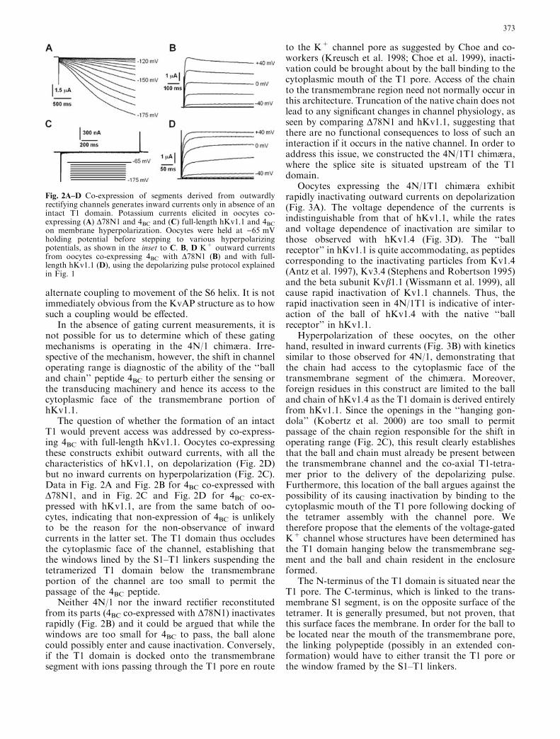
The question of whether the formation of an intactT1 would prevent access was addressed by co-express-ing 4BC with full-length hKv1.1. Oocytes co-expressingthese constructs exhibit outward currents, with all thecharacteristics of hKv1.1, on depolarization (Fig. 2D)but no inward currents on hyperpolarization (Fig. 2C).Data in Fig. 2A and Fig. 2B for 4BC co-expressed withD78N1, and in Fig. 2C and Fig. 2D for 4BC co-ex-pressed with hKv1.1, are from the same batch of oo-cytes, indicating that non-expression of 4BC is unlikelyto be the reason for the non-observance of inwardcurrents in the latter set. The T1 domain thus occludesthe cytoplasmic face of the channel, establishing thatthe windows lined by the S1–T1 linkers suspending thetetramerized T1 domain below the transmembraneportion of the channel are too small to permit thepassage of the 4BC peptide.
Neither 4N/1 nor the inward rectifier reconstitutedfrom its parts (4BC co-expressed with D78N1) inactivatesrapidly (Fig. 2B) and it could be argued that while thewindows are too small for 4BC to pass, the ball alonecould possibly enter and cause inactivation. Conversely,if the T1 domain is docked onto the transmembranesegment with ions passing through the T1 pore en route
to the K+ channel pore as suggested by Choe and co-workers (Kreusch et al. 1998; Choe et al. 1999), inacti-vation could be brought about by the ball binding to thecytoplasmic mouth of the T1 pore. Access of the chainto the transmembrane region need not normally occur inthis architecture. Truncation of the native chain does notlead to any significant changes in channel physiology, asseen by comparing D78N1 and hKv1.1, suggesting thatthere are no functional consequences to loss of such aninteraction if it occurs in the native channel. In order toaddress this issue, we constructed the 4N/1T1 chimæra,where the splice site is situated upstream of the T1domain.
Oocytes expressing the 4N/1T1 chimæra exhibitrapidly inactivating outward currents on depolarization(Fig. 3A). The voltage dependence of the currents isindistinguishable from that of hKv1.1, while the ratesand voltage dependence of inactivation are similar tothose observed with hKv1.4 (Fig. 3D). The ‘‘ballreceptor’’ in hKv1.1 is quite accommodating, as peptidescorresponding to the inactivating particles from Kv1.4(Antz et al. 1997), Kv3.4 (Stephens and Robertson 1995)and the beta subunit Kvb1.1 (Wissmann et al. 1999), allcause rapid inactivation of Kv1.1 channels. Thus, therapid inactivation seen in 4N/1T1 is indicative of inter-action of the ball of hKv1.4 with the native ‘‘ballreceptor’’ in hKv1.1.
Hyperpolarization of these oocytes, on the otherhand, resulted in inward currents (Fig. 3B) with kineticssimilar to those observed for 4N/1, demonstrating thatthe chain had access to the cytoplasmic face of thetransmembrane segment of the chimæra. Moreover,foreign residues in this construct are limited to the balland chain of hKv1.4 as the T1 domain is derived entirelyfrom hKv1.1. Since the openings in the ‘‘hanging gon-dola’’ (Kobertz et al. 2000) are too small to permitpassage of the chain region responsible for the shift inoperating range (Fig. 2C), this result clearly establishesthat the ball and chain must already be present betweenthe transmembrane channel and the co-axial T1-tetra-mer prior to the delivery of the depolarizing pulse.Furthermore, this location of the ball argues against thepossibility of its causing inactivation by binding to thecytoplasmic mouth of the T1 pore following docking ofthe tetramer assembly with the channel pore. Wetherefore propose that the elements of the voltage-gatedK+ channel whose structures have been determined hasthe T1 domain hanging below the transmembrane seg-ment and the ball and chain resident in the enclosureformed.
The N-terminus of the T1 domain is situated near theT1 pore. The C-terminus, which is linked to the trans-membrane S1 segment, is on the opposite surface of thetetramer. It is generally presumed, but not proven, thatthis surface faces the membrane. In order for the ball tobe located near the mouth of the transmembrane pore,the linking polypeptide (possibly in an extended con-formation) would have to either transit the T1 pore orthe window framed by the S1–T1 linkers.
Fig. 2A–D Co-expression of segments derived from outwardlyrectifying channels generates inward currents only in absence of anintact T1 domain. Potassium currents elicited in oocytes co-expressing (A) D78N1 and 4BC and (C) full-length hKv1.1 and 4BCon membrane hyperpolarization. Oocytes were held at )65 mVholding potential before stepping to various hyperpolarizingpotentials, as shown in the inset to C. B, D K+ outward currentsfrom oocytes co-expressing 4BC with D78N1 (B) and with full-length hKv1.1 (D), using the depolarizing pulse protocol explainedin Fig. 1
373

The aberrant interactions which bring about the shiftin operating range of the chimæra perturbs the structureof the channel globally as the reversal potential of thechimæra is shifted from close to )90 mV for hKv1.1 to)35 mV in the chimæra, indicative of reduced selectivityfor K+ (Chanda et al. 1999a). However, the channel isstill relatively selective for K+, as the reversal potentialfor Xenopus oocytes equally selective for Na+ and K+ isestimated to be about +2 mV in the ND96 bathingmedium. Toxin sensitivity of the chimæra is also affected
as IC50 for dendrotoxin (DTX) is 11 nM for the inwardcurrents of 4N/1T1 chimæra (Varshney and Mathew2003a) versus 3 nM for hKv1.1 (Hopkins et al. 1999).However, since DTX does not affect other Kv channelseven at the micromolar concentration range (Hopkinset al. 1999), it may be concluded that the structuralperturbation, while observable, is small.
In order to narrow down the portion of the trans-planted hKv1.4 ball and chain responsible for the shift inoperating range, we have constructed another chimæricchannel, the 4Nb/1T1 chimæra, where the ball is at-tached to the T1 domain by a linker of only 22 residues(144 residues deleted), as opposed to 166 residues in 4N/1T1 (Fig. 3C, inset). Oocytes expressing this constructexhibit rapidly inactivating outward currents on depo-larization (Fig. 3C) but no inward currents on hyper-polarization (Fig. 3C, inset). The voltage dependence ofboth activation and inactivation are similar to thoseobserved for 4N/1T1 (Fig. 3D). The absence of inwardcurrents in the 4Nb/1T1 construct suggests that theresidues involved in the aberrant interaction lie either inthe deleted portion of the chain, or that such a largedeletion restricts the remaining residues from participa-tion in these interactions. Identification of the mem-brane-embedded partners in the interaction couldprovide a handle to identifying the machinery underly-ing the transduction of voltage sensing to channelopening. More immediately germane is the rapid inac-tivation of the outward currents. This would suggestthat the distance from the anchor site on the T1 domainto the mouth of the channel pore is comparable to or lessthan the length of an extended 22-residue polypeptide.
Fig. 3A–D Deletion of linker between T1 domain and inactivatingball generates outwardly rectifying channels with intact inactiva-tion properties. A Rapidly inactivating outward currents throughthe 4N/1T1 chimæra (inset shows a schematic of the chimæra), inresponse to depolarizing pulses. No hyperpolarizing prepulse wasused prior to depolarizing to potentials indicated against the traces;instead, an inter-episode time of 10 s was used to ensure completerecovery from inactivation. B Inward currents through 4N/1T1channels. Oocytes expressing the 4N/1T1 chimæra were held at)65 mV and stepped to hyperpolarizing potentials from )65 to)175 mV in 10 mV steps, as shown in the inset to Fig. 2B. CCurrents through 4Nb/1T1 chimæra (schematically shown in inseti), Currents elicited on depolarizing oocytes from a holdingpotential of )80 mV to the indicated depolarizing potentialsfollowing a 1 s hyperpolarizing prepulse of )120 mV. Inset ii:currents observed on hyperpolarizing to a series of potentials from)65 mV to )175 mV in )10 mV steps, from a holding potential of)65 mV. D Boltzmann function fits of the voltage dependence ofactivation (filled symbols) and inactivation (open symbols) of 4Nb/1T1 and 4N/1T1 channels compared to those of hKv1.1 andhKv1.4. The V1/2 values used for the activation fits (black lines) are)29.2 mV (hKv1.1, filled squares), )30.5 mV (4N/1T1, filled uptriangles) and )28.3 mV (4Nb/1T1, filled down triangles); V1/2 ofinactivation (gray lines): )42.8 mV (4Nb/1T1, open down triangles),)45.7 mV (4N/1T1, open up triangles) and )51.4 mV (hKv1.4, opencircles)
374

The model proposed here is at odds with those pro-posed elsewhere with the ball and chain suspended fromthe T1 domain into the cytoplasm (Aldrich 2001; Gulbiset al. 2000). Such a model is consistent with data on theinactivation of full-length Kv1.1 caused by peptidescorresponding to the inactivating particles from Kv1.4,Kv3.4, ShB and Kvb1.1 (Stephens and Robertson 1995;Antz et al. 1997; Wissmann et al. 1999). It is possiblethat the ‘‘gondola window’’ is large enough to permit thepassage of such small peptides (ranging from 37 to 62amino acid residues in size), but too small to permit thepassage of the ball and chain from hKv1.4 (210 residuesin all). Inactivating particles attached to the beta su-bunits by long chains are also able to snake in throughthe basket weave (Zhou et al. 2001). More pertinent thanthe length of the peptides concerned would be thestructures adopted by the different peptides. The struc-ture of 4BC could be particularly bulky, given the recentNMR structure of two tandemly linked inactivatingparticles in the Kv1.4 channel (Wissmann et al. 2003).The size of the opening could, in principle, be estimatedfrom sieving experiments using variants of the 4BCpeptide.
The model we propose is also at odds with thatproposed earlier by Choe and co-workers (Kreusch et al.1998), which has the T1 domain docking onto thecytoplasmic mouth of the channel pore and the ballbinding, in turn, to the cytoplasmic mouth of the T1pore. However, rapid N-type inactivation appears to becompetitive with TEA, which binds near the selectivityfilter as shown by a variety of experimental approaches(see Yellen 1998 for review). Moreover, fast inactivationby inactivation-peptides is unaffected by removal of theT1 domain of Shaker (Kobertz and Miller 1999), dem-onstrating that their binding site lies elsewhere.
Finally, results of chain deletion experiments onKv1.4 indicate that the rate of inactivation is relativelyinsensitive to the length of the chain between T1 and theball domain (Tseng-Crank et al. 1993). The model pro-posed here, with the ball residing in the restricted vol-ume enclosed by the T1 and the transmembrane domain,would be consistent with these results. The model ofChoe and co-workers would also be consistent withthese data, but is ruled out by the insensitivity of inac-tivation to the loss of T1 (Kobertz and Miller 1999).Models with the ball and chain suspended in the cytosolrequire the chain to be long enough to snake around thebulky T1 assembly and present the ball to the pore. Thismay be inconsistent with the observed length indepen-dence of inactivation, particularly for large deletions.Tseng-Crank et al. (1993) report that deletions of up to60 residues in the chain do not significantly reduce therate of inactivation), while the 4Nb/1T1 construct has achain of just 22 residues between the T1 and ball.Interestingly, Kv3.4 channels inactivate rapidly (Rudyet al. 1999), with a chain of just five residues connectingthe T1 tetrameric structure (Bixby et al. 1999) and theball structure solved by NMR (Antz et al. 1997). Weestimate the length of a contour from the N-terminus of
a Shaker T1 subunit radially to its outer edge and thenvertically up the side of the subunit to be �50 A (basedon the crystal structure of the T1 domain). The contourlength estimated from the cytoplasmic mouth of the T1domain to the mouth of the channel pore in the ShakerEM-reconstructed structure (Sokolova et al. 2001) is�75 A. Determining the corresponding contour on thehigher resolution EM structure of the Kv1 channel iscomplicated by the associated b subunits, and our bestestimate is �100 A. Note that the T1 domain in thisstructure has a vertical dimension of almost 50 A (Orl-ova et al. 2003).
Our data using physiology to assess accessibility ofthe cytoplasmic face of the channel to perturbing pep-tides clearly demonstrates that bulky peptides such as4BC are prevented from interacting with cytoplasmicresidues by an assembled T1 domain. However, the samepeptides when covalently attached N-terminal to the T1domain can perturb channel physiology. We proposethat the N-terminal ‘‘ball and chain’’ of voltage-gatedpotassium channels could be trapped within the‘‘hanging gondola’’ formed by the suspended T1 (Ko-bertz et al. 2000) at the time of assembly of the channels.
Acknowledgements A.V. acknowledges support from the KanwalRekhi Scholarship of the TIFR Endowment Fund. The authorsthank Ms. Kavitha S. for her help with experiments. This work wassupported by internal funds from NCBS.
References
Aldrich RW (2001) Fifty years of inactivation. Nature 411:643–644
Antz C, Geyer M, Fakler B, Schott MK, Guy HR, Frank R,Ruppersberg JP, Kalbitzer HR (1997) NMR structure ofinactivation gates from mammalian voltage-dependent potas-sium channels. Nature 385:272–275
Bezanilla F (2000) The voltage sensor in voltage-dependent ionchannels. Physiol Rev 80:555–592
Bixby KA, Nanao MH, Shen NV, Kreusch A, Bellamy H, Pfaf-finger PJ, Choe S (1999) Zn2+ binding and molecular deter-minants of tetramerization in voltage gated K+ channels. NatStruct Biol 6:38–43
Chanda B, Tiwari JK, Varshney A, MathewMK (1999a) Exploringthe architecture of potassium channels using chimæras to revealsignal transduction. Biosci Rep 9:301–306
Chanda B, Tiwari JK, Varshney A, Mathew MK (1999b) Trans-planting the N-terminus from Kv1.4 to Kv1.1 generates an in-wardly rectifying K+ channel. Neuroreport 10:237–241
Choe S, Kreusch A, Pfaffinger PJ (1999) Towards the three-dimensional structure of voltage-gated potassium channels.Trends Biochem Sci 24:345–349
Doyle DA, Cabral JM, Pfuetzner RA, Kuo A, Gulbis JM, CohenSL, Chait BT, MacKinnon R (1998) The structure of thepotassium channel: molecular basis of K+ conduction andselectivity. Science 280:69–77
Gulbis JM, Zhou M, Mann S, MacKinnon R (2000) Structure ofthe cytoplasmic beta subunit-T1 assembly of voltage- depen-dent K+ channels. Science 289:123–127
Hopkins WF, Allen M, Tempel BL (1999) Interactions of snakedendrotoxins with potassium channels. Methods Enzymol294:649–661
Jiang Y, Lee A, Chen J, Cadene M, Chait BT, MacKinnon R(2002) Crystal structure and mechanism of a calcium-gatedpotassium channel. Nature 417:515–522
375

Jiang Y, Lee A, Chen J, Ruta V, Cadene M, Chait BT, MacKinnonR (2003a) X-ray structure of a voltage-dependent K+ channel.Nature 423:33–41
Jiang Y, Ruta V, Chen J, Lee A, MacKinnon R (2003b) Theprinciple of gating charge movement in a voltage-dependentK+ channel. Nature 423:42–48
Kobertz WR, Miller C (1999) K+ channels lacking the �tetramer-ization� domain: implications for pore structure. Nat Struct Biol6:1122–1125
Kobertz WR, Williams C, Miller C (2000) Hanging gondolastructure of the T1 domain in a voltage-gated K+ channel.Biochemistry 39:10347–10352
Kreusch K, Pfaffinger PJ, Stevens CF, Choe S (1998) Crystalstructure of the tetramerization domain of shaker potassiumchannel. Nature 392:945–948
Kuo A, Gulbis JM, Antcliff JF, Rahman T, Lowe ED, Zimmer J,Cuthbertson J, Ashcroft FM, Ezaki T, Doyle DA (2003)Crystal structure of the potassium channel KirBac1.1 in theclosed state. Science 300:1922–1926
Latorre R (2003) Structure and function of potassium channels inplants: some inferences about the molecular origin of inwardrectification in KAT1 channels. Mol Membr Biol 20:19–25
MacKinnon R (1995) Pore loops: an emerging theme in ionchannel structure. Neuron 14:889–892
Mannikko R, Elinder F, Larsson HP (2002) Voltage-sensingmechanism is conserved among ion channels gated by oppositevoltages. Nature 419:837–841
Miller AG, Aldrich RW (1996) Conversion of a delayed rectifierK+ channel to a voltage-gated inward rectifier K+ channel bythree amino acid substitutions. Neuron 16:853–858
Minor DL, Lin YF, Mobley BC, Avelar A, Jan YN, Jan LY,Berger JM (2000) The polar T1 interface is linked to confor-mational changes that open the voltage-gated potassiumchannel. Cell 102:657–670
Orlova EV, Papakosta M, Booy FP, van Heel M, Dolly JO (2003)Voltage-gated K+ channel from mammalian brain: 3D struc-ture at 18 A of the complete a4b4 complex. J Mol Biol326:1005–1012
Ramaswami M, Gautam M, Kamb A, Rudy B, Tanouye MA,Mathew MK (1990) Human potassium channel genes: molec-ular cloning and functional expression. Mol Cell Neurosci1:214–223
Rudy B, Chow A, Lau D, Amarillo Y, Ozaita A, Saganich M,Moreno H, Nadal MS, Hernandez-Pineda R, Hernandez-CruzA, Erisir A, Leonard C, Vega-Saenz de Miera E (1999) Con-tributions of Kv3 channels to neuronal excitability. Ann NYAcad Sci 868:304–343
Sather WA, Yang J, Tsien RW (1994) Structural basis of ionchannel permeation and selectivity. Curr Opin Neurobiol4:313–323
Sesti F, Rajan S, Gonzalez-Colaso R, Nikolaeva N, Goldstein SA(2003) Hyperpolarization moves S4 sensors inward to openMVP, a methanococcal voltage-gated potassium channel. NatNeurosci 6:353–361
Shen NV, Pfafinger PJ (1995) Molecular recognition and assemblysequences involved in the subfamily-specific assembly of volt-age-gated K+ channel subunit proteins. Neuron 14:625–633
Sigworth FJ (2003) Structural biology: life�s transistors. Nature423:21–22
Smith PL, Baukrowitz T, Yellen G (1996) The inward rectificationmechanism of the HERG cardiac potassium channel. Nature379:833–836
Sokolova O, Kolmakova-Partensky L, Grigorieff N (2001) Three-dimensional structure of a voltage-gated potassium channel at2.5 nm resolution. Structure 9:215–220
Stephens GJ, Robertson B (1995) Inactivation of the clonedpotassium channel mouse Kv1.1 by the human Kv3.4 �ball�peptide and its chemical modification. J Physiol (Lond) 484:1–13
Stuhmer W, Ruppersberg JP, Schroter KH, Sakmann B, StockerM, Giese KP, Perschke A, Baumann A, Pongs O (1989)Molecular basis of functional diversity of voltage-gated potas-sium channels in mammalian brain. EMBO J 8:3235–3244
Tseng-Crank J, Yao JA, Berman MF, Tseng GN (1993) Functionalrole of the NH2-terminal cytoplasmic domain of a mammalianA- type K+ channel. J Gen Physiol 102:1057–1083
Varshney A, Mathew MK (2003a) Inward and outward potassiumcurrents through the same chimera human Kv channel. EurBiophys J 32:113–121
Varshney A, Mathew MK (2003b) A tale of two tails: cytosolictermini and K+ channel function. Prog Biophys Mol Biol83:153–170
Varshney A, Kavitha S, MathewMK (2002) Modulation of voltagesensitivity by N-terminal cytoplasmic residues in human Kv1.2channels. Eur Biophys J 31:365–372
Wissmann R, Baukrowitz T, Kalbacher H, Kalbitzer HR, Rup-persberg JP, Pongs O, Antz C, Fakler B (1999) NMR structureand functional characteristics of the hydrophilic N-terminus ofthe potassium channel beta-subunit Kvbeta1.1. J Biol Chem274:35521–35525
Wissmann R, Bildl W, Oliver D, Beyermann M, Kalbitzer HR,Bentrop D, Fakler B (2003) Solution structure and function ofthe �tandem-inactivation domain� of the neuronal A-typepotassium channel Kv1.4. J Biol Chem 278:16142–16150
Xu J, Yu W, Jan YN, Jan LY, Li M (1995) Assembly of voltage-gated potassium channels. Conserved hydrophilic motifsdetermine subfamily-specific interactions between the alpha-subunits. J Biol Chem 270:24761–24768
Yellen G (1998) The moving parts of voltage-gated ion channels. QRev Biophys 31:239–295
Zagotta WN, Hoshi T, Aldrich RW (1990) Restoration of inacti-vation in mutants of shaker potassium channels by a peptidederived from ShB. Science 250:568–571
Zhou M, Morais-Cabral JH, Mann S, MacKinnon R (2001)Potassium channel receptor site for the inactivation gate andquaternary amine inhibitors. Nature 411:657–661
376