AQA A-Level Psychologyhub.chase.worcs.sch.uk/wp-content/uploads/2019/06/Psych-2019... · Synaptic...
24
Biopsychology AQA A-Level Psychology Paper 2: Biopsychology Candidates should be able to: - I have class notes on this I have revision notes on this I have revised this 1. Divisions of the nervous system; central and peripheral (somatic & autonomic) 2. Structure and function of sensory, relay and motor neurons. Synaptic transmission, neurotransmitters, excitation and inhibition 3. Function of the endocrine system: glands and hormones 4. Fight or flight response including the role of adrenaline 5. Localisation of function in the brain and hemispheric lateralisation; motor, somatosensory, visual, auditory and language centres; Broca's and Wernicke's areas, split brain research. Plasticity and functional recovery of the brain after trauma. 6. Ways of studying the brain; Scanning - PET, fMRI, EEG and event- related potentials (ERP). Post-mortem 7. Biological rhythms, circadian, infradian and ultradian. Endogenous and exogenous zeitgebers. Name: Teacher: Miss Mesoudi
Transcript of AQA A-Level Psychologyhub.chase.worcs.sch.uk/wp-content/uploads/2019/06/Psych-2019... · Synaptic...

Biopsychology AQA A-Level Psychology
Paper 2: Biopsychology Candidates should be able to: -
I have class notes on
this
I have revision notes on
this
I have revised
this
1. Divisions of the nervous system; central and peripheral (somatic & autonomic)
2. Structure and function of sensory, relay and motor neurons. Synaptic transmission, neurotransmitters, excitation and inhibition
3. Function of the endocrine system: glands and hormones 4. Fight or flight response including the role of adrenaline 5. Localisation of function in the brain and hemispheric lateralisation; motor, somatosensory, visual, auditory and language centres; Broca's and Wernicke's areas, split brain research. Plasticity and functional recovery of the brain after trauma.
6. Ways of studying the brain; Scanning - PET, fMRI, EEG and event-related potentials (ERP). Post-mortem
7. Biological rhythms, circadian, infradian and ultradian. Endogenous and exogenous zeitgebers.
Name:
Teacher: Miss Mesoudi

Date: ___________________
2
Localisation of function in the brain
Localisation versus holistic theory:
During the 19th century, scientists such as Paul Broca and Karl Wernicke discovered that specific
areas of the brain are associated with particular physical and psychological functions.
Before these investigations, scientists generally took a holistic view and thought all parts of the brain
were involved in the processing of thought and action.
In contrast Broca and Wernicke both thought that there was a localisation of function. This is the
idea that different parts of the brain perform different tasks and are involved with different parts of
the body. It follows that if someone had an illness or an injury to a certain area of the brain the
function in that area will also be affected.
Hemispheres of the brain and the cerebral
cortex:
The brain is divided into two symmetrical halves
called the left and right hemispheres. Some of
our physical and psychological functions are
dominated by a particular hemisphere
(lateralisation). As a general rule activity on the
right-hand side of the body is controlled by the
left hemisphere and activity on the left-hand
side of the body is controlled by right
hemisphere of the brain.
The outer layer of each of the hemispheres is called the cerebral cortex. It covers the inside of the
brain, it is around 3mm thick. This is what separates us from other animals as this is much more
developed in humans. The cortex appears grey (hence the term ‘grey matter’) due to the location of
cell bodies.
The motor, somatosensory, visual and auditory centres:
The cortex of both hemispheres is subdivided into four lobes,
which are named after the bones under which they lie: the
frontal lobe, the parietal lobe, the occipital lobe and the
temporal lobe. Each lobe deals with different functions.
The back of the frontal lobe, in both hemispheres, is the
motor area. This deals with movement on the in the opposite
side of the body. Damage here would lead to a possible loss
of fine motor movement.
At the front of both parietal lobes is the somatosensory area, this is separated from the motor area
by a ‘valley’ called the central sulcus. The somatosensory area deals with sensory information from
the skin is represented. The amount of somatosensory area devoted to a particular body part shows

Date: ___________________
3
the sensitivity of that area. For example receptors for our face and hands take up over half of the
somatosensory area.
The occipital lobe deals visual information, so is known as the visual area or visual cortex.
Information from the right visual field sends information to the left visual cortex and the left visual
field to the right visual cortex. This means that damage to the right visual cortex can cause blindness
in parts of the left visual field.
Finally the temporal lobes house the auditory area. This deals with speech-based information.
Damage here my result in hearing loss; the more damage the more the loss of hearing.
The language area of the brain:
Unlike the areas explained previously the language areas of the brain are concentrated in the left
hemisphere of the brain. In the 1880’s, a surgeon named Paul Broca identified an area in the left
frontal lobe (Broca’s area) that deals with speech production. Damage to this area results in Broca’s
aphasia which is characterised by slow speech and also lacks fluency.
Around the same time Karl Wernicke identified patients that had no problem producing language
but severe difficulty understanding it. So the speech they produced was fluent but meaningless.
Wernicke identified an area in the left temporal lobe (Wernicke’s area) as being responsible for
understanding language which would result in Wernicke’s aphasia when damaged. Patients with this
will produce nonsense words (neologisms) as part of the content of their speech.
Evaluation:
Brain scan evidence:
There is a lot of evidence providing support for the different areas of the brain dealing with different
functions. For instance Peterson et al. (1988) scanned brains to demonstrate Wernicke’s area being
active during a listening task, and Broca’s area active during a reading task. Suggesting these areas of
the brain have different functions.
Tulving et al. (1994) found that semantic and episodic memories are in different parts of the
prefrontal cortex. There are now many sophisticated methods of scanning and measuring activity in
the brain, which provide sound scientific evidence for localisation.
Neurological evidence:
The practice of surgically removing or destroying parts of the brain developed in the 1950’s. This is
known as the lobotomy. These were brutal and imprecise and severed connections on the frontal
lobe, in an attempt to control aggressive behaviour.
Controversially neurosurgery is still used today, albeit sparingly, in extreme cases of OCD and
depression. Dougherty et al (2002) found that when OCD patients had undergone an operation
known as a cingulotomy. Post-surgical follow ups found that a third of participants had met the
criteria for successful response to the surgery, and 14 % had a partial response. This suggests that
behaviour and symptoms of major mental disorders are localised.

Date: ___________________
4
Case study
evidence: Unique
cases also support
localisation, as
described below.
Check it
1. Using an example, explain what is meant by localisation of function (3 marks)
2. Describe one study in which localisation of brain function was investigated. Include details of
what the psychologists did and what was found (3 marks)

Date: ___________________
5
3. State the location in the brain of each of the following: (3 marks)
The motor area -
The visual area -
The auditory area –
Plasticity and functional recovery of the brain after trauma
Brain plasticity:
The brain would appear to be ‘plastic’ meaning that it has the ability to change throughout life.
When we are babies there is a rapid growth in the number of synaptic connections it has, it peaks to
around 15,000 at age 2-3 years. This equates to twice as many that are in the adult brain. As we age,
rarely used connections are deleted and others are strengthened.
Originally it was thought that when we are adults that the changes stop. However recent research
suggests that at any time in our lives existing neural connections can change and new ones can be
formed, this results from learning new things and also from our experiences (plasticity)
Research:
Maguire et al. (2000) studied the brains of London taxi driver. They found more grey matter in the
posterior hippocampus than in a matched control group. This part of the brain is associated with the
spatial and navigational skills in humans. As part of their training cabbies take a test called ‘the
knowledge’. This is a process in which they have to learn all the streets and possible routes. This
learning alters the structure of the brain in the taxi drivers. Also it was noted that the longer they
were in the job the more the structure of the brain changed.
Draganski et al (2006) found similar results in medical students. They tested them 3 months before
and after their exams. Learning-induced changes were seen in the posterior hippocampus.
Functional recovery of the brain after trauma:
Following physical injury, or other forms of trauma such as a stroke, has seen unaffected areas of the
brain are able to take over and compensate for the damaged areas. This functional recovery is
another example of plasticity. Neuroscientists suggests this can happen quickly after trauma and
then slow down, where a person may have to undergo rehabilitation to help further their recovery.
What happens during recovery?
The brain is able to rewire itself and reorganise by forming new synaptic connections. Secondary
neural pathways, that wouldn’t normally carry out those functions are activated to enable
functioning to continue, often like before. This is supported by some structural changes on the brain:

Date: ___________________
6
Axonal sprouting: The growth of new nerve endings which connect with other undamaged
nerve cells to form new pathways.
Reformation of blood vessels
Recruitment of homologous (similar) areas on the opposite side of the brain to perform
specific tasks. An example would be if Broca’s area was damaged on the left side of the
brain, the right side would then would carry out its function. After recovery it would then
shift back.
Evaluation:
Practical application:
Understanding the process of plasticity has helped in our understanding of neurorehabilitation. This
means that after spontaneous recovery slows down after a few weeks forms of physical therapy can
be introduced to help improve functioning. Techniques may include movement therapy and also
electrical stimulation of the brain for some stroke sufferers to help improve cognitive function and
physical movement. This shows the brain can fix itself to a point but will need extra intervention to
help further.
Negative plasticity:
Sometimes the brains ability to rewire itself can be maladaptive. Prolonged drug use has been seen
in showing poorer cognitive ability as well as an increased risk of dementia in later life. Also 60-80%
of amputees have been known to develop phantom limb syndrome-the sensation that the severed
limb is still there. This results in pain and unpleasant feelings. This is thought to be due to cortical
reorganisation in the somatosensory cortex.
Age and plasticity:
Functional plasticity tends to reduce with age. It reorganises itself quicker in childhood as there is
more to learn. However Bezzola et al (2012) saw that 40 hours of golf training produced changes in
the neural representation of movement in p’s aged 40-60. Using fMRI, the researchers saw reduced
motor cortex activity in the novice golfers compared to a control group. This seems to show that
there were more efficient neural representations after training. Showing neural plasticity does
change with age.

Date: ___________________
7
Check it
1. Define what is meant by plasticity in the brain (3 marks)
2. Briefly outline research into functional recovery of the brain after injury (5 marks)
Extra:
Research the case of Pedro Bach-Y-Rita and explain what it shows about brain plasticity and
functional recovery:

Date: ___________________
8
Split-brain research into hemispherical lateralisation
Split Brain research:
Hemispheric lateralisation:
As we have seen the ability to produce and understand language is usually controlled by the left
hemisphere of the brain. This suggests that for most of us, language is subject to hemispheric
lateralisation. Psychologists wanted to find out if this may be the case with other processes. This was
investigated by Roger Sperry who carried out a number of experiments called split-brain research.
He won the Nobel Prize in 1981 as a consequence of his research.
Sperry examined neurological, behavioural and psychological effects of disconnecting the
hemispheres by severing the nerve fibres (corpus callosum) which join them (split-brain procedure).
This procedure was used on people with severe epilepsy to stop seizures spreading across the two
hemispheres. Cutting the corpus callosum stops any cross-talk between the hemispheres.
METHOD: Design: Quasi experiment. Participants: 11 patients. Apparatus/materials: A setup which
allowed testing of (a) right and left halves of visual field and (b) right and left hands and legs, with
vision excluded. The apparatus could present information to one hemisphere (each hemisphere
receives information from, and controls, the opposite side of the body). Procedure: The patient sits
at the table with their hands under a screen or visual stimuli is projected onto the screen. The screen
divides the left and right halves of visual fields. Visual stimuli are projected for 1/10th of a second
which is too fast for eye movements (so information cannot go into the ‘wrong’ half of the visual
field). The patient keeps their eyes focused on the centre (one eye covered). Many different tests
were done on vision, emotion, olfaction, consciousness, and so on.
RESULTS: Disconnection does not affect ordinary behaviour, intelligence or personality. However,
there were some STM deficits, orientation problems and mental fatigue. The tests showed that the
hemispheres have different abilities and functions, and that one hemisphere does not know what
the other has seen or felt. For example, Sperry found that the patients have two separate visual
inner worlds, one for each hemisphere. Some test results: (a) an object was recognised again only if
it was presented to the same half of visual field; (b) when information was presented to the left
visual field (right hemisphere) they could not name what they had seen; (c) they had no trouble
pointing to a matching picture using their left hand (right hemisphere) instead of saying what it is;
(d) patients could only re-identify objects with the same hand that first identified it and could not
name objects picked up in their left hand; (e) if two different figures were flashed simultaneously to
right and left visual fields, patients could draw figure seen on left half with their left hand, but when
asked what they have drawn, they identify what they have seen in the right visual field; (f) patient
reports seeing nothing when nude photo flashed to left half but grin, blush and giggle (they have no
idea why).
CONCLUSION: The hemispheres have different functions; split-brain patient has a ‘divided
consciousness’. Studies support localisation of function.

Date: ___________________
9
Evaluation of Sperry and Gazzaniga’s study
Evaluation:
Demonstrated lateralised brain functions:
Sperry’s research has provided us with an impressive and sizeable body of research findings. The
main conclusion is that the left hemisphere is more geared to analytic and verbal tasks whilst the
right is more adept at performing spatial tasks and music. The right hemisphere can only produce
rudimentary words and phrases but contributes emotional and holistic content to language.
Research support:
Support from the Wada test, which is an anaesthetic is injected into an artery which anaesthetises
one hemisphere. The individual is then asked to read aloud and once the left hemisphere is
anaesthetised, reading becomes impaired. This shows the left hemisphere is used for speech and
reading.
Research limitations:
There is a lack of modern research on split brain patients, as the procedure is rarely carried out
today. This is due to the development and progression of drugs for epilepsy meaning patients don’t
need invasive surgical procedures.
Experimental method &
sampling
A very small sample (11 patients). The experiment focused on
abnormal features of behaviour (i.e., severe epileptics,
commissurotomy). Advantage of experiment: provides insight into
how the normal brain functions. Disadvantage: people with these
abnormal features are a minority (see generalisations).
Validity It is a natural (quasi) experiment, so there is a lack of control over
the variables (e.g., mental ability before operation, severity of the
corpus callosum cutting).
Usefulness of research Implications for helping people who have brain damage.
Generalisations Limited generalisation as the study used mainly right-handers and
split-brain patients are in the minority.

Date: ___________________
10
Check it
1. Outline one research procedure used to investigate split-brain patients (3 marks)
2. Briefly evaluate research using split-brain patients to investigate hemispheric lateralisation
(4 marks)
Ways of investigating the brain
Scanning and other techniques:
Functional magnetic resonance imaging (fMRI): Works by detecting changes in blood oxygenation
and flow that occur due to neural activity. When an area of the brain is more active it uses more
oxygen and blood flow is directed there. They produce 3D images and show which parts of the brain
are involved in what processes. This has helped further our understanding of localisation of function.
Electroencephalogram (EEG): Measure electrical activity in the brain via electrodes that are fixed to
the individuals scalp. The scan records brainwave patterns. EEG’s are often used by clinicians to
identify unusual patterns of activity that may indicate neurological abnormalities, such as epilepsy,
tumours or disorders of sleep.
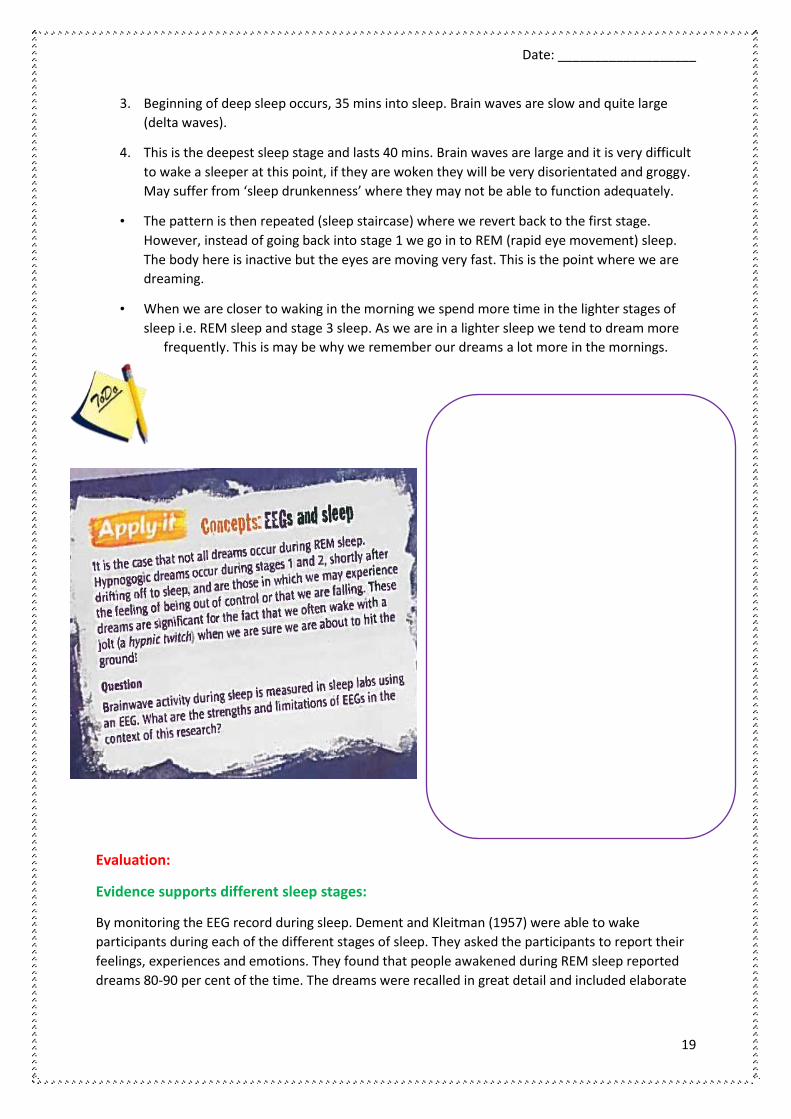
Event-related potentials (ERPs): Although there are many applications of EEG’s, it is still seen to be a
crude and overly general measure of brain activity. However in the recordings from the EEG there is
lots of specific data. They are associated with specific neural, sensory and motor responses. As a
result there are ways of teasing out and isolating this information. The technique will filter out
information and leave the relevant responses that relate to specific processes and tasks. What
remains is an event-related potential: these are types of brainwaves that are triggered by specific
events. Research has shown different types of ERP and how these are linked to various processes.
Post-mortem examinations: Are the analysis of the brain after someone has died. Individuals whose
brains are studied tend to have rare illnesses or defects that can be studied. Areas are examined
after death to try and identify the possible causes of the affliction the person suffered from. They
may also compare the brain to an unaffected brain to see the differences.

Date: ___________________
11
Evaluation:
fMRI:
Strengths- One advantage is that it doesn’t use radiation, unlike a PET scan. It is virtually risk free
and straightforward to use. The images are high in resolution, and show a great deal of detail and
also show a clear picture of localisation in the brain.
Weaknesses- However it is expensive, and can only get a clear image if the person remains perfectly
still. It can only measure blood flow to the brain and not specific neuronal activity. So it can be
difficult to see exactly what type of brain activity is being represented on screen.
EEG:
Strengths- It has proven invaluable in diagnosing disorders, such as epilepsy. The random bursts of
activity can be easily detected using this technique. It has also contributed much in our
understanding of the stages of sleep. EEG technology can also detect brain activity at a resolution of
a single millisecond.
Weaknesses - The main drawback is that it is too general. It isn’t useful in pinpointing the specific
source of neuronal activity. So researchers can’t tell where some activities are specifically based in
the brain.
ERP:
Strengths- EEG weaknesses are partly addressed here. ERP’s bring more specific measurement of the
neural processes at work. As ERP information is derived from EEG information it has excellent
temporal resolution. Therefore it has led to widespread use in in looking at cognitive functions and
deficits.
Weaknesses- Critics have pointed out that the methodology of the ERP isn’t standardised, so it is
difficult to confirm findings. Another issue is that to get an accurate reading background noise and
other extraneous material need to be eliminated, which isn’t easy to do.
Post-mortems:
Strengths- The evidence was key in our early understanding of how the brain worked. Broca and
Wernicke both relied on post-mortem evidence in their research. They provide medical knowledge
and help generate hypotheses for further study.
Weaknesses- Observed damage may not be linked to the actual deficit of the individual, but could be
caused by other traumas or brain decay. Also there is an issue of consent. Some patients whose
brains are being studied after death may not have been in the position of being able to give their full
consent. For example HM, whose memory was damaged, couldn’t give fully informed consent, but
his brain was studied anyway.

Date: ___________________
12
Check your understanding:
Strengths Limitations
fMRI
EEG
ERP
Post-Mortems
Check it
1. Outline one difference between EEGs and ERPs as ways of investigating the brain (2
marks)

Date: ___________________
13
2. Briefly evaluate post-mortem examinations as a way of investigating the brain (4 marks)

Date: ___________________
14
Biological Rhythms: Circadian Rhythms
Circadian Rhythms:
Biological rhythms:
A biological rhythm can be defined as any change in a biological activity (such as sleep and waking)
that repeats periodically. These rhythms are most often synchronised with daily, monthly, or annual
cyclical changes in the environment.
Lots of behaviours occur regularly in cycles. These cycles may be of different lengths.
Circadian – Occurs approx. every 24 hours. E.g. Sleep-Wake cycle
All biological rhythms are governed by two things: the body’s internal biological clocks, which are
called endogenous pacemakers and external changes in the environment which are known as
exogenous zeitgebers.
The sleep/wake cycle:
The fact that we feel drowsy when it’s night-time and alert during the day
demonstrates the effect of daylight-an important exogenous zeitgeber- on
our sleep/wake cycle. However if our body clock was left to ‘free-run’,
without the influence of light and other external sues, would we be able to
fall asleep and wake up at regular intervals. There have been several studies
to test this.
Siffre’s (pronounced Seef) cave study:
Aim: This study investigated what would happen to people’s circadian
rhythms if they were cut off from all zeitgebers (signals from outside the body that tell us about the
time of day – such as light and dark, clocks), and had to rely on their endogenous pacemaker
(internal body clock) to tell them when to eat and sleep. Would we still stick to a natural 24-hour
rhythm?
Method: Michel Siffre, a French cave explorer, spent over six months living in a cave in Texas, deep
under the ground, with no light, or anything else to tell him what time of day it was. His biological
clock was allowed to ‘free-run’, that is, he just followed his body’s inclinations, eating and sleeping
whenever he chose, with no fixed timetable. He was wired up so that some of his body functions
could be recorded; he had a telephone link to the outside world, and was monitored by video
camera.
Results: Siffre had a fairly erratic sleep-wake pattern at first, but it settled down to a pattern that
averaged just over 25 hours, instead of 24 hours.
Conclusion: We do have an internal mechanism that regulates our sleep/wake cycle, but it shifts to
a length of approximately 25 hours if we do not have external zeitgebers to reset it.

Date: ___________________
15
Evaluation:
This is a one-participant study, so may not be generalisable to all humans. Also Siffre’s living
conditions were unusual in other ways than simply lacking time signals, and other factors such as
loneliness could have affected his behaviour.
Similar studies have been done with rats, isolating them from daylight (Groblewski), and found a
similar increase in the sleep-wake cycle, which supports the findings from the Siffre study. A
strength of the study is that it lasted a long time, allowing Siffre’s rhythms to settle down into a
natural pattern.
Other research:
Folkard et al. (1985): Studied a group of 12 participants who lived in a cave for 3 weeks. They had to
go to bed when the clock said 11.45 pm. And they had to get up at 7.45 am. Over the course of the
study, the researchers gradually speeded up the clock (unbeknown to the participants) so an
apparent 24 hour day was reduced to 22. It was found that not one of the participants adjusted well
to the new regime. This would suggest the existence of strong free-running circadian rhythm that
can’t easily be overridden by changes by changes in the external environment.
Evaluation:
Practical application to shift work:
Knowledge of circadian rhythms has led to researchers understanding the detrimental consequences
of shift work. For example shit workers often suffer from a reduction in concentration at around 6
am. Knutsson (2003) said that shift workers are also prone to poor health, with over a third more
likely to suffer from heart disease. This may be due to the stress of the shift in their circadian
rhythm. Because they are getting poor quality sleep.
Practical application to drug therapy:
Circadian rhythms co-ordinate with a number of other bodily processes. These include heart-rate,
digestion and hormone levels. This in turn has an effect on the action of drugs on the body and how
well they are absorbed and distributed.
Research in to this has found that there are times during the day or night when drugs are most
effective. This has led to guidelines as to when to take the drugs for a whole range of medications
including anticancer, cardiovascular, respiratory and anti-epileptic drugs.

Date: ___________________
16
Check it
1. With reference to an example, define what is meant by a circadian rhythm (3 marks)
2. Describe one study that investigated a circadian rhythm (4 marks)

Date: ___________________
17
Biological rhythms: Infradian and ultradian rhythms
Ultradian - Occurs more than once a day. E.g. eating
Infradian – Lasts longer than 24 hours and can be weekly, monthly or annually. E.g. menstrual
cycle
Infradian rhythms:
Seasonal affective disorder (SAD):
SAD (Seasonal Affective Disorder) is a type of winter depression which affects millions of people
every winter between September and April, in particular during December, January and February. It
is a type of infradian rhythm known as a circannual rhythm, as it is subject to a yearly cycle.
SAD is caused by a biochemical imbalance, this is thought to be melatonin, in the hypothalamus due
to the shortening of daylight hours and the lack of sunlight in winter.
For many people SAD is a seriously disabling illness, preventing them from functioning normally
without continuous medical treatment. For others, it is a milder condition.
Symptoms
• a desire to oversleep and difficulty staying awake, but in some cases, disturbed sleep and
early morning wakening;
• feeling fatigue and an inability to carry out normal routine;
• a craving for carbohydrates and sweet foods, usually resulting in weight gain;
• feelings of misery, guilt and loss of self-esteem, sometimes hopelessness and despair,
sometimes apathy and loss of feelings;
• an irritability and desire to avoid social contact;
• a tension and inability to tolerate stress;
• a decreased interest in sex and physical contact
SAD symptoms usually reoccur regularly each winter, starting between September and November
and continuing until March or April; a diagnosis can be made after 2 or more consecutive winters of
symptoms.
SAD symptoms disappear in spring, either suddenly with a few weeks of hypomania/hyperactivity, or
gradually, depending on the intensity of sunlight in the spring/early summer.
SAD may begin at any age, but the main age of onset is 18-30. It occurs throughout the northern and
southern hemispheres but is rare in those living within 30 degrees of the Equator, where daylight
hours are long, constant and extremely bright.

Date: ___________________
18
The menstrual cycle:
This is seen as an example of an infradian rhythm as it only occurs once every month. This is
characterised by a change in hormone levels which regulate ovulation. A typical cycle lasts for 28
days. During the cycle rising levels of oestrogen cause the ovary to develop an egg and release it
(ovulation). After ovulation progesterone helps the womb grow thicker, preparing for pregnancy. If
pregnancy doesn’t occur then the egg is absorbed and the womb lining comes away.
Research study:
Although it is an endogenous system, there are thought to be some exogenous factors. Stern and
McClintock (1998) found that women’s menstrual cycles may start to synchronise as a result of
female pheromones. The sample were 29 women with a history of irregular periods. Samples of
pheromones of 9 women at different stages in their menstrual cycles were taken, by putting pads in
their armpits. They were worn for around 8 hours of the day to ensure the pheromones were picked
up. The pads were treated with alcohol then frozen, to be rubbed on the upper lip of the other
participants.
On day one, pads from the start of the menstrual cycle were applied to all 20 women, on day two
they were all given a pad from the second day of the cycle and so on. McClintock found that 68% of
the women experienced changes to their cycle which brought them closer to their ‘odour donor’.
Ultradian rhythms:
Stages of sleep:
• The introduction of the electroencephalograph (EEG) in the 1930’s was crucial in helping us
understand how sleep works.
• Dement and Kleitman in the
1950’s used this to show that
there are many stages of sleep
that we go through in the night
(see key study).
Slow-wave sleep stages
• Slow-wave sleep or (SWS) has
4 stages, this is also called
NREM sleep. The stages we
pass through when we are
asleep are an example of an
ultradian rhythm.
1. This is where we fall asleep. Breathing is slower, blood pressure falls and blood flow is
reduced. Brain waves are slower and irregular-these are called alpha waves. It is easy to be
woken up in this stage.
2. This stage lasts 20 mins. Gradually move in to a deeper sleep. Large brain waves occur and
occasional quick spikes of activity. Body functions slow even more.
Date: ___________________
19
3. Beginning of deep sleep occurs, 35 mins into sleep. Brain waves are slow and quite large
(delta waves).
4. This is the deepest sleep stage and lasts 40 mins. Brain waves are large and it is very difficult
to wake a sleeper at this point, if they are woken they will be very disorientated and groggy.
May suffer from ‘sleep drunkenness’ where they may not be able to function adequately.
• The pattern is then repeated (sleep staircase) where we revert back to the first stage.
However, instead of going back into stage 1 we go in to REM (rapid eye movement) sleep.
The body here is inactive but the eyes are moving very fast. This is the point where we are
dreaming.
• When we are closer to waking in the morning we spend more time in the lighter stages of
sleep i.e. REM sleep and stage 3 sleep. As we are in a lighter sleep we tend to dream more
frequently. This is may be why we remember our dreams a lot more in the mornings.
Evaluation:
Evidence supports different sleep stages:
By monitoring the EEG record during sleep. Dement and Kleitman (1957) were able to wake
participants during each of the different stages of sleep. They asked the participants to report their
feelings, experiences and emotions. They found that people awakened during REM sleep reported
dreams 80-90 per cent of the time. The dreams were recalled in great detail and included elaborate

Date: ___________________
20
visual images. Only 7 per cent of the awakenings from NREM sleep led to dream recall. Dement had
found the point during the ultradian cycle when people dream.
However, they also found that sleepers, when awoken in REM sleep, were not always dreaming.
The importance of the REM link is that it potentially provides a way to identify when someone is
dreaming – for example, Hobson and McCarley proposed that dreams are just random electrical
signals typical of REM sleep. However, such theories of dreaming are based on the erroneous
assumption that REM activity = dreaming.
Evolutionary basis of the menstrual cycle:
There is evidence to suggest that menstrual synchrony (McClintock) has an evolutionary benefit. If
our ancestors all menstruated together then they would fall pregnant at similar times, therefore
when they had children they could all be cared for collectively. So there would be more chance of
survival.
However there is an argument to suggest that it would be maladaptive to synchronise the menstrual
cycle. Being synchronised would mean that there would be more competition for mates and
therefore the fitness of the offspring would be lowered and they may not survive. So it would make
more sense to avoid synchrony.
Methodological problems:
Early studies on menstrual synchrony may have suffered from confounding variables that may not
have been considered. The changes on a woman’s menstrual cycle could be due to other factors
such as stress, changes on diet, exercise etc. Research also only focuses on small samples of women
and relies on the participants self-reporting the onset of their own cycles.
Check it
1. With reference to an example, define what is meant by an ultradian rhythm (3 marks)
2. Describe one study that investigated an infradian rhythm (4 marks)

Date: ___________________
21
Endogenous pacemakers and Exogenous zeitgebers
Endogenous pacemakers and the sleep/wake cycle:
Pineal gland and melatonin:
• Is a small pea shaped gland that contains light sensitive cells.
• When light is sensed the production of a hormone, melatonin is inhibited.
• When light levels fall the production of melatonin increases in the pineal gland.
• Melatonin induces sleep.
• Thus light, the pineal gland and melatonin regulate the sleep-wake cycle.
• The Pineal gland is important to birds and reptiles. It is located just below the skull (for
mammals it is deep within the brain). In fact many lizards have a ‘third eye’ (to sense light),
near the pineal gland, that protrudes from the skull.
Role of the suprachiasmatic nucleus (SCN):
• In mammals the pathways are more complex.
• For mammals the main biological clock appears to be a small area in the hypothalamus
(positioned just behind the eyes)-the suprachiasmatic nucleus (SCN).
• The SCN has an in-built circadian rhythmic firing pattern.
• When the SCN of a rat is lesioned (damaged by cutting) its circadian rhythm including
sleeping and feeding patterns is totally disrupted.
• When working properly in mammals it regulates the release of melatonin in the pineal gland
via an interconnecting pathway.
• Another pathway links the SCN to the retina of each eye.
• This then helps to maintain the amount of light falling on the retina and therefore indirectly
influence the release of melatonin in the pineal gland.
Animal studies and the SCN:
DeCoursey et al. (2000) destroyed the SCN connections in the brains of 30 chipmunks. They were
returned to their natural habitat and monitored for 80 days. The sleep/wake cycle disappeared, by
the end of the study a significant proportion had been killed by predators.
Ralph et al (1990) bred ‘mutant’ hamsters with a 20 hour sleep/wake cycle. When foetal tissue was
transferred to into the brains of normal hamsters, the cycles of the second group defaulted to 20
hours.
Both studies emphasise the role of the SCN in establishing and maintaining the circadian sleep/wake
cycle.

Date: ___________________
22
Exogenous zeitgebers and the sleep/wake cycle:
They help us to understand when the seasons change. It deals with external cues such as the
changing of the seasons (exogenous). These exogenous cues are known as zeitgebers from the
German word ‘time giver’. Entrainment is a process of resetting the biological clock with exogenous
zeitgebers.
Light:
This is the dominant zeitgeber in humans. This is due to discovery of melatonin. See above. Artificial
light is less effective. Light does not just effect the SCN. It has been found that certain proteins in the
body detect changes in light (Hall, 2000).
Light is important to us as studies on blind people have shown. They find it very difficult to have
regular sleep patterns because of their inability to detect light, even when given social cues, such as
clocks. Some have to take sedatives and stimulants to regulate their natural rhythm (Miles et al
1977). This can be used as evaluation.
Social cues:
In babies the sleep/wake cycle is pretty much random. However at 6 weeks the circadian rhythms
begin and by about 16 weeks, most babies are entrained. The schedules imposed by the parents are
a key influence. For example set times for meals and bed times. Research also suggests that adapting
to local times for eating and sleeping, is an effective way of entraining circadian rhythms and beating
jet lag when travelling long distances.
Evaluation:
Ethics in animal studies:
In the DeCoursey et al study the animals were exposed to considerable harm, and therefore risk
when they were returned to their natural environment. Whether what we learn from these studies
justifies the harsh treatment of the animals is a matter of debate.
Beyond the master clock:
Research has found numerous circadian rhythms in many organs and cells in the body. They can be
found in adrenal, lungs, liver, pancreas, spleen, thymus and skin. Although they are highly influenced
by the SCN, they can act independently. Damiola aet al. (2000) demonstrated how changing feeding
patterns in mice could alter the circadian rhythms of cells in the liver by up to 12 hours, whilst
leaving the rhythm of the SCN unaffected. This suggests that there may be many other complex
influences on the sleep/wake cycle, aside from the master clock (SCN).

Date: ___________________
23
Exam Questions (A Level)
1) Read the item and then answer the questions that follow.
Figure 1 shows the left hemisphere of the human brain. Six areas of cortical specialisation are
labelled A, B, C, D, E and F.
Figure 1: Left hemisphere of the human brain
Using your knowledge of localisation of function in the brain, identify the area of cortical
specialisation. Shade one box only for each area.
. Broca’s area
. Somatosensory cortex
. Visual cortex
. Wernicke’s area
. Motor cortex
[1 mark] [1 mark] [1 mark] [1 mark] [1 mark]
2) The electroencephalogram (EEG) and event-related potentials (ERPs) both involve recording the
electrical activity of the brain.
Outline one difference between the EEG and ERPs. [2 marks]

Date: ___________________
24
3) Read the item and then answer the question that follows.
Sam is a police officer. She has just started working the night shift and after a week, she finds that
she has difficulty sleeping during the day and is becoming tense and irritable. Sam is also worried
that she is less alert during the night shift itself.
Using your knowledge of endogenous pacemakers and exogenous zeitgebers, explain Sam’s
experiences. [4 marks]
4) The human female menstrual cycle is an example of one type of biological rhythm; it is called a:
A circadian rhythm
B infradian rhythm
C ultradian rhythm
[1 mark]
6) Split brain patients show unusual behaviour when tested in experiments. Briefly explain how
unusual behaviour in split brain patients could be tested in an experiment. [2 marks]
7) Briefly evaluate research using split brain patients to investigate hemispheric lateralisation of
function. [4 marks]