Approaches to ab initio molecular replacement of [alpha ...
12
research papers Acta Cryst. (2017). D73, 985–996 https://doi.org/10.1107/S2059798317016436 985 Received 14 June 2017 Accepted 15 November 2017 Edited by Q. Hao, University of Hong Kong Keywords: transmembrane proteins; ab initio phasing; ab initio modelling; predicted contacts. Supporting information: this article has supporting information at journals.iucr.org/d Approaches to ab initio molecular replacement of a-helical transmembrane proteins Jens M. H. Thomas, a Felix Simkovic, a Ronan Keegan, b Olga Mayans, c Chengxin Zhang, d Yang Zhang d and Daniel J. Rigden a * a Institute of Integrative Biology, University of Liverpool, Liverpool L69 7ZB, England, b Research Complex at Harwell, STFC Rutherford Appleton Laboratory, Didcot OX11 0FA, England, c Fachbereich Biologie, Universita ¨t Konstanz, D-78457 Konstanz, Germany, and d Department of Computational Medicine and Bioinformatics, Department of Biological Chemistry, Medical School, University of Michigan, 100 Washtenaw Avenue, Ann Arbor, MI 48109-2218, USA. *Correspondence e-mail: [email protected] -Helical transmembrane proteins are a ubiquitous and important class of proteins, but present difficulties for crystallographic structure solution. Here, the effectiveness of the AMPLE molecular replacement pipeline in solving -helical transmembrane-protein structures is assessed using a small library of eight ideal helices, as well as search models derived from ab initio models generated both with and without evolutionary contact information. The ideal helices prove to be surprisingly effective at solving higher resolution structures, but ab initio- derived search models are able to solve structures that could not be solved with the ideal helices. The addition of evolutionary contact information results in a marked improvement in the modelling and makes additional solutions possible. 1. Introduction Transmembrane proteins are an important class of proteins that are estimated to comprise about 30% of the proteome (Tusna ´dy et al. , 2004). They reside, at least partly and often predominantly, within the hydrophobic cell membrane, sand- wiched between the aqueous cell interior and exterior. Transmembrane proteins come in two main forms, -helical and -barrel, with the overwhelming majority being of the -helical form (White & Wimley, 1999). Estimates of the number of transmembrane proteins encoded in the human genome vary. Most studies agree that roughly 26% of proteins are transmembrane proteins, but this includes a large number of single-pass transmembrane proteins. Polytopic proteins, as studied here, are thought to represent around 14% of the human proteome (Alme ´n et al., 2009; Fagerberg et al., 2010). Conventional X-ray crystallography is still the predominant mode of structure solution for transmembrane proteins. Unfortunately, their hydrophobic nature means that trans- membrane proteins are particularly challenging to work with experimentally. Detergents are usually required to extract the protein from the membrane environment. These surround the protein, mimicking the membrane environment and forming a water-soluble protein–detergent complex (PDC). The deter- gent coating of the PDC reduces the number of protein– protein contacts available to guide crystal formation, resulting in large fragile crystals with a high solvent content (Moraes et al., 2014). The nature of these crystals means that they often diffract poorly and are unstable in an X-ray beam. As a result of these complications, of the more than 130 000 protein structures currently deposited in the PDB (Berman et al., 2000), fewer than 3% (3084) are classified as transmembrane ISSN 2059-7983 Konstanzer Online-Publikations-System (KOPS) URL: http://nbn-resolving.de/urn:nbn:de:bsz:352-2-tie4zyqlg9458 Erschienen in: Acta crystallographica Section D : Structural biology ; 73 (2017), Pt 12. - S. 985-996 http://dx.doi.org/10.1107/S2059798317016436
Transcript of Approaches to ab initio molecular replacement of [alpha ...
research papers
Acta Cryst. (2017). D73, 985–996 https://doi.org/10.1107/S2059798317016436 985
Received 14 June 2017
Accepted 15 November 2017
Edited by Q. Hao, University of Hong Kong
Keywords: transmembrane proteins; ab initio
phasing; ab initio modelling; predicted contacts.
Supporting information: this article has
supporting information at journals.iucr.org/d
Approaches to ab initio molecular replacement ofa-helical transmembrane proteins
Jens M. H. Thomas,a Felix Simkovic,a Ronan Keegan,b Olga Mayans,c Chengxin
Zhang,d Yang Zhangd and Daniel J. Rigdena*
aInstitute of Integrative Biology, University of Liverpool, Liverpool L69 7ZB, England, bResearch Complex at Harwell,
STFC Rutherford Appleton Laboratory, Didcot OX11 0FA, England, cFachbereich Biologie, Universitat Konstanz,
D-78457 Konstanz, Germany, and dDepartment of Computational Medicine and Bioinformatics, Department of
Biological Chemistry, Medical School, University of Michigan, 100 Washtenaw Avenue, Ann Arbor, MI 48109-2218,
USA. *Correspondence e-mail: [email protected]
�-Helical transmembrane proteins are a ubiquitous and important class of
proteins, but present difficulties for crystallographic structure solution. Here, the
effectiveness of the AMPLE molecular replacement pipeline in solving �-helical
transmembrane-protein structures is assessed using a small library of eight ideal
helices, as well as search models derived from ab initio models generated both
with and without evolutionary contact information. The ideal helices prove to
be surprisingly effective at solving higher resolution structures, but ab initio-
derived search models are able to solve structures that could not be solved with
the ideal helices. The addition of evolutionary contact information results in a
marked improvement in the modelling and makes additional solutions possible.
1. Introduction
Transmembrane proteins are an important class of proteins
that are estimated to comprise about 30% of the proteome
(Tusnady et al., 2004). They reside, at least partly and often
predominantly, within the hydrophobic cell membrane, sand-
wiched between the aqueous cell interior and exterior.
Transmembrane proteins come in two main forms, �-helical
and �-barrel, with the overwhelming majority being of the
�-helical form (White & Wimley, 1999). Estimates of the
number of transmembrane proteins encoded in the human
genome vary. Most studies agree that roughly 26% of proteins
are transmembrane proteins, but this includes a large number
of single-pass transmembrane proteins. Polytopic proteins, as
studied here, are thought to represent around 14% of the
human proteome (Almen et al., 2009; Fagerberg et al., 2010).
Conventional X-ray crystallography is still the predominant
mode of structure solution for transmembrane proteins.
Unfortunately, their hydrophobic nature means that trans-
membrane proteins are particularly challenging to work with
experimentally. Detergents are usually required to extract the
protein from the membrane environment. These surround the
protein, mimicking the membrane environment and forming a
water-soluble protein–detergent complex (PDC). The deter-
gent coating of the PDC reduces the number of protein–
protein contacts available to guide crystal formation, resulting
in large fragile crystals with a high solvent content (Moraes et
al., 2014). The nature of these crystals means that they often
diffract poorly and are unstable in an X-ray beam. As a result
of these complications, of the more than 130 000 protein
structures currently deposited in the PDB (Berman et al.,
2000), fewer than 3% (3084) are classified as transmembrane
ISSN 2059-7983
Konstanzer Online-Publikations-System (KOPS) URL: http://nbn-resolving.de/urn:nbn:de:bsz:352-2-tie4zyqlg9458
Erschienen in: Acta crystallographica Section D : Structural biology ; 73 (2017), Pt 12. - S. 985-996
http://dx.doi.org/10.1107/S2059798317016436
proteins by the TMDET algorithm (Tusnady et al., 2004). The
low number of structures means that the probability of finding
homologous structures to use for molecular replacement
(MR) can often be low, so that the use of unconventional
approaches may be required. These can include methods
based on fragments or libraries of fragments, such as that
employed by ARCIMBOLDO, ARCIMBOLDO_LITE and
ARCIMBOLDO_BORGES (Rodrıguez et al., 2009; Sammito
et al., 2013, 2015; Millan et al., 2015), or those that employ ab
initio structure prediction (Bibby et al., 2012; Shrestha et al.,
2011; Shrestha & Zhang, 2015; Wang et al., 2016).
Ab initio protein-structure prediction is the process of
determining the tertiary structure of a protein starting purely
from its sequence, and not relying on the use of an existing
structure as a template. Popular examples of ab initio
modelling software include ROSETTA, QUARK and SAINT2
(Simons et al., 1997; Rohl et al., 2004; Xu et al., 2012; Ellis et al.,
2010). Ab initio modelling for globular proteins, however, is
accurate only for modest chain lengths. For example, in the
absence of additional information, reliable modelling with
ROSETTA is still limited to sequences of up to 130 residues
(Tai et al., 2014), so that models of larger proteins are not
usually sufficiently accurate to be suitable for MR.
Our program AMPLE uses a cluster-and-truncate proce-
dure to extract ensembles for MR from the initial decoys
generated by ab initio programs (Bibby et al., 2012). AMPLE
constructs MR search ensembles by generating 1000 ab initio
models and then taking up to 200 models from the top
SPICKER cluster (Zhang & Skolnick, 2004) and truncating
them into 20 evenly-spaced size bins based on per-residue
structural variance measurements. The truncated models are
then subclustered under 1, 2 or 3 A radius r.m.s.d. thresholds,
before being subjected to three different side-chain treat-
ments: polyalanine (removal of all side chains), ‘reliable’
(retaining side chains for residues with fewer rotamers) and
all-atom (no editing). The search ensembles are subjected to
MR with Phaser (McCoy et al., 2007; Read & McCoy, 2016)
and MR-positioned models are subjected to density modifi-
cation and automated main-chain tracing using SHELXE
(Thorn & Sheldrick, 2013). Further structure rebuilding of the
SHELXE chain traces with ARP/wARP (Langer et al., 2008)
and/or Buccaneer (Cowtan, 2006) can also be undertaken.
Despite the usually poor overall quality of the models for
larger proteins (which means that the individual models are
unsuitable for MR), the AMPLE algorithm is able to generate
successful ensembles for MR, and we have seen successes with
targets of up to 250 residues in length for �-helical coiled-coil
proteins (Thomas et al., 2015) and up to 221 residues for cases
where contact predictions (see below) were available to assist
the modelling of gobular proteins (Simkovic et al., 2016). The
more regular and ordered nature of �-helices, as opposed to �-
sheets or loops, facilitates ab initio modelling for structures
containing these secondary-structure elements. In addition,
the membrane-spanning helices of an �-helical transmem-
brane protein can be reliably predicted and assumed to remain
within a layer of finite width. This helpfully limits the
conformational space to be explored during the ab initio
modelling, aiding model accuracy and potentially raising the
upper size limit of tractable targets. Thus, ab initio modelling
might be particularly suitable for transmembrane proteins
(Yarov-Yarovoy et al., 2006) and make the resulting predic-
tions good candidates for solution with AMPLE.
The accuracy of ab initio modelling may also be improved
by the incorporation of additional information, one source of
which is evolutionary covariance. In recent years, there has
been a step change in the accuracy of residue–residue contact
predictions generated from sequence information alone
(Simkovic, Ovchinnikov et al., 2017). These methods infer
which residues are in physical contact by looking at the
evolutionary covariance signal within an alignment of a family
of homologous sequences. We have previously demonstrated
the benefits of using such predictions in addressing challenging
globular domains with AMPLE (Simkovic et al., 2016).
Contact-prediction algorithms can generally be divided into
two distinct categories: evolutionary coupling analysis and
supervised machine learning. The former derives contact
predictions by detecting evolutionary covariance amongst
homologous sequences, and various implementations attempt
this by employing a cooperative statistical model. Examples of
implementations that employ a pseudo-likelihood maximiza-
tion model can be found in applications such as GREMLIN
(Ovchinnikov et al., 2014) and CCMPRED (Seemayer et al.,
2014). Other implementations include sparse covariance
matrix-inversion models such as PSICOV (Seemayer et al.,
2014; Jones et al., 2012) or mean-field direct coupling analysis
models such as EVFold–mfDCA (Kajan et al., 2014).
Supervised machine-learning algorithms are not as effective
as most evolutionary coupling analysis algorithms; however, in
recent studies combining the two has proven to be the most
effective use of both. These meta-predictors have been
developed to combine predictions from a number of different
prediction methods. MetaPSICOV is one such meta-predictor
that uses a neural network to combine predictions from,
amongst others, the PSICOV, mfDCA and CCMPRED scores
(Jones et al., 2015). A related approach is MEMBRAIN, which
is a contact predictor optimized for transmembrane proteins
(Jones et al., 2015; Xiao & Shen, 2015). MEMBRAIN uses a
neural network to combine a covariance-based approach
(PSICOV) with the combination of a number of maximum-
likelihood approaches, each of which trains a statistical model
using sequence-derived features such as residue position along
the transmembrane helix or sequence separation of residues.
Most recently, NeBcon was proposed to combine multiple
contact predictors from both coevolution and machine-
learning techniques through naıve Bayes classifier and neural
network training, which shows an advantage over the best
individual predictors (He et al., 2017).
Here, we explore the ability of AMPLE to solve trans-
membrane-protein structures using MR search ensembles
derived from ab initio models from a variety of sources.
Attempts were made to solve structures using models derived
from ROSETTA’s established ROSETTAMEMBRANE
protocol and the QUARK modelling protocol, both of which
do not use contact information by default. Solution was then
research papers
986 Thomas et al. � Ab initio MR of �-helical transmembrane proteins Acta Cryst. (2017). D73, 985–996
attempted with ROSETTA models created using contact
information derived from GREMLIN, CCMPRED, Meta-
PSICOV or MEMBRAIN to test whether the additional
contact information improved the success rate. As in our
earlier work (Thomas et al., 2015), we also tried to solve the
structures with a library of short, ideal �-helices ranging in size
from five to 40 residues. These were chosen for comparison
with the highly truncated but model-derived �-helix-rich
search models of a similar size that AMPLE frequently
generates. These latter, unlike the ideal helices, are ensembles
and can contain bent or kinked helices as well as other irre-
gular features. The comparison therefore illuminates the
contribution of the modelling and ensembling to MR success.
2. Methods
2.1. Test-set selection
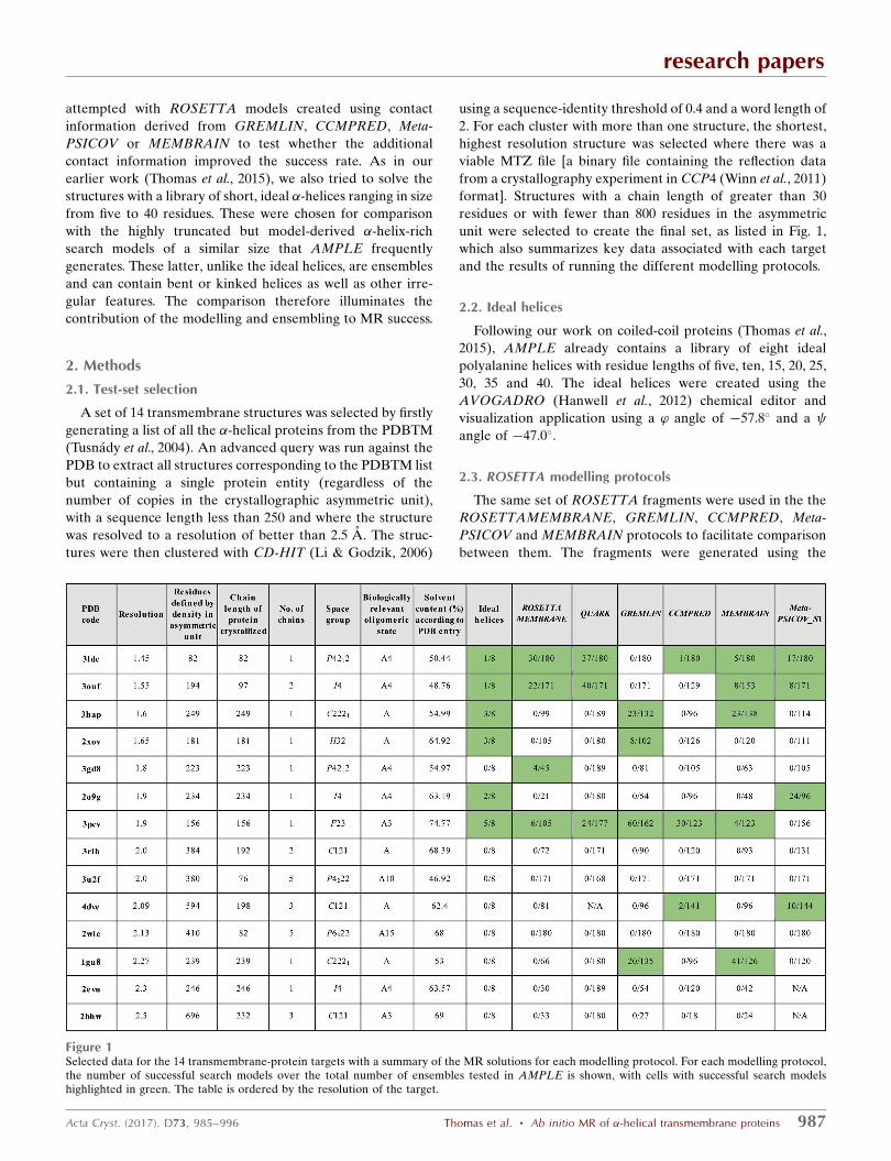
A set of 14 transmembrane structures was selected by firstly
generating a list of all the �-helical proteins from the PDBTM
(Tusnady et al., 2004). An advanced query was run against the
PDB to extract all structures corresponding to the PDBTM list
but containing a single protein entity (regardless of the
number of copies in the crystallographic asymmetric unit),
with a sequence length less than 250 and where the structure
was resolved to a resolution of better than 2.5 A. The struc-
tures were then clustered with CD-HIT (Li & Godzik, 2006)
using a sequence-identity threshold of 0.4 and a word length of
2. For each cluster with more than one structure, the shortest,
highest resolution structure was selected where there was a
viable MTZ file [a binary file containing the reflection data
from a crystallography experiment in CCP4 (Winn et al., 2011)
format]. Structures with a chain length of greater than 30
residues or with fewer than 800 residues in the asymmetric
unit were selected to create the final set, as listed in Fig. 1,
which also summarizes key data associated with each target
and the results of running the different modelling protocols.
2.2. Ideal helices
Following our work on coiled-coil proteins (Thomas et al.,
2015), AMPLE already contains a library of eight ideal
polyalanine helices with residue lengths of five, ten, 15, 20, 25,
30, 35 and 40. The ideal helices were created using the
AVOGADRO (Hanwell et al., 2012) chemical editor and
visualization application using a ’ angle of �57.8� and a angle of �47.0�.
2.3. ROSETTA modelling protocols
The same set of ROSETTA fragments were used in the the
ROSETTAMEMBRANE, GREMLIN, CCMPRED, Meta-
PSICOV and MEMBRAIN protocols to facilitate comparison
between them. The fragments were generated using the
research papers
Acta Cryst. (2017). D73, 985–996 Thomas et al. � Ab initio MR of �-helical transmembrane proteins 987
Figure 1Selected data for the 14 transmembrane-protein targets with a summary of the MR solutions for each modelling protocol. For each modelling protocol,the number of successful search models over the total number of ensembles tested in AMPLE is shown, with cells with successful search modelshighlighted in green. The table is ordered by the resolution of the target.
ROSETTA make_fragments.pl script supplying the
-nohoms flag to ensure that no homologous fragments were
used and so that no structural information from homologous
structures was included in any modelling.
2.4. ROSETTAMEMBRANE modelling protocol
The original ROSETTAMEMBRANE protocol (Yarov-
Yarovoy et al., 2006) was used. Yarov-Yarovoy and coworkers
recommend only using SAM (Sequence Alignment and
Modeling System; Katzman et al., 2008) to predict the
secondary structure, as the default JUFO (Meiler & Baker,
2003) and PSIPRED (Jones, 1999) predictors often incorrectly
predict the secondary structure for transmembrane proteins.
However, the recommended method for AMPLE users to
generate the fragment database required by ROSETTA is to
use the online ROBETTA server (Kim et al., 2004), which
does not support the use of SAM. Standard ROSETTA frag-
ments, obtained using the default secondary-structure
prediction protocol as used by the ROBETTA server, were
therefore used as this replicates the procedure that will be
followed by AMPLE users. In addition, this allowed the use of
the same fragment databases with all the modelling protocols,
as described above.
The OCTOPUS server (July 2015; Viklund & Elofsson,
2008) was used to predict transmembrane regions and a
lipophilicity prediction was generated using the run_lips.pl
script, which undertakes a PSI-BLAST (Altschul et al., 1997)
multiple sequence alignment of the target sequence against
the NR database (O’Leary et al., 2016). The OCTOPUS and
lipophilicity predictions were then used with the ROSETTA-
MEMBRANE executable to generate the models. ROSETTA
v.2015.22.57859 was used for the ROSETTAMEMBRANE
modelling and all subsequent ROSETTA modelling in this
work.
2.5. QUARK modelling
The online QUARK server is a particularly easy way for
AMPLE users to generate ab initio models, as it does not
require them to install any additional software or use their
local machine for the time-consuming model-generation stage
(Keegan et al., 2015). Although the QUARK modelling
protocol (Xu et al., 2012) does not feature any membrane-
specific protocols, previous work by the developers has
demonstrated success in modelling transmembrane proteins in
Escherichia coli (Xu & Zhang, 2013). The QUARK decoys
were generated on 20 November 2015. For each target, ten
independent replica-exchange Monte Carlo (REMC) simula-
tions were performed by QUARK, where each REMC runs 40
replicas in parallel at different temperatures with each replica
containing 500 Monte Carlo cycles of simulations. This process
output 50 000 decoys from the ten lowest-temperature replicas
(i.e. ten REMC simulations� ten lowest-temperature replicas
� 500 Monte Carlo cycles). From the full decoy set, a subset of
5000 decoys was randomly selected for further consideration.
To reconstruct the full-atomic model from each of the selected
C� decoy structures, an initial all-atom representation was
generated using PULCHRA 3.06 (Rotkiewicz & Skolnick,
2008) or MaxSprout 2006.10 (Holm & Sander, 1991) if
PULCHRA was unsuccessful. Side chains were then added
using SCWRL 4.0 (Krivov et al., 2009).
In order to ensure that no homologous fragments were used
in the modelling, PDB structures with a sequence identity of
>30% to the target or that were detectable by PSI-BLAST (a
criterion used by earlier ab initio folding benchmark tests;
Simons et al., 2001; Zhang et al., 2003) were excluded from the
QUARK fragment library.
2.6. ROSETTA modelling with GREMLIN predicted contacts
The transmembrane-modelling protocol for modelling
transmembrane proteins using contact predictions generated
by the GREMLIN server (http://gremlin.bakerlab.org/) was
developed by Ovchinnikov et al. (2015). Contact predictions
generated by the GREMLIN server in November 2015 were
used to generate the models. This method presents an
attractive alternative to the original ROSETTAMEMBRANE
protocol, as it only requires a user to generate the contact
information using the GREMLIN server and then run the
standard ROSETTA executables. There is no need to perform
OCTOPUS transmembrane prediction or the lipophilicity
prediction, with the associated need for BLAST and the NR
database to be installed, as there is with ROSETTA-
MEMBRANE. The protocol is described further in xS1.
2.7. Contact modelling (excluding GREMLIN)
For each target, a multiple sequence alignment (MSA) was
generated using the conkit-predict script from the
CONKIT package (Simkovic, Thomas et al., 2017) and using
HHblits v.2.0.16 (Alva et al., 2016) against UniProt20
v.2016_02 (The UniProt Consortium, 2017). A contact meta-
prediction using MetaPSICOV v.1.04 (Jones et al., 2015) was
generated, which in turn used the following contact-prediction
pipelines: CCMPRED v.0.3.2 (Seemayer et al., 2014), Free-
Contact v.1.0.21 (Kajan et al., 2014) and PSICOV v.2.1b3
(Jones et al., 2012). The predictions from MetaPSICOV stage 1
(MetaPSICOV_S1) were used as the contact predictions for
MetaPSICOV as recommended in Jones et al. (2015). The
CCMPRED predictions generated by MetaPSICOV were
used as the CCMPRED predictions. A set of contacts was also
generated using the MEMBRAIN server v.2015-03-15.
For each set of contact predictions, the top L contact pairs
were then turned into a set of restraints for ROSETTA, where
L is the number of residues in the target sequence. The FADE
energy function was employed with parameters identical to
those reported in Michel et al. (2014). Modelling was then
carried out with the ROSETTA ABINITIO protocol.
2.8. Molecular replacement
All of the models were run in the automated MR pipeline
AMPLE v.1.0 using default parameters (Bibby et al., 2012) and
with the CCP4 suite v.6.5.13 (Winn et al., 2011), SHELXE
v.2014/14 and ARP/wARP v.7.5.
research papers
988 Thomas et al. � Ab initio MR of �-helical transmembrane proteins Acta Cryst. (2017). D73, 985–996
To be considered a successful MR solution, the positioned
models were required to yield a SHELXE correlation coeffi-
cient (CC) of at least 25.00 and an average chain length (ACL)
of greater than 10.00. We imposed as an additional condition
of success that structure rebuilding of the SHELXE chain
traces with ARP/wARP (Langer et al., 2008) and/or Buccaneer
(Cowtan, 2006) resulted in an Rfree value of 0.45 or better. A
further validation was provided by measuring the weighted
mean phase error between each rebuilt model and the crystal
structure.
3. Results
The weighted mean phase error for all of the final rebuilt
models was measured to confirm that the structure had been
correctly determined. In all cases the error was less than 30�.
3.1. Ideal helices
Attempting solution with the AMPLE library of ideal
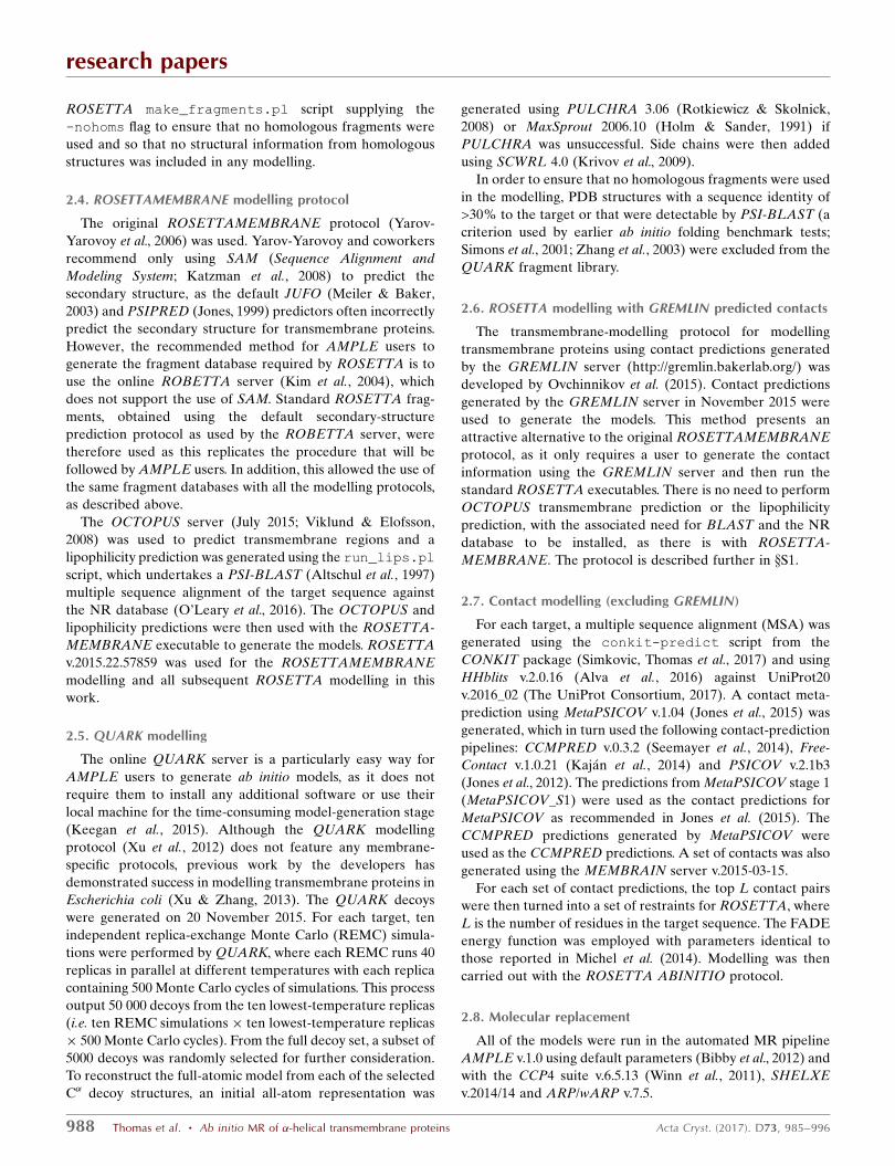
helices solved six of the targets, as shown in Figs. 1 and 2.
The library of ideal helices was able to solve six targets in
total. All targets smaller than 261 residues and with a reso-
lution better than 1.9 A were solved using this protocol, with
the exception of target 3gd8. Considering that transmembrane
proteins are considered to be relatively hard targets to solve,
that so many of this set can be solved using a small library of
ideal helices and a simple MR protocol is an encouraging
result to set alongside other advances in the use of ideal
helices (Millan et al., 2015).
3.2. ROSETTAMEMBRANE
Solution was then attempted using the ROSETTA-
MEMBRANE protocol. Fig. 1 shows that ROSETTA-
MEMBRANE performs more poorly than the AMPLE library
of ideal helices, solving four targets. Three of the solved
targets could be solved with the ideal helices. The single
exception is 3gd8, which could now be solved for the first time.
Targets 3hap, 2xov and 2o9g were not solved, despite being
solvable with the ideal helices.
An analysis of the quality of the models was then made to
determine how the effectiveness of the modelling affected the
ability of AMPLE to solve the targets using ROSETTA-
MEMBRANE. The TM score of the complete model that
became the centroid of the search ensemble was used as a
metric for the quality of the models within the ensemble. A
TM score of greater than 0.5 is generally considered to indi-
cate correct prediction of the overall fold. The results of the
analysis are displayed in Supplementary Fig. S2. The results
show that there was no correlation between the quality of the
models and the success of the ensembles, with the successful
search models all coming from ensembles where the TM score
of the complete centroid model was between 0.21 and 0.33.
Targets 3u2f and 2wie failed to solve, despite some ensembles
being derived from models with TM scores of 0.739 and 0.715,
respectively.
Target 3gd8 could be solved with four ensembles, despite
none of the successful ensembles being derived from a model
with a TM score of better than 0.227. Selected data for the
ensembles are listed in Supplementary Table S1. The RIO
score (Thomas et al., 2015), which assesses the in-sequence and
research papers
Acta Cryst. (2017). D73, 985–996 Thomas et al. � Ab initio MR of �-helical transmembrane proteins 989
Figure 2Results for attempting solution of transmembrane proteins with idealhelices mapped against target resolution and number of residues in theasymmetric unit of the crystallographic cell. Successes are in blue andfailures are in red.
Figure 3Successful solutions from ensembles c1_tl11_r2_allatom andc1_tl6_r3_reliable (blue and magenta, respectively) overlaid on thecrystal structure of PDB entry 3gd8 (green).
out-of-sequence register overlap of the placed search-model
residues (fragments of at least three residues) with the
corresponding crystal structure, was zero for all solutions,
indicating that the helices were not placed correctly with
regard to sequence. Although the successful ensembles
(c1_t11_r3_polyAla, c1_t11_r2_allatom, c1_t6_r3_reliable and
c1_t6_r2_allatom) had undergone different subcluster radii
and side-chain treatment, the final models for the successful
search models c1_t11_r3_polyAla and c1_t11_r2_allatom were
almost identical, as were those for c1_t6_r3_reliable and
c1_t6_r2_allatom. [Ensembles in AMPLE are named using a
quartet of identifiers separated by underscores. The first
identifier is the number of the SPICKER cluster that the
models were derived from, prefixed with a c, the second the
truncation level, prefixed with a t, the third the subcluster
radius, prefixed with an r, and the last the side-chain mode].
Fig. 3 shows the first of each pair (c1_t11_r2_allatom and
c1_t6_r3_reliable) overlaid on the crystal structure.
The two solutions appeared to be largely straight helices of
lengths of 25 and 14 residues, respectively. It is therefore
interesting that the ideal helices of lengths 25 and 15 were
unable to solve target 3gdb. An analysis of the placement
of the helical segment in the
two solutions of 3gd8 with
HELANAL (Kumar & Bansal,
2012) identified the search
models as being ‘curved’ and the
helix of 3gd8 as being ‘kinked’
(Bansal et al., 2000). A ‘kink’ is
defined by the authors when the
bending angle of a given residue
is greater than 20� but less than
60�. The search ensembles
generated by AMPLE contained
between 19 and 30 search models,
so it appears that the approx-
imation of the slightly kinked
helix by the slightly curved search
model and/or diversity within the
search-model ensembles is what
enabled the AMPLE search
ensemble to succeed where the
AMPLE set of ideal helices
failed. The HELANAL analysis
and models of the search ensem-
bles are displayed in Supplemen-
tary Table S2 and Supplementary
Fig. S3, respectively.
3.3. QUARK
Solution was then attempted
with QUARK models. The results
are displayed in Fig. 1, with a
graphical summary in Supple-
mentary Fig. S4 and an analysis of
the TM scores in Supplementary
Fig. S5. QUARK was able to
solve three targets, all of which
could be solved with the AMPLE
library of ideal helices. An
analysis of the TM scores for the
models similar to that undertaken
for ROSETTAMEMBRANE
showed that the quality of the
models again had little effect on
solution. The QUARK models for
target 3u2f were of even better
research papers
990 Thomas et al. � Ab initio MR of �-helical transmembrane proteins Acta Cryst. (2017). D73, 985–996
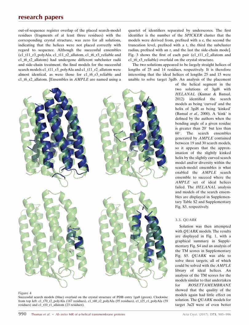
Figure 4Successful search models (blue) overlaid on the crystal structure of PDB entry 1gu8 (green). Clockwisefrom top left: c1_t70_r2_polyAla (167 residues), c1_t40_r2_polyAla (95 residues), c1_t25_r1_polyAla (59residues) and c1_t10_r1_allatom (23 residues).
quality than those for ROSETTAMEMBRANE (maximum
TM score of 0.792), but the target could still not be solved.
Overall, the quality of the QUARK models was rather better
than for ROSETTAMEMBRANE (median TM score across
all models of 0.385 as opposed to 0.263), but as the general
quality of the modelling is poor, even the better models rarely
seem to have the potential to generate solutions.
3.4. GREMLIN
Solution was then attempted with models generated by the
GREMLIN modelling protocol. The results are displayed in
Fig. 1, with a graphical summary shown in Supplementary Fig.
S6 and an analysis of the TM scores shown in Supplementary
Fig. S7. GREMLIN was able to solve four targets, including
1gu8, which could not be solved with AMPLE’s simple
approach to ideal helices, QUARK or ROSETTA-
MEMBRANE. However, targets 3ldc, 3ouf and 2o9g were not
solved, all of which could be solved with the ideal helices, and
neither was 3gd8, which could be solved with ROSETTA-
MEMBRANE. The contact information used by GREMLIN
dramatically improved the quality of the models, so that the
median TM score across all models was 0.667, as opposed to
0.263 for ROSETTAMEMBRANE and 0.385 for QUARK. It
also appears that the better models are contributing more to
the solutions. 20 successful models were generated for target
1gu8, with a wide range of model sizes (23, 35, 47, 59, 71, 83,
95, 107, 119 and 167 residues). A selection of four of the
successful solutions, covering the whole span of sizes, is shown
in Fig. 4.
Fig. 4 shows that all of the solutions were derived from the
same cluster, with the smaller ensembles being heavily trun-
cated versions of the larger ones. The models for target 1gu8
are much better than with any of the previous methods (a
maximum TM score for successful/unsuccessful of 0.829/0.856)
and this is what appears to have made solution possible. The
modelling has captured the packing and helical curvature of
six of the helices. It is interesting that for the smallest solution
(c1_t10_r1_allatom with 23 residues), just two short helical
segments correctly packed against each other are sufficient to
elicit a solution, whereas an ideal helical segment of the same
length cannot. Interestingly the ensemble derived from the
untruncated cluster of models, the centroid of which had a TM
score of 0.856, was unable to solve the target.
3.5. CCMPRED, MEMBRAIN and MetaPSICOV_S1
Solution was then attempted with models built with
the assistance of contact predictions from CCMPRED,
MEMBRAIN or METAPSICOV_S1. The results are
summarized in Fig. 1, with graphical summaries of the results
shown in Supplementary Figs. S8, S10 and S12, and analyses of
the TM scores shown in Supplementary Figs. S9, S11 and S13.
The successes across the three modelling protocols were
mixed. CCMPRED solved three targets, MEMBRAIN solved
five and METAPSICOV_S1 solved four; thus, none solved as
many targets as the ideal helix run. However, CCMPRED and
METAPSICOV_S1 both solved target 4dve, which could not
be solved with any other method, and METAPSICOV_S1 also
solved target 2o9g, which had previously only been solved
with the AMPLE library of ideal helices.
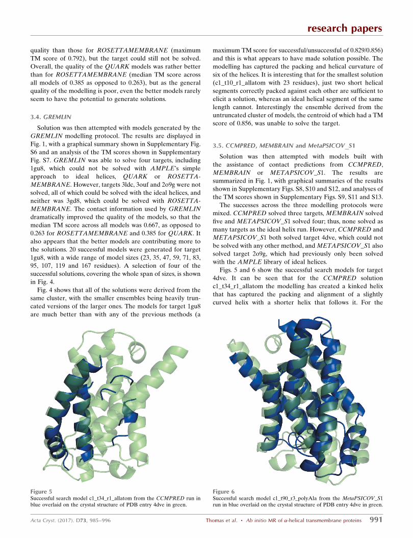
Figs. 5 and 6 show the successful search models for target
4dve. It can be seen that for the CCMPRED solution
c1_t34_r1_allatom the modelling has created a kinked helix
that has captured the packing and alignment of a slightly
curved helix with a shorter helix that follows it. For the
research papers
Acta Cryst. (2017). D73, 985–996 Thomas et al. � Ab initio MR of �-helical transmembrane proteins 991
Figure 5Successful search model c1_t34_r1_allatom from the CCMPRED run inblue overlaid on the crystal structure of PDB entry 4dve in green.
Figure 6Successful search model c1_t90_r3_polyAla from the MetaPSICOV_S1run in blue overlaid on the crystal structure of PDB entry 4dve in green.
METAPSICOV_S1 search model c1_t90_r3_polyAla, the
modelling performed extremely well (TM score of 0.7201 for
the full model used as the ensemble centroid) and has
captured both the overall packing and curvature of the helices.
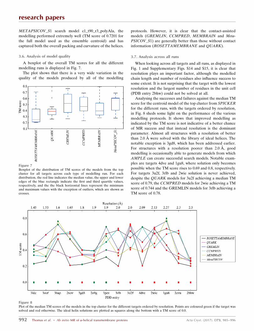
3.6. Analysis of model quality
A boxplot of the overall TM scores for all the different
modelling runs is displayed in Fig. 7.
The plot shows that there is a very wide variation in the
quality of the models produced by all of the modelling
protocols. However, it is clear that the contact-assisted
models (GREMLIN, CCMPRED, MEMBRAIN and Meta-
PSICOV_S1) are generally better than those without contact
information (ROSETTAMEMBRANE and QUARK).
3.7. Analysis across all runs
When looking across all targets and all runs, as displayed in
Fig. 1 and Supplementary Figs. S14 and S15, it is clear that
resolution plays an important factor, although the modelled
chain length and number of residues also influence success to
some extent. It is not surprising that the target with the lowest
resolution and the largest number of residues in the unit cell
(PDB entry 2bhw) could not be solved at all.
Analysing the successes and failures against the median TM
score for the centroid model of the top cluster from SPICKER
for the different runs, with the targets ordered by resolution,
in Fig. 8 sheds some light on the performance of the various
modelling protocols. It shows that improved modelling as
indicated by the TM score is not indicative of a better chance
of MR success and that instead resolution is the dominant
parameter. Almost all structures with a resolution of better
than 2.0 A were solved with the library of ideal helices. The
notable exception is 3gd8, which has been addressed earlier.
For structures with a resolution poorer than 2.0 A, good
modelling is occasionally able to generate models from which
AMPLE can create successful search models. Notable exam-
ples are targets 4dve and 1gu8, where solution only becomes
possible when the TM score rises to 0.69 and 0.8, respectively.
For targets 3u2f, 3rlb and 2wie solution is never achieved,
despite the QUARK models for 3u2f achieving a median TM
score of 0.79, the CCMPRED models for 2wie achieving a TM
score of 0.744 and the GREMLIN models for 3rlb achieving a
TM score of 0.78.
research papers
992 Thomas et al. � Ab initio MR of �-helical transmembrane proteins Acta Cryst. (2017). D73, 985–996
Figure 7Boxplot of the distribution of TM scores of the models from the topcluster for all targets across each type of modelling run. For eachdistribution, the red line indicates the median value, the upper and loweredges of the blue rectangle indicate the first and third quartile values,respectively, and the the black horizontal lines represent the minimumand maximum values with the exception of outliers, which are shown ascrosses.
Figure 8Plot of the median TM scores of the models in the top cluster for the different targets ordered by resolution. Points are coloured green if the target wassolved and red otherwise. The ideal helix solutions are plotted as squares along the bottom with a TM score of 0.0.
That targets 3u2f, 3rlb and 2wie were not solved despite
reasonable models being generated could be an indication that
the crystallographic data were particularly poor. However, an
analysis of crystallographic quality metrics in Supplementary
Table S3, such as the redundancy, Rmerge and I/�(I), did not
indicate that any of these targets differed systematically from
the others. An example of such an analysis is Supplementary
Fig. S16, which shows the TM score plotted against the I/�(I)
of the highest resolution shell and demonstrates that there is
no particular pattern between the data quality as measured by
this metric, the quality of the models and what could be
solved.
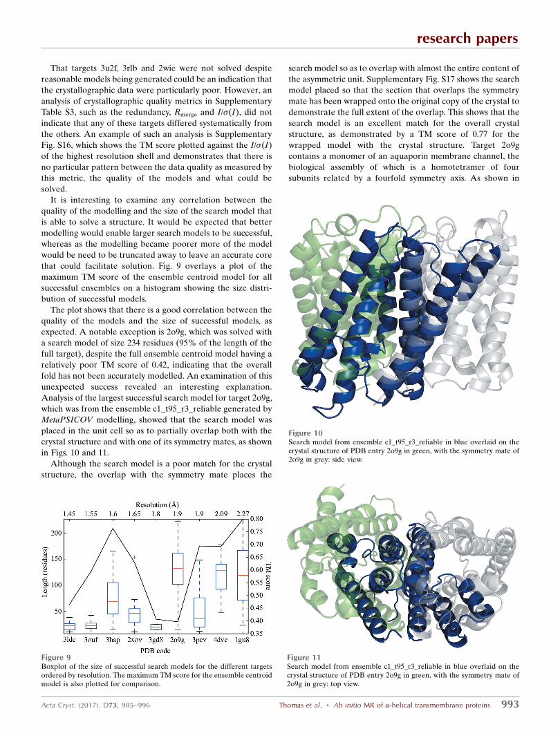
It is interesting to examine any correlation between the
quality of the modelling and the size of the search model that
is able to solve a structure. It would be expected that better
modelling would enable larger search models to be successful,
whereas as the modelling became poorer more of the model
would be need to be truncated away to leave an accurate core
that could facilitate solution. Fig. 9 overlays a plot of the
maximum TM score of the ensemble centroid model for all
successful ensembles on a histogram showing the size distri-
bution of successful models.
The plot shows that there is a good correlation between the
quality of the models and the size of successful models, as
expected. A notable exception is 2o9g, which was solved with
a search model of size 234 residues (95% of the length of the
full target), despite the full ensemble centroid model having a
relatively poor TM score of 0.42, indicating that the overall
fold has not been accurately modelled. An examination of this
unexpected success revealed an interesting explanation.
Analysis of the largest successful search model for target 2o9g,
which was from the ensemble c1_t95_r3_reliable generated by
MetaPSICOV modelling, showed that the search model was
placed in the unit cell so as to partially overlap both with the
crystal structure and with one of its symmetry mates, as shown
in Figs. 10 and 11.
Although the search model is a poor match for the crystal
structure, the overlap with the symmetry mate places the
search model so as to overlap with almost the entire content of
the asymmetric unit. Supplementary Fig. S17 shows the search
model placed so that the section that overlaps the symmetry
mate has been wrapped onto the original copy of the crystal to
demonstrate the full extent of the overlap. This shows that the
search model is an excellent match for the overall crystal
structure, as demonstrated by a TM score of 0.77 for the
wrapped model with the crystal structure. Target 2o9g
contains a monomer of an aquaporin membrane channel, the
biological assembly of which is a homotetramer of four
subunits related by a fourfold symmetry axis. As shown in
research papers
Acta Cryst. (2017). D73, 985–996 Thomas et al. � Ab initio MR of �-helical transmembrane proteins 993
Figure 9Boxplot of the size of successful search models for the different targetsordered by resolution. The maximum TM score for the ensemble centroidmodel is also plotted for comparison.
Figure 10Search model from ensemble c1_t95_r3_reliable in blue overlaid on thecrystal structure of PDB entry 2o9g in green, with the symmetry mate of2o9g in grey: side view.
Figure 11Search model from ensemble c1_t95_r3_reliable in blue overlaid on thecrystal structure of PDB entry 2o9g in green, with the symmetry mate of2o9g in grey: top view.
Supplementary Fig. S18, the search model sits perfectly on the
interface between two monomers.
Where a protein is active as an obligate oligomer, evolu-
tionary covariance will emerge in an intermolecular fashion at
the subunit interfaces. Since there are no reliable methods
to distinguish between intramolecular and intermolecular
contact pairs in homo-oligomers, and the latter were also
present in the sets used to drive modelling, the folding will try
to satisfy all corresponding restraints. The aquaporin chain
contains two subdomains that are structurally similar and are
considered to have arisen by evolutionary duplication (Park &
Saier, 1996). The METAPSICOV_S1 model represents two
subdomains spanning a subunit interface, rather than two
subdomains within a single chain, yet the accurate interface
packing unexpectedly captured by the modelling allows
successful structure solution.
In order to test our hypothesis that intermolecular contact
predictions drove the successful METAPSICOV_S1 model-
ling, we compared the set of predictions against the crystal
structure. A comparison of the predicted METAPSICOV_S1
contact pairs against the contact pairs extracted from the
monomer at 8 A distance between C� and C� (C� in case of
Gly) atoms indicated a high precision of 62.4%. Indeed,
looking only at the dimer interface, 21 contacts in the top L
pairs were predicted correctly with an average confidence
score of 0.435. In particular, two hotspot residues alone, 45
and 102, cover seven correctly predicted contacts (Supple-
mentary Fig. S19). Similarly, in the CCMPRED top L contact
pairs, 23 contacts were found in the dimer interface, although
the corresponding models were unsuccessful. The
MEMBRAIN server did not predict a single contact pair
across the dimer interface.
The placing of a search model so as to overlap with a
symmetry mate is something that has been observed
previously with AMPLE. In part, it was inaccurate positioning
of long helical fragments so as to overlap with a neighbouring
symmetry mate in the first AMPLE paper (Bibby et al., 2012)
that prompted our work on coiled-coil proteins (Thomas et al.,
2015), where we also observed this phenomenon. In these
cases, though, it was largely the fortuitous placement of a
fragment that happened to cross the boundary of the asym-
metric unit that facilitated solution, although correct generic
helical packing modes were correctly captured. The solution
of target 2o9g is different as the addition of the contact
information has explicitly resulted in the modelling, albeit in
an intramolecular fashion, of the intermolecular interface.
4. Discussion
This exploration of the ability of AMPLE to solve �-helical
transmembrane proteins was prompted by our earlier
successes solving small globular proteins, where 80% of the
entirely �-helical structures could be solved (Bibby et al.,
2012), and with coiled-coil proteins (Thomas et al., 2015),
where again 80% of the structures could be solved. A notably
positive outcome of this work is the solution of all bar one of
the targets with a resolution of better than 2.0 A using a small
library of eight ideal helices.
Using ideal helical fragments to solve structures has
been demonstrated before by programs such as ARCIM-
BOLDO_LITE (Sammito et al., 2015), and a transmembrane
structure was solved with helical fragments using ARCIM-
BOLDO_BORGES (Sammito et al., 2013; Millan et al., 2015),
for example. However, these approaches use a more
sophisticated MR procedures than AMPLE does, and
ARCIMBOLDO_BORGES employs a curated library of
fragments derived from existing structures. That a simple MR
approach with a small library of ideal helices can solve so
many of this test set is encouraging, as it shows that neither
laborious experimental phasing nor relatively computationally
expensive modelling or MR protocols may be required to
solve transmembrane proteins with a resolution better than
2.0 A.
For the current target set, where the resolution is poorer
than 2.0 A some form of ab initio modelling is required in
order to generate a sufficiently large search model to enable
solution. The two contact-free modelling protocols,
ROSETTAMEMBRANE and QUARK, were able to generate
solutions, but the overall quality of the modelling was poor
and the protocols could only solve one structure that could not
be solved with AMPLE’s set of ideal helices.
The addition of inter-residue contact pairs results in a
marked increase in the overall quality of the models and
facilitates the solution of two structures that could not be
solved using any other protocol. These two structures were
both at moderate resolution (2.09 A for target 4dve and
2.27 A for target 1gu8), and target 4dve, with 594 residues, was
the second largest by number of residues in the asymmetric
unit. This improvement of the modelling when contact-
prediction information is included is especially encouraging, as
contact prediction is a fast-moving field that is evolving and
improving rapidly: deep learning, for example, seems to hold
particular promise (Wang et al., 2017), and metagenomic data
will inevitably spread the availability of this information
across more protein families (Ovchinnikov et al., 2017).
Transmembrane proteins seem to offer both particular chal-
lenges and opportunities. On the one hand their low packing
densities and water-containing cavities are likely to weaken
the covariance signal that contact prediction relies on (Rose et
al., 2014) and the relatively limited number of transmembrane-
protein structures available limits the accuracy with which
biophysical parameters relevant to methods development can
be determined (Li et al., 2017). On the other hand, the
fundamentally limited range of intramembrane packing
interactions facilitates the development of bespoke trans-
membrane-protein methods of contact prediction (see, for
example, Li et al., 2017). For the same reason, there are good
reasons to think that specific transmembrane-protein ab initio
modelling protocols will produce better results than general
methods. It is unfortunate that the one attempted here
(ROSETTAMEMBRANE) does not appear to be being
actively developed, but other methods are in development
(Law et al., 2017).
research papers
994 Thomas et al. � Ab initio MR of �-helical transmembrane proteins Acta Cryst. (2017). D73, 985–996
A particularly interesting feature of the contact modelling
was the generation of a homodimer interface for the target
2o9g by METAPSICOV_S1. Although this facilitated the
solution of target 2o9g, it demonstrates the current inability of
contact-generation algorithms to separate the intramolecular
and intermolecular contacts. Although either set of contacts
can be useful for different purposes (Simkovic, Ovchinnikov et
al., 2017), until it becomes possible to separate them the
modelling will attempt to satisfy both sets at the same time,
which can confidently be predicted to result in poorer models
than if a single set were used. Curiously, high-quality models
with TM scores of around 0.8 were calculated for this target,
but clusters containing them were not sources of successful
search models.
As well as highlighting the value of a putative method to
distinguish intramolecular and intermolecular contact
predictions, the results offer other pointers towards future
productive algorithmic developments. When the modelling
performs well, AMPLE is often able to generate an ensemble
that can solve the structure; this applies to both high-
resolution and low-resolution structures. With high-resolution
structures, SHELXE is able to trace up to a full structure, even
when only a small fragment has been placed correctly, as is the
case for ideal helices. With high-resolution structures, when
the modelling is relatively poor, AMPLE must prune away
enough of the incorrect structure to leave a sufficiently
accurate substructure suitable for MR and tracing with
SHELXE. Sometimes the truncation algorithm in AMPLE is
able to perform this (examples include targets 3ouf and
2xov), although at other times it fails and the truncated
ensemble is unable to solve the structure, whereas
an ideal helix of the same length can. In these cases, it
will be of value to explore whether bespoke truncation
protocols for transmembrane proteins could improve
performance.
All of this work was undertaken with CCP4 v.6.5.3
(including SHELXE v.2014/14 and ARP/wARP v.7.5), which
was released on 3 July 2015. This was necessary to ensure that
the different runs of AMPLE with the different modelling
protocols were comparable. As of the time of writing CCP4 is
at v.7.0.045 (including SHELXE v.2017/1) and considerable
improvements have been made in the CCP4 software
packages that AMPLE relies on. In related work (unpub-
lished) we have run a number of the cases in this work with a
newer version of CCP4 and observed significant improve-
ments in our success rate. We therefore expect that a user of
AMPLE using the very latest version of CCP4 would have an
even better chance of solving their structure than the work in
this paper suggests.
In summary, this work shows that AMPLE can already
solve many (9/14) of a set of �-helical transmembrane
proteins. Higher resolution cases can be attempted first using
ideal helices, while others yield to the ability of AMPLE to
construct MR search ensembles from ab initio models. Targets
for which contact predictions can be calculated show a distinct
benefit from their use. As data volume and methods devel-
opment push contact prediction to better performance in the
future, the overall success rate of AMPLE can be expected to
improve further.
Funding information
This work was supported by BBSRC grant BB/L009544/1
‘CCP4 Grant Renewal 2014–2019: Question-driven crystallo-
graphic data collection and advanced structure solution’. FS is
supported by BBSRC DTP PhD scholarships, as was JT at the
time of the work.
References
Almen, M., Nordstrom, K. J. V., Fredriksson, R. & Schioth, H. B.(2009). BMC Biol. 7, 50.
Altschul, S. F., Madden, T. L., Schaffer, A. A., Zhang, J., Zhang, Z.,Miller, W. & Lipman, D. J. (1997). Nucleic Acids Res. 25, 3389–3402.
Alva, V., Nam, S.-Z., Soding, J. & Lupas, A. N. (2016). Nucleic AcidsRes. 44, W410–W415.
Bansal, M., Kumart, S. & Velavan, R. (2000). J. Biomol. Struct. Dyn.17, 811–819.
Berman, H. M., Westbrook, J., Feng, Z., Gilliland, G., Bhat, T. N.,Weissig, H., Shindyalov, I. N. & Bourne, P. E. (2000). Nucleic AcidsRes. 28, 235–242.
Bibby, J., Keegan, R. M., Mayans, O., Winn, M. D. & Rigden, D. J.(2012). Acta Cryst. D68, 1622–1631.
Cowtan, K. (2006). Acta Cryst. D62, 1002–1011.Ellis, J. J., Huard, F. P. E., Deane, C. M., Srivastava, S. & Wood, G. R.
(2010). BMC Bioinformatics, 11, 172.Fagerberg, L., Jonasson, K., von Heijne, G., Uhlen, M. & Berglund, L.
(2010). Proteomics, 10, 1141–1149.Hanwell, M. D., Curtis, D. E., Lonie, D. C., Vandermeersch, T., Zurek,
E. & Hutchison, G. R. (2012). J. Cheminform. 4, 17.He, B., Mortuza, S. M., Wang, Y., Shen, H.-B. & Zhang, Y. (2017).
Bioinformatics, 33, 2296–2306.Holm, L. & Sander, C. (1991). J. Mol. Biol. 218, 183–194.Jones, D. T. (1999). J. Mol. Biol. 292, 195–202.Jones, D. T., Buchan, D. W. A., Cozzetto, D. & Pontil, M. (2012).
Bioinformatics, 28, 184–190.Jones, D. T., Singh, T., Kosciolek, T. & Tetchner, S. (2015).
Bioinformatics, 31, 999–1006.Kajan, L., Hopf, T. A., Kalas, M., Marks, D. S. & Rost, B. (2014). BMC
Bioinformatics, 15, 85.Katzman, S., Barrett, C., Thiltgen, G., Karchin, R. & Karplus, K.
(2008). Bioinformatics, 24, 2453–2459.Keegan, R. M., Bibby, J., Thomas, J., Xu, D., Zhang, Y., Mayans, O.,
Winn, M. D. & Rigden, D. J. (2015). Acta Cryst. D71, 338–343.Kim, D. E., Chivian, D. & Baker, D. (2004). Nucleic Acids Res. 32,
W526–W531.Krivov, G. G., Shapovalov, M. V. & Dunbrack, R. L. Jr (2009).
Proteins, 77, 778–795.Kumar, P. & Bansal, M. (2012). J. Biomol. Struct. Dyn. 30, 773–783.Langer, G., Cohen, S. X., Lamzin, V. S. & Perrakis, A. (2008). Nature
Protoc. 3, 1171–1179.Law, E. C., de Oliveira, S. H. P., Kelm, S., Shi, J. & Deane, C. M.
(2017). Biophys. J. 112, 61a.Li, W. & Godzik, A. (2006). Bioinformatics, 22, 1658–1659.Li, Z., Wang, S., Yu, Y. & Xu, J. (2017). arXiv:1704.07207.McCoy, A. J., Grosse-Kunstleve, R. W., Adams, P. D., Winn, M. D.,
Storoni, L. C. & Read, R. J. (2007). J. Appl. Cryst. 40, 658–674.Meiler, J. & Baker, D. (2003). Proc. Natl Acad. Sci. USA, 100, 12105–
12110.Michel, M., Hayat, S., Skwark, M. J., Sander, C., Marks, D. S. &
Elofsson, A. (2014). Bioinformatics, 30, i482–i488.Millan, C., Sammito, M. & Uson, I. (2015). IUCrJ, 2, 95–105.Moraes, I., Evans, G., Sanchez-Weatherby, J., Newstead, S. & Shaw
Stewart, P. D. (2014). Biochim. Biophys. Acta, 1838, 78–87.
research papers
Acta Cryst. (2017). D73, 985–996 Thomas et al. � Ab initio MR of �-helical transmembrane proteins 995
O’Leary, N. A. et al. (2016). Nucleic Acids Res. 44, D733–D745.Ovchinnikov, S., Kamisetty, H. & Baker, D. (2014). Elife, 3, e02030.Ovchinnikov, S., Kinch, L., Park, H., Liao, Y., Pei, J., Kim, D. E.,
Kamisetty, H., Grishin, N. V. & Baker, D. (2015). Elife, 4, e09248.Ovchinnikov, S., Park, H., Varghese, N., Huang, P.-S., Pavlopoulos,
G. A., Kim, D. E., Kamisetty, H., Kyrpides, N. C. & Baker, D.(2017). Science, 355, 294–298.
Park, J. H. & Saier, M. H. Jr (1996). J. Membr. Biol. 153, 171–180.Read, R. J. & McCoy, A. J. (2016). Acta Cryst. D72, 375–387.Rodrıguez, D. D., Grosse, C., Himmel, S., Gonzalez, C., de Ilarduya,
I. M., Becker, S., Sheldrick, G. M. & Uson, I. (2009). NatureMethods, 6, 651–653.
Rohl, C. A., Strauss, C. E. M., Misura, K. M. S. & Baker, D. (2004).Methods Enzymol. 383, 66–93.
Rose, A., Theune, D., Goede, A. & Hildebrand, P. W. (2014). NucleicAcids Res. 42, D347–D351.
Rotkiewicz, P. & Skolnick, J. (2008). J. Comput. Chem. 29, 1460–1465.Sammito, M., Millan, C., Frieske, D., Rodrıguez-Freire, E., Borges,
R. J. & Uson, I. (2015). Acta Cryst. D71, 1921–1930.Sammito, M., Millan, C., Rodrıguez, D. D., de Ilarduya, I. M., Meindl,
K., De Marino, I., Petrillo, G., Buey, R. M., de Pereda, J. M., Zeth,K., Sheldrick, G. M. & Uson, I. (2013). Nature Methods, 10, 1099–1101.
Seemayer, S., Gruber, M. & Soding, J. (2014). Bioinformatics, 30,3128–3130.
Shrestha, R., Berenger, F. & Zhang, K. Y. J. (2011). Acta Cryst. D67,804–812.
Shrestha, R. & Zhang, K. Y. J. (2015). Acta Cryst. D71, 304–312.Simkovic, F., Ovchinnikov, S., Baker, D. & Rigden, D. J. (2017).
IUCrJ, 4, 291–300.Simkovic, F., Thomas, J. M. H., Keegan, R. M., Winn, M. D., Mayans,
O. & Rigden, D. J. (2016). IUCrJ, 3, 259–270.
Simkovic, F., Thomas, J. M. H. & Rigden, D. J. (2017). Bioinformatics,33, 2209–2211.
Simons, K. T., Kooperberg, C., Huang, E. & Baker, D. (1997). J. Mol.Biol. 268, 209–225.
Simons, K. T., Strauss, C. & Baker, D. (2001). J. Mol. Biol. 306, 1191–1199.
Tai, C.-H., Bai, H., Taylor, T. J. & Lee, B. (2014). Proteins, 82, Suppl. 2,57–83.
The UniProt Consortium (2017). Nucleic Acids Res. 45, D158–D169.
Thomas, J. M. H., Keegan, R. M., Bibby, J., Winn, M. D., Mayans, O. &Rigden, D. J. (2015). IUCrJ, 2, 198–206.
Thorn, A. & Sheldrick, G. M. (2013). Acta Cryst. D69, 2251–2256.Tusnady, G. E., Dosztanyi, Z. & Simon, I. (2004). Bioinformatics, 20,
2964–2972.Viklund, H. & Elofsson, A. (2008). Bioinformatics, 24, 1662–1668.Wang, S., Sun, S., Li, Z., Zhang, R. & Xu, J. (2017). PLoS Comput.
Biol. 13, e1005324.Wang, Y., Virtanen, J., Xue, Z., Tesmer, J. J. G. & Zhang, Y. (2016).
Acta Cryst. D72, 616–628.White, S. H. & Wimley, W. C. (1999). Annu. Rev. Biophys. Biomol.
Struct. 28, 319–365.Winn, M. D. et al. (2011). Acta Cryst. D67, 235–242.Xiao, F. & Shen, H.-B. (2015). J. Chem. Inf. Model. 55, 2464–2474.Xu, D., Rahman, N. A. B. D., Othman, R., Hu, P. & Huang, M. (2012).
Proteins, 80, 2154–2168.Xu, D. & Zhang, Y. (2013). Sci. Rep. 3, 1895.Yarov-Yarovoy, V., Schonbrun, J. & Baker, D. (2006). Proteins, 62,
1010–1025.Zhang, Y., Kolinski, A. & Skolnick, J. (2003). Biophys. J. 85, 1145–
1164.Zhang, Y. & Skolnick, J. (2004). J. Comput. Chem. 25, 865–871.
research papers
996 Thomas et al. � Ab initio MR of �-helical transmembrane proteins Acta Cryst. (2017). D73, 985–996