Prevalence of anemia and iron deficiency anemia in Chinese ...

APPROACH TO ANEMIA
William Lamb Jr. D.O., FACP Director Osteopathic Medical Education, UPMC Shadyside
Definition
• Males Hgb 14-17 • Females Hgb 12-16

Causes
• Blood loss • Decreased production • Increased destruction (hemolysis)
RBC FORMATION
• Production requires: • erythropoietin • iron (red meat, poultry, fish) • B 12
History
• Family hx of anemia • Drugs, use or abuse • Last Hgb?

Symptoms
• Asymptomatic • Tachycardia • DOE, chest pain • Fatigue, malaise • pallor

Exam
• Pallor • Lymphadenopathy • Petichiae • Heme + stool • Glossitis • Stomatitis
Reticulocyte count
• Key to initial classification of anemia • Residual RNA in newly made RBC • Normal is 23,000 – 90,000 • > 110,000 suggests normal marrow
response to anemia • 2-3 x normal is expected within 10 days of
onset of anemia.

Reticulocyte production Index
• Retic count x (measured Hgb/14)/2 Divided by 2 for maturation time correction,
in the face of anemia, reticulocytes are released from the marrow prematurely, so this additional correction may be needed
• If polychromasia (normoblasts) are not seen on smear, this correction is not needed

Physiologic Classification of Anemia
High ------------------------- Retic count -------------------Low or NL l l Bleeding---yes---Blood loss Smear no l l l l Smear Micro Normo
Macro Schistocytes IDA Aplasia B12 Spherocytes Thal Marrow infilt
Folate Sickle cells sideroblast Renal dx MDS Bite cells Inflammation Drug Tox Target cells Chronic dx Etoh Inclusions

• Mayo clinic algorithm

HYPOCHROMIC MICROCYTIC
• Iron Deficiency • Thalassemia • Chronic Disease, Inflammatory (usually
normo, normo) • Myelodysplastic syndrome (ringed
sideroblasts seen on bone marrow)
Iron Deficiency
• Low Fe , high TIBC, low Ferritin. High RDW
• Reduced transferrin saturation (Fe/TIBC) • If ferritin < 15, essentially no Fe stores • Iron absorbed proximal small bowel:
celiac dx, IBD, resection can affect • May see accompanying thrombocytosis
Iron Deficiency • Signs and symptoms • fatigue, malaise • headaches • pallor • pagophagia (ice chewing) • pica • glossitis • stomatitis • koilonychia (spooning of nails) • chronic blood loss is always suspect in adults
Iron Deficiency Treatment • Correct underlying problem if blood loss • FeSO4 325 mg tid is least expensive, watch for
nausea and constipation • Slow Fe preparations not advised as absorption
is markedly reduced • Parenteral Fe therapy available for the rare
patient who does not tolerate oral • Duration: when Hgb normalizes, treat for
additional 3-6 months to replenish the Fe stores
Thalassemia
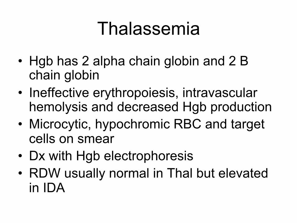
• Hgb has 2 alpha chain globin and 2 B chain globin
• Ineffective erythropoiesis, intravascular hemolysis and decreased Hgb production
• Microcytic, hypochromic RBC and target cells on smear
• Dx with Hgb electrophoresis • RDW usually normal in Thal but elevated
in IDA

Thalassemia
A single gene deletion is an asymtomatic carrier state that is normal clinically and hematologically
Alpha Thal Minor or Alpha Thal trait is a two gene deletion and is usually asymptomatic, mild anemia, microcytosis
• African, Mediterranean, SE Asia, Middle Eastern descent
• No treatment is needed

Thalassemia
• Hemoglobin H disease is caused by deletion of 3 alpha genes
• Severe anemia with CHF and hypoxia • Hydrops fetalis or Hemoglobin Bart is
caused by 4 gene deletion of the alpha chain and usually causes intrauterine demise
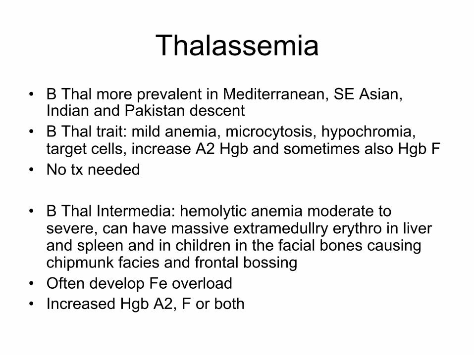
Thalassemia • B Thal more prevalent in Mediterranean, SE Asian,
Indian and Pakistan descent • B Thal trait: mild anemia, microcytosis, hypochromia,
target cells, increase A2 Hgb and sometimes also Hgb F • No tx needed
• B Thal Intermedia: hemolytic anemia moderate to severe, can have massive extramedullry erythro in liver and spleen and in children in the facial bones causing chipmunk facies and frontal bossing
• Often develop Fe overload • Increased Hgb A2, F or both
Thalassemia
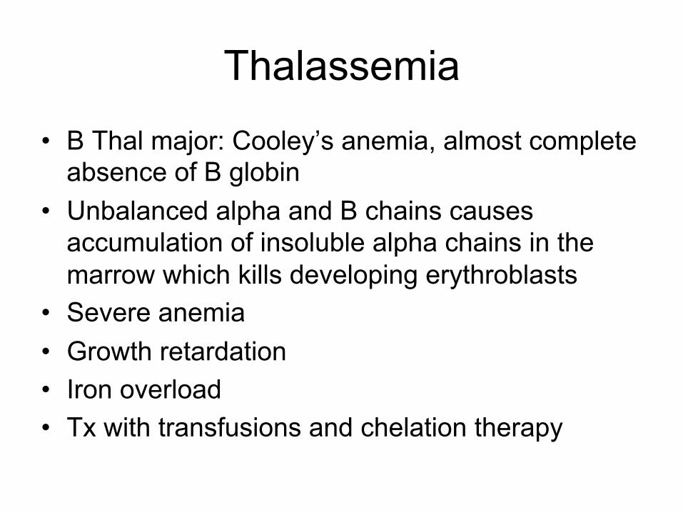
• B Thal major: Cooley’s anemia, almost complete absence of B globin
• Unbalanced alpha and B chains causes accumulation of insoluble alpha chains in the marrow which kills developing erythroblasts
• Severe anemia • Growth retardation • Iron overload • Tx with transfusions and chelation therapy
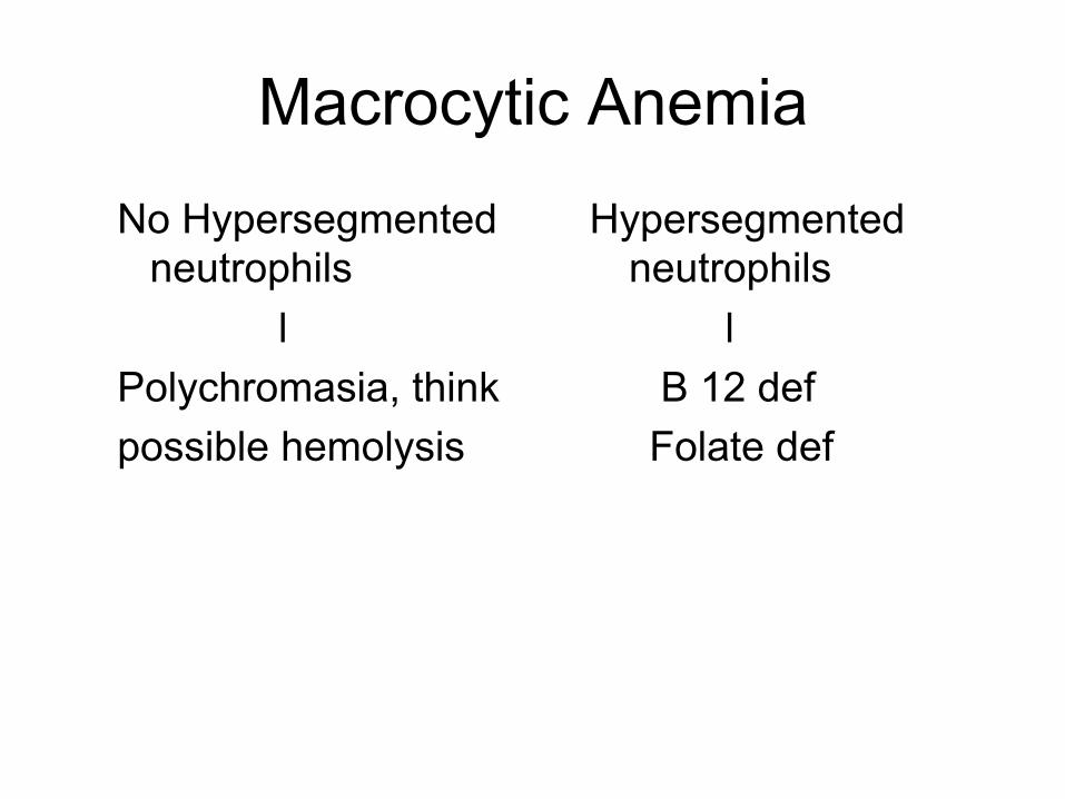
Macrocytic Anemia
No Hypersegmented Hypersegmented neutrophils neutrophils
l l Polychromasia, think B 12 def possible hemolysis Folate def
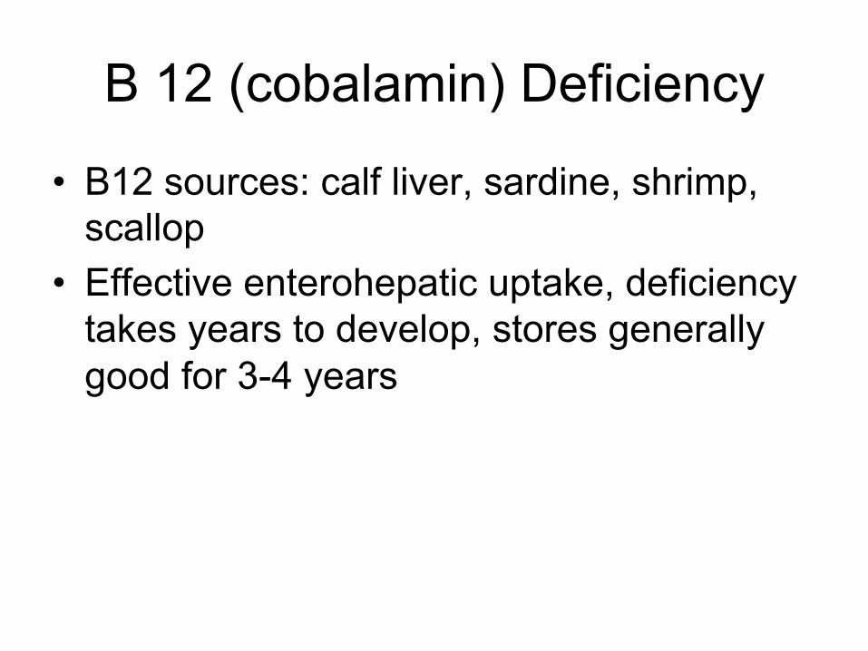
B 12 (cobalamin) Deficiency
• B12 sources: calf liver, sardine, shrimp, scallop
• Effective enterohepatic uptake, deficiency takes years to develop, stores generally good for 3-4 years

B 12 deficiency
• Decreased absorption: • Pernicious anemia (antibody directed
destruction directed at parietal cells) • Aging with achlorhydria • Celiac disease • Pancreatic insufficiency • Bacterial overgrowth

B12 deficiency exam
• Glossitis • Pallor • Jaundice possible due to ineffective
erythropoiesis • Neuropathy • Spastic ataxia • Dementia • Psychosis
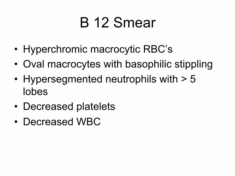
B 12 Smear
• Hyperchromic macrocytic RBC’s • Oval macrocytes with basophilic stippling • Hypersegmented neutrophils with > 5
lobes • Decreased platelets • Decreased WBC

B 12 Diagnosis
• B12 level • Methylmalonic acid level is more sensitive
than measuring the B 12 level and will be high with B 12 deficiency (expensive test)
• Homocysteine level should be high • Haptoglobin level may be decreased • LDH may be high • Indirect Bili may be high

B 12 Treatment
• B12 1000 mcg IM per month, 6 injections spaced every 3-7 days should replenish stores
• Oral replacement has been shown to be as effective and less costly at 1000-2000Mcg/ day
• Hemoglobin levels may take several months to normalize, if they do not, consider alternative diagnosis such as MDS
• Neuropsychiatric disorders take longer to resolve and ultimately may not

Folate Deficiency
• Sources: green leafy vegs., melons, lemons, bananas, fortified grains
• Dietary deficiency is unusual • Stores are only 3-4 months • Triamterene, Phenytoin accelerate folate
metabolism • Alcohol decreases folate absorption • It is best to measure erythrocyte folate levels,
serum folate may go up quickly with a meal
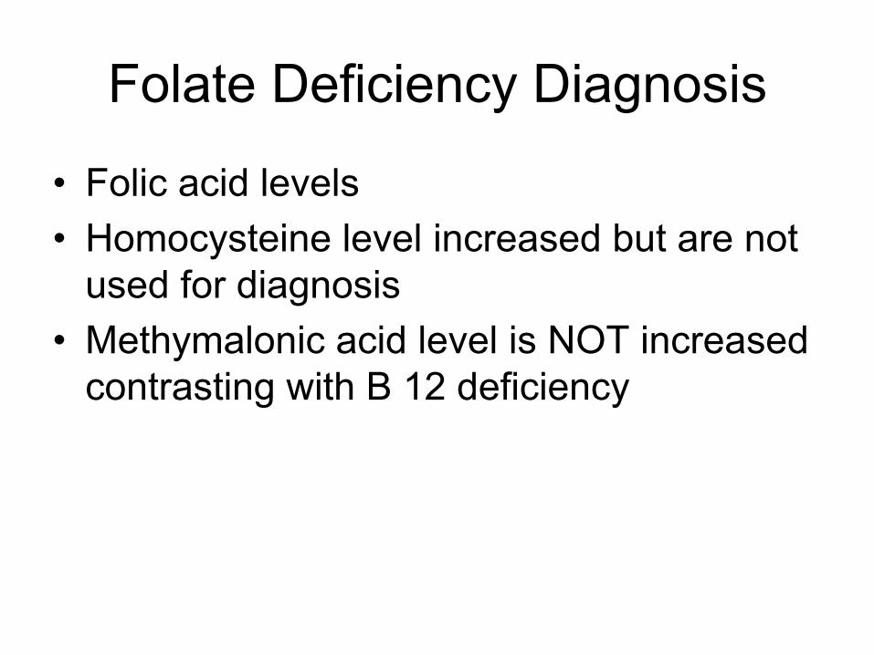
Folate Deficiency Diagnosis
• Folic acid levels • Homocysteine level increased but are not
used for diagnosis • Methymalonic acid level is NOT increased
contrasting with B 12 deficiency

Folate Deficiency treatment
• Folic acid 5-15 mg daily • Will take about 4 months to treat • Always check B 12 level before giving the
Folic acid, if this is low and not corrected, the anemia will improve but not the Neuropsych symptoms
• Long term therapy may be needed for patients with a degree of hemolysis or those on dialysis

INFLAMMATORY ANEMIA
• Inflammation • Infection • Tissue injury • Other conditions (cancer for one) causing
release of inflammatory cytokines such as TNF alpha, IL 6, IL 1, and interferon
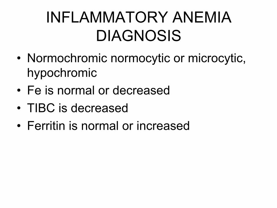
INFLAMMATORY ANEMIA DIAGNOSIS
• Normochromic normocytic or microcytic, hypochromic
• Fe is normal or decreased • TIBC is decreased • Ferritin is normal or increased
INFLAMMATORY ANEMIA TREATMENT
• No treatment needed in general • Use Epo carefully as this may cause Htn
and increase risk of thrombosis

ANEMIA WITH NORMOCHROMIC NORMOCYTIC INDICES
• Renal disease: level of anemia correlates with degree of renal failure
• Normo, normo • Fe levels, TIBC, ferritin all normal • Those on dialysis may also develop Fe
deficiency • Epo levels low

ANEMIA WITH NORMOCHROMIC NORMOCYTIC INDICES
• Liver disease • Smear: spur cells, stomatocytes • If Alcoholic, may have also Folate deficit
and may have blood loss so check the Iron levels also

HEMOLYTIC ANEMIA
• Signs: DOE, pallor, jaundice, gallstones, splenomegaly
• Labs: LDH increased Indirect Bili increased Haptoglobin decreased
HEMOLYTIC ANEMIA
CONGENITAL • Erythrocyte membrane – spherocytosis • Erythrocyte enzyme – G6PD • Defective Hgb structure – Sickle,
Thalassemias

HEMOLYTIC ANEMIA CONGENITAL
• G6PD deficiency – Male > Female, AA most common
• Episodic hemolysis in response to stressors (infection, TMP-SMX, Nitrofurantoin), fava beans
• Heterozygotes protected to some degree against P. falciparum
• Smear : Bite cells • Tx: supportive, withdraw offending agent
HEMOLYTIC ANEMIA ACQUIRED
Autoimmune: Warm – IgG 80% mild splenomegaly Direct Coombs + 90%, weakly + C3 spherocytes may be seen Treatment: Prednisone 1 mg/kg splenectomy Rituximab Cytoxan

HEMOLYTIC ANEMIA ACQUIRED • Autoimmune: Cold agglutinin– IgM 20% Direct Coombs neg, positive C3 clumped RBC may develop weeks after EBV or mycoplasma infection Treatment: Warm clothes Chlorambucil, Cytoxan, Rituxan Steroids and splenectomy not effective Plasmapheresis for acute
HEMOLYTIC ANEMIA ACQUIRED
• Microangiopathic smear shows helmet cells or schistocytes
• TTP, DIC, HUS, aged mechanical heart valve
• Plasma exchange may be lifesaving for TTP and HUS

HEMOLYTIC ANEMIA ACQUIRED
PNH • stem cell disorder with hemolytic anemia,
pancytopenia and atypical thrombosis (mesenteric, cerebral).
• Dx based on flow cytometry • Tx with anticoagulants, Eculizimab helpful,
steroids may help occasionally but ultimately require immunosuppressants or allogeneic BMT
• Median survival 10-15 years
HEMOLYTIC ANEMIA ACQUIRED
• Exposures – Malaria, Arsine gas, Babesiosis (Nantucket, Cape Cod, NC), Clostridial sepsis
• Venoms – brown recluse spider bite, snakes, massive wasp or bee stings
• Drugs – Cyclosporine, Tacrolimus, Clopidogrel, Ticlopidine
• Copper toxicity – Wilson’s disease • Severe burns, Radiation

SMEARS • Microcytosis : Fe def, Inflammatory, Thal • Macrocytes: B12, Folate, MDS • Spherocytes: Hereditary Spherocytosis • Target cells: Hemoglobinopathy, Liver disease,
splenectomy • Burr cells: Kidney dx • Bite cells: G6PD • Spur cells: severe liver dx • Sickle cells: SCD • Nucleated RBC: marrow stress (hemolysis, hypoxia) • Teardrop cells: Fibrosis, infiltrative marrow dx, marrow
granuloma
(from Harrison’s Principles of Internal Medicine)


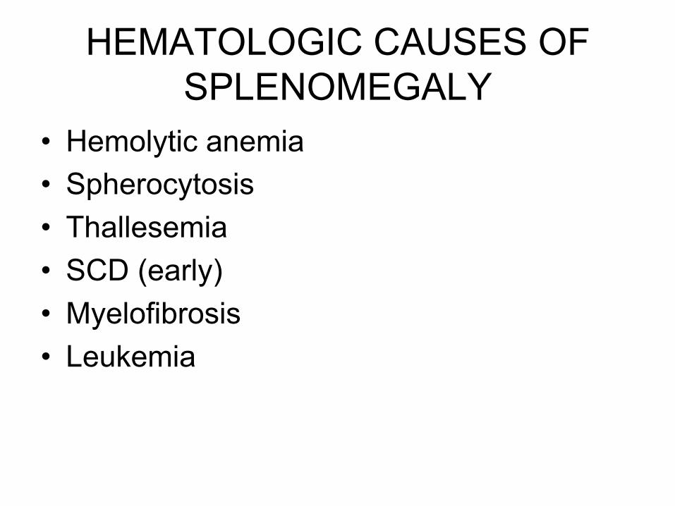
HEMATOLOGIC CAUSES OF SPLENOMEGALY
• Hemolytic anemia • Spherocytosis • Thallesemia • SCD (early) • Myelofibrosis • Leukemia

HEMATOLOGIC CAUSES OF MASSIVE SPLENOMEGALY
>1000 grams Thallesemia CML CLL Lymphoma Hairy Cell Leukemia P. Vera Myelofibrosis Autoimmune Hemolytic Anemia
QUESTIONS
• 1. In B12 Deficiency, the Haptoglobin level may be decreased
• A. True • B. False
QUESTIONS
2. In Fe deficiency anemia: • A. it will take 3-6 months after the Hgb is
normalized to replace Fe stores • B. Slow Fe is preferred replacement • C. Thrombocytosis is often present • D. RDW is usually normal 1. A, B, C 2. A, C 3. B,D 4. all of the above
QUESTIONS
3. In Inflammatory Anemia • A. cells are Normo/normo or micro/hypo • B. Fe is normal or decreased • C. TIBC is decreased • D. Ferritin is low • 1. A,B, and C 2. A and C 3. B and D 4. All
of the above.