
Approach To A Patient With Single Red Hot...
51
Approach To A Patient With Single Red Hot Joint Dr. Syed Atiqul Haq Professor & Chairman, Deptt of Rheumatology, BSM Medical University, Dhaka Treasurer, APLAR (Asia Pacific League of Associations for Rheumatology) Vice President, Bangladesh Rheumatology Society
Transcript of Approach To A Patient With Single Red Hot...

Approach To A Patient With
Single Red Hot Joint
Dr. Syed Atiqul Haq Professor & Chairman, Deptt of Rheumatology, BSM
Medical University, Dhaka Treasurer, APLAR (Asia Pacific League of Associations
for Rheumatology) Vice President, Bangladesh Rheumatology Society

Scenario 1…
• A 38-year-old man presented with severe pain at the root of
his right great toe that started at midnight and awakened him
– It reached maximum intensity over 6 hours
• He had an episode of painful swelling of left ankle 2 yrs ago
– That episode resolved with 1 day self-treatment with diclofenac
• No FH of joint, skin or eye disease

Scenario 1: Examination
• Mildly overweight. BP 150/95. GE unremarkable

Important Features in Dx of Gout
• Past history of acute monoarthritis
• History of spontaneous resolution of pain
• Presence of tophi
• MSU crystals in SF

Scenario 2
• A 48-year-old man with well-controlled type 2 DM presented
with acute painful swelling of the left knee for 3 days
– The pain reached its peak over about a day
– Unabated despite intake of multiple doses of pain killers
• The patient never experienced similar joint pain in the past
• No FH of joint, skin or eye disease
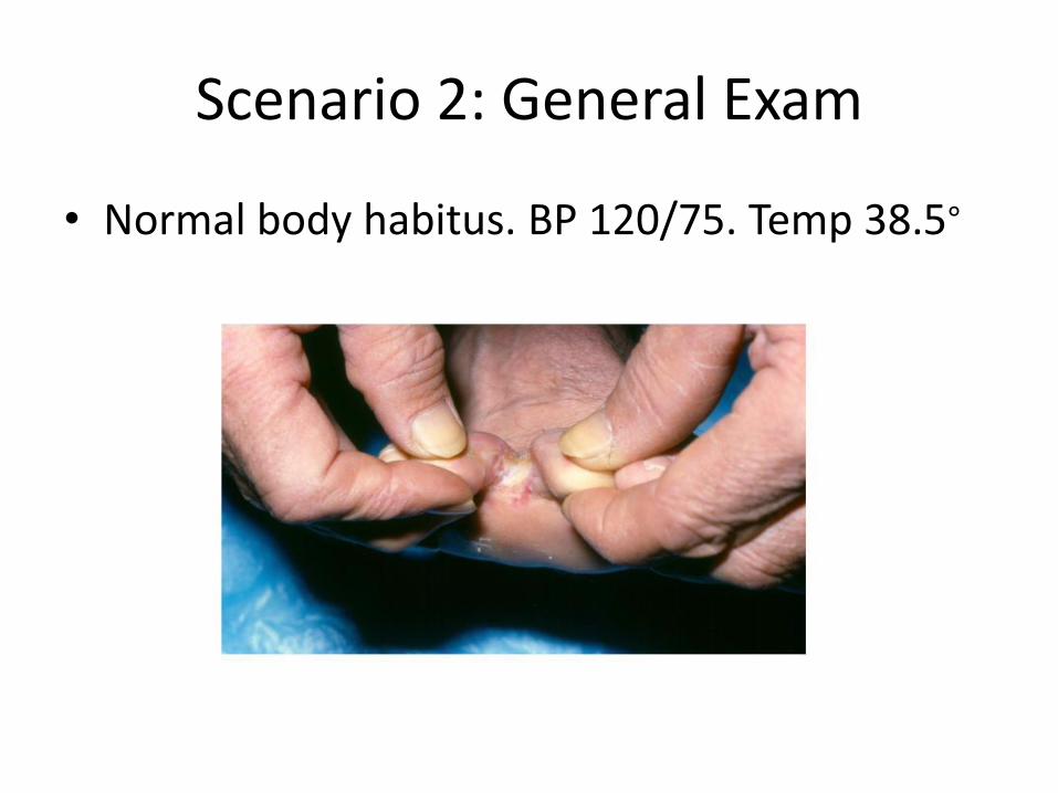
Scenario 2: General Exam
• Normal body habitus. BP 120/75. Temp 38.5∘

Scenario 2: Systemic
• Examination of other
systems and MSK
system other than the lt
knee: unremarkable
• CBC: TWBC 17,500; Poly
90%

Joint aspiration

Findings Important for Septic Arthritis
• No past episode
• No spontaneous decrease in pain
• Presence of fever and redness over the joint
• H/O recent joint injury
• SF TWBC >50X109/L, 90% of WBCs PMN

Organisms Causing Septic Arthritis
• Staph. aureus (↑ MRSA), S. epidermidis
• Streptococci
• Neisseria gonorrheae
• Gram –ve aerobic/anerobic organisms

Consequences of Delay in Dx
Bacteremia
Sepsis
Prolonged suffering
Joint destruction
Disability
A need for prosthetic joint surgery
Death

Scenario 3
• A 58-year-old woman with pre-existing chronic
mechanical knee pain presented with acute painful
swelling of her right knee for 7 days:
– Her knee hit the table shelf 1 day before the onset
– She had two episodes of milder exacerbation of pain
lasting 3 & 5 days during preceding 1 month
– Past and FH unrevealing

Scenario 3: Physical & Lab
• Obese
• Afebrile
• MSK other than
knee normal
• Other systems:
normal
• TWBC: 12,300, P 78%
• S. Uric acid 7.3 mg/dl

Scenario 4: Clinical
• A 28-year-old man has presented with severe pain in
PIP of right middle finger since previous afternoon
– It reached peak in 1 hr
– Suffering from such episodes in 1 to 3 joints over last 9 months
– Duration 2 hours to 7 days, mostly 1-3 days, intervals variable
– Affects mostly upper limb joints
– Points to periarticular areas and muscles as sites in past episodes
– No residual pain or disability in between
– No preceding loose motions, urethral discharge

Exam and Lab
• No abnormality on GE, other systems, other
parts of MSK system
• ESR 47, RF weakly +ve
• CBC, S uric acid, ACPA, HLA B 27, SI jts normal

Other Causes of Acute Monoarthritis
Trauma: sprain
Hemarthrosis
Osteoarthritis
Reactive arthritis
Psoriatic arthritis
Rheumatoid arthritis
Sarcoidosis
SLE


Steps of Evaluation
History
Physical Examination
Investigation

“The initial approach to diagnosing acute
monoarthritis should involve the completion
of both a history and a physical examination”
Baker DG & Schumaker HR Jr.
Acute monoarthritis. NEJM 1993; 329: 1013—20.


History • Socio-demographic background
• Pre-existing chronic illnesses and treatment
• History of present illness:
– Full description of the pain
– Summary of other symptoms
– Systemic enquiry
• Past history
• Family history
• Personal history
• Menstrual history

Socio-demographic Background
• Gout: middle aged men, low SE class, alcohol
• Pseudogout: elderly
• Ankle sprain: young men, sport
• Hemophilia: young boys

Pre-existing Chronic Illnesses and Treatment
• Gout: psoriasis, CKD, myeloproliferative disease,
cyanotic heart diseases, diuretics, pyrazinamide, anti-
cancer drugs
• Septic arthritis: immunocomromised state including
DM, RA, joint injection, septic foci
• Pseudogout: osteoarthritis, primary
hyperparathyroidism, hemochromatosis, Wilson’s

Summary of Other Symptoms
• Fever: septic arthritis
• Cough: sarcoidosis, TB, pneumonia with septic arthritis
• Painful red eye: spondyloarthropathies, sarcoidosis
• Conjunctivitis, preceding diarrhea, urethral discharge: reactive
• Chronic GI symptoms: enteropathic arthritis
• Generalized aches, fatigue, anorexia, polyuria, ureteric colic:
hyperparathyroidism with pseudogout

Past History
• Painful red eyes: spondarthropathies
• Inflammatory back pain: SpAs
• Similar episodes:
– Same joint: sprain, hemophilia, PVNS
– Same or different joints: gout, pseudogout, PR

Family History
• Rheumatic diseases: SpAs, hemophilia,
uncommon forms of gout
• Painful red eyes: SpAs
• Psoriasis: gout, PsoA
• TB: tubercular arthritis


Physical Examination
General examination
Examination of other systems
Examination of the musculo-skeletal system

General Examination
• Obesity: gout, pseudogout
• Cachexia: TB
• Cyanosis: cyanotic heart disease
• Polycythemia: polycythemia vera
• Lymphadenopathy: septic, sarcoidosis, TB,
neoplastic

Tophi

Systemic Examination
• CVS: Tachycardia and hypotension: sepsis, soft S1, murmurs
• Lungs: tachypnea--sepsis
• Liver, spleen, abdominal lumps, testicular enlargement: neoplastic, gout
• Cranial neuropathies: SLE, sarcoidosis, vasculitis
• GPN
• Mononeuritis multiplex

GALS

Screening MSK Exam
• Posture and gait
• Metacarpal and metatarsal squeeze
• SchÖber test
• Detailed examination of affected joint

“Must verify that the pain is truly monoarticular”
Ensworth S. Rheumatology: 1. Is it arthritis? CMAJ 2000; 1011--16


SchÖber Test

• Up to 20% of patients presenting with acute
knee monoarthritis progress to develop RA
-- Tenaka et al. Mod Rheuma 2001; 11: 61—64
• Up to 25% of IBD patients present with acute
lower limb large joint monoarthritis
-- Holden et al. Rheum Dis Clin N Am 2003; 29: 513—30


Principles • Choice of investigations depends on history and
examination
– No set of tests is routine for all scenarios
• Cost-effectiveness
• May not require investigations
– acute monoarthritis in an established case of gout or
clinically obvious ankle sprain ankle sprain

Somewhat Routine
• CBC
• Synovial fluid study: most valuable:
– Naked eye inspection
– Cell counts
– Crystals: polarized light
– Gram stain
– CS

“Acute monoarthritis should be considered infectious until proven
otherwise”
Goldenberg DL. Septic arthritis. Lancet 1998; 197--202
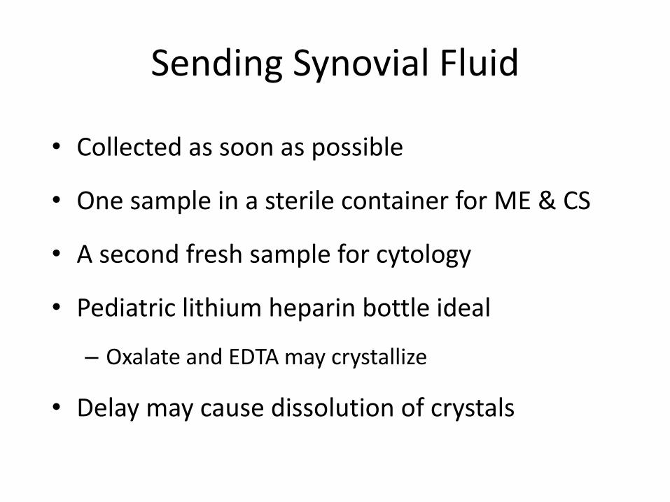
Sending Synovial Fluid
• Collected as soon as possible
• One sample in a sterile container for ME & CS
• A second fresh sample for cytology
• Pediatric lithium heparin bottle ideal
– Oxalate and EDTA may crystallize
• Delay may cause dissolution of crystals

Selective
• Serum uric acid
• X-ray of the joint with contralateral
• Chest X-ray
• X-ray/ MRI SI joints
• Other imaging, e.g., USG, CT scan
• HLA B27, RF, ACPA, ANA
• Factors VIII, IX


Customizable, Not Routine Protocol
• Scenario 1: CBC, S. uric acid, SF study
• Scenario 2: CBC, SF study incl. CS, blood CS, CS
of swab from interdigital infection
• Scenario 3: CBC, SF study
• Scenario 4: CBC, SF study
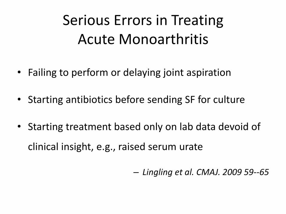
Serious Errors in Treating Acute Monoarthritis
• Failing to perform or delaying joint aspiration
• Starting antibiotics before sending SF for culture
• Starting treatment based only on lab data devoid of
clinical insight, e.g., raised serum urate
– Lingling et al. CMAJ. 2009 59--65

Conclusions
• Considered infectious unless proved otherwise
• Redness indicates infection or crystal
• S. fluid study mandatory if infection suspected
• Acute monoarthritis is a medical emergency:
– must be investigated and treated promptly
• Cibere J. Rheumatology: 4. Acute monoarthritis. CMAJ 2000;
1577—83


![Recent and Current Positions - Texas Tech Universityp3e.ttu.edu/personnel/bayneCV.pdf · 2017-02-27 · [28] Sandeep Nimmagadda*, Atiqul Islam*, Stephen B. Bayne, R.P. Walker, Lourdes](https://static.fdocuments.in/doc/165x107/5f6cbededd182342b46e1400/recent-and-current-positions-texas-tech-2017-02-27-28-sandeep-nimmagadda.jpg)


