Appraisal, work-up and diagnosis of anterior uveitis · Anterior uveitis is the term used for the...
18
Page 1 sur 18 Appraisal, work-up and diagnosis of anterior uveitis A practical approach Dr. Carl P. Herbort, MD Ophthalmologist FMH Privat-docent (PD) & former Master of Education and Research (fMER) at the University of Lausanne Private Centre : Retinal, Inflammatory and External Eye Diseases Centre for Specialised Ophthalmic Care (COS) 6, Rue de la Grotte CH-1003 LAUSANNE Switzerland Fax : +41 21 648 60 10 E-mail : [email protected]
Transcript of Appraisal, work-up and diagnosis of anterior uveitis · Anterior uveitis is the term used for the...

Page 1 sur 18
Appraisal, work-up and diagnosis of anterior uveitis A practical approach
Dr. Carl P. Herbort, MD Ophthalmologist FMH Privat-docent (PD) & former Master of Education and Research (fMER) at the University of Lausanne Private Centre : Retinal, Inflammatory and External Eye Diseases Centre for Specialised Ophthalmic Care (COS) 6, Rue de la Grotte CH-1003 LAUSANNE Switzerland
Fax : +41 21 648 60 10
E-mail : [email protected]

Page 2 sur 18
Introduction The diagnostic yield in uveitis has significantly improved in the last 15 to 20 years, in part because
a clear classification is available to the clinician but even more so because diagnostic tests have
greatly improved lately.
With a systematic approach, based on history and clinical examination and guided by laboratory
tests and other investigational tests, a diagnosis can be expected in about 70% of cases.
A compilatory approach such as it is exposed in most uveitis textbooks has to be avoided. (Figure
1a & 1b) Endless lists of diseases the exposure to which the patient is supposed to tick on a check-
list are not contributing to a diligent work-up of uveitis and are even counterproductive as this may
push the clinician into the wrong direction.
Fig. 1a & 1b Example of the traditional lists that are
featured in most uveitis books
It is much more productive to use a comprehensive approach that avoids a detailed anamnesis (history) at
the beginning of the consultation but limits the initial anamnesis (history) to the immediate complaints of the
patient followed immediately with a thorough clinical examination. Once the the clinical signs have been
established and have allowed to characterize the type of uveitis (granulomatous versus non-granulomatous)
the anamnesis (history) can be resumed by asking oriented questions. The clinician should then try to
classify the uveitis more precisely, refer to the local epidemiological data and come up with a working
diagnosis, which he should always be prepared to change. At this point targeted investigations and
laboratory tests should be performed. The result is a diagnosis which has higher degree of probability that
can be followed by appropriate therapy. In case no dignosis can be suspected non specific therapy may
have to be started or therapy can be withheld. In a proportion of cases the evolution of the disease will lead
to the diagnosis. (Flow-chart 1)
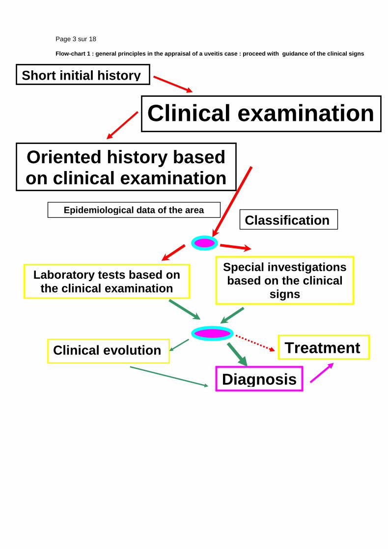
Page 3 sur 18
Flow-chart 1 : general principles in the appraisal of a uveitis case : proceed with guidance of the clinical signs
Short initial history
Clinical examination
Oriented history based on clinical examination
Classification Epidemiological data of the area
Laboratory tests based on the clinical examination
Special investigations based on the clinical
signs
Clinical evolution
Diagnosis
Treatment

Page 4 sur 18
The work-up of anterior uveitis
Definition
The anatomical classification of uveitis into anterior, intermediate and posterior forms is
very useful to conduct the work-up and eventually to reach a diagnosis, even though inflammation does not
always respect these anatomical boundaries. Anterior uveitis is the term used for the group of inflammatory
disorders for which the preponderant part of the inflammation is situated at the level of the pars plicata of
the ciliary body, the retroiridal space, the iris and the anterior chamber.
Symptoms and signs of anterior uveitis
The severity of symptoms in anterior uveitis ranges from no symptoms in chronic disease such as anterior
uveitis related to juvenile idiopathic arthritis (JIA) to very severe symptoms in acute uveitis such as HLA-
B27 related uveitis. Symptoms of acute anterior uveitis include photophobia, redness, pain, decreased
vision and tearing in the absence of discharge.
The signs of anterior uveitis are listed in table 1
____________________________________________________________________
Table 1. Signs of anterior uveitis.
1.1.Conjunctival injection
1.2. Keratic precipitates
1.3. Aqueous flare / fibrinous clots (Figure 2)
1.4. Posterior synechiae between the iris and the capsule of the lens (Figure 3)
1.5. Aqueous cells / hypopyon (Figure 4)
1.6. Iris rubeosis (usually reversible)
1.7. Iris nodules (Koeppe / Busacca) (Figures 5a & 5b)
1.8. Iris atrophy (herpes uveitis and Fuchs’ uveitis)
1.9. Intraocular pressure changes (hypotony in severe acute anterior non-granulomatous uveitis; hypertony
in granulomatous uveitis)
____________________________________________________________________
Figure 2. Fibrinous clot in anterior chamber in typical case of HLA-B27 related uveitis.

Page 5 sur 18
Figure 3. Posterior irido-lenticular synechiae. The ring of pigment deposits on the cristalline lens show
where the iris was attached (synechiae). There is one remaining synechia at six o’clock on the verge of
detaching itself following the administration of massive dilating drops.
Figure 4. Hypopyon. Sedimented white cells form a level at the bottom of the anterior chamber, associated
with a ring of fibrine on the surface of the cristalline lens (top) indicating broken synechiae. One remaining
synechia on the meridian of 7 o’clock.
Figure 5a. Koeppe nodules. Two Koeppe nodules at the border of the iris associted with synechiae.

Page 6 sur 18
Figure 5b. Busacca Nodules. UBM picture showing nodule within the stroma in a patient with tuberculous
uveitis.
1. The conjunctival injection in anterior uveitis can be diffuse or localized circumferentially at the limbus
(perikeratic injection) or mixed (diffuse and perikeratic injection).
2. The morphology of keratic precipitates (KPs) is very useful to help distinguish non-granulomatous from
granulomatous uveitis. Small diffuse KPs causing dusting of the endothelium are characteristic for non-
granulomatous uveitis such as HLA-B27 related acute anterior uveitis. When KPs become larger than
endothelial dust they can be individualised and correspond to granulomatous KPs. The morphology and
distribution of granulomatous KPs is often useful in orienting towards a more specific diagnosis within
granulomatous causes (table 2). Medium and large size KPs are called "mutton fat" KPs. There is a lot of
confusion regarding the characterisation of KPs. In many textbooks only mutton-fat KPs are termed
granulomatous and small granulomatous KPs as those found in Fuchs’ uveitis are erroneously termed as
non granulomatous. Therefore Fuchs’ uveitis is mistakenly classified as non-granulomatous in many
textbooks. It is useful to distinguish between fresh and chronic (old) muton-fat KPs. Old mutton-fat KPs
tend to be less white, pigmented and less dense in the centre.
____________________________________________________________________

Page 7 sur 18
Table 2. Granulomatous keratic precipitates (KPs)
Type of KP Clinical entity to suspect
Small stellate, even distribution no gravitation Fuchs' (heterochromic) uveitis (Figure 6)
Small gravitational or inferior random distribution CMV uveitis (Figure 7)
Small/medium/ size, focal spherical distribution
under stromal keratitis h.simplex/zoster keratouveitis (Figure 8)
Medium/large gravitational distribution
(mutton-fat) sarcoidosis, tuberculosis, toxoplasmosis (Figure 9)
Very few (2-5)small/medium/large size,
inferior peripheral (iridocorneal angle) Posner-Schlossmann syndrome
____________________________________________________________________
Figure 6. Fuchs uveitis. Small stellate non-gravitationally distributed keratic precipitates (KPs) typical of Fuchs’ uveitis.
Figure 7. KPs in CMV anterior uveitis. Scarce unilateral KPs of different sizes (small to medium) in a case of cytomegalovirus (CMV) anterior uveitis.
Figure 8a. Herpes KPs. Usually herpes/zoster KPs are arranged in a round disciform pattern and are small
to medium sized..

Page 8 sur 18
Figure 8b. Herpes KPs. Usually arranged in a round disciform pattern very well visible against
retroillumination. Note also sectorial iris atrophy at the top of the picture at the level of the superior iris.
Figure 9. Mutton-fat KPs. This type of gravitational large KPs can be seen in tuberculosis and sarcoidosis.
In toxoplasmosis the anterior inflammation sometimes associated with the retinitis is also made of large
mutton-fat KPs, although the KPs are usually less numerous.
3. Anterior chamber flare is caused by exudation of proteins into the normally clear aqueous humor from
iris vessels or accross the ciliary body epithelium following the breakdown of the blood-aqueous barrier. The
intensitiy of flare is measured in a standard fashion following the grading system proposed by the Proctor
Group in San Francisco in 1959 (table 3.)1 that is however only qualitative. A beam 1mm wide and 3mm
long with the highest light intensity and 16X magnification on the BQ Haag-Streit slitlamp is used. When the
concentration of proteins in the aqueous is very high, they agglomerate and form fibrinous clots, a finding
more common in acute non-granulomatous uveitis. (Figure 2)
4. Depending on the amount and composition of aqueous inflammatory proteins adherences between the
iris and anterior capsule of the cristalline lens can form (posterior synechiae). (Figures 3 & 4)
Table 3. Slit-lamp grading of aqueous flare (1 mm X 3mm beam)
No flare 0
faint , just detectable +
moderate, iris details clear ++

Page 9 sur 18
marked, iris details hazy +++
Intense, fibrin ++++
____________________________________________________________________
Since a few years it is now possible to measure flare in a quantitative and objective fashion, using laser
flare photometry (LFP). (Figure 10) This new technology makes flare the only quantitative parameter to
measure intraocular inflammation. So far cells were juged more accurate to measure inflammatory activity
in uveitis. At best this measurement is however only semi-quantitative. LFP was shown to be more sensitive
than slit-lamp assessement of cells to measure the evolution of inflammatory activity, making flare the new
gold standard to assess intraocular inflammation. LFP allows to detect subclinical flare intensities and
changes, that can be predictive of clinical recurrence. It allows to detect resistance of inflammation to
treatment and closer follow-up of therapy, often leading to corticosteroid sparing in the treatment. When
available, laser flare photometry certainly allows improved management of uveitis.2
5. Aqueous cells used to be the reference parameter for inflammatory activity because their evaluation
was quantifiable by slit-lamp examination. Nowadays this is no more true when LFP is available. LFP is the
quantifiable gold standard to measure inflammatory activity even in chronic inflammation with chronic
breakdown of the hemato-ocular barriers, as it has been shown that even when there are no cells, LFP can
detect active inflammation that responds to therapy. Grading of cells in the anterior chamber has been
standardized by Hogan et al. at the Proctor Foundation in 1959.1(table 4)
____________________________________________________________________
Table 4. Slit-lamp grading of aqueous cells (1 mm X 3-4mm beam)
No cells 0
1-5 cells ±
6-10 cells +
11-20 cells ++
21-50 cells +++
> 50 cells ++++
____________________________________________________________________
It is important to make the difference between pigment clumps and inflammatory cells and to examine the
anterior chamber prior to mydriasis as cells and especially pigment dispersion can sometimes be seen after
pupillary dilatation.
When the quantity of cells is very dense they sediment and cause a hypopyon, a sign more often seen in
HLA-B27 related uveitis, Behçet's uveitis and uveitis related to juvenile idiopathic arthritis (JIA). (Figure 4)
6. In severe and longstanding uveitis iris rubeosis can develop. It is in fact most often a pseudo-rubeosis
that is reversible after introduction of anti-inflammatory treatment. Even when a real rubeosis has

Page 10 sur 18
developed, it is usually situated at the pupillary border of the iris and is much less agessive and proliferative
than ischemic rubeosis iridis. In Fuchs’ uveitis with extensive iris atrophy iridal vessels can be seen and
correspond to a pseudo-rubeosis.
7. Iris nodules (Figures 5a & 5b)
In granulomatous uveitis two types of iris nodules can develop. When situated at the pupillary margin
(and on the surface of the iris) they are called Koeppe nodules, have a fluffy appearance and a size going
from very small barely visible excresences to frank nodules. When nodules are situated in the body of the
iris stroma they are called Bussaca nodules.
8. Sectorial or widespread iris atrophy is a sign rather specific for herpes simplex or herpes zoster uveitis
and is a useful diagnostic help. Diffuse atrophy is very often seen in Fuchs’ uveitis.
9. Intraocular pressure changes due to uveitis can present either as hypotension or hypertension.
Hypotony is usually measured in severe uveitis involving the ciliary body such as acute anterior non-
granulomatous HLA-B-27 related uveitis. Hypertony is usually associated with granulomatous uveitis,
especially herpes simplex or herpes zoster uveitis but also sarcoidosis or spill-over anterior uveitis
associated with toxoplasmic retinochoroiditis. In recent years, CMV anterior uveitis, a newly recognised
usually unilateral entity very often causing hypertony has been described. 3
The work-up of anterior uveitis
The anatomical diagnosis of anterior uveitis has first to be verified by excluding spill-over inflammation
associated with uveitis of the posterior segment (intermediate or posterior uveitis). To exclude posterior
involvement pupil dilatation is mandatory in all cases. Secondly, the type of clinical presentation has to
be characterized as non-granulomatous or granulomatous in order to correctly orient work-up and
differential diagnosis.
Non-granulomatous uveitis is characterised mainly by the type of keratic precipitates that presents as fine
KPs producing endothelial dusting. In severe cases fibrinous clotting or hypopyon can occur depending on
whether protein influx or cellular infiltration is predominant. In case of severe inflammation it is also
common to find posterior synechiae and pressure tends to be more often decreased than increased.
____________________________________________________________________
Table 5. Common causes of non-granulomatous anterior inflammation
in anterior uveitis or panuveitis
HLA-B27 related uveitis
Behçet's uveitis
Juvenile rhumatoid arthritis related uveitis
Uveitis associated with scleritis
Uveitis associated with streptococcal infection

Page 11 sur 18
____________________________________________________________________
Granulomatous uveitis is characterized by KPs that are larger than the dusty KPs of non-granulomatous
uveitis. They are better individualized but their size varies depending on the inflammatory process. The
medium and large size granulomatous KPs are called mutton-fat KPs. (Figure 9) Other characteristic
features of granulomatous uveitis are Koeppe and Bussaca nodules. (Figures 5a & 5b) Synechiae are
common in more pronounced inflammation. Pressure changes when present are usually characterized by
increased intraocular pressure.
Although this distinction is a very useful working classification, the subdivision is not an absolute one. A
granulomatous uveitis may initially present as non-granulomatous when dusty KPs are very thick so that
before taking its granulomatous aspect. Conversely in rare cases a non-granulomatous uveitis can take an
aspect that might be qualified as granulomatous. In most cases the distinction is however rather well
delineated
____________________________________________________________________
Table 6. Common causes of granulomatous anterior inflammation
1. in anterior uveitis
Fuchs' uveitis Herpetic (herpes simplex, varicella-zoster, Epstein-Barr) uveitis CMV anterior uveitis Posner-Schlossmann syndrome Sarcoidosis Tuberculosis Syphilis
2. in panuveitis
Sarcoidosis Vogt-Koyanagi -Harada syndrome, sympathetic ophthalmia Tuberculosis Syphilis Toxoplasmic retinochoroiditis Necrotizing herpetic retinopathies (acute retinal necrosis) Post-surgical inflammatiom (propionibacterium acnes)
____________________________________________________________________
Work-up of non-granulomatous anterior uveitis
In case of simple, fibrinous or hypopyon nongranulomatous uveitis, the only first-line work-up test we
presently perform is the detection of the HLA-B27 antigen. HLA-B27 testing is performed even if the
inflammation is only moderate. If the test is positive it will avoid further unnecessary testing during a
subsequent episode and it is reassuring for the patient and the doctor to know the specific diagnosis,
especially when it is a benign disease. In case of a positive result no further investigation is performed at
the ophthalmological level. It is however recommended to take an oriented history that will allow, with the
help of the internist or rhumatologist when necessary, to subclassify the affection into ankylosing
spondylarhritis, Reiter's syndrome, Crohn's disease, ulcerative colitis or simply into HLA-B27 uveitis without
systemic associated disease. About 50-55% of acute anterior non-granulomatous uveitis are HLA-B27

Page 12 sur 18
positive in Europe, but this varies from one geographical area to another, being for instance quite low in
Japan. In the remaining 45-50%of cases a specific diagnosis is more difficult to establish.4
If the episode of HLA-B27 negative non-granulomatous uveitis is of limited severity
and / or responds readily to classical topical corticosteroid therapy no further investigation is performed.
Flow-chart 2 : diagnostic steps in non-granulomatous uveitis
Diagnostic approach : non-granulomatous anterior uveitis
HLA-B27 antigen
Resolution
Ankylosing spondylithis, Reiter, HURC,
Crohn,
Psorriasis
Positive
Non-granulomatous
anterior uveitis
Behçet’s uveitis
HLA-B27
uveitis
Negative
Juvenile idiopathic arthritis
(JIA)
No further
work-up
Recurrence
In children
Hypopyon Aphtae, genital
ulceration
( HLA-B51)
Work-up as for granulomatous
uveitis
Examine posterior
segment !!!
ANA
Bilaterality
Renal pathology Β-2-micro- globulin
TINU-syndrome
ANA = antinuclear antibodies ; HURC = haemorhagic ulcerative recto-colitis; TINU = Tubulo-interstitial nephritis and uveitis

Page 13 sur 18
In case of an anterior uveitis with hypopyon signs and symptoms found in Behçet's syndrome should be
searched for, in particular oral and/or genital ulcerations, cutaneous signs such as erythema nodosum and
pustules, arthralgies, thrombophlebitis or central nervous system involvement. If Behçet's uveitis is
suspected we find it useful to look for the HLA-B51 antigen that, when present, represents an aditional
argument for the diagnosis of Behçet's uveitis, especially in the milder forms of Behçet's uveitis seen in the
european caucasian population. Isolated anterior Behçet's uveitis can occur but posterior involvement
should be searched for by funduscopy and is best investigated by performing a fluorescein angiography
looking for retinal vasculitis.
In case of non-granulomatous uveitis in children (with or without band keratopathy) history should be
directed towards juvenile idiopathic arthritis (JIA). Inflammatory symptoms can be completely absent,
contrasting with the severe signs of uveitis such as hypopyon and extensive synechiae that can
characterise JIA related uveitis. Uveitis is usually associated with the pauciarticular form of JIA and testing
should include anti-nuclear antibodies (ANA) that are present in up to 70% of JRA patients with uveitis. In
elderly children it is also useful to test for the presence of HLA-B27 antigen.
A bilateral non granulomatous uveitis in children, but also in adults, should prompt to search or exclude
tubulointerstitial nephritis and uveitis syndrome (TINU), an often neglected diagnosis. Renal function should
be tested, starting with the dosage of creatininemia requiring sometimes renal biopsy and urinalysis should
be performed looking for glucosuria and dosage of beta-2-microglobulin which is found to be elevated
inTINU.5
In children pars planitis can initially present with a pronounced anterior participation and can be mistaken
for an anterior uveitis if the posterior segment is not carefully analyzed.
In case of non-responding HLA-B27 negative anterior uveitis or in case of recurrence we pursue the work-
up in the same fashion as for a granulomatous uveitis
Work-up of granulomatous uveitis
Before starting the work-up of granulomatous uveitis, it is important to exclude Fuchs' heterochromic uveitis
because this condition, when sufficiently typical, does not need any work-up. Furthermore corticosteroid
treatment should be withheld in Fuchs’ uveitis to avoid the side-effects of a treatment that usually has no
impact on the inflammatory process. Characteristic findings of Fuchs' uveitis include fine stellate
granulomatous keratic precipitates that usually do not accumulate inferiorly by gravitation but are more
uniformly distributed over the whole surface of the endothelium,( Figure 6) fine Koeppe nodules at the
pupillary edge of the the iris, prominent vessels in the irido-corneal angle seen by gonioscopy and absence
of posterior synechiae. Heterochromia is only present in fair-coloured irises but not in dark irises. Therefore
the name of this entity should not include any more the term of heterochromic and should simply be called
Fuchs’ uveitis. It should be kept in mind that involvement can be bilateral. Laser flare photometry shows a

Page 14 sur 18
very moderate breakdown of blood-aqueous barrier of 10.2±3.5 ph/ms (normal 3.5-4.0 ph/ms) that rermains
relatively stable over time and ususally does not respond significantly to anti-inflammatory treatment.
Spill-over anterior granulomatous uveitis as it can occur in very inflammatory toxoplasmic retinochoroiditis
has to be excluded by performing a detailed examination of the posterior segment with funduscopy.
Flow-chart 3 : diagnostic steps in granulomatous uveitis.
Granulomatous uveitis : systematic diagnostic approach 1
Step 1 exclude Fuchs’
uveitis • small stellate KPs, non gravitational, regularly distributed over the cornea
• vitreous infiltration
• usually unilateral
• Koeppe nodules possible
• polar posterior cataract
• heterochromia
Step 2 : first-line lab. tests Lysozyme (L)
Angiotensin converting enzyme (ACE)
Tuberculin skin test Multiple herpetic serologies
(polyclonal activation)
Serum L & ACE elevated
DTH anergy Herpetic serologies show
polyclonal activation
Serum L & ACE elevated
Mantoux®/PPD skin test/TB +++; γ-IFN-releasing assays +
Tuberculosis
probable
Sarcoidosis
most probable
Chest X-ray,
TB cultures
Chest X-ray &/or CT-scan, BAL, Biopsy,
Ga-scan
Granulomatous
uveitis
BAL = bronchio-alveolar lavage ; DTH = delayed type hypersensitivity ; Ga-scan = galium scan ; IFN = interferon; TB = tuberculosis
Page 15 sur 18
The first-line laboratory tests performed to investigate granulomatous uveitis are serum angiotensine
converting enzyme (ACE) and lysozyme, products indicating the presence of granulomatous lesions. ACE
can be normally elevated in children and serum lysozyme levels tend to be progressively more elevated in
elderly persons. It is therefore important to perform both tests.
The second step is to differentiate between sarcoid, tuberculous or other granulomatous causes. In order to
differentiate between tuberculosis and sarcoidosis, multiple skin tests, measuring delayed type
hypersensitivity reaction to several antigens to which the adult patient should be normally reacting
(MultiMérieux® containing tuberculin, streptococcus, diphteria, tetanos, tricophyton and candida antigens)
are performed to search for anergy, a strong argument for sarcoidosis. This test is however rarely
performed nowadays as the Merieux® multi-antigen applicator is difficult to find. In a patient vaccinated for
tuberculosis or that is known to have been exposed to a tuberculous infection a PPD skin test that has
become negative has the same diagnostic value. We find it also useful to look for polyclonal antibody
activation that is present in up to 85% of the patients with sarcoidosis6,7 and was also found in patients with
ocular sarcoidosis.8 For this purpose, serologies to four herpes viruses to which most of the adult
population has been exposed (herpes simplex, herpes zoster, cytomegalovirus and Epstein-Barr) are
performed. ELISA serology detects exposure to these viruses and complement fixation serology is done to
establish whether the antibody titers are elevated. An isolated elevated titer to one virus only might be
indicative of a viral etiology. Polyclonal activation however is an additional element for sarcoidosis. This
non-specific antibody elevation is the cause of some of the false-positive diagnosis of presumed infectious
uveitis, relying only on a serology such as Lyme borreliosis. A positive serology is not a confirmation of
ocular Lyme disease. We followed five cases with uveitis and a positive Lyme serology that had negative
anterior chamber antibody ratios (Goldmann-Witmer coefficient) and for whom the diagnosis was finally
sarcoidosis. Patients with a compatible clinical picture and positive ACE & lysozyme tests in presence of
cutaneous anergy have a probability of over 95% to have ocular sarcoidosis.9
On the other hand when the PPD tuberculin skin test is hyperpositive this should raise the suspicion of a
tuberculous granlomatous uveitis. Nowadays the next test to be performed is one of the gamma-interferon
releasing assay that are testing blood lymphocytes of patients in order to detect whether there are
lymphocytes reacting in vitro when put in presence with specific proteins coming from Mycobacterium
Tuberculosis. When the patient’s lymphocytes release gamma-Interferon, it means that the patient has
been exposed to the bacteria and tuberculosis should be actively researched.10
Syphilis serology is performed either routinely or in case of a positive history. In case of undefined
diagnosis, serology for Lyme borreliosis is performed with the known limitations of the value of a positive
serology.
Toxoplasmic retinochoroiditis can sometimes present as a granulomatous (hypertensive) anterior spill-over
uveitis. The presence of a retinal focus orients clearly into this direction. In order to make the diagnosis
possible toxoplasmic serology should be performed shown simply the presence IgG antibodies indicating
that the patient has been in contact once in his life with Toxoplasma Gondii.

Page 16 sur 18
In case of negative ACE / lysozyme test and a non-contributary skin test a herpetic uveitis should be
suspected. Clinical signs that are very suggestive of herpes simplex / zoster uveitis are ocular hypertension
and iris atrophy (found both in herpes simplex and varicella-zoster uveitis). Laboratory confirmation of
herpes simplex / zoster anterior uveitis can be obtained by the detection of intraocular production of
antibodies in the aqueous humor (Goldmann-Witmer coefficient). Aqueous paracenthesis is however not
performed routinely in these cases but reserved for sight-threatening diseases such as necrotic herpetic
retinopathies (NHR) that include acute retinal necrosis. It is also performed in uveitis suspected to be
herpetic but that does not reapond to classical combined systemic antiviral and topical antiviral therapy to
detect CMV DNA in the aqueous.
A condition that can be associated with anterior granulomatous uveitis is multiple sclerosis (MS). In most
cases postrior segment findings such as priphlebitis and vitritis are usually present. In patients with a history
compatible with MS investigations should be directed towards MS beginning with a cerebral MRI.11
Figure 10. Laser flare photometry. This machine is measuring the content of proteins in the anterior
chamber and hence the level of inflammation in an objective and quantitative fashion. The instrument
shines a laser into the anterior chamber and the electronic detector is counting the number of photons that
are back-scattered. Back-scattered photons are proportional to the amount of particles (proteins) and hence
proportional to the intraocular inflammation

Page 17 sur 18
Flow-chart 4 : differential diagnosis of granulomatous uveitis.
Granulomatous uveitis : systematic diagnostic approach 2
first-line laboratory tests Lysozyme (L)
Angiotensin converting enzyme (ACE)
PPD TB skin test Multiple herpetic serologies
(polyclonal activation)
Sarcoidosis
Tuberculous uveitis (chorioretinitis)
Lyme uveitis
Syphilitic uveitis
(chorioretinitis)
Syphilis serology
Lyme serologies
Granulomatous
uveitis
L & ACE normal DTH non-
contributive +tive herpes serology
Intraocular HT Iris atrophy
Unilateral
HSV/VZV uveitis probable
R-choroidal focus
Toxopl. Gondii Abs +
Toxoplasmic retinochoroiditis
Exclude Fuchs
Uveitis !!! ACE & L positive + + anergy
-
PPD ++++ and +tive G-IFN releasing test
ACE, L negative
- -
Resits usual ttt :
CMV uveitis
CMV = cytomegalovirus; DTH = delayed type hypersensitivity ; G-IFN = gamma-interferon; HT = hypertension; TB = tuberculosis; ttt = treatment

Page 18 sur 18
References 1. Hogan MJ, Kimura SJ, Thygeson P. Signs and symptoms of uveitis. 1. Antrior uveitis. Amer J. Ophthalmol 1959; 47:155-170. 2. Herbort CP, Guex-Crosier Y, de Ancos E, Pittet N. Use of laser flare photometry to assess and monitor inflammation in uveitis. Ophthalmology 1997; 104:64-72. 3.De Schryver I, Rozenberg F, Cassoux N, Michelson S, Kestelyn P, LeHoang P, Davis JL, Bodaghi B. Diagnosis and treatment of cytomegalovirus iridocyclitis without retinal necrosis. Br J Ophthalmol 2006; 90:852-5. 4. Tran VT, Auer C, Guex-Crosier Y, Pittet N, Herbort CP. Epidemiological characteristics of uveitis in Switzerland. Internat Ophthalmol 1995; 18: 293-298. 5. Mackensen F, Smith JR, Rosenbaum JT. Enhanced recognition, treatment and prognosis of tubulointerstitial nepritis and uveitis syndrome. Ophthalmology 2007; 114:995-9. 6. James DG, Williams WJ. Immunology of sarcoidosis. Amer J Med 1982; 72:5-8. 7. Daniele RP, Dauber JH, Rossman MD. Immunologc abnormalties in sarcoidosis. Ann Int Med 1980; 92:406-416. 8. Berthoud-Kündig JF, Keller A, Herbort CP. Elévation polyclonale des immunoglobulines : une aide plutôt qu'un piège dans le diagnostic des uvéites causées par la sarcoidose. Klin Montsbl Augenheilkd 1994; 204:323-329. 9. Baarsma GS, La Hey E, Glasius E, de Vries J, Kijlstra A. The predictive value of serum converting enzyme and lysozyme levels in the diagnosis of ocular sarcoidosis. Am J Ophthalmol 1987; 104:211-7. 10. Itty S, Bakri SJ, Pulido JS, Herman DC, Faia LJ, Tufty GT, Bennett SR, Falk NS. Initial results of Quantiferon-TB Gold testing in patients with uveitis. Eye 2009; 23:904-9. 11. Lim JI, Tessler HH, Goodwin JA. Anterior granulomatous uveitis in patients with multiple sclerosis. Ophthalmology 1991; 98:142-145.


![The Management of Acute Anterior Uveitis Complicating ...downloads.hindawi.com/journals/bmri/2018/9460187.pdf · acute anterior uveitis (AAU) []. e latter is commonly reported as](https://static.fdocuments.in/doc/165x107/5f0b5eb77e708231d4302ec5/the-management-of-acute-anterior-uveitis-complicating-acute-anterior-uveitis.jpg)