Applications of Immune Responses Chapter 17. Principles of Immunization Naturally acquired immunity...
25
Applications of Immune Responses Chapter 17
-
Upload
augustus-newton -
Category
Documents
-
view
215 -
download
0
Transcript of Applications of Immune Responses Chapter 17. Principles of Immunization Naturally acquired immunity...
Applications of Immune Responses
Chapter 17

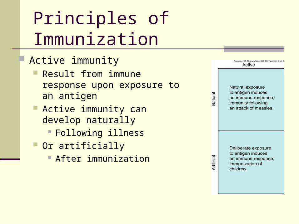
Principles of Immunization Naturally acquired immunity is
acquisition of adaptive immunity through natural events
Immunization mimics these events by inducing artificially acquired immunity
Natural or artificial immunity can be divided into Active immunity Passive immunity
Principles of Immunization
Active immunity Result from immune response
upon exposure to an antigen Active immunity can develop
naturally Following illness
Or artificially After immunization

Principles of Immunization
Passive Immunity Occurs naturally during pregnancy
IgG from mother crosses placenta Inferres protection to the baby
Occurs naturally as result of breast feeding
IgA antibodies in breast milk given to child
Artificial passive immunity involves transfer of antibodies produced by another person or animal
Can be used to prevent disease before or after likely exposure
Vaccines and Immunization
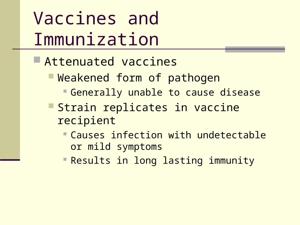
Attenuated vaccines Weakened form of pathogen
Generally unable to cause disease Strain replicates in vaccine recipient
Causes infection with undetectable or mild symptoms
Results in long lasting immunity
Vaccines and Immunization
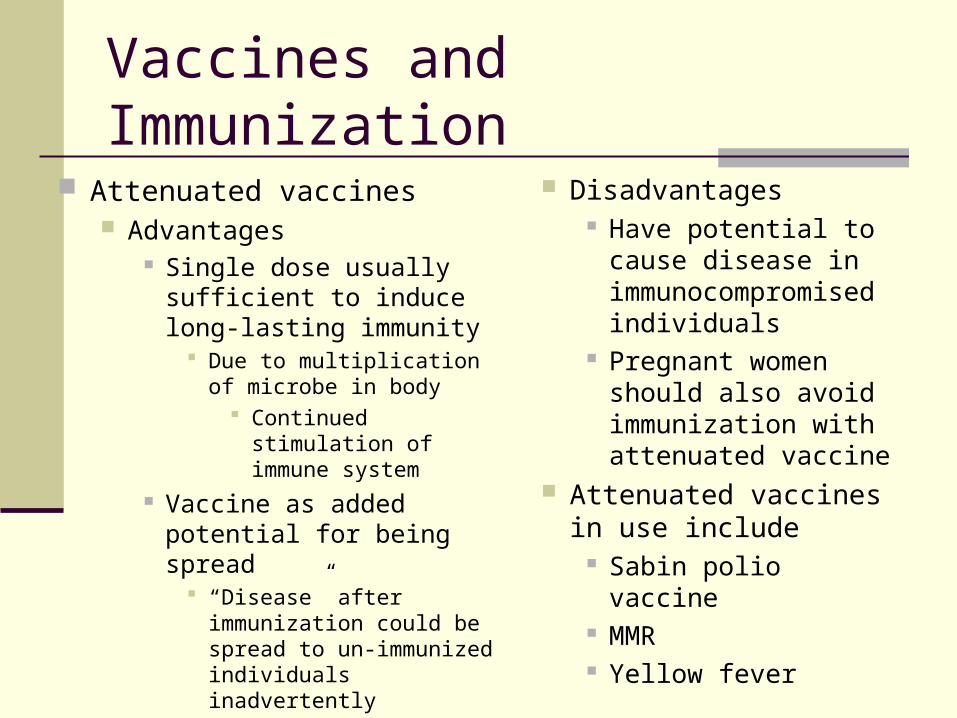
Attenuated vaccines Advantages
Single dose usually sufficient to induce long-lasting immunity
Due to multiplication of microbe in body
Continued stimulation of immune system
Vaccine as added potential for being spread
“Disease” after immunization could be spread to un-immunized individuals inadvertently
Disadvantages Have potential to
cause disease in immunocompromised individuals
Pregnant women should also avoid immunization with attenuated vaccine
Attenuated vaccines in use include
Sabin polio vaccine MMR Yellow fever
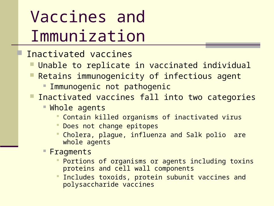
Inactivated vaccines Unable to replicate in vaccinated individual Retains immunogenicity of infectious agent
Immunogenic not pathogenic Inactivated vaccines fall into two categories
Whole agents Contain killed organisms of inactivated virus Does not change epitopes Cholera, plague, influenza and Salk polio are whole agents
Fragments Portions of organisms or agents including toxins proteins and
cell wall components Includes toxoids, protein subunit vaccines and polysaccharide
vaccines
Vaccines and Immunization
Principles of Immunological Testing
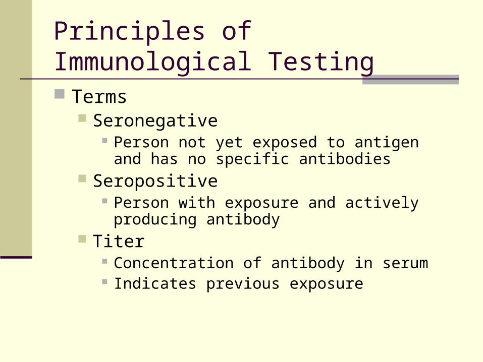
Terms Seronegative
Person not yet exposed to antigen and has no specific antibodies
Seropositive Person with exposure and actively producing
antibody Titer
Concentration of antibody in serum Indicates previous exposure

Obtaining antibody Serum is fluid portion of blood with no clotting
factors Plasma is fluid portion with clotting factors Laboratory animals are used to produce known
antibodies Animal is immunized with antigen and produces
specific antibodies Antibodies are retrieved by harvesting animal’s
serum
Principles of Immunological Testing

Obtaining antibody Certain serological tests bind human IgG
Antibodies are termed anti-human IgG These can be produced in animals immunized
with IgG from human serum
Principles of Immunological Testing
Principles of Immunological Testing
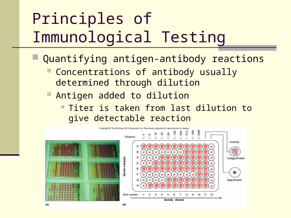
Quantifying antigen-antibody reactions Concentrations of antibody usually determined through
dilution Antigen added to dilution
Titer is taken from last dilution to give detectable reaction
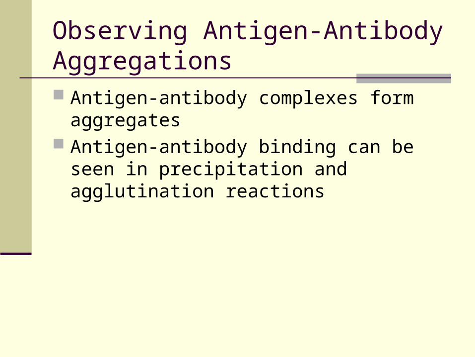
Observing Antigen-Antibody Aggregations Antigen-antibody complexes form aggregates Antigen-antibody binding can be seen in
precipitation and agglutination reactions
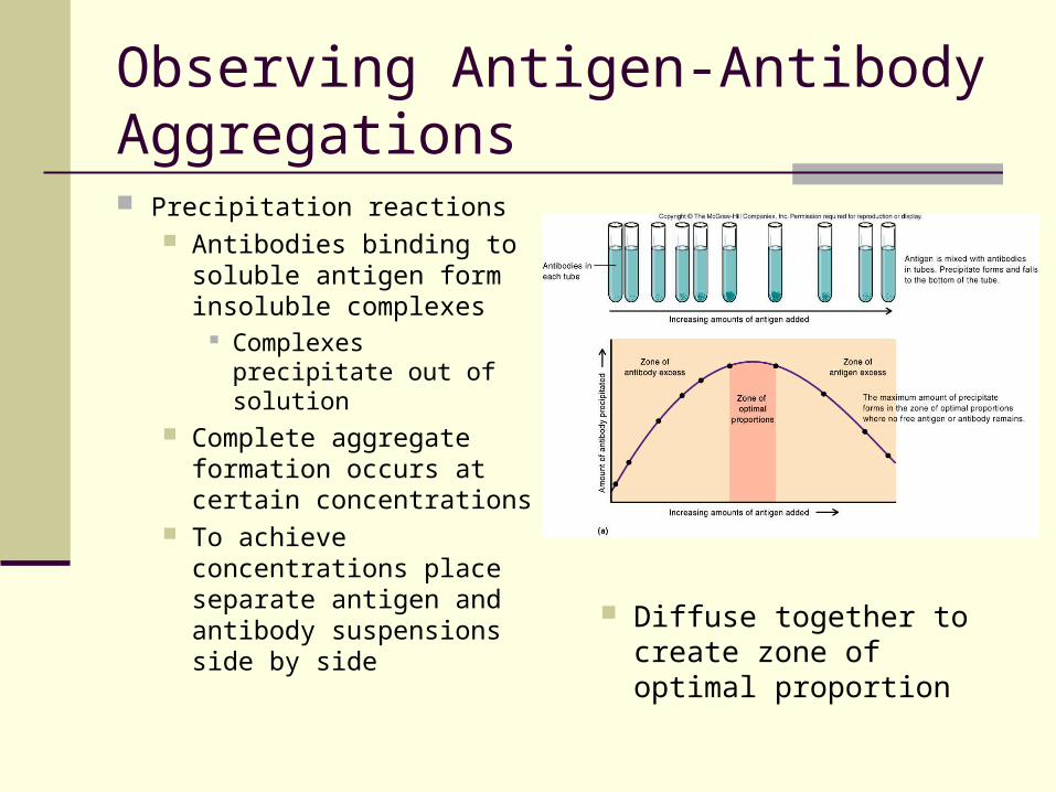
Observing Antigen-Antibody Aggregations Precipitation reactions
Antibodies binding to soluble antigen form insoluble complexes
Complexes precipitate out of solution
Complete aggregate formation occurs at certain concentrations
To achieve concentrations place separate antigen and antibody suspensions side by side
Diffuse together to create zone of optimal proportion
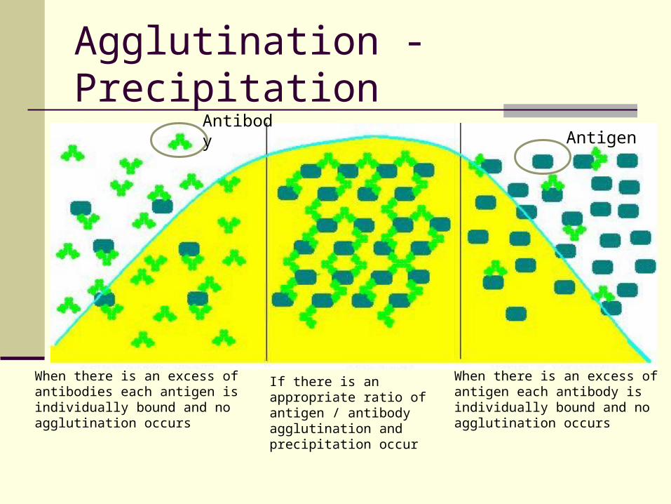
Agglutination - Precipitation
When there is an excess of antibodies each antigen is individually bound and no agglutination occurs
When there is an excess of antigen each antibody is individually bound and no agglutination occurs
If there is an appropriate ratio of antigen / antibody agglutination and precipitation occur
AntibodyAntigen
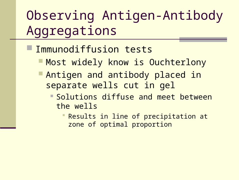
Observing Antigen-Antibody Aggregations Immunodiffusion tests
Most widely know is Ouchterlony Antigen and antibody placed in separate wells
cut in gel Solutions diffuse and meet between the wells
Results in line of precipitation at zone of optimal proportion

Ouchterlony Double Diffusion
Note: A line of precipitation has formed between the center well and wells 3 & 5. This indicates there is antigen/antibody specificity between the center well and these two wells.
Antigens and antibodies will diffuse and at some point optimal concentrations will occur and if the antigen is specific for the antibody a precipitate line will form.
Example: Has this patient ever had rubella, rubeolla, or diptheria? If they have their serum will contain antibodies against the disease. Put patient serum in the center. Put the disease agents (antigens) in wells 1 – 5, and allow to diffuse. A precipitation line between wells indicates that the patient has had that disease
Usually a known antigen or known antibody is placed in the center and test serum is placed in the peripherial wells.
Observing Antigen-Antibody Aggregations
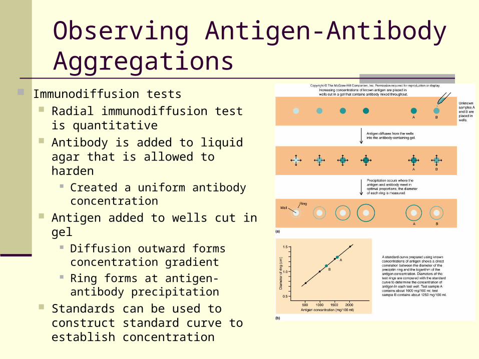
Immunodiffusion tests Radial immunodiffusion test is
quantitative Antibody is added to liquid agar
that is allowed to harden Created a uniform antibody
concentration Antigen added to wells cut in gel
Diffusion outward forms concentration gradient
Ring forms at antigen-antibody precipitation
Standards can be used to construct standard curve to establish concentration
Observing Antigen-Antibody Aggregations
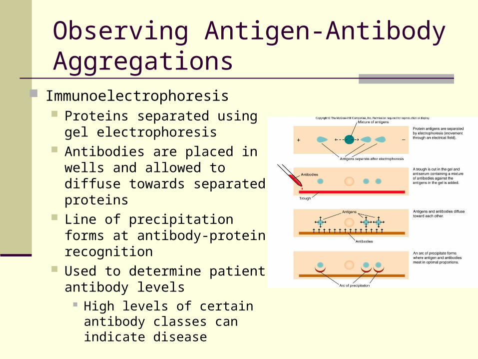
Immunoelectrophoresis Proteins separated using gel
electrophoresis Antibodies are placed in wells
and allowed to diffuse towards separated proteins
Line of precipitation forms at antibody-protein recognition
Used to determine patient antibody levels
High levels of certain antibody classes can indicate disease

Observing Antigen-Antibody Aggregations Agglutination reactions
Large insoluble particles are involved Obvious aggregations are formed
Makes the easier to see Direct agglutination
Specific antibody mixed with insoluble antigen Readily visible clumping indication of positive result
Indirect agglutination Amplifies aggregation formation
Antibody attached to latex bead Agglutination of these beads much easier to see
Using Labeled Antibodies to Detect Interactions
Detectable markers can be attached to specific antibodies Marked antibodies used to detect presence of
given antigen Test include
Fluorescent Antibody (FA) test Enzyme Linked Immunosorbant Assay (ELISA) Western blotting Fluorescence Activated Cell Sorter (FACS)
Using Labeled Antibodies to Detect Interactions
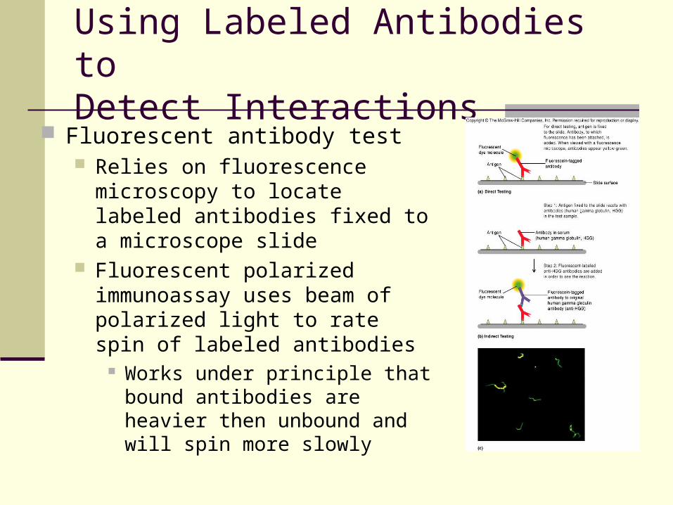
Fluorescent antibody test Relies on fluorescence microscopy to
locate labeled antibodies fixed to a microscope slide
Fluorescent polarized immunoassay uses beam of polarized light to rate spin of labeled antibodies
Works under principle that bound antibodies are heavier then unbound and will spin more slowly
Using Labeled Antibodies to Detect Interactions
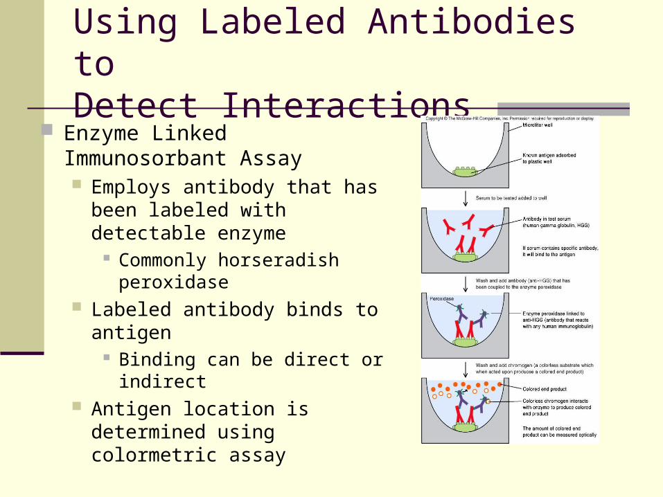
Enzyme Linked Immunosorbant Assay Employs antibody that has been
labeled with detectable enzyme Commonly horseradish
peroxidase Labeled antibody binds to
antigen Binding can be direct or
indirect Antigen location is determined
using colormetric assay

Enzyme Linked Immunosorbant Assay Direct ELISA
Looks for specific antigen Specimen placed in wells of microtiter plate
Wells treated with antibody for antigen
Indirect ELISA Looks of antibody in patient serum
Human IgG Wells of plate treated with known antigen
Using Labeled Antibodies to Detect Interactions
Using Labeled Antibodies to Detect Interactions
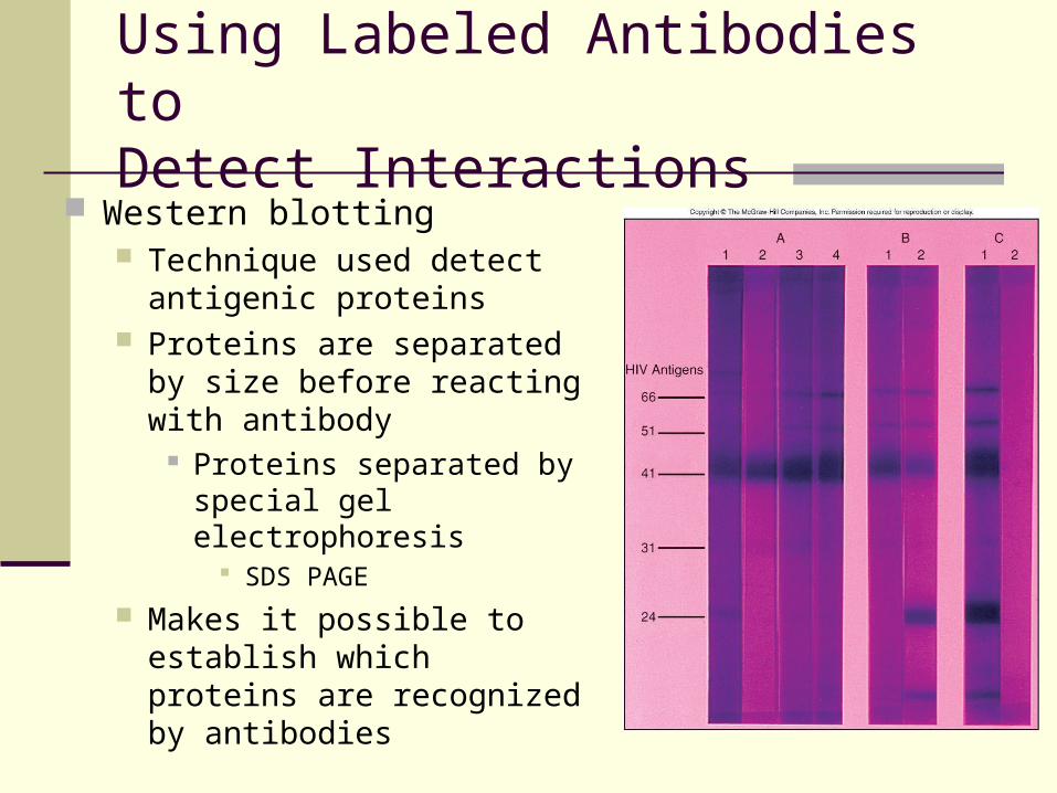
Western blotting Technique used detect
antigenic proteins Proteins are separated by size
before reacting with antibody Proteins separated by
special gel electrophoresis SDS PAGE
Makes it possible to establish which proteins are recognized by antibodies

Using Labeled Antibodies to Detect Interactions
Fluorescence Activated Cell Sorter (FACS) Special version of flow cytometry counts cells
labeled with fluorescent antibodies Used to count subsets of T cells
CD4 and CD8 cell especially Antibodies are attached to the CD4 and CD8 markers
Cells with fluorescently labeled markers are counted