
Apoptosis signal-regulating kinase 1-mediated … · Citation Cellular Microbiology, 7(9):...
43
Instructions for use Title Apoptosis signal-regulating kinase 1-mediated sustained p38 mitogen-activated protein kinase activation regulates mycoplasmal lipoprotein- and staphylococcal peptidoglycan-triggered Toll-like receptor 2 signalling pathways Author(s) Into, Takeshi; Shibata, Ken-ichiro Citation Cellular Microbiology, 7(9): 1305-1317 Issue Date 2005-09 Doc URL http://hdl.handle.net/2115/30270 Rights The definitive version is available at www.blackwell-synergy.com Type article (author version) File Information CM7-9.pdf Hokkaido University Collection of Scholarly and Academic Papers : HUSCAP
Transcript of Apoptosis signal-regulating kinase 1-mediated … · Citation Cellular Microbiology, 7(9):...

Instructions for use
Title Apoptosis signal-regulating kinase 1-mediated sustained p38 mitogen-activated protein kinase activation regulatesmycoplasmal lipoprotein- and staphylococcal peptidoglycan-triggered Toll-like receptor 2 signalling pathways
Author(s) Into, Takeshi; Shibata, Ken-ichiro
Citation Cellular Microbiology, 7(9): 1305-1317
Issue Date 2005-09
Doc URL http://hdl.handle.net/2115/30270
Rights The definitive version is available at www.blackwell-synergy.com
Type article (author version)
File Information CM7-9.pdf
Hokkaido University Collection of Scholarly and Academic Papers : HUSCAP

CMI-05-0056
Apoptosis signal-regulating kinase 1 -mediated sustained p38 mitogen-activate protein kinase
activation regulates mycoplasmal lipoprotein- and staphylococcal peptidoglycan-triggered
Toll-like receptor 2 signaling pathways
Takeshi Into,† and Ken-ichiro Shibata*
Laboratory of Oral Molecular Microbiology, Department of Oral Pathobiological Science,
Hokkaido University Graduate School of Dental Medicine, Nishi 7, Kita 13, Kita-ku, Sapporo
060-8586, Japan
*For correspondence: Ken-ichiro Shibata, Department of Oral Pathobiological Science,
Hokkaido University Graduate School of Dental Medicine, Nishi 7, Kita 13, Kita-ku, Sapporo
060-8586, Japan.
E-mail: [email protected]; Tel. +81(11)706-4240. Fax +81(11)706-4901.
†Present address: Laboratory of Oral Disease Research, National Institute for Longevity
Sciences (NILS), Aichi, Japan
Running title: Roles of ASK1 in TLR2 signaling
Key words: TLR2; ASK1; p38 MAPK; AP-1; NF-κB; caspase
1

Summary
Toll-like receptor (TLR) 2 functions as a sensor for detecting various microbial components
conserved in bacteria or fungi in innate immunity. TLR2 induces several signaling pathways
linking to activation of the transcriptional factors NF-κB and AP-1 as well as induction of cell
death. In human embryonic kidney 293 cells expressed human TLR2, mycoplasmal lipoproteins
(MLP) or staphylococcal peptidoglycans (PGN) induced sustained phosphorylation of p38
MAPK, accompanied by generation of reactive oxygen species. This observation encouraged us
to examine roles of apoptosis signal-regulating kinase 1 (ASK1) in TLR2 signaling, because
ASK1 is an upstream activator of p38 MAPK during exposure to oxidative stress and other
stressful stimuli. A kinase-inactive mutant of ASK1 greatly impaired the sustained
phosphorylation of p38 MAPK induced by MLP or PGN. This mutant also attenuated MLP- or
PGN-induced transcriptional activities of NF-κB and AP-1 via inhibition of p38 MAPK
activation. MLP- or PGN-induced cell death reactions, including DNA fragmentation and
caspase-3/7 activation, were also downregulated by the ASK1 mutant via p38 MAPK inhibition.
Furthermore, TLR2 signaling had a potential to phosphorylate and dephosphorylate ASK1 at
Ser83 residue. Thus, MLP and PGN have capabilities to induce ASK1-dependent signaling
pathways which regulate p38 MAPK activation through TLR2, leading to activation of NF-κB
and AP-1 as well as induction of cell death.
2

Introduction
Toll-like receptors (TLRs) represent a family of type I transmembrane proteins characterized by
multiple copies of leucine-rich repeats in the extracellular domain and a cytoplasmic Toll/IL-1R
homology (TIR) domain (Medzhitov et al., 1997). At least eleven TLRs have so far been
identified in mammalian species, and they are capable of sensing broad classes of pathogens,
including viruses, bacteria, fungi and protozoan parasites, by recognizing of specific and
distinct pathogen-associated molecular patterns (PAMPs) as cognate ligands (Akira and Takeda,
2004; Janeway and Medzhitov, 1999; Takeda et al., 2003; Zhang et al., 2004a). Among all
members of the TLR protein family, TLR2 detects the broadest spectrum of agonistic PAMPs
from viruses to protozoans, such as several bacterial lipoproteins/lipopeptides, peptidoglycans
(PGN), lipoarabinomannans, porins, glycoproteins and fimbriae, fungal zymosan, viral core
proteins and hemagglutinins (HA) and protozoan glycosylphosphatidylinositol anchors
(Kirschning and Schmann, 2002; Takeda et al., 2003).
It has been shown that TLR2 first initiates signaling by recruiting the TIR
domain-containing adaptors TIRAP (also known as Mal) and MyD88 after recognition of
cognate PAMPs (Akira and Takeda, 2004; Horng et al., 2002; Mansell et al., 2004; O’Neill et
al., 2003; Yamamoto et al., 2002). The receptor complex is then joined by the members of
IL-1R-associated kinases (IRAKs) IRAK1 and IRAK4 via their death domains (DDs) (Akira
and Takeda, 2004; Janssens and Beyaert, 2003; Suzuki et al., 2002). After phosphorylation,
IRAK1 and IRAK4 dissociate from TLR-associated receptor complex and then interact with
TNFR-associated factor (TRAF) 6 (Akira and Takeda, 2004). TRAF6 possesses a unique
molecule-binding specificity, especially for TAK1 and TAB1/2, that results in triggering
3

activation of the cascades of transcriptional factors NF-κB and AP-1 families (Akira and
Takeda, 2004; Bradley and Pober, 2001; Ninomiya-tsuji et al., 1999; Shuto et al., 2001;
Takaesu et al., 2000). It is now thought that TRAF6 is a key mediator in the MyD88-dependent
TLR/IL-1R signaling pathway leading to activation of a wide variety of innate immune
responses (Gohda et al., 2004). MyD88 is also known to recruit a well-known death receptor’s
adaptor, Fas-associated death domain (FADD), downstream of TLR2 via DD-DD interaction
(Aliprantis et al., 2000). After interaction with MyD88, FADD is thought to activate a death
initiator cysteine protease, caspase-8, to initiate subsequent proapoptotic reactions. Indeed, we
have recently shown that mycoplasmal lipoproteins (MLP) induce apoptotic cell death via
MyD88, FADD and caspase-8 downstream of TLR2 (Into et al., 2002b and 2004). Thus,
MyD88 can also initiate the so-called death signaling pathway. However, the implication of
MyD88 dependence of these partially ambivalent signaling pathways is still controversial.
Additionally, several MyD88-independent pathways, including Rac-1, RhoA, protein kinase C,
PI3K and Akt/protein kinase B, have also been shown to participate in TLR2-mediated
signaling (Arbibe et al., 2000; Strassheim et al., 2004; Teusch et al., 2004).
One of the critical functions of TRAF6 protein is to mediate the activation of AP-1, the
activity of which is stimulated by mitogen-activated protein kinases (MAPKs) through either
direct phosphorylation or transcription of the AP-1 components c-fos and c-jun (Bradley and
Pober, 2001; Dong et al., 2002; Tanoue and Nishida, 2003). MAPKs, which include Ser/Thr
kinases such as p38 MAPKs, JNKs and ERKs, are located at the downstream end of a
three-tiered system that also contains MAPK kinases (MAPKKs) and MAPK kinase kinases
(MAPKKKs). Among MAPK members, p38 MAPKs and JNKs are known as stress-activated
protein kinases that are activated by a wide variety of stressors. JNKs are activated by at least
4

two MAPKKs, SEK1/MKK4 and MKK7, whereas p38 MAPKs are activated by MKK3 and
MKK6 (Dong et al., 2002; Tanoue and Nishida, 2003). The activation of these two MAPK
cascades is involved in the promotion of both cell survival and apoptotic cell death (Dong et al.,
2002; Takeda et al., 2002; Tanoue and Nishida, 2003). Although we recently raised the
possibility that TLR2-mediated p38 MAPK activation participated in MLP-induced apoptotic
cell death (Into et al., 2004), the regulatory mechanisms and detail functions of MAPKs in
TLR2-mediated cell death reactions have not been clarified yet.
In this study, we found that one of the MAPKKK members, apoptosis signal-regulating
kinase 1 (ASK1; also known as MEKK5), the activation of which is associated with the
generation of intracellular reactive oxygen species (ROS), is a key mediator in regulation of the
MLP- or PGN-induced p38 MAPK activation downstream of TLR2. We further show that
ASK1 is required for the regulation of MLP- or PGN-induced cell death reactions as well as for
the activation of transcriptional activity of AP-1 and NF-κB downstream of TLR2.
5
Results
MLP- or PGN-induced phosphorylation of stress-activated MAPKs
MLP and PGN are known as crude immunostimulatory compounds and function as potent
agonistic ligands for TLR2. To analyze TLR2-specific signaling events induced by these
components, we utilized human embryonic kidney (HEK) 293 cell-derived transfectants,
because this cell line lacks endogenous TLR2 expression and is nonresponsive to at least MLP
and PGN until TLR2 gene transfection (Fujita et al., 2003; Into et al., 2004). MLP or PGN
activated the transcriptional activities of both AP-1 and NF-κB in HEK293 cells stably
transfected with a construct of C-terminal V5 epitope-tagged TLR2 cloned into the pEF6 vector
(293/TLR2-V5 cells) in a dose-dependent manner (Fig. 1A and B). Expectedly, HEK293 cells
stably transfected with an empty pEF6 vector (293/pEF6 cells) did not respond to both MLP and
PGN at the all concentrations used (Fig. 1A and B). 293/TLR2-V5 cells also responded to
zymosans as another TLR2 ligand, but the activity was extremely weak when compared with
those of MLP and PGN.
Using 293/TLR2-V5 cells, we then examined the phosphorylation state of the
stress-activated MAPKs p38 MAPK and JNK by Western blot analyses. MLP induced the
phosphorylation of both p38 MAPK and JNK at all concentrations used (Fig. 1C). The
phosphorylation of p38 MAPK was detected 15 min after stimulation, reached a maximum at 30
min, and was then gradually reduced (Fig. 1C). The phosphorylation of JNK was detected 60,
30 and 15 min after stimulation with 0.1, 1 and 10 μg ml-1 of MLP, respectively, reached a
maximum at 30 to 60 min, and was then gradually reduced (Fig. 1C). MLP induced robust
phosphorylation of these MAPKs at concentrations of 1 and 10 μg ml-1, and the phosphorylation
6

state was sustained for at least 180 min after stimulation (Fig. 1C). Similar results for
PGN-induced phosphorylation of p38 MAPK and JNK were also obtained in the cells (data not
shown). Furthermore, we confirmed that 10 μg ml-1 of MLP or PGN induced sustained
phosphorylation of p38 MAPK and JNK in promonocytic THP-1 cells used as a non-transfected
cell line, which constitutively express a functional TLR2 (Into et al., 2002a) (data not shown).
Thus, MLP and PGN activate TLR2-mediated stress-activated MAPKs, and the higher
concentration of these compounds could induce sustained phosphorylation of these MAPKs.
Induction of TLR2-mediated oxidative stress by MLP or PGN
It has been reported that various kinds of cellular stressors can induce generation of intracellular
ROS. The ROS-associated redox systems are known to affect activation of MAPKs, especially
p38 MAPK and JNK (Martindale and Holbrook, 2002; Takeda et al., 2002; Torres and Forman,
2003). Furthermore, it has been reported that stimulation of THP-1 cells with TLR2 ligands
induces ROS generation (Aliprantis et al., 1999). Therefore, flow cytometric analysis was
carried out to determine whether challenge with MLP or PGN induced ROS generation in
293/TLR2-V5 cells. The stimulation of the cells induced a significant increase in
CM-H2DCFDA fluorescence after 1-h stimulation with MLP or PGN in a dose-dependent
manner (Fig. 2), indicating that both MLP and PGN have a capability to induce the generation
of intracellular ROS. MLP and PGN did not induce ROS generation in 293/pEF6 cells (data not
shown). Thus, challenge with TLR2 ligands induces oxidative stress in TLR2-expressing cells.
Regulation of MLP- or PGN-induced sustained p38 MAPK phosphorylation by ASK1
7

ASK1 was first discovered by Ichijo et al. (Ichijo et al., 1997) as a MAPKKK member that
mediated activation of p38 MAPK and JNK and apoptosis in response to oxidative stress and
TNF via its catalytic functions. By using ASK1-/- cells, their additional studies have
demonstrated that ASK1 was required for sustained activation of p38 MAPK and JNK induced
by oxidative stress and TNFR1 (Takeda et al., 2002; Tobiume et al., 2001). Thus, ASK1 has
been thought to be an important molecule regulating stress-activated protein kinase pathways
and proapoptotic pathways downstream of TNFR1. Importantly, it has been suggested that the
TLR2 signaling pathways are analogous to those triggered by TNFR1 (Aliprantis et al., 2000;
Into et al., 2004). As described above, this study demonstrated that TLR2 stimulation induced
sustained activation of p38 MAPK and JNK and oxidative stress. Therefore, we examined
whether ASK1 was involved in MLP- or PGN-induced TLR2 signaling pathways.
The expression of endogenous ASK1 was observed in both parental HEK293 cells and
293/TLR2-V5 cells (Fig. 3A). We constructed a kinase-inactive mutant of ASK1 cloned in the
pcDNA3 vector (Lys709Met; ASK1-KM) and stably transfected the construct into
293/TLR2-V5 cells (293/TLR2-V5/ASK1-KM cells; Fig. 3A). 293/TLR2-V5 cells stably
transfected with a pcDNA3 empty vector (293/TLR2-V5/pcDNA3 cells) were also established
as control cells. Stimulation of 293/TLR2-V5/ASK1-KM cells with MLP (0.1, 1 and 10 μg ml-1)
resulted in significantly impaired phosphorylation states of both p38 MAPK and JNK when
compared with those in 293/TLR2-V5/pcDNA3 cells (Fig. 3B and C; P <0.01). Notably,
MLP-induced sustained phosphorylation of p38 MAPK was diminished, but the transient
phosphorylation still occurred in 293/TLR2-V5/ASK1-KM cells (Fig. 3B). In contrast to this,
MLP-induced sustained JNK phosphorylation was still observed in the cells (Fig. 3C). To
8

confirm whether the effects of ASK1-KM on phosphorylation of these MAPKs were specific,
we then performed experiments using THP-1 cells stably transfected with ASK1-KM
(THP-1/ASK1-KM cells) and pcDNA3 empty vector (THP-1/pcDNA3 cells). Stimulation of
THP-1/ASK1-KM cells with 10 μg ml-1 of MLP resulted in impaired sustained p38 MAPK
phosphorylation, which was occurred 1 h after stimulation, when compared with those in
THP-1/pcDNA3 cells (Fig. 3D). Distinct differences between pcDNA3 and ASK1-KM were not
observed in MLP-induced JNK phosphorylation.
These observations suggest that ASK1 is at least an important molecule to mediate the
sustained p38 MAPK phosphorylation downstream of TLR2 via its catalytic functions.
Regulation of MLP- or PGN-induced activation of AP-1 and NF-κB by ASK1
We next investigated whether ASK1 regulated MLP- or PGN-induced activation of AP-1 and
NF-κB. The activity of AP-1 was significantly decreased in MLP- or PGN-stimulated
293/TLR2-V5/ASK1-KM cells compared with that in 293/TLR2-V5/pcDNA3 cells (Fig. 4A,
left panel). It was likely that this decrease was partially due to the decrease in the
ASK1-mediated p38 MAPK activation, because the specific inhibitor for p38 MAPK SB203580
slightly decreased AP-1 activity induced by 10 μg ml-1 of MLP (Fig. 4A, right panel).
MLP-induced JNK activation also participated in the AP-1 activation, because the specific
inhibitor for JNK SP600125 partially decreased the activity (Fig. 4A, right panel). Furthermore,
we found that the activity of NF-κB was also significantly decreased in MLP- or
PGN-stimulated 293/TLR2-V5/ASK1-KM cells compared with that in 293/TLR2-V5/pcDNA3
cells (Fig. 4B, left panel). This decrease was mainly due to the decrease in ASK1-mediated p38
9

MAPK activation, but JNK activation, because SB203580, rather than SP600125, showed
greater inhibition of NF-κB activity induced by 10 μg ml-1 of MLP (Fig. 4B, right panel).
Thus, ASK1 was found to regulate TLR2-mediated activation of p38 MAPK via its catalytic
function leading to activation of two major transcriptional factors, AP-1 and NF-κB.
Regulation of MLP- or PGN-induced cell death by ASK1
TLR2-mediated signaling pathways are linked to the death signal pathway (Aliprantis et al.,
2000; Into et al., 2004). We have recently reported that MLP induced several caspase-dependent
cell death reactions such as apoptotic morphological changes, externalization of
phosphatidylserine, DNA fragmentation and cleavage of poly(ADP-ribose)polymerase through
TLR2-, MyD88-, FADD- and p38 MAPK-dependent machineries (Into et al., 2004). In addition,
PGN (and MLP, but not zymosan) also induced TLR2-, caspase-8- and caspase-3-dependent cell
death in THP-1 cells (Fig. 5A) and other cells (Abrahams et al., 2004). Therefore, experiments
were carried out to determine whether ASK1 is involved in MLP- or PGN-induced cell death
reactions, especially activation of caspases. Stimulation of 293/TLR2-V5/pcDNA3 cells with
MLP or PGN induced DNA fragmentation in a dose-dependent manner (Fig. 5B). Activations of
caspase-8, caspase-9 and caspase-3/7 were also clearly observed in the stimulated cells (Fig.
5D-F, left panels). On the other hand, stimulation of 293/TLR2-V5/ASK1-KM cells with MLP
or PGN resulted in impaired cell death reactions, mainly in DNA fragmentation and caspase-3/7
activation, and partially in caspase-8 and -9 activations (Fig. 5C-F, left panels). Thus, the
catalytic activity of ASK1 is also important for the regulation of TLR2-mediated apoptotic
reactions. Notably, pretreatment of 293/TLR2-V5/pcDNA3 cells with SB203580 mainly
10

attenuated MLP-induced DNA fragmentation and caspase-3/7 activation, whereas pretreatment
of the cells with SP600125 mainly attenuated the activations of caspase-8 and caspase-9 (Fig.
5C-F, right panels).
Thus, ASK1 is likely to play important roles in MLP- or PGN-induced cell death reactions
mainly by regulation of p38 MAPK-mediated caspase-3 activation downstream of TLR2.
Regulation of phosphorylation of ASK1 by MLP-induced TLR2 signaling
This study demonstrated that the catalytic activity of ASK1 played an important role in the
regulation of MLP- or PGN-induced stress and death signaling downstream of TLR2. Recent
findings have suggested that the potential of catalytic activity of ASK1 is neutralized by the
phosphorylation of several serine residues, including Ser83 and Ser967 (Fujii et al., 2004;
Goldman et al., 2004; Kim et al., 2001). Therefore, we examined whether stimulation of TLR2
with MLP affected the phosphorylation state of these serine residues. MLP clearly induced
phosphorylation of Ser83 residue in endogenous ASK1 at the concentration of 0.1 μg ml-1 in
293/TLR2-V5 cells (Fig. 6). However, the phosphorylated residue was dephosphorylated by
stimulation with 1 μg ml-1 of MLP in a time-dependent fashion (Fig. 6). Additionally,
stimulation with 10 μg ml-1 of MLP clearly induced phosphorylation of the residue at the earlier
time points (5–15 min; data not shown), and then induced complete dephosphorylation of the
residue at least 1 h after stimulation (Fig. 6). In contrast to these, MLP did not affect the
phosphorylation state of Ser967 residue at all concentrations tested (Fig. 6).
Thus, it is likely that TLR2-mediated signaling has a potential to regulate the catalytic
function of ASK1 via phosphorylation and dephosphorylation at the Ser83 residue in ASK1.
11

Discussion
In this study, we demonstrated that stimulation of TLR2 with microbial components, MLP or
PGN, initiated activation of stress-activated MAPKs, which was accompanied by intracellular
ROS generation (Figs 1 and 2). Several studies have already shown that stimulation of TLR2
with synthetic bacterial lipopeptides induced ROS generation, or oxidative burst, that is thought
to be mediated by the NAD(P)H oxidase system (Aliprantis et al., 1999 and 2001). In relation to
this, a recent study has indeed demonstrated that the NAD(P)H oxidase member Nox4, which
was highly expressed in HEK293 cells (Shiose et al., 2001), was involved in TLR4-induced
ROS generation (Park et al., 2004). Elevated ROS are thought to serve as a second messenger to
control a broad range of physiological and pathological processes, including cell proliferation,
inflammation and apoptosis (Martindale and Holbrook, 2002; Torres and Forman, 2003; Ueda et
al., 2002; Yodoi et al., 2001). It is now thought that ASK1 emerges as a major regulator of
ROS-initiated signaling in response to various kinds of cellular stressors, including several
ligand-receptor systems (Chen et al., 2003; Takeda et al., 2002; Yodoi et al., 2001). ASK1 is
present in cytosol as an inactive complex with thioredoxin (Trx), and then dissociate from Trx
leading to active form after Trx oxidization by ROS (Takeda et al., 2002; Yodoi et al., 2001).
ASK1 then forms a stable complex by interaction with TRAFs followed by activation of
specific MAPKKs (Nishitoh et al., 1998; Takeda et al., 2002). Downstream of TLR2, ASK1
therefore appears to be activated by NAD(P)H oxidase system-induced ROS, and then mediates
activation of MAPKs, especially sustained activation of p38 MAPK.
It is well researched that TLR2 recruits MyD88 as an adopter molecule to initiate
subsequent signaling. In this study, we demonstrated that ASK1 participated in MLP- or
12

PGN-induced TLR2 signaling pathways. Although it remains unclear whether the
ROS/ASK1-mediated responses are dependent on MyD88-mediated machineries, it is
speculated that ASK per se is not absolutely indispensable for the initiation of TLR2 signaling,
judging from the results that ASK1-KM did not abrogate TLR2-mediated activations of AP-1
and NF-κB (Fig 4) and the machinery of ASK1 activation as described above. This speculation
is also supported by the previous findings that ASK1-mediated MAPK cascades are inessential
for the death receptor-induced proapoptotic signaling (Takeda et al., 2002, Tobiume et al.,
2001). However, Takeda et al previously described that ASK1 appears to function as a
multifunctional stress-sensing kinase to control cell survival and death in response to various
kinds and strengths of stresses (Takeda et al., 2002). Our present findings may support these
multiple functions of ASK1 in microbial stimulus-initiated TLR2 signaling.
We demonstrated that ASK1-mediated activation of p38 MAPK downstream of TLR2 could
regulate the transcriptional activity of both AP-1 and NF-κB (Fig. 4). It is well known that AP-1
activity is tightly regulated by three MAPK members, p38 MAPK, JNK and ERK (Bradley and
Pober, 2001; Dong et al., 2002; Tanoue and Nishida, 2003). We examined the effects of
SB203580 and SP600125 on TLR2-mediated AP-1 activity and expectedly found that AP-1
activation was blocked by both of these inhibitors (Fig. 4A). In addition, we also found that the
MLP-induced NF-κB activation could be partially blocked by SB203580 and, to a lesser extent,
SP600125 (Fig. 4B). These are supported by several reports which have demonstrated that the
activation of p38 MAPK and JNK can induce NF-κB activation (Carter et al., 1999;
Schulze-Osthoff et al., 1997; Vanden Berghe et al., 1998). Therefore, it can be considered that
ASK1-mediated p38 MAPK-dependent signaling cascades directly affect TLR2-mediated
activation of both AP-1 and NF-κB, which promotes cell survival and activation.
13

We further demonstrated that the ASK1-mediated MAPK activation could also affect the
induction of TLR2-mediated apoptotic reactions (Fig. 5). This observation is also supported by
the previous findings that ASK1 is involved in the regulation of the apoptotic pathways
downstream of TNFR1 and Fas (Hatai et al., 2000; Ichijo et al., 1997; Takeda et al., 2002).
ASK1 could evidently affect TLR2-induced DNA fragmentation and caspase-3/7 activation, but
weakly affected activation of caspase-8 and -9, mainly via p38 MAPK activation. Therefore,
ASK1 appears to function upstream of caspase-3/7 in TLR2-mediated death signaling that may
be originally triggered through MyD88 and FADD. These findings further suggest that ASK1
mainly modulates the execution of TLR2-mediated apoptotic reactions by regulating
caspase-3/7, which are known as executioner caspases, but not by regulating initiator caspases.
Indeed, ASK1 has been shown to affect pro- and anti-apoptotic molecules, such as Bcl-2, Bax
and CIIA, which function upstream of the executioner caspases (Cho et al., 2003; Takeda et al.,
2002; Yuan et al., 2003).
The functions of ASK1 are regulated by multiple mechanisms. The prominent mechanisms
by which the catalytic activity of ASK1 is regulated are thought to be protein-protein
interactions. The major up-regulatory factors of ASK1, including TRAFs, Daxx, AIP1/DAB2IP,
JSAP1/JIP3 and hD53L1, have already been reported (Chang et al., 1998; Cho et al., 2002;
Matsuura et al., 2002; Nishitoh et al., 1998; Takeda et al., 2002; Zhang et al., 2004b; Zhang et
al., 2003). In contrast to this, Trx, 14-3-3 proteins, glutaredoxin, GSTM1-1, Hsp72 and Raf-1
were shown to be down-regulators (Chen et al., 2001; Goldman et al., 2004; Park et al., 2002;
Takeda et al., 2002). The activation, oligomerization and protein-protein interactions of ASK1
are thought to be largely dependent on the phosphorylation at several Ser/Thr residues in ASK1.
Indeed, phosphorylation at Thr838 residue in ASK1 is known to initiate the activation of ASK1
14

(Takeda et al., 2002). On the other hand, the binding of 14-3-3 proteins to ASK1 is dependent
on phosphorylation at Ser967 residue in ASK1, leading to inhibition of ASK1 activity
(Goldman et al., 2004; Zhang et al., 2003). Furthermore, Akt/protein kinase B phosphorylates
Ser83 residue in ASK1 to neutralize the catalytic functions of ASK1 (Kim et al., 2001; Yuan et
al., 2003). Recently, the phosphorylation at Ser1034 residue in ASK1 was also reported to
participate in the inhibition of ASK1 (Fujii et al., 2004). In this study, we demonstrated that
stimulation of TLR2 with a low dose of MLP induced phosphorylation at Ser83 residue in
ASK1, whereas stimulation with a high dose of MLP induced dephosphorylation of the residue
(Fig. 6). This observation indicates that TLR2 signaling regulates ASK1 activity, which may be
due to the activity of Akt and an unknown protein phosphatase, and allusively shows the reason
why ASK1 is only involved in sustained, but transient, activation of MAPKs (p38 MAPK in this
study). In addition, this our finding supports the evidence that ASK1 senses strengths of
TLR2-mediated stresses. Further study should be carried out to elucidate the extended
regulatory mechanisms of ASK1, including oligomerization and protein-protein interactions,
downstream of TLR2.
In Fig. 7, we showed schematic of ASK1 functions in TLR2 signaling pathways. Our
observations suggest that ASK1 works as an important intermediate in microbial
stimulus-induced TLR2 signaling through its regulatory effects on p38 MAPK, transcriptional
factors and apoptotic reactions (Fig. 7). Originally, TLR2 is expressed on the surface of various
types of cells to detect microbes (and/or their components) followed by initiation of
MyD88-dependent innate immune reactions and apoptosis, whereas ASK1 is expressed in the
cytosol of various types of cells to detect intracellular ROS following initiation of stress
reactions, including the activation of stress-activated MAPKs and apoptosis. After ligation of
15
cognate PAMPs such as MLP and PGN with TLR2, ASK1 may function to control the
MyD88-dependent divergent signaling pathways, which are largely dependent on the amount of
microbes (and/or microbial components) and generated ROS. Therefore, our future study will
focus on these regulatory mechanisms and the function of ASK1 in TLR2-mediated multiple
cellular responses, innate immune responses and microbial infectious diseases.
16

Experimental procedures
Reagents, chemicals and antibodies (Abs)
PGN derived from Staphylococcus aureus was obtained from Fluka Production. Zymosan A was
purchased from Sigma. Selective inhibitors for p38 MAPK (SB203580) and JNK (SP600125)
were purchased from Calbiochem. All of mAbs to human MAPKs (phospho-specific p38, p38α,
phospho-specific JNK and JNK1/2) were obtained from BD Pharmingen. Abs to V5-epitope tag
and HA-epitope tag were purchased from Invitrogen and Santa Cruz Biotecnology, Inc.,
respectively. Abs to human ASK1 and phospho-ASK1 (Ser83 and Ser967) were obtained from
Cell Signaling Technology, Inc. An Ab to extracellular domain of human TLR2 was described
previously (Fujita et al., 2003).
All of the other chemicals were obtained from commercial sources and were of analytical or
reagent grade.
Preparation of MLP
Mycoplasma fermentans ATCC 19989 was grown in PPLO broth supplemented with 20% (v/v)
horse serum, 1% (w/v) yeast extract, 1% (w/v) D-glucose and penicillin G (1000 U ml-1). When
there was a fall in pH of 1 unit, the cells were collected by centrifugation at 15,000 g for 15 min,
washed three times with PBS, and suspended in PBS. The cells were treated with Triton X-114
to extract MLP according to the method described previously (Into et al., 2002c; Shibata et al.,
17

2000). MLP were dissolved in PBS containing 5% (v/v) DMSO at the concentration of 3 mg
ml-1 and stored at -80ºC.
DNA expression vectors
The cDNA construct of C-terminal V5-epitope tagged human TLR2 cloned in the pEF6 vector
was described previously (Into et al., 2004). The construct containing HA-tagged human ASK1
cloned in the pcDNA3 vector was kindly provided by Prof. Hidenori Ichijo (Tokyo University,
Japan), and kinase-inactive version of ASK1 (Lys709Met; ASK1-KM) (Ichijo et al., 1997;
Takeda et al., 2002) was generated by using a Quick Change site directed mutagenesis kit
(Stratagene) according to the manufacturer’s instructions.
Cell culture and transfection
HEK293 cells were obtained from ATCC and grown at 37ºC and in 5% CO2 in DMEM (Sigma)
supplemented with 10% FBS (Sigma) and penicillin (100 U ml-1) / streptomycin (100 μg ml-1)
(Sigma). A TLR2-V5 construct or a pEF6 empty vector was transfected into HEK293 cells
using Metafectene lipofection reagent (Biontex Laboratories) according to the manufacturer’s
instructions. Stable transfectants were selected in the presence of blasticidin (50 μg ml-1;
Invitrogen), followed by immunoblot analysis to confirm the expression of TLR2-V5 using Abs
to either TLR2 or V5-epitope. An HA-tagged ASK1-KM construct or a pcDNA3 empty vector
was transfected into 293/TLR2-V5 cells using Metafectene lipofection reagent. Stable
transfectants were selected in the presence of both G418 (800 μg ml-1; Sigma) and blasticidin
18

(50 μg ml-1), followed by immunoblot analysis to confirm the expression of ASK1-KM and
TLR2-V5 using Abs to ASK1, HA-epitope and V5-epitope. Cells of the human promonocytic
cell line THP-1 were obtained from Health Science Research Resources Bank and grown at
37ºC and in 5% CO2 in RPMI 1640 (Sigma) supplemented with 10% FBS and
penicillin/streptomycin. An HA-tagged ASK1-KM construct or a pcDNA3 empty vector was
transfected into THP-1 cells using Metafectene lipofection reagent. Stable transfectants were
selected in the presence of G418 at the concentration of 800 μg ml-1.
Luciferase reporter gene assay
The luciferase reporter gene assay in HEK293-derived transfectants was carried out essentially
by the method described previously (Fujita et al., 2003; Into et al., 2004). Briefly, the cells were
plated at 5 × 10⁴ cells/well in poly-L-lysine-coated 24-well plates before DNA transfection. The
cells were transiently transfected by Metafectene lipofection reagent with 50 ng of an NF-κB
reporter plasmid (pNF-κB-Luk; Stratagene) or an AP-1 reporter plasmid (pAP-1-Luc;
Stratagene) together with 5 ng of a construct directing expression of Renilla luciferase under the
control of a constitutively active thymidine kinase promoter (pRL-TK; Promega). After 24 h of
incubation, the cells were stimulated with MLP or PGN in media containing 1% FBS for 6 h.
Then the cells were lysed and luciferase activity was measured by using a Dual-Luciferase
reporter assay system (Promega) according to the manufacturer’s instructions. Results are
expressed as means ± SD of three determinations.
Western blot analysis
19

HEK293 or THP-1 cell-derived transfectants were stimulated with or without MLP. Then the
cells were collected and lysed in a buffer consisting of 20 mM Tris-hydrochloride (pH 7.2), 150
mM sodium chloride, 5 mM EDTA and 1% Nonident P-40 in the presence of protease inhibitors
(Roche) at 4ºC for 15 min followed by clarification by centrifugation at 12,000 × g for 10 min.
These cell lysates were diluted by an equal volume of SDS sample buffer consisting of 0.5 M
Tris-hydrochloride (pH 7.2), 10% glycerol, 2% SDS, 5% 2-mercaptoethanol and 0.05%
bromphenol blue. Samples were boiled for 5 min and separated under reducing conditions on
10% SDS-PAGE gels and then transferred onto polyvinylidene fluoride membranes (Sigma).
Membranes were blocked at room temperature for 1 h with 10% nonfat skim milk in PBS and
then reacted with the Abs for 1 h. Immunoreactive bands were visualized by an ECL system
(Amersham Pharmacia Biotech) after being treated with an Ab to HRP-conjugated anti-mouse
or anti-rabbit IgG. All experiments were performed at least three times, and representative
results are shown in the figures.
Detection of ROS
The generation of intracellular ROS was quantified fluorometrically using a cell-permeable
oxidation-sensitive fluorescent probe, CM-H2DCFDA (Molecular Probes). 293/TLR2-V5 cells
(2.5 × 10⁵ cells) were plated in 6-well plates and pretreated at 37ºC with DMEM containing 5
μM CM-H2DCFDA for 30 min. The cells were gently washed once with DMEM and then
stimulated with MLP or PGN in DMEM containing 2.5% FBS. After stimulation for 1 h, the
cells were harvested, washed twice with DMEM, resuspended in DMEM, and analyzed on a
20

FACSCalibur flow cytometer (BD Biosciences). Dead cells and debris were excluded from the
analysis by electronic gating of forward and side scatters. Experiments were performed at least
three times, and representative results are shown in the figures.
Determination of the activities of p38 MAPK and JNK by ELISA
HEK293-derived transfectants (2 × 10⁴ cells) were seeded in poly-L-lysine-coated 96-well
plates. The cells were stimulated with MLP and then assayed for determination of the activity of
p38 MAPK or JNK using FACETM ELISA Kits (Active Motif) according to the manufacturer’s
instructions. In this study, we used a mAb to phospho-p38 MAPK (T180/Y182) or
phospho-JNK (T183/Y185) as a primary Ab to detect activated p38 MAPK or JNK.
Colorimetric reaction and crystal violet cell staining were measured by absorbance on a
spectrophotometer at 450 nm and 595 nm, respectively. The measured OD450 readings were
corrected for cell number by dividing the OD450 reading for a given well by the OD595 reading
for that well. Results were calculated as fold increase when the control cells were taken as 1 and
are expressed as means ± SD of three determinations.
Cytotoxicity assay
Cytocidal activity of MLP, PGN and zymosan in THP-1 cells was assessed essentially by the
method described previously (Into et al., 2004). The caspase inhibitors Ac-IETD-CHO and
Ac-DMQD-CHO were obtained from Calbiochem. Results are expressed as means ± SD of
three determinations.
21

Detection of cell apoptosis by ELISA
The generation of mono- and oligonucleosomes due to DNA fragmentation during the apoptotic
process was detected using a Cell Death Detection ELISA kit (Roche). HEK293 cell-derived
transfectants were seeded at 4 × 10⁴ cells/well in poly-L-lysine-coated 24-well plates before
stimulation. After stimulation with MLP or PGN for 24 h, the cells were lysed using lysis
solution provided in the kit for 20 min on ice. The cell extracts were centrifuged for 10 min at
14,000 × g. The supernatants containing mono- and oligonucleosomes were equally diluted for
all samples and assayed according to the manufacturer’s instructions. Briefly, 96-well plates
were coated with an anti-histone Ab. After blocking the wells using a buffer provided in the kit,
samples were added and incubated for 90 min. Then an anti-DNA Ab conjugated with HRP was
introduced. Colorimetric changes were developed by adding the substrate of peroxidase (ABTS),
and optical density was measured at 405 nm with a spectrophotometer. The results are expressed
as enrichment factor (the ratio of the OD readings from treated cells and the OD values from
corresponding control cells). Results are expressed as means ± SD of three determinations.
Caspase activity assay
Caspase activity was determined using the Caspase-Glo assay (Promega). Briefly,
HEK293-derived transfectants (1 × 10⁴ cells) were seeded in poly-L-lysine-coated 96-well
plates and stimulated with 10 μg/ml of MLP or PGN for 24 h. Then either the caspase-3/7, -8 or
22
-9 substrate was added to the culture and incubated at room temperature in the dark for 30
minutes. Following incubation, luminescence was measured. The amount of luminescence
detected as relative light units was proportional to each caspase activity. Results were calculated
as fold increase in the case when the control cells were taken as 1 and are expressed as means ±
SD of three determinations.
Statistical analysis
All values were evaluated by statistical analyses. For the comparison of MLP- or PGN-treated
versus untreated cells or ASK1-KM-transfected versus mock-transfected cells, data were
analyzed using Student’s t-test. To test the effect of inhibitor treatment, ANOVA
multiple-group analysis was used. Differences were considered to be statistically significant at
the level of P < 0.01.
23

References
Abrahams, V.M., Bole-Aldo, P., Kim, Y.M., Straszewski-Chavez, S.L., Chaiworapongsa, T.,
Romero, R., and Mor, G. (2004) Divergent trophoblast responses to bacterial products
mediated by TLRs. J Imunol 173: 4286-4296.
Akira, S., and Takeda, K. (2004) Toll-like receptor signaling. Nat Rev Immunol 4: 499-511.
Aliprantis, A.O., Weiss, D.S., and Zychlinsky, A. (2001) Toll-like receptor-2 transduces signals
for NF-κB activation, apoptosis and reactive oxygen species production. J Endotoxin Res 7:
287-291.
Aliprantis, A.O., Yang, R.B., Mark, M.R., Suggett, S., Devaux, B., Radolf, J.D., et al. (1999)
Cell activation and apoptosis by bacterial lipoproteins through Toll-like receptor-2. Science
285: 736-739.
Aliprantis, A.O., Yang, R.B., Weiss, D.S., Godowski, P., and Zychlinsky, A. (2000) The
apoptotic signaling pathway activated by Toll-like receptor 2. EMBO J 19: 3325-3336.
Arbibe, L., Mira, J.P., Teusch, N., Kline, L., Guha, M., Mackman, N., et al. (2000) Toll-like
receptor 2-mediated NF-κB activation requires Rac1-dependent pathway. Nat Immunol 1:
533-540.
Bradley, J.R., and Pober, J.S. (2001) Tumor necrosis factor receptor-associated factors (TRAFs).
Oncogene 20: 6482-6491.
Carter, A.B., Knudtson, K.L. Monick, M.M., and Hunninghake, G.W. (1999) The p38
mitogen-activated protein kinase is required for NF-κB-dependent gene expression: the role of
TATA-binding protein (TBP). J Biol Chem 274: 30858-30863.
Chang, H.Y., Nishitoh, H., Yang, X., Ichijo, H., and Baltimore, D. (1998) Activation of
24
apoptosis signal-regulating kinase 1 (ASK1) by the adaptor protein Daxx. Science 281:
1860-1863.
Chen, J., Fujii, K., Zhang, L., Roberts, T., and Fu, H. (2001) Raf-1 promotes cell survival by
antagonizing apoptosis signal-regulating kinase 1 through a MEK-ERK independent
mechanism. Proc Nat Acad Sci USA 98: 7783-7788.
Chen, M.C., Hwang, M.J., Chou, Y.C., Chen, W.H., Cheng, G., Nakano, H., et al. (2003) The
role of apoptosis signal-regulating kinase 1 in lymphotoxin-β receptor-mediated cell death. J
Biol Chem 278: 16073-16081.
Cho, S., Ko, H.M., Kim, J.M., Lee, J.A., Park, J.E., Jang, M.S., et al. (2002) Positive regulation
of apoptosis signal-regulating kinase 1 by hD53L1. J Biol Chem 279: 16050-16056.
Cho, S.G., Kim, J.W., Lee, Y.H., Hwang, H.S., Kim, M.S., Ryoo, K., et al. (2003) Identification
of a novel antiapoptotic protein that antagonizes ASK1 and CAD activities. J Cell Biol 163:
71-81.
Dong, C., Davis, R.J., and Flavell, R.A. (2002) MAP kinase in the immune responses. Annu Rev
Immunol 20: 55-72.
Fujii, K., Goldman, E.H., Park, H.R., Zhang, L., Chen, J., and Fu, H. (2004) Negative control of
apoptosis signal-regulating kinase 1 through phosphorylation of Ser-1034. Oncogene 23:
5099-5104.
Fujita, M., Into, T., Yasuda, M., Okusawa, T., Hamahira, S., Kuroki, Y., et al. (2003)
Involvement of leucine residues at positions 107, 112, and 115 in a leucine-rich repeat motif of
human Toll-like receptor 2 in the recognition of diacylated lipoproteins and lipopeptides and
Staphylococcus aureus peptidoglycans. J Immunol 171: 3675-3683.
Gohda, J., Matsumura, T., and Inoue, J. (2004) TNFR-associated factor (TRAF) 6 is essential
25

for MyD88-dependent pathway but not Toll/IL-1 receptor domain-containing adaptor-inducing
IFN-β (TRIF)-dependent pathway in TLR signaling. J Immunol 173: 2913-2917.
Goldman, E.H., Chen, L., and Fu, H. (2004) Activation of apoptosis signal-regulating kinase 1
by reactive oxygen species through dephosphorylation at serine 967 and 14-3-3 dissociation. J
Biol Chem 279: 10442-10449.
Hatai, T., Matsuzawa, A. Inoshita, S., Mochida, Y., Kuroda, T., Sakamaki, K., et al. (2000)
Execution of apoptosis signal-regulating kinase 1 (ASK1)-induced apoptosis by the
mitochondria-dependent caspase activation. J Biol Chem 275: 26576-26581.
Horng, T., Barton, G.M., Flavell, R.A., and Medzhitov, R. (2002) The adaptor molecule TIRAP
provides signalling specificity for Toll-like receptors. Nature 420: 329-333.
Ichijo, H., Nishida, E., Irie, K., ten Dijke, P., Saitoh, M., Moriguchi, T., et al. (1997) Induction
of apoptosis by ASK1, a mammalian MAPKKK that activates SAPK/JNK and p38 signaling
pathways. Science 275: 90-94.
Into, T., Fujita, M., Okusawa, T., Hasebe, A., Morita, M., and Shibata, K. (2002a) Synergic
effects of mycoplasmal lipopeptides and extracellular ATP on activation of macrophages.
Infect Immun 70: 3586-3591.
Into, T., Kiura, K., Yasuda, M., Kataoka, H., Inoue, N., Hasesbe, A., et al. (2004) Stimulation
of human Toll-like receptor (TLR) 2 and TLR6 with membrane lipoproteins of Mycoplasma
fermentans induce apoptotic cell death after NF-κB activation. Cell Microbiol 6: 187-199.
Into, T., Nodasaka, Y., Hasebe, A., Okuzawa, T., Nakamura, J., Ohata, N., and Shibata, K.
(2002b) Mycoplasmal lipoproteins induce caspases- and Toll-like receptor 2-mediated cell
death in lymphocytes and monocytes. Microbiol Immunol 46: 265-276.
Into, T., Okada, K., Inoue, N., Yasuda, M., and Shibata, K. (2002c) Extracellular ATP regulates
26

cell death of lymphocytes and monocytes induced by membrane-bound lipoproteins of
Mycoplasma fermentans and Mycoplasma salivarium. Microbiol Immunol 46: 667-675.
Janeway, C.A. Jr, and Medzhitov, R. (1999) Lipoproteins take their Toll on the host. Curr Biol
9: R879-882.
Janssens, S., and Beyaert, R. (2003) Functional diversity and regulation of different
interleukin-1 receptor-associated kinase (IRAK) family members. Mol Cell 11: 293-302.
Kim, A.H., Khursigara, G., Sun, X., Franke, T.F., and Chao, M.V. (2001) Akt phosphorylates
and negatively regulates apoptosis signal-regulating kinase 1. Mol Cell Biol 21: 893-901.
Kirschning, C.J., and Schumann, R.R. (2002) TLR2: cellular sensor for microbial and
endogenous molecular patterns. Curr Top Microbiol Immunol 270: 121-144.
Mansell, A., Brint, E., Gould, J.A., O’Neill, L.A., and Hertzog, P.J. (2004) Mal interacts with
tumor necrosis factor-associated factor (TRAF)-6 to mediate NF-κB activation by Toll-like
receptor (TLR) 2 and TLR4. J Biol Chem 279: 37227-37230.
Martindale, J.L., and Holbrook, N.J. (2002) Cellular response to oxidative stress: signaling for
suicide and survival. J Cell Physiol 192: 1-15.
Matsuura, H., Nishitoh, H., Takeda, K., Matsuzawa, A., Amagasa, T., Ito, M., et al. (2002)
Phosphorylation-dependent scaffolding role of JSAP1/JIP3 in the ASK1-JNK signaling
pathway. A new mode of regulation of the MAP kinase cascade. J Biol Chem 277:
40703-40709.
Medzhitov, R., Preston-Hurlburt P., and Janeway C.A. Jr. (1997) A human homologue of the
Drosophila Toll protein signals activation of adaptive immunity. Nature 388: 394-397.
Ninomiya-tsuji, J., Kishimoto, K., Hiyama, A., Inoue, J., Cao, Z., and Matsumoto, K. (1999)
The kinase TAK1 can activate the NIK-IκB as well as the MAP kinase cascade in the IL-1
27

signalling pathway. Nature 398: 252-256.
Nishitoh, H., Saitoh, M., Mochida, Y., Takeda, K., Nakano, H., Rothe, M., et al. (1998) ASK1
is essential for JNK/SAPK activation by TRAF2. Mol Cell 2: 389-395.
O’Neill, L.A., Dunne, A., Edjeback, M., Gray, P., Jefferies, C., and Wietek, C. (2003) Mal and
MyD88: adaptor proteins involved in signal transduction by Toll-like receptors. J Endotoxin
Res 9: 55-59.
Park, H.S., Cho, S.G., Kim, C.K., Hwang, H.S., Noh, K.T., Kim, M.S., et al. (2002) Heat shock
protein Hsp72 is a negative regulator of apoptosis signal-regulating kinase 1. Mol Cell Biol 22:
7721-7730.
Park, H.S., Jung, H.Y., Park, E.Y., Kim, J., Lee, W.J., and Bae, Y.S. (2004) Direct interaction of
TLR4 with NAD(P)H oxidase 4 isozyme is essential for lipopolysccharide-induced production
of reactive oxygen species and activation of NF-κB. J Immunol 173: 3589-3593.
Schulze-Osthoff, K., Ferrari, D., Riehemann, K., and Wesselborg, S. (1997) Regulation of
NF-κB activation by MAP kinase cascades. Immunobiology 198: 35-49.
Shibata, K., Hasebe, A., Into, T., Yamada, M., and Watanabe, T. (2000) The N-terminal
lipopeptide of 44-kDa membrane-bound lipoprotein of Mycoplasma salivarium is responsible
for the expression of intercellular adhesion molecule-1 on the cell surface of normal gingival
fibroblasts. J Immunol 165: 6538-6544.
Shiose, A., Kuroda, J., Tsuruya, K., Hirai, M., Hirakata, H., Naito, S., et al. (2001) A novel
superoxide-producing NAD(P)H oxidase in kidney. J Biol Chem 276: 1417-1423.
Shuto, T., Xu, H., Wang, B., Han, J., Kai, H., Gu, X.X., et al. 2001. Activation of NF-κB by
nontypeable Hemophilus influenzae is mediated by Toll-like receptor 2-TAK1-dependent
NIK-IKKα/β-IκBα and MKK3/6-p38 MAP kinase signaling pathways in epithelial cells. Proc
28

Nat Acad Sci USA 98: 8774-8779.
Strassheim, D., Asehnoune, K., Park, J.S., Kim, J.Y., He, Q., Richard, D., et al. (2004)
Phosphoinositide 3-kinaase and Akt occupy central roles in inflammatory responses of
Toll-like receptor 2-stimulated neutrophils. J Immunol 172: 5727-5733.
Suzuki, N., Suzuki, S., and Yeh, W.C. (2002) IRAK-4 as the central TIR signaling mediator in
innate immunity. Trends Immunol 23: 503-506.
Takaesu, G., Kishida, S., Hiyama, A., Yamaguchi, K., Shibuya, H., Irie, K., et al. (2000) TAB2,
a novel adaptor protein, mediates activation of TAK1 MAPKKK by linking TAK1 to TRAF6 in
the IL-1 signal transduction pathway. Mol Cell 5: 649-658.
Takeda, K., Kaisho, T. and Akira, T. (2003) Toll-like receptors. Annu Rev Immunol 21: 335-376.
Takeda, K., Matsuzawa, A., Nishitoh, H., and Ichijo, H. (2002) Roles of MAPKKK ASK1 in
stress-induced cell death. Cell Struct Funct 28: 23-29.
Tanoue, T., and Nishida, E. (2003) Molecular recognitions in the MAP kinase cascades. Cell
Signal 15: 455-462.
Teusch, N., Lombardo, E., Eddleston, J., and Knaus, U.G. (2004) The low molecular weight
GTPase RhoA and atypical proten kinase Cζ are required for TLR2-mediated gene
transcription. J Immunol 173: 507-514.
Tobiume, K., Matsuzawa, A., Takahashi, T., Nishitoh, H., Morita, K., Takeda, K., et al. (2001)
ASK1 is required for sustained activation of JNK/p38 MAP kinase and apoptosis. EMBO Rep
2: 222-228.
Torres, M., and Forman, H.J. (2003) Redox signaling and the MAP kinase pathways. Biofactors
17: 287-296.
Ueda, S., Masutani, H., Nakamura, H., Tanaka, T., Ueno, M., and Yodoi J. (2002) Redox
29

control of cell death. Antioxid Redox Signal 4: 405-414.
Vanden Berghe, W., Plaisance, S., Boone, E., Bosscher, K., Schmitz, M.L., Fiers, W., and
Haegeman, G. (1998) p38 and extracellular signal-regulated kinase mitogen-activated protein
kinase pathways are required for nuclear factor-κB p65 transactivation mediated by tumor
necrosis factor. J Biol Chem 273: 3285-3290.
Yamamoto, M., Sato, S., Hemmmi, H., Sanjo, H., Uematsu, S., Kaisho, T., et al. (2002)
Essential roles for TIRAP in activation of the signaling cascade shared by TLR2 and TLR4.
Nature 420: 324-329.
Yodoi, J., Masutani, H., and Nakamura H. (2001) Redox regulation by the human thioredoxin
system. Biofactors 15: 107-111.
Yuan, Z.Q., Feldman, R.I. Sussman, G.E., Coppola, D. Nicosia, S.V., and Cheng, J.Q. (2003)
AKT2 inhibition of cisplatin-induced JNK/p38 and Bax activation by phosphorylation of
ASK1: implication of AKT2 in chemoresistance. J Biol Chem 278: 23432-23440.
Zhang, D., Zhang, G, Hayden, M.S., Greenblatt, M.B., Bussey, C., Flavell, R.A., and Ghosh, S.
(2004a) A Toll-like receptor that prevents infection by uropathogenic bacteria. Science
303:1522-1526.
Zhang, H., Zhang, R., Luo, Y., D’Alessio, A., Pober, J.S., and Min, W. (2004b) AIP/DA2IP, a
novel member of the Ras-GAP family, transduces TRAF2-induced ASK1-JNK activation. J
Biol Chem 279: 44955-44965.
Zhang, R., He, X., Liu, W., Lu, M., Hsieh, J.T., and Min, W. (2003) AIP1 mediates
TNF-α-induced ASK1 activation by facilitating dissociation of ASK1 from its inhibitor 14-3-3.
J Clin Invest 111: 1933-1943.
30

Figure legends
Fig. 1. TLR2-mediated activation of stress-activated MAPKs induced by MLP and PGN.
A and B. 293/pEF6 and 293/TLR2-V5 cells were transiently transfected with an AP-1 (A) or an
NF-κB (B)-driven luciferase reporter construct. Then the cells were stimulated for 6 h with
MLP, PGN or zymosan at the concentrations indicated. Cells were lysed and assayed for
luciferase activities. Results, expressed as the means ± SD of values for triplicate wells, are
representative of three separate experiments.
C. 293/TLR2-V5 cells were stimulated with MLP at the indicated concentrations for the
indicated period. Then the cells were lysed and assayed for the phosphorylation states of p38
MAPK and JNK1/2 by Western blotting using phosphorylation-specific Abs (p-p38 and
p-JNK1/2). Immunoblot analyses for phosphorylation-independent MAPKs (p38α and JNK1/2)
were also carried out as loading controls. The result shown is representative for three
independent experiments.
Fig. 2. TLR2-mediated generation of ROS induced by MLP and PGN. An oxidation-sensitive
fluorescence probe, CM-H2DCFDA, was loaded into 293/TLR2-V5 cells. Then the cells were
stimulated for 1 h with MLP (A) or PGN (B) at the indicated concentrations and assayed for
flow cytometry. The shaded histogram represents the cells cultured without stimulators.
Representative results of three separate experiments are shown.
Fig. 3. Involvement of ASK1 in TLR2-mediated stress-activated MAPK activation.
A. Parental 293, 293/TLR2-V5/pcDNA3 and 293/TLR2-V5/ASK1-KM cells were prepared by
31

the method described in “Materials and Methods”. Then the cells (1 × 10⁶ cells/sample) were
analyzed for the expression of endogenous ASK1, HA-tagged ASK1-KM and TLR2-V5 using
an Ab to ASK1, HA-epitope and V5-epitope, respectively, by Western blotting (WB).
B and C. 293/TLR2-V5/pcDNA3 and 293/TLR2-V5/ASK1-KM cells were stimulated with MLP
at the indicated concentrations for the indicated period. MLP-induced phosphorylation of p38
MAPK (B) and JNK (C) was determined by an ELISA method. Results are expressed as the
means ± SD of values for triplicate wells. See “Experimental procedures” for details. All values
were analyzed for statistical significance using Student’s t-test by comparison of ASK1-KM
versus pcDNA3 in the respective periods. Differences were considered at the level of P < 0.01.
D. THP-1/pcDNA3 and THP-1/ASK1-KM cells (1 × 10⁶ cells/sample) were stimulated with 10
μg ml-1 of MLP for the indicated period. Then the cells were lysed and assayed for the
phosphorylation states of p38 MAPK and JNK1/2 by Western blotting using
phosphorylation-specific Abs (p-p38 and p-JNK1/2). The result shown is representative for
three independent experiments.
Fig. 4. Involvement of ASK1 in activation of AP-1 and NF-κB induced MLP and PGN.
293/TLR2-V5/pcDNA3 cells and 293/TLR2-V5/ASK1-KM cells were transiently transfected
with an AP-1 (A) or an NF-κB (B)-driven luciferase reporter construct. Then the cells were
stimulated for 6 h with MLP or PGN at the indicated concentrations. In the case of using MAPK
inhibitors, 293/TLR2-V5/pcDNA3 cells were pretreated for 1 h with 25 μM of SB203580 or
SP600125 and then stimulated with or without 10 μg ml-1 of MLP. The cells were lysed and
assayed for luciferase activities. Results, expressed as the means ± SD of values for triplicate
wells, are representative of three separate experiments. *P < 0.01, pcDNA3 versus ASK1-KM
32
(Student’s t-test). **P < 0.01, versus inhibitor untreated (one-way AVOVA).
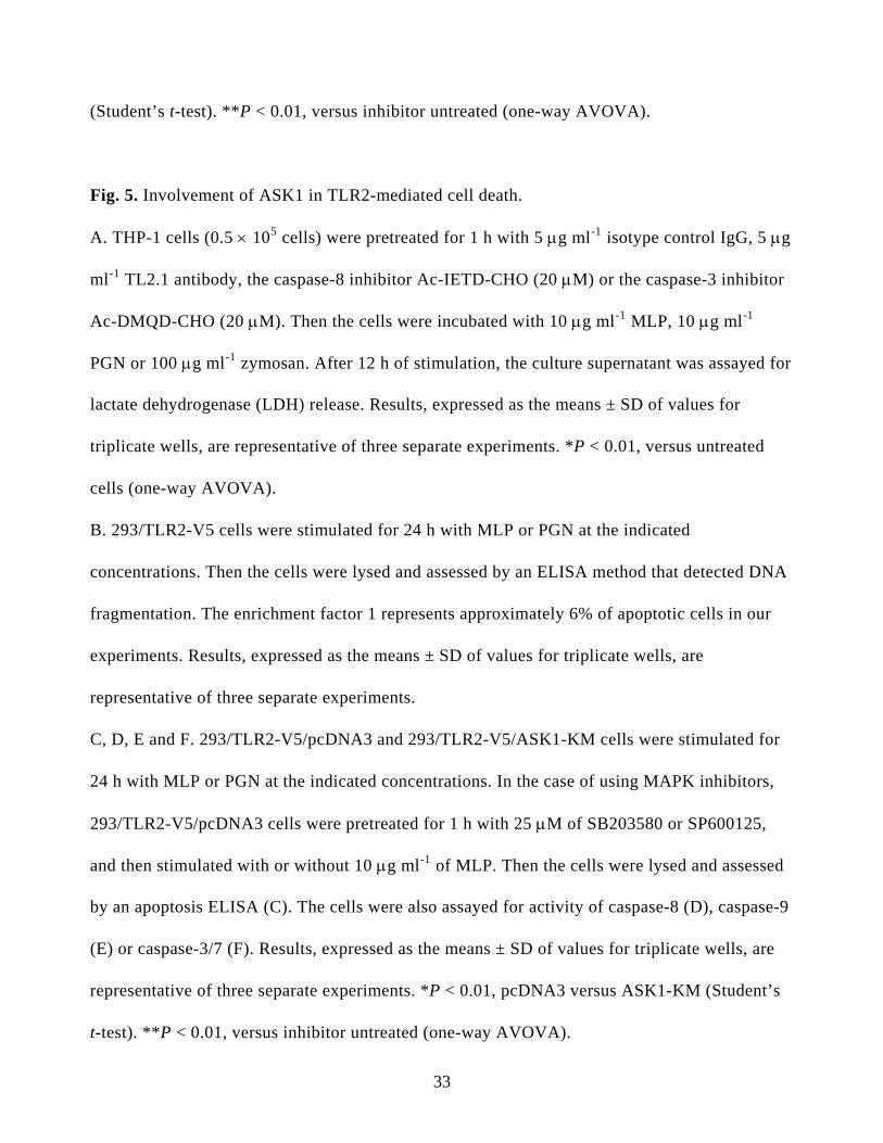
Fig. 5. Involvement of ASK1 in TLR2-mediated cell death.
A. THP-1 cells (0.5 × 10⁵ cells) were pretreated for 1 h with 5 μg ml-1 isotype control IgG, 5 μg
ml-1 TL2.1 antibody, the caspase-8 inhibitor Ac-IETD-CHO (20 μM) or the caspase-3 inhibitor
Ac-DMQD-CHO (20 μM). Then the cells were incubated with 10 μg ml-1 MLP, 10 μg ml-1
PGN or 100 μg ml-1 zymosan. After 12 h of stimulation, the culture supernatant was assayed for
lactate dehydrogenase (LDH) release. Results, expressed as the means ± SD of values for
triplicate wells, are representative of three separate experiments. *P < 0.01, versus untreated
cells (one-way AVOVA).
B. 293/TLR2-V5 cells were stimulated for 24 h with MLP or PGN at the indicated
concentrations. Then the cells were lysed and assessed by an ELISA method that detected DNA
fragmentation. The enrichment factor 1 represents approximately 6% of apoptotic cells in our
experiments. Results, expressed as the means ± SD of values for triplicate wells, are
representative of three separate experiments.
C, D, E and F. 293/TLR2-V5/pcDNA3 and 293/TLR2-V5/ASK1-KM cells were stimulated for
24 h with MLP or PGN at the indicated concentrations. In the case of using MAPK inhibitors,
293/TLR2-V5/pcDNA3 cells were pretreated for 1 h with 25 μM of SB203580 or SP600125,
and then stimulated with or without 10 μg ml-1 of MLP. Then the cells were lysed and assessed
by an apoptosis ELISA (C). The cells were also assayed for activity of caspase-8 (D), caspase-9
(E) or caspase-3/7 (F). Results, expressed as the means ± SD of values for triplicate wells, are
representative of three separate experiments. *P < 0.01, pcDNA3 versus ASK1-KM (Student’s
t-test). **P < 0.01, versus inhibitor untreated (one-way AVOVA).
33

Fig. 6. Effect of stimulation of TLR2 with MLP on the phosphorylation of serine residues in
ASK1. 293/TLR2-V5 cells (1 × 10⁷ cells/sample) were prepared and stimulated with MLP at the
indicated concentrations for the indicated period. The phosphorylated serine residues (Ser83 and
Ser967) in ASK1 (p-ASK1) were examined by Western blotting. Immunoblot analysis for
phosphorylation-independent endogenous ASK1 was also carried out as a loading control. The
result shown is representative for three independent experiments.
Fig. 7. Schematic of MLP- or PGN-induced TLR2siginaling pathways. See the text for details.
34

Acknowledgements
We would like express our hearty thanks to Prof. Hidenori Ichijo, Tokyo University, Japan for
providing an ASK1-HA construct. This work was supported by Grants-in-Aid for Science
Research (no. B(2)15390549, 16791102 and B17390498) provided by the Ministry of Education,
Culture, Sports, Science and Technology, Japan and by a grant from the Akiyama Foundation.
35

Fig. 1
A B
C
Fig.1

Fig. 2

Fig. 3
B
C
D
TLR2-V5/pcDNA3
TLR2-V5/pcDNA3
TLR2-V5/ASK1-KM
TLR2-V5/ASK1-KM
A

Fig. 4
A
** *
* * ** **
B
** *
**
***
Fig. 5
B
C
***
**
*
*
*
E
***
A
* **
D
*
F
**
** **
**
**
*

Fig. 6
MLP (μg ml-1):
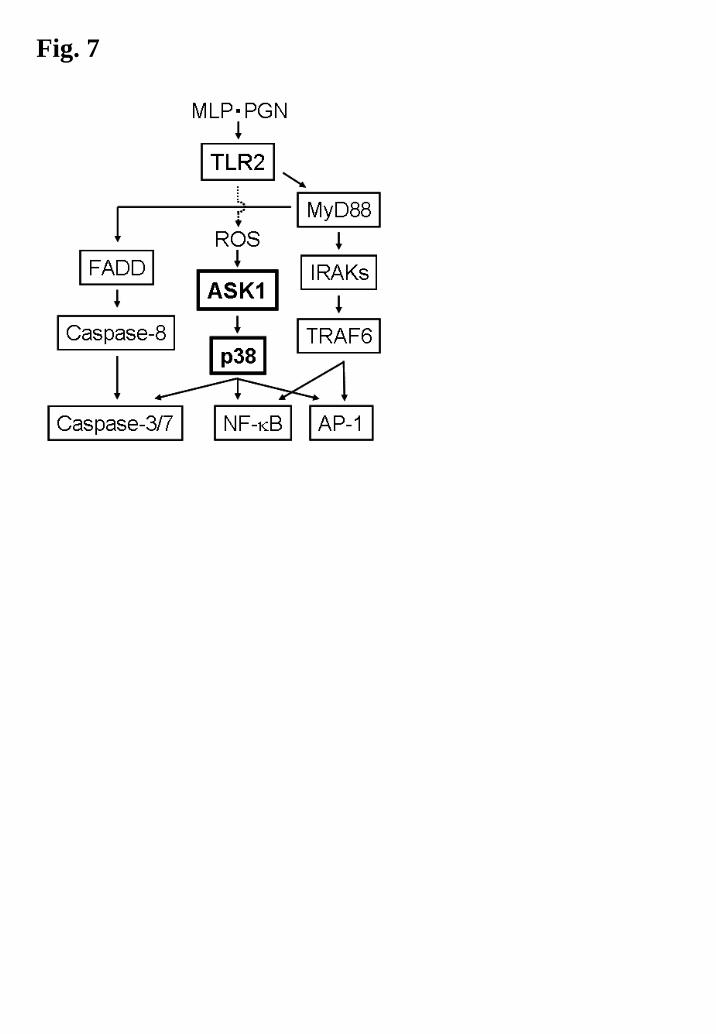
Fig. 7


![Supply of Goods talkW - David Bartos · Gmbh [1999] 1 W.L.R. 1305, 1317) In short for a Scottish court to have jurisdiction under place of performance under ... article 5 (1) (b)](https://static.fdocuments.in/doc/165x107/5b8520f37f8b9ae5498dbe71/supply-of-goods-talkw-david-gmbh-1999-1-wlr-1305-1317-in-short-for.jpg)