Apoptosis: A Functional Paradigm for Programmed Plant Cell Death ...
17
The Plant Cell, Vol. 8, 375-391, March 1996 O 1996 American Society of Plant Physiologists Apoptosis: A Functional Paradigm for Programmed Plant Cell Death lnduced by a Host-Selective Phytotoxin and lnvoked during Development Hong Wang,a Juan Li,a Richard M. Bostock,a9b and David G. Gilchrista7bi’ a Center for Engineering Plants for Resistance Against Pathogens (CEPRAP), University of California, Davis, California 95616 Department of Plant Pathology, University of California, Davis, California 95616 The host-selective AAL toxins secreted by Alternaria alternata f sp lycopersici are primary chemical determinants in the Alternaria stem canker disease of tomato. The AAL toxins are members of a new class of sphinganine analog mycotoxins that cause cell death in both animals and plants. Here, we report detection of stereotypic hallmarks of apoptosis during cell death induced by these toxins in tomato. DNA ladders were observed during cell death in toxin-treated tomato pro- toplasts and leaflets. The intensity of the DNA ladders was enhanced by Ca2+and inhibited by Zn2+. The progressive delineation of fragmented DNA into distinct bodies, coincident with the appearance of DNA ladders, also was observed during death of toxin-treated tomato protoplasts. In situ analysis of cells dying during development in both onion root caps and tomato leaf tracheary elements revealed DNA fragmentation localired to the dying cells as well as the additional formation of apoptotic-llke bodies in sloughing root cap cells. We conclude that the fundamental elements of apoptosis, as characterized in animais, are conserved in plants. The apoptotic process may be expressed during some developmen- tal transitions and is the functional process by which symptomatic lesions are formed in the Alternaria stem canker disease of tomato. Sphinganine analog mycotoxíns may be used to characterize further signaling pathways leading to apoptosis in plants. INTRODUCTION An unresolved question in plant biology is whether conserved signal transduction pathways lead to cell death by specifically ordered metabolic changes during normal development, en- vironmental stress, or pathogen attack. The concept of a functionally conserved, gene-directed program for the main- tenance of cellular homeostasisthat is dependent on localized cell death is established in animal systems, including humans, nematodes, and insects (Tomei and Cope, 1991, 1994; Gerschenson and Totello, 1992; Hengartner and Horvitz, 1994). This orderly process of programmed cell death (pcd), known as apoptosis, depends on the active participation of the dying cell and is regulated by a number of well-characterizedgenes (Schwartzmanand Cidlowski, 1993; Williams and Smith, 1993). Functionally, apoptosis is a distinct form of pcd that differs from degenerative death or necrosis in its nature and biological sig- nificance (Martin, 1993; Vaux et al., 1994; Kerr et al., 1995; Steller, 1995). Although apoptosis is a process in which the cell directs its own death, necrosis is essentially the opposite. It is the outcome of severe injurious changes in the environ- ment of affected cells and is not an active gene-dependent form of cell death (Kerr et al., 1995). To whom correspondence should be addressed. Apoptosis, invoked in morphological development and in cir- cumstances in which cells are unnecessaryor would otherwise prove harmful to the organism (Tomei and Cope, 1991), has been legitimized scientifically by both genetic and molecular studies of the process (Alison and Sarraf, 1992; Williams and Smith, 1993; Whyte and Evan, 1995). Although apoptosis may have evolved for morphogenetic and developmental reasons, it appears to represent both a positive effector against disease and a target of active suppression by many successful patho- gens (Savill, 1994; Vaux et al., 1994). Apoptosis also can be invoked as a suicide program after the appearance or disap- pearance of many different signals, including pathogens (Carson and Ribeiro, 1993; Martin, 1993; Barr and Tomei, 1994; Tomei and Cope, 1994) and toxins (Corcoran et al., 1994). Con- versely, in disease situations in which immortalizationof a cell occurs, the expression of various proto-oncogenesand virus- encoded genes that actively suppress apoptosis appears to be widespread (Raff, 1992; Smith et al., 1993; Cartier et al., 1994). In each of these cases, it is clear that multiple, distinct signaling pathways are involved in the induction of apoptosis, but eventually there is convergence in the seminal events that give rise to the phenotype of apoptosis. There is a series of morphologicaland biochemical hallmarks of apoptosis in animais (Tomei and Cope, 1991,1994).Animal
Transcript of Apoptosis: A Functional Paradigm for Programmed Plant Cell Death ...
The Plant Cell, Vol. 8, 375-391, March 1996 O 1996 American Society of Plant Physiologists
Apoptosis: A Functional Paradigm for Programmed Plant Cell Death lnduced by a Host-Selective Phytotoxin and lnvoked during Development
Hong Wang,a Juan Li,a Richard M. Bostock,a9b and David G. Gilchrista7bi’ a Center for Engineering Plants for Resistance Against Pathogens (CEPRAP), University of California, Davis, California 95616
Department of Plant Pathology, University of California, Davis, California 95616
The host-selective AAL toxins secreted by Alternaria alternata f sp lycopersici are primary chemical determinants in the Alternaria stem canker disease of tomato. The AAL toxins are members of a new class of sphinganine analog mycotoxins that cause cell death in both animals and plants. Here, we report detection of stereotypic hallmarks of apoptosis during cell death induced by these toxins in tomato. DNA ladders were observed during cell death in toxin-treated tomato pro- toplasts and leaflets. The intensity of the DNA ladders was enhanced by Ca2+ and inhibited by Zn2+. The progressive delineation of fragmented DNA into distinct bodies, coincident with the appearance of DNA ladders, also was observed during death of toxin-treated tomato protoplasts. In situ analysis of cells dying during development in both onion root caps and tomato leaf tracheary elements revealed DNA fragmentation localired to the dying cells as well as the additional formation of apoptotic-llke bodies in sloughing root cap cells. We conclude that the fundamental elements of apoptosis, as characterized in animais, are conserved in plants. The apoptotic process may be expressed during some developmen- tal transitions and is the functional process by which symptomatic lesions are formed in the Alternaria stem canker disease of tomato. Sphinganine analog mycotoxíns may be used to characterize further signaling pathways leading to apoptosis in plants.
INTRODUCTION
An unresolved question in plant biology is whether conserved signal transduction pathways lead to cell death by specifically ordered metabolic changes during normal development, en- vironmental stress, or pathogen attack. The concept of a functionally conserved, gene-directed program for the main- tenance of cellular homeostasis that is dependent on localized cell death is established in animal systems, including humans, nematodes, and insects (Tomei and Cope, 1991, 1994; Gerschenson and Totello, 1992; Hengartner and Horvitz, 1994). This orderly process of programmed cell death (pcd), known as apoptosis, depends on the active participation of the dying cell and is regulated by a number of well-characterized genes (Schwartzman and Cidlowski, 1993; Williams and Smith, 1993). Functionally, apoptosis is a distinct form of pcd that differs from degenerative death or necrosis in its nature and biological sig- nificance (Martin, 1993; Vaux et al., 1994; Kerr et al., 1995; Steller, 1995). Although apoptosis is a process in which the cell directs its own death, necrosis is essentially the opposite. It is the outcome of severe injurious changes in the environ- ment of affected cells and is not an active gene-dependent form of cell death (Kerr et al., 1995).
To whom correspondence should be addressed.
Apoptosis, invoked in morphological development and in cir- cumstances in which cells are unnecessary or would otherwise prove harmful to the organism (Tomei and Cope, 1991), has been legitimized scientifically by both genetic and molecular studies of the process (Alison and Sarraf, 1992; Williams and Smith, 1993; Whyte and Evan, 1995). Although apoptosis may have evolved for morphogenetic and developmental reasons, it appears to represent both a positive effector against disease and a target of active suppression by many successful patho- gens (Savill, 1994; Vaux et al., 1994). Apoptosis also can be invoked as a suicide program after the appearance or disap- pearance of many different signals, including pathogens (Carson and Ribeiro, 1993; Martin, 1993; Barr and Tomei, 1994; Tomei and Cope, 1994) and toxins (Corcoran et al., 1994). Con- versely, in disease situations in which immortalization of a cell occurs, the expression of various proto-oncogenes and virus- encoded genes that actively suppress apoptosis appears to be widespread (Raff, 1992; Smith et al., 1993; Cartier et al., 1994). In each of these cases, it is clear that multiple, distinct signaling pathways are involved in the induction of apoptosis, but eventually there is convergence in the seminal events that give rise to the phenotype of apoptosis.
There is a series of morphological and biochemical hallmarks of apoptosis in animais (Tomei and Cope, 1991,1994). Animal

376 The Plant Cell
cells undergoing apoptosis exhibit shrinkage, loss of cell-to- cell contact in organized tissues, and orderly fragmentation of nuclear DNA at internucleosomal sites. The fragmented DNA may then be organized into sharply defined membrane-bound bodies referred to as apoptotic bodies. In animal cells, these apoptotic bodies are taken up by adjacent cells and degraded within minutes to hours (Bursch et al., 1990). lnternucleosomal cleavage of DNA is catalyzed by an endogenous Ca2+- dependent endonuclease that generates 180-bp fragments and multiples of these units, which are resolved by gel electropho- resis as a laddered pattern of DNA that corresponds to the nucleosomal fragments (Collins et al., 1992; Cohen et al., 1994). Fragmentation of DNA during apoptosis also can be detected in situ by reagents that react with the exposed 3' hydroxyls on the nucleosomal units (Gavrieli et al., 1992; Gold et al., 1994; Li et al., 1995). The assay procedure involves end labeling the DNA fragments by terminal deoxynucleotidyl transferase (IdT) with UTP conjugated to a detectable marker (Gorczyca et al.,
Phenotypically analogous death-enforced changes in ho- meostasis occur in plants both during development (Woodson et al., 1992; ONeill et al., 1993; Zhang and ONeill, 1993; Chasan, 1994; Smart, 1994) and in response to pathogens (Keen, 1990; Lamb, 1994). Recently, there has been consid- erable speculation that hypersensitive resistance-associated cell death in severa1 incompatible plant-microbe interactions may follow a death pattern similar to apoptosis in animals (Bachmair et al., 1990; Greenberg and Ausubel, 1993; Dietrich et al., 1994; Greenberg et ai., 1994; Lamb, 1994; Vaux et al., 1994; Mittler et al., 1995). Because much is known about both the process and modulating genes in animals, the search for functional homologs of these genes in plants may prove use- ful if aspects of the process have been conserved in plants as they appear to be in animals. The stereotypic hallmarks of apoptosis, including DNA ladders and apoptotic bodies (Wyllie, 1995), and homologs of genes shown to regulate apop- tosis in animals have not been reported in plants. However, recent reports, based on in situ detection of DNA fragmenta- tion in dying cells, presumably resulting from specific endonuclease cleavage of nuclear DNA at internucleosomal sites during xylem differentiation and disease resistance ex- pression, have suggested that an initial event consistent with pcd in animals can occur in plants (Mittler et al., 1995).
The metabolic and genetic bases of the functionally signifi- cant localized death of plant cells associated with necrotrophic disease (susceptibility), the hypersensitive reaction (HR) (re- sistance), reproduction, tissue differentiation, and natural senescence have not been characterized (see Ryerson and Heath, 1996). We have been studying the process of cell death in plants undergoing a susceptible response to pathogens to identify gene products that may modulate the process. This is part of a long-term effort to detect potential targets for genetic engineering of nove1 resistance genes by modifying the ex- pression of host genes that either facilitate or fail to block susceptibility.
1993).
One of the systems we have been using to look for genetic and cellular markers of disease-dependent cell death is the Alternaria stem canker disease (Grogan et al., 1975; Gilchrist et al., 1992, 1995; Moussatos et al., 1993a, 19934 1994) of tomato caused by Alternaria alternata f sp lycopersici. Key fea- tures of this system include (1) expression by the pathogen of the host-selective toxins (AAL toxins) that are primary de- terminants of the disease (Siler and Gilchrist, 1983; Clouse and Gilchrist, 1986) and (2) expression by the host of the Asc (Alternaria stem canker) gene that determines susceptibility (asdasc) and resistance (Asc/Asc) to the pathogen and to the AAL toxins (Clouse and Gilchrist, 1986). Cell death in tomato leaflets, caused by either the pathogen or purified AAL toxin, appears as localized spots of dead cells (Gilchrist and Grogan, 1976). Protoplasts of Asc isolines also react to the AAL toxins in a genotype-specific, toxin concentration-dependent manner, indicating that the alleles of the Asc gene are functionally active in protoplasts (Moussatos et al., 1993a). Toxin-induced cell death in protoplasts and leaflets requires between 36 to 48 hr, during which toxin-treated cells appear to maintain integrity of the plasma membrane until symptoms of death are observed (Gilchrist, 1983).
AAL toxins are a family of monoesters of tricarballylic acid and polyhydric alcohols (Bottini et al., 1981; Caldas et ai., 1994, 1995) structurally related to sphinganine, a precursor of the chemical backbone of all sphingolipids. The AAL toxins and the fumonisins, a group of chemically related congeners pro- duced by Fusarium moniliforme (Nelson et al., 1993), are potent and widespread mycotoxins with important health implications (Sydenham et al., 1991; Hopmans and Murphy, 1993; Merrill et al., 1993). The most abundant and active toxin congeners are AAL toxin TA and fumonisin B1 (FB,). The spectrum of biological effects induced by these sphinganine analog myco- toxins (SAMs) includes cell death in plants (Gilchrist and Grogan, 1976; Clouse and Gilchrist, 1986; Gilchrist et al., 1992, 1995; Abbas et al., 1994) and maladies in animals ranging from cancer (Gelderblom et al., 1992; Syndenham et al., 1990) to renal, neural, and hepatic degenerative diseases (Ross et al., 1990; Norred et al., 1992; Thiel et al., 1992; Norred, 1993).
Functionally, these toxins are potent inhibitors of ceramide synthase in plants (Abbas et al., 1994; Gilchrist et ai., 1995) and animals (Wang et al., 1990,1992; Norred et al., 1992; Merrill et al., 1993; Schroeder et al., 1994), which suggests that changes in cellular levels of sphingoid bases may be linked to cell death. As second messengers, sphingolipids and sphin- goid bases now are known to regulate cell behavior at many levels, including cell communication, growth factor receptors, growth, differentiation, and transformation (Ghosh et al., 1994; Jarvis et al., 1994a, 1994b; Natarajan et al., 1994; Bose et al., 1995; Chao, 1995; Jones and Murray, 1995; Khan et ai., 1995; Merrill et al., 1995; Obeid and Hannun, 1995; Ohata et a1.,1995). Recently, we observed that fumonisins and AAL toxins induce cellular hallmarks of apoptosis in African green monkey kid- ney (CV-1) cells (Wang et al., 1996; Winter et al., 1996), most likely through interdiction of lipid-based signaling pathways.
Apoptotic Characters in Plant Cell Death 377
Given the relationship between ceramides and apoptosis inanimals, SAMs and ceramide metabolism, and our observa-tion that SAMs induce apoptosis in CV-1 cells, we reasonedthat asc/asc tomato cells might be a useful system to ascer-tain whether stereotypical hallmarks of the apoptotic processoccur during toxin-induced cell death.
Evidence for the existence in plants of a pcd process thatis available for use during developmental transitions and thatcould be triggered during disease would likely be dependenton genes of interest to both plant biologists and pathologists.Protoplasts and intact tomato leaflet tissue were analyzed forcharacteristics of apoptosis following treatment with host-selective SAMs. Simultaneously, we histologically examinedtomato leaf sections during formation of tracheary elementsand onion root cap cells undergoing sloughing during root elon-gation for evidence of developmentally regulated apoptoticcharacteristics. We report here (1) the presence of DMA cleav-age at internucleosomal linker sites during toxin-induced celldeath, using the TdT end-labeling (TUNEL) method; (2) Ca2+-dependent DNA ladders of nuclear DNA from toxin-treated pro-toplasts and leaflets; and (3) the formation of distinct bodies,resembling apoptotic bodies, containing fragmented DNA inboth SAM-treated protoplasts and sloughing root cap cells.
M 1 6 7 8
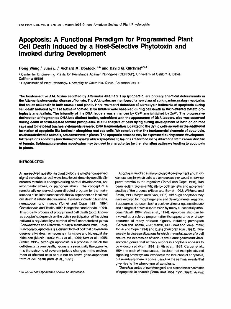
Figure 1. Agarose Gel Analysis of Tomato Protoplasts for DNAFragmentation.
Lane 1 contains protoplasts that were incubated in TM-2 medium con-taining 3.3 mM CaCI2; lane 2, medium plus 20 \iM AAL toxin; lane3, medium plus 20 nM FB,; lane 4, medium plus 20 \iM FB, and 50liM cycloheximide; lane 5, medium plus 5 mM Fe2SO4; lane 6,medium containing 1 mM CaCI2 plus 20 nM FB,; lane 7, medium con-taining 10 mM CaCI2 plus 20 nM FB,; lane 8, medium containing 10mM Ca2' and 10 mM ZnS04 plus 20 nM FB,; and lane M, DNA mark-ers of 1-kb fragments. DNA was isolated 48 hr after treatment, separatedon a 1.5% agarose gel by electrophoresis, stained with ethidium bro-mide, and photographed under UV illumination.
RESULTS
Detection of ONA Ladders after Toxin Treatment ofasc/asc Protoplasts
Protoplasts were preincubated in TM-2 medium (Shahin, 1985)for 6 hr and subjected to gentle centrifugation in 0.6 M sucroseto remove cells that had died during protoplast isolation; thehealthy protoplasts were then treated with 20 u,M TA for 48hr. This toxin concentration was chosen because it led to thedeath of ~50% of the total number of treated cells within 48hr in both leaflets and protoplasts. This level of cell death per-mitted detection of both increases and decreases in death dueto various treatments imposed in these studies.
As shown in Figure 1, agarose gel analysis of DNA isolatedfrom TA-treated cells revealed DNA ladders consisting of frag-ments increasing in size by multiples of 180 bp when stainedwith ethidium bromide (Figure 1, lane 2). Healthy control cells,maintained in culture medium alone, did not show DNA frag-mentation under these conditions (Figure 1, lane 1). Theaddition of 20 u.M FB, to the protoplasts induced DNA laddersequivalent to those induced by TA (Figure 1, lane 2). Additionaltreatments that induced DNA ladders during cell death, in-cluding arachidonic acid, a fungal elicitor of the HR fromPhytophthora infestans (Bostock et al., 1981), and KCN (seeRyerson and Heath, 1996), are listed in Table 1.
Another frequent observation regarding the induction of DNAladders in animals is that apoptosis is inhibited by cyclohexi-mide, suggesting that the process requires gene expression,at least in some situations, to go to completion (Schwartzmanand Cidlowski, 1993). In tomato protoplasts, the addition of 50nM cycloheximide reduced substantially the degree of DNAfragmentation induced by 20 u,M FB, (Figure 1, lane 4) or TA(data not shown). The reduction of Ca2+ (as CaCI2) from 3.3to 1 mM in the TM-2 medium (Figure 1, lane 6) led to a reduc-tion in DNA fragmentation, whereas an increase to 10 mMCa2+ (Figure 1, lane 7) increased the intensity of the DNA lad-ders induced by 20 u.M FB,. The activating effect of Ca2+ onDNA fragmentation was completely negated by the additionof 10 mM ZnSO4 (Figure 1, lane 8). The concentration- depen-dent activation by Ca2+ and inhibition by Zn2+ of toxin-inducedDNA fragmentation of the plant DNA are consistent with thewidely recognized effect of these divalent cations on the en-dogenous endonuclease responsible for cleavage of DNAduring apoptosis in animal systems (Barry and Eastman, 1992;Lohmann and Beyersmann, 1993; Peitsch et al., 1993).Whereas cell death in the tomato protoplast system generatedDNA ladders from many effectors (Table 1), we found that 5mM Fe2SO4 killed 90 to 100% of the protoplasts within 12 hrbut did not induce DNA fragmentation (Figure 1, lane 5;Schwartzman and Cidlowski, 1993).

378 The Plant Cell
Table 1. Compounds or Treatments Tested in TomatoProtoplasts or Leatlets for Induction of DMA Ladders3
CompoundsAAL toxin TAFB,KCN"KCNArachidonic acidHeat shockHeat shockStaurosporineC-2 ceramideFe2SO4
Concentration11
or Duration5 nM5 nM1 nM
300 nM100 nM4 hr at 42°C5 min at 65°C500 nM
20 nM5 mM
CellDeathc
303375
100305510203095
DNAFragmentation"1
Protoplasts Leaflets+ + + ++++ + + + + ++ + + + NT1
+ + + + ++ + + NT+ + + NTNT 0'+ + NT+ + NT0 0
a All tissues were incubated for 48 hr at 23°C before DNA isolationand vital staining.b Each concentration listed is the lowest concentration tested that in-duced ladders or the highest concentration tested that did not induceladders.c Percentage of cell death is based on fluorescein diacetate vitalstaining.d Relative intensity of DNA ladders compared with Figure 1 is shown:+, lane 4 to + + + + , lane 7.
8 KCN-induced protoplast DNA fragmentation was at concentrationsas low as 1 nM. DNA ladders could be resolved within 4 hr at 100nM KCN.1 NT, not tested; 0, tested but not detected.
Detection of DNA Ladders by DNA Gel Blot Analysisafter Treatment of Leaflets with SAMs
Detached leaflets were treated with TA by the standard leafletbioassay (Gilchrist and Grogan, 1976) in which toxin is takenup for 36 hr through the petiole of the leaflet from filter paperimpregnated with 20 u,M TA or FB, in H2O. At this toxin con-centration, typical lesions covered ~50 to 60% of the totalleaflet, indicating that DNA from the dead areas would be com-bined with a nearly equal amount of DNA from apparentlyhealthy areas outside the lesions when the entire leaflet wasextracted. Preliminary experiments indicated that gel blot anal-ysis was more sensitive than ethidium bromide staining indetecting DNA ladders. Hence, to maximize visualization ofladders from treated leaflets containing both lesion and healthyregions, DNA gel blot analysis was performed on DNA thatwas isolated by the nonphenol method (Tai and Tanksley, 1991)and then separated on 1.5% agarose gel. The separated DNAwas transferred to a nylon membrane and hybridized with 32P-labeled tomato total genomic DNA. As shown under these con-ditions in Figure 2, both toxins (Figure 2, lanes 1 and 2) inducedDNA fragmentation in the treated leaflets; this fragmentationappeared as a series of bands corresponding to increasingmultiples of 180- to 200-bp fragments. No fragmentation wasobserved in the water control tissue (Figure 2, lane C). In ad-
dition, identical results were obtained with DNA isolated fromleaflets taken directly from plants infected with the A. a. lycoper-sici that induced typical symptoms of the Alternaria stem cankerdisease (Gilchrist and Grogan, 1976).
In Situ Detection of Nuclear Fragmentation by theTUNEL Method in Protoplasts
Apoptosis is further revealed in animals by the orderly com-partmentation of DNA into distinct membrane-bound apoptoticbodies that persist for varying periods of time. The bodies con-taining fragmented DNA eventually move to the periphery ofthe affected cells, which then become distorted in shapearound these bodies, a process called blebbing (Tomei andCope, 1991). In situ detection of nuclear DNA fragmentationand visualization of apoptotic bodies involves using a TUNELof the free 3' -OH groups on the nucleosomal fragments witha detectable marker (Gorczyca et al., 1993). The TUNELmethod was used to determine whether fragmentation was re-stricted to nuclear DNA of TA-treated protoplasts and whetherthe fragmented DNA was then compartmentalized into sev-eral distinct bodies, as occurs in animal systems. In theseexperiments, the asc/asc protoplasts were incubated insideplastic rings directly on microscope slides for 24 hr in TM-2medium containing 20 nM TA and compared with cells in-cubated in TM-2 medium alone after application of the TUNELmethod and counterstaining the cells with Hoechst 33342 fortotal DNA.
As shown in Figure 3, light microscopy revealed the loca-tion and shape of the TA-treated cells compared with the control
C 1 2
Figure 2. Toxin-Induced DNA Fragmentation in Tomato Leaflets.Excised tomato leaflets were treated for 36 hr with H2O (lane C), 20liM AAL toxin (lane 1), or 20 nM FB, (lane 2). DNA was isolated andseparated as described in Methods. Gel blot analysis was performedby hybridizing the DNA with a 32P-labeled tomato genomic DNA. Thearrowheads indicate 500- and 1000-bp DNA markers.
Apoptotic Characters in Plant Cell Death 379
s
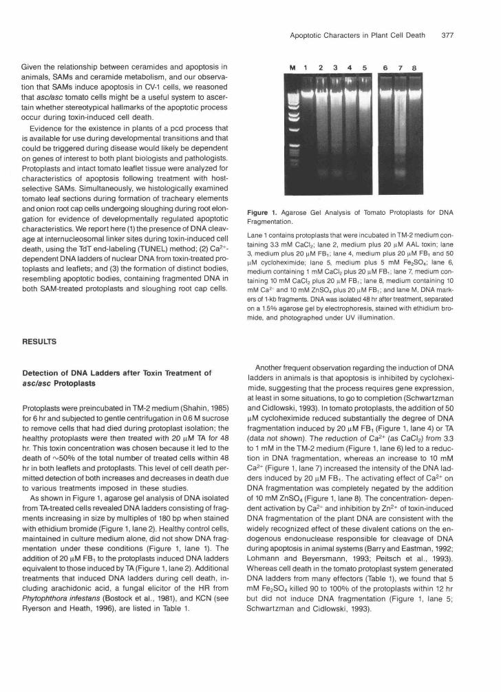
Figure 3. In Situ Detection of Apoptosis in Tomato Protoplasts.
(A) to (C) Protoplasts incubated for 24 hr in TM-2 medium (control).(D) to (F) Protoplasts incubated for 24 hr in TM-2 medium containing 20 nM AAL toxin.Nomarski optics were used for (A) and (D). In (B) and (E), Hoechst 33342 was used to stain the DNA. In (C) and (F), the TUNEL assay wasused to detect DNA fragmentation. Bar in (F) = 20 urn.

380 The Plant Cell
protoplasts (Figures 3A and 3D) and permitted distinction of cells that were becoming chlorotic, darkened, and slightly dis- torted in shape after a 24-hr exposure to the toxin. DNA staining revealed single nuclei within the individual cells that appeared to be centrally located and of similar size and shape in control cells (Figure 38). In contrast, cells treated with TA exhibited nuclei with more variable size and shape, frequently with mul- tiple distinct bodies containing DNA that often were associated with the periphery of the cell (Figure 3E).
Application of the TUNEL method to end-labeled fragmented DNA with dUTP-digoxigenin followed by treatment with anti- digoxigenin antibody and fluorescein isothiocyanate (FITC)- labeled anti-goat antibody revealed fragmented nuclear DNA in toxin-treated cells (Figures 3C and 3F). Together, these tech- niques permitted viewing the exact same cells for general appearance with light microscopy (Nomarski), localization of nuclear DNA (Hoechst 33342), and detection of fragmented nuclear DNA (TUNEL), using different wavelengths of UV light. The TUNEL method revealed that -50% of the cells were posi- tive for excess 3' - 0 H groups and that some of these cells contained multiple TUNEL-positive, distinctly separated bod- ies. The size, shape, and location of the Hoechst-stained DNA in each of the TUNEL-positive cells corresponded exactly with the TUNEL-positive bodies (compare Figures 3F and 3E) in the TA-treated cells. No FITC-derived fluorescence was ob- served when the TdT, the dUTP substrate, or the respective antibodies were omitted from the reaction. Vital staining of dy- ing protoplasts with fluorescein diacetate indicated that these distinct bodies, even when separated from the cell, were bounded by an intact membrane, although we did not conduct electron microscopy studies to confirm this point. These results are strikingly consistent with the characteristics of apoptosis in animals cells, in which similar techniques have been used as in situ strategies to visualize nuclei undergoing fragmenta- tion during apoptosis and the subsequent formation of apoptotic bodies (Gorczyca et al., 1993).
In Situ Detection of Nuclear Fragmentation in TA-Treated Tomato Leaflets
Tomato leaflets (asc/asc) were allowed to take up 20 pM TA for 24 hr in the standard detached leaflet bioassay procedure. Localized dead areas and surrounding areas at the lesion mar- gin without symptoms were fixed in 10% buffered formalin, cut into small pieces, imbedded in HistoPrep (Fisher), and frozen in liquid nitrogen before sectioning with a freezing microtome at a thickness of 10 pm. Areas of dead cells were readily detected by their brown color under normal light as well strong autofluorescence in the dead areas under UV light. In addition, there was a complete loss of cell-to-cell contact, which is seen in Figures 48 and 4D. Strong autofluorescence in the lesion areas quenched FlTC fluorescence in contrast to protoplasts. This problem was circumvented by using anti- digoxigenin antibody conjugated to alkaline phosphatase with
leaflet tissue, although the TUNEL-positive color stain could block the Hoechst stain. As shown in Figure 4, the areas con- taining dead cells consistently exhibited a positive TUNEL reaction that was limited to the area of the lesion (designated as dc in Figure 48). The TUNEL-positive area did not extend beyond the margin of the lesion, nor did TUNEL-positive cells appear in the water-treated controls (Figure 4C). The same section (Figure 4A) counterstained with Hoechst 33342 re- vealed that DNA was present in all cells, but intact nuclei were confined to cells in areas that did not express a positive alka- line phosphatase reaction.
Dependence on end labeling by TdT in the lesion areas was confirmed by the results of negative control reactions con- ducted in the absence of the transferase (Figure 4D) that showed no color development in the area of the dead cells. In addition, the cells in the lesion area appeared to have lost cell-to-cell contact and exhibited distorted margins but retained the fragmented DNA within the cell margins, even though the DNA was no longer confined to a single nuclear body, as can be seen in DNA counterstained cells outside the lesion area (compare Figures 4A and 48). All of these results are consis- tent with the morphological characteristics of apoptosis in organized animal tissues (Gorczyca et al., 1993).
Quantification of TA-lnduced Cell Death in asc/asc Protoplasts
The positive association between detection of DNA ladders and TUNEL-positive DNA in protoplasts treated with 20 pM TA or FB, suggested that most protoplasts may have died in a manner consistent with the induction of an apoptotic process. We examined this relationship further in additional experiments in which the protoplasts were isolated in the same manner as those in Figures 3 but were cultured at 24OC for 48 hr in 250 pL of TM-2 medium with or without either toxin in 24-well plates with a concentration of 105 protoplasts per mL. Viable cells, detected by staining with fluorescein diacetate, and total cells were viewed on an inverted microscope. For in situ applica- tion of the TUNEL assay, protoplasts from the same isolations as shown in Figure 3 were cultured directly on slides, and the TUNEL reaction was run with alkaline phosphatase conjugated to the anti-digoxigenin antibody. Use of alkaline phosphatase was necessary because FlTC fluorescence faded too quickly to permit accurate counting.
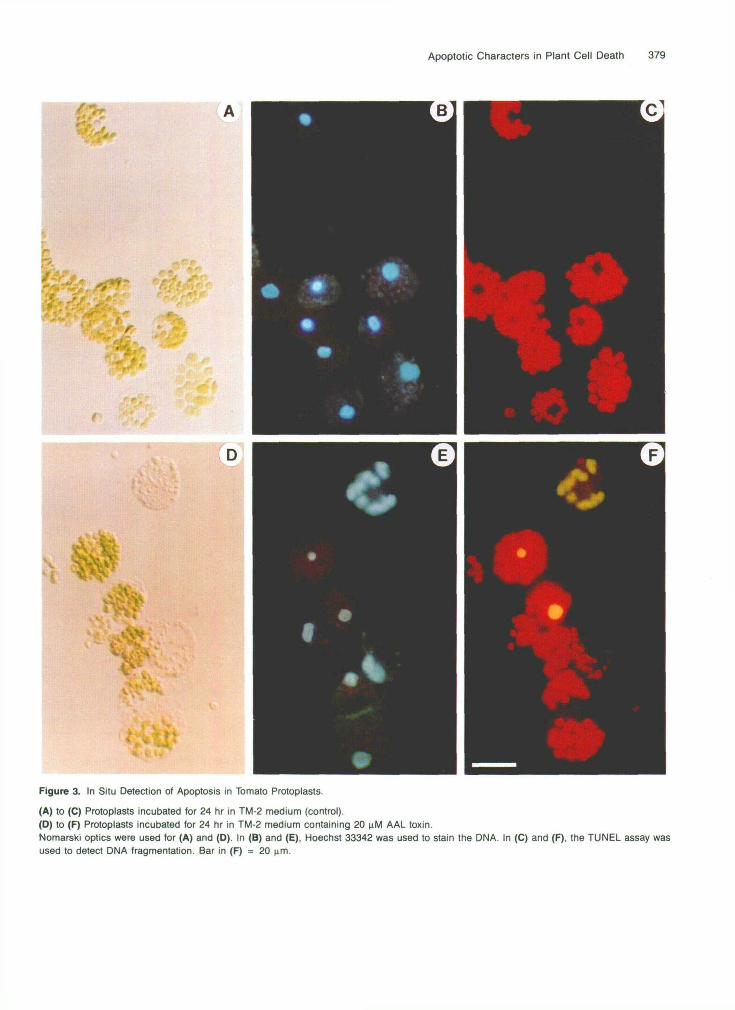
Approximately 50% of the cells died in the TA and FB, treat- ments; of these, 85 and 95%, respectively, were TUNEL positive (Figures 5A and 58). The addition of 10 mM Ca2+ to the TA treatment resulted in 100% of the dead cells reacting TUNEL positive, in contrast to the addition of 10 mM Zn2+, which did not protect against cell death but reduced the frag- mentation detectable by the TUNEL reaction to <10% of the total dead cells. Cycloheximide treatment resulted in a reduc- tion of both TA-associated death and TUNEL-positive cells to the respective control values, suggesting that inhibition of gene

Apoptotic Characters in Plant Cell Death 381
B
•- \
Figure 4. In Situ Detection of Apoptosis in Tomato Leaflets.
Tomato leaflets in (A), (B), and (D) were treated for 24 hr with AAL toxin (20 nM). Tomato leaflets in (C) were treated for 24 hr with H2O.(A) Toxin-treated tomato leaf stained for DMA with Hoechst 33342.(B) The same field of tomato leaf section as shown in (A) stained by using the TUNEL method as described in Methods.(C) Section from a water-treated (control) tomato leaflet stained by using the TUNEL method.(D) AAL toxin-treated tomato leaf tissue stained as shown in (B), except for the absence of TdT.Note that the detection of fragmented DNA in dead cell (dc) areas was TdT dependent. Bar in (A) = 50 urn.
expression reduced the absolute amount of cell death result-ing from toxin treatment. These data also suggest that the effectof 10 mM Ca2+ added to the TM-2 medium was not to increasethe number of dead cells but to increase the activity of theendonuclease in those cells that had been triggered to die.
Progression of Nuclear Changes during TA-lnducedCell Death in asc/asc Protoplasts
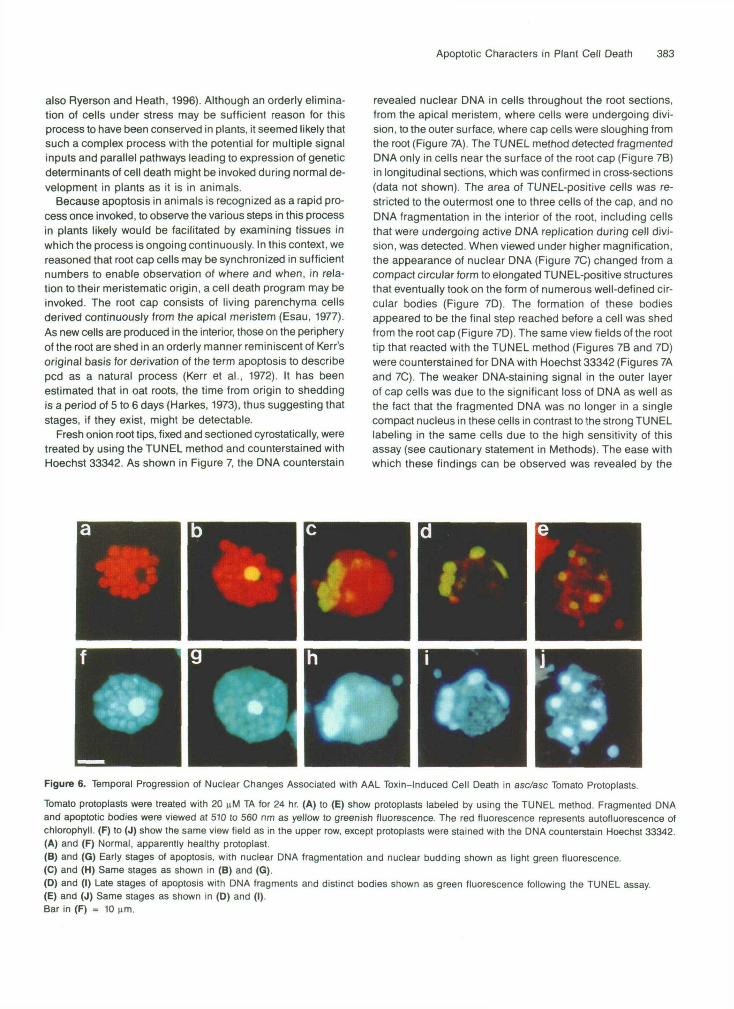
In the course of viewing thousands of protoplasts harvestedat different times after treatment with TA that were undergo-ing cell death, we observed that the changes in nuclear DNAfollowed a consistent pattern once the nuclear material first
reacted positively in the TUNEL procedure. As shown in Fig-ure 6, this progression of changes began with an intact nucleus(Figures 6A and 6F) that maintained its shape and size butreacted TUNEL positive with the DNA localized in a single nu-clear body (Figures 6B and 6G). This was followed by anexpansion or spreading of the nuclear material, which thenbegan to show delineation into several nuclear bodies (Figures6C and 6H). These bodies maintained a generally round shapebut sometimes took on a more elongated form as the processproceeded (Figures 6D and 61). Following migration of thesebodies to the periphery of the cell, the final stage appearedto be one in which the cell began to lose shape (blebbing) andthen disintegrate, at which point the distinct bodies containingthe fragmented DNA were released into the medium (Figures
382 The Plant Cell
A
50 u)
u) 5 40
$ 30 U m
*
c.
; 20 10
s
O
B
6o 1 f ([I 5 o j
1 2 3 4 5 6
Treatments Figure 5. Quantification of AAL Toxin-lnduced pcd in Tomato Protoplasts.
(A) Protoplasts were isolated and cultured in 250 pL of treatment so- lution in a 24-well plate ata concentration of 105 protoplasts per mL. Plates were incubated at 24OC for 48 hr. Viable cells were counted in three wells with 10 views per well and each view containing 4 0 0 cells. (8) Protoplasts were cultured directly on slides and fixed, as described in Methods. After in situ TdT-digoxigenin end labeling, the slides were stained with anti-digoxigenin antibody followed by alkaline phospha- tase-labeled anti-goat antibody. Apoptotic protoplasts were counted in two slides with 10 views per slide and each view containing ~ 2 0 0 cells. Means and SEM from three experiments are indicated above the respec- tive bars: (1) indicates the control; (2), treatment with 20 pM TA; (3, 20 pM FB,; (4), 20 pM TA plus 10 mM Ca2+; (5), 20 pM TA plus 10 mM Caz+ and 10 mM Zn2+; and (6), 20 pM TA and 15 pM cycloheximide.
6E and 6J). After the bodies moved outside the cell, many appeared to remain intact, retained their shape, and remained closely associated with each other in the high osmotic protoplast medium.
We were unable to estimate how long it took for a cell to traverse these changes once fragmentation begins, because the death process in these protoplasts is not fully synchronized and all determinations are made on destructively sampled cells. However, we were able to detect stages equivalent to those illustrated here from 6 to 36 hr under standard assay condi- tions. In animal cells undergoing apoptosis, analogous events can occur in minutes to a few hours. In some cases, discrete stages are not readily or consistently detectable because the events proceed so quickly. Condensation and budding to form apoptotic bodies can be completed within minutes (Matter, 1979) and may remain visible for only a few hours (Bursch et al., 1990), prornpting Kerr et al. (1991) to comment that apop- totic bodies are remarkably inconspicuous histologically. We suspect that the tomato protoplast system is biased toward de- tection of stages, because we are able to sample periodically during the 24- to 48-hr death window. This likely improved our chances of seeing progressive stages, in contrast to viewing similar events in organized tissue undergoing either toxin- induced or developmentally triggered death by this process.
Effect of AAL Toxin on Tomato Protoplasts and Leaflet Tissue in Resistant Asc lsolines
60th protoplasts and leaflets of the AsdAsc isoline were treated with TA at 5 to 20 pM under the same conditions as the asc/asc isoline. There was no apparent toxin-induced death observed in either situation, nor were there any DNA ladders or TUNEL- positive reactions observed, and the tissues did not differ from untreated controls. However, both protoplasts and leaflets pro- duced DNA ladders in the Asc/Asc isoline when treated with other inducers of cell death (Table 1). These other effectors with potential to induce apoptosis based on animal studies included sphinganine metabolites (C-2 ceramide), a lipid-based elicitor of cell death (arachidonic acid), the protein kinase in- hibitor staurosporine, and heat shock. Concentrations used were based on previous reports in the literature on animals. Among the chemicals tested, many induced DNA ladders and cell death in both genotypes, but only the SAMs were geno- type specific in toxicity and DNA fragmentation. However, Fe2S04 killed cells but did not induce the formation of DNA ladders and therefore was used as a negative control for nonapoptotic cell death.
Detection of TUNEL-Positive Cells and Apoptotic-like Bodies in Sloughing Root Cap Cells
Detection of morphological characteristics of apoptosis in tomato cells challenged by a number of negative externa1 stimuli has confirmed the existence of severa1 functional ele- ments of the apoptotic process in plant cells under stress (see
Apoptotic Characters in Plant Cell Death 383
also Ryerson and Heath, 1996). Although an orderly elimina-tion of cells under stress may be sufficient reason for thisprocess to have been conserved in plants, it seemed likely thatsuch a complex process with the potential for multiple signalinputs and parallel pathways leading to expression of geneticdeterminants of cell death might be invoked during normal de-velopment in plants as it is in animals.
Because apoptosis in animals is recognized as a rapid pro-cess once invoked, to observe the various steps in this processin plants likely would be facilitated by examining tissues inwhich the process is ongoing continuously. In this context, wereasoned that root cap cells may be synchronized in sufficientnumbers to enable observation of where and when, in rela-tion to their meristematic origin, a cell death program may beinvoked. The root cap consists of living parenchyma cellsderived continuously from the apical meristem (Esau, 1977).As new cells are produced in the interior, those on the peripheryof the root are shed in an orderly manner reminiscent of Kerr'soriginal basis for derivation of the term apoptosis to describepcd as a natural process (Kerr et al., 1972). It has beenestimated that in oat roots, the time from origin to sheddingis a period of 5 to 6 days (Harkes, 1973), thus suggesting thatstages, if they exist, might be detectable.
Fresh onion root tips, fixed and sectioned cyrostatically, weretreated by using the TUNEL method and counterstained withHoechst 33342. As shown in Figure 7, the DMA counterstain
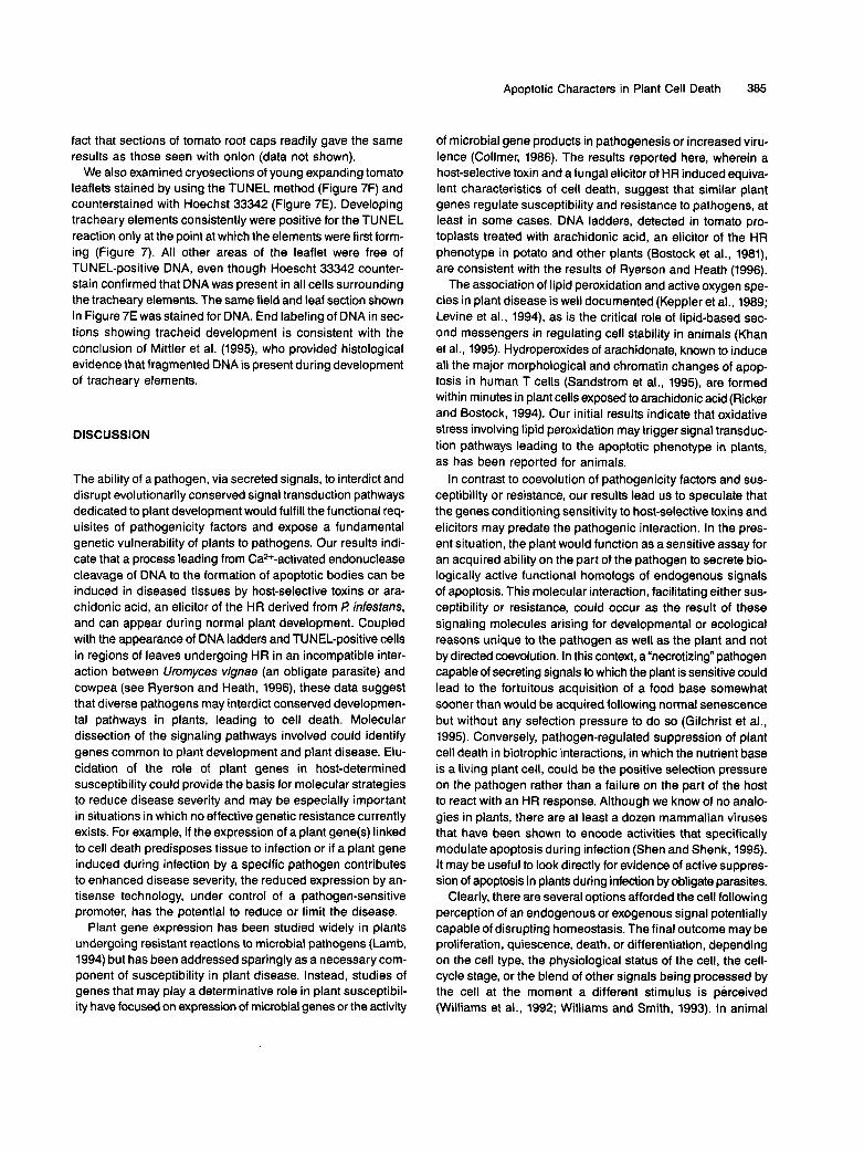
revealed nuclear DNA in cells throughout the root sections,from the apical meristem, where cells were undergoing divi-sion, to the outer surface, where cap cells were sloughing fromthe root (Figure 7A). The TUNEL method detected fragmentedDNA only in cells near the surface of the root cap (Figure 7B)in longitudinal sections, which was confirmed in cross-sections(data not shown). The area of TUNEL-positive cells was re-stricted to the outermost one to three cells of the cap, and noDNA fragmentation in the interior of the root, including cellsthat were undergoing active DNA replication during cell divi-sion, was detected. When viewed under higher magnification,the appearance of nuclear DNA (Figure 7C) changed from acompact circular form to elongated TUNEL-positive structuresthat eventually took on the form of numerous well-defined cir-cular bodies (Figure 7D). The formation of these bodiesappeared to be the final step reached before a cell was shedfrom the root cap (Figure 7D). The same view fields of the roottip that reacted with the TUNEL method (Figures 7B and 7D)were counterstained for DNA with Hoechst 33342 (Figures 7Aand 7C). The weaker DNA-staining signal in the outer layerof cap cells was due to the significant loss of DNA as well asthe fact that the fragmented DNA was no longer in a singlecompact nucleus in these cells in contrast to the strong TUNELlabeling in the same cells due to the high sensitivity of thisassay (see cautionary statement in Methods). The ease withwhich these findings can be observed was revealed by the
Figure 6. Temporal Progression of Nuclear Changes Associated with AAL Toxin-Induced Cell Death in asc/asc Tomato Protoplasts.
Tomato protoplasts were treated with 20 |iM TA for 24 hr. (A) to (E) show protoplasts labeled by using the TUNEL method. Fragmented DNAand apoptotic bodies were viewed at 510 to 560 nm as yellow to greenish fluorescence. The red fluorescence represents autofluorescence ofchlorophyll. (F) to (J) show the same view field as in the upper row, except protoplasts were stained with the DNA counterstain Hoechst 33342.(A) and (F) Normal, apparently healthy protoplast.(B) and (G) Early stages of apoptosis, with nuclear DNA fragmentation and nuclear budding shown as light green fluorescence.(C) and (H) Same stages as shown in (B) and (G).(D) and (I) Late stages of apoptosis with DNA fragments and distinct bodies shown as green fluorescence following the TUNEL assay.(E) and (J) Same stages as shown in (D) and (I).Bar in (F) = 10 urn.

384 The Plant Cell
Figure 7. Apoptosis during Plant Development.
(A) Longitudinal section of onion root tip stained for DNA with Hoechst 33342.(B) Same section as shown in (A) but labeled by using the TUNEL assay. Shown are root cap cells undergoing apoptosis.(C) Root cap cells stained for DNA with Hoechst 33342.(D) The same field of root cap cells as shown in (C) stained by using the TUNEL method. Shown is the formation of apoptotic bodies.(E) Cross-section of young expanding tomato leaflet stained for DNA with Hoechst 33342(F) The same field of leaf section as shown in (E) stained by using the TUNEL method. Shown are developing tracheids undergoing DNA fragmentation.Bar in (B) = 50 |jm tor (A), (B), (E), and (F); bar in (D) = 2 jam for (C) and (D).
Apoptotic Characters in Plant Cell Death 385
fact that sections of tomato root caps readily gave the same results as those seen with onion (data not shown).
We also examined cryosections of young expanding tomato leaflets stained by using the TUNEL method (Figure 7F) and counterstained with Hoechst 33342 (Figure 7E). Developing tracheary elements consistently were positive for the TUNEL reaction only at the point at which the elements were first form- ing (Figure 7). All other areas of the leaflet were free of TUNEL-positive DNA, even though Hoescht 33342 counter- stain confirmed that DNA was present in all cells surrounding the tracheary elements. The same field and leaf section shown in Figure 7E was stained for DNA. End labeling of DNA in sec- tions showing tracheid development is consistent with the conclusion of Mittler et al. (1995), who provided histological evidence that fragmented DNA is present during development of tracheary elements.
DlSCUSSlON
The ability of a pathogen, via secreted signals, to interdict and disrupt evolutionarily conserved signal transduction pathways dedicated to plant development would fulfill the functional req- uisites of pathogenicity factors and expose a fundamental genetic vulnerability of plants to pathogens. Our results indi- cate that a process leading from ca*+-activated endonuclease cleavage of DNA to the formation of apoptotic bodies can be induced in diseased tissues by host-selective toxins or ara- chidonic acid, an elicitor of the HR derived from P infestans, and can appear during normal plant development. Coupled with the appearance of DNA ladders and TUNEL-positive cells in regions of leaves undergoing HR in an incompatible inter- action between Uromyces vignae (an obligate parasite) and cowpea (see Ryerson and Heath, 1996), these data suggest that diverse pathogens may interdict conserved developmen- tal pathways in plants, leading to cell death. Molecular dissection of the signaling pathways involved could identify genes common to plant development and plant disease. Elu- cidation of the role of plant genes in host-determined susceptibility could provide the basis for molecular strategies to reduce disease severity and may be especially important in situations in which no effective genetic resistance currently exists. For example, if the expression of a plant gene(s) linked to cell death predisposes tissue to infection or if a plant gene induced during infection by a specific pathogen contributes to enhanced disease severity, the reduced expression by an- tisense technology, under control of a pathogen-sensitive promoter, has the potential to reduce or limit the disease.
Plant gene expression has been studied widely in plants undergoing resistant reactions to microbial pathogens (Lamb, 1994) but has been addressed sparingly as a necessary com- ponent of susceptibility in plant disease. Instead, studies of genes that may play a determinative role in plant susceptibil- ity have focused on expression of microbial genes or the activity
of microbial gene products in pathogenesis or increased viru- lence (Collmer, 1986). The results reported here, wherein a host-selective toxin and a funga1 elicitor of HR induced equiva- lent characteristics of cell death, suggest that similar plant genes regulate susceptibility and resistance to pathogens, at least in some cases. DNA ladders, detected in tomato pro- toplasts treated with arachidonic acid, an elicitor of the HR phenotype in potato and other plants (Bostock et al., 1981), are consistent with the results of Ryerson and Heath (1996).
The association of lipid peroxidation and active oxygen spe- cies in plant disease is well documented (Keppler et al., 1989; Levine et al., 1994), as is the critical role of lipid-based sec- ond messengers in regulating cell stability in animals (Khan et al., 1995). Hydroperoxides of arachidonate, known to induce all the major morphological and chromatin changes of apop- tosis in human T cells (Sandstrom et al., 1995), are formed within minutes in plant cells exposed to arachidonic acid (Ricker and Bostock, 1994). Our initial results indicate that oxidative stress involving lipid peroxidation may trigger signal transduc- tion pathways leading to the apoptotic phenotype in plants, as has been reported for animals.
In contrast to coevolution of pathogenicity factors and sus- ceptibility or resistance, our results lead us to speculate that the genes conditioning sensitivity to host-selective toxins and elicitors may predate the pathogenic interaction. In the pres- ent situation, the plant would function as a sensitive assay for an acquired ability on the part of the pathogen to secrete bio- logically active functional homologs of endogenous signals of apoptosis. This molecular interaction, facilitating either sus- ceptibility or resistance, could occur as the result of these signaling molecules arising for developmental or ecological reasons unique to the pathogen as well as the plant and not by directed coevolution. In this context, a "necrotizing" pathogen capable of secreting signals to which the plant is sensitive could lead to the fortuitous acquisition of a food base somewhat sooner than would be acquired following normal senescence but without any selection pressure to do so (Gilchrist et al., 1995). Conversely, pathogen-regulated suppression of plant cell death in biotrophic interactions, in which the nutrient base is a living plant cell, could be the positive selection pressure on the pathogen rather than a failure on the part of the host to react with an HR response. Although we know of no analo- gies in plants, there are at least a dozen mammalian viruses that have been shown to encode activities that specifically modulate apoptosis during infection (Shen and Shenk, 1995). It may be useful to look directly for evidence of active suppres- sion of apoptosis in plants during infection by obligate parasites.
Clearly, there are severa1 options afforded the cell following perception of an endogenous or exogenous signal potentially capable of disrupting homeostasis. The final outcome may be proliferation, quiescence, death, or differentiation, depending on the cell type, the physiological status of the cell, the cell- cycle stage, or the blend of other signals being processed by the cell at the moment a different stimulus is perceived (Williams et al., 1992; Williams and Smith, 1993). In animal

386 The Plant Cell
medicine, both the signals that regulate gene expression and the impact of altered gene expression on apoptosis (Wyllie, 1995) are the subject of intense interest in basic research in human diseases ranging from cancer (Anderson et al., 1994; Fisher, 1994) to autoimmune diseases (Critchfield and Lenardo, 1995; Thompson, 1995).
Interestingly, the term apoptosis was derived from the Greek by J.F.R. Kerr in 1972 to reflect the process by which leaves fall from trees to emphasize the natural process of a pro- grammed form of cell death he was studying in liver tissue (Kerr et al., 1972). Analogous to animals, the confirmation and manipulation of a gene expression-dependent apoptotic pro- cess in plants will require linkage to specific genes, sets of interacting genes, or gene products that respond to the sig- nals transduced by the range of stimuli that elicit this form of pcd. For now the principal criteria for apoptosis in plants are those used by animal biologists, namely, rapid changes in chro- matin structure, in situ labeling of fragmenting DNA, DNA ladders, and the formation of apoptotic bodies.
We are cognizant of the limitations of relying solely on a sin- gle criterion such as DNA fragmentation to ascribe cell death to an apoptotic process (Collins et al., 1992); hence, we have examined additional facets and metabolic connections between the events observed in plants and those reported in animals. Cloning of plant genes functionally homologous to those shown to affect apoptosis in animals will be necessary to resolve fur- ther the similarities and the differences in cross-kingdom conservation of this process (Takayama et al., 1994). Unfortu- nately, homology at the DNA leve1 is often <200/0 between different animal species, and dilution of expressed messages in populations of differentially responding plant cells could be another difficulty (Hengartner and Horvitz, 1994; Shen and Shenk, 1995). For induced genes, the number of cells in an organized tissue undergoing apoptosis may be small compared with the total cell mass, the process may be asynchronous, and the number of cells induced to die apoptotically compared with those that actually complete the stereotypical stages of the process before metabolic collapse may represent only a fraction of the total.
A corollary issue relates to the basis by which particular cell groups (spots) are targeted for death in leaf spot diseases, HR- induced spots, and lesion mimic spots. The fact that only cer- tain areas die, even in situations in which cells outside the responding area are exposed simultaneously to the stimulus (elicitor, toxin, infiltrated pathogen, stress, or inappropriate developmental signals), suggests that either temporal or per- manent diversity exists in elements controlling the response of individual cells to different stimuli in organized tissue networks.
The fate of the apoptotic-like bodies containing fragmented DNA is unknown. The role of apoptotic bodies in animal tis- sues is thought to facilitate phagocytosis of the debris from a dying cell as an efficient means to protect the surrounding tissues from potentially harmful cell contents. In the absence of phagocytic cells, the contents of a plant cell, packaged in
apoptotic bodies similar to the role of vacuoles but containing additional constituents, could represent a prophylactic func- tion during release of cellular contents from a dying cell into tissue. What is clear is that the mechanism for organizing this material into stable bodies, equivalent to apoptotic bodies in animal cells, is conserved in plants, as evidenced by the for- mation of such bodies during the final phases of sloughing of root cap cells during onion root growth.
Whether the full program for apoptosis or only phases of the program occur in plant cells that are programmed to die is not known. Also, the types and number of pathways that may converge to give the stereotypic hallmarks are unknown. If the situation in plants were like apoptosis in animals, the induction of pcd in one or a few cells in a given region or tis- sue may trigger the rapid death of surrounding cells; the latter cells may not show the apoptotic phenotype but die nonethe- less (Collins et al., 1992). The most salient conclusion we draw from these studies and those of Ryerson and Heath (1996) is that the functional properties of apoptosis occur in plants, and the role of pcd in plants appears to encompass that proposed in animals, both in health and sickness.
METHODS
Plant Materials
Tomato (Lycopersicon esculentum) plants used for leaflet assays were grown from seed of Alternaria stem canker (Asc) isolines (Clouse and Gilchrist, 1986). Plants were grown in soil under greenhouse condi- tions for 4 weeks, transferred to growth chambers (25OC/2loC daylnight, 16-hr photoperiod) at the five-leaf stage, and used in experiments at the seven-leaf stage. Plants were illuminated with a combination of cool-white fluorescent and incandescent lamps giving an intensity of 112 W m-2 irradiance at the leaf canopy. Excised tomato leaflets were put on a filter paper saturated with treatment solutions in a glass Petri dish and placed under continuous light for 36 to 48 hr at 25OC, as de- scribed previously (Moussatos et al., 1993b).
Fumonisin, AAL Toxin, and Other Chemical Treatments
Fumonisin 8, (FB,) was purchased from Sigma (>95O/o pure). AAL (Al- ternaria alternata f sp lycopefsici) toxin TA was produced and purified as described previously (Caldas et al., 1994). The preparation used was >98% pure based on analysis by HPLC and electrospray mas spectrometry. C-2 ceramide was obtained from Calbiochem (La Jolla, CA), and staurosporine and arachidonic acid, prepared as an emul- sion in water (Bostock et al., 1981), were from Sigma.
Protoplast Preparation
Tomato leaf protoplasts were isolated from Asc isoline plants grown under aseptic conditions in Murashige and Skoog medium (Moussatos et al., 1993a). After 3 to 4 weeks of growth, the plants were transferred to a dark chamber at 24OC for 2 days, followed by 6 to 8 hr of dark

Apoptotic Characters in Plant Cell Death 387
at 4OC. After the cold treatment, leaflets were hawested, and protoplasts were isolated as described earlier (Moussatos et al., 1993a). After iso- lation, protoplasts were freed from cellular debris by flotation on a 0.6 M sucrose solution, using low-speed centrifugation (1009 for 5 min). Viable protoplasts were collected at the interface and washed three times by centrifugation in 10% mannitol solution before they were resuspended in culture medium. The fractionated protoplasts were preincubated for 12 hr in TM-2 medium containing 3.3 mM Caz+ with no added hormones (Shahin, 1985) at a density of 2 x 10⁵ protoplasts per mL. After preincubation, protoplasts were centrifuged on a sucrose cushion as given above, and viable protoplasts were washed and resuspended in the same incubation medium before exposure to the treatment as indicated. After treatment, protoplasts were centrifuged at l O O g for 5 min. The pelleted protoplasts were placed immediately in liquid Nz and stored at -8OOC for DNA analysis. For in situ detec- tion of DNA fragmentation. protoplasts were incubated directly on Super Frost Plus microscope slides (Fisher Scientific) inside an Incu-Ring (Polyscientific, Inc., Warrington, PA) at 24OC under continuous light.
lsolation of Nuclear DNA for Fragmentation Analysis
Protoplast incubations were performed at 24OC in TM-2 medium in the presence of the respective toxins or other amendments at the con- centrations indicated. Protoplast DNAwas isolated first in 100 mM Tris, 50 mM EMA, 1% SDS, pH 7.5, as the extraction buffer, followed by extraction in phenol, chloroform, and a final ethanol precipitation. Af- ter RNase digestion at 100 pg1mL for 1 hr at V C , identical amounts of DNA were separated on a 15% agarose gel by electrophoresis. Stan- dard leaflet bioassays were performed on AsdAsc and asc/asc leaflets, as described earlier (Gilchrist and Grogan, 1976), at 20 pM AAL toxin to induce .V50% cell death on a leaf area basis. Toxin-treated tomato leaf DNA was isolated using the nonphenol method described by Tai and Tanksley (1991), followed by 100 pg/mL Rnase A digestion for 1 hr at 37%. Agarose gel electrophoresis was performed immediately af- ter DNA isolation. Finally, DNA was transferred onto a Nytran Plus membrane (Scheicher & Schuell) and hybridized with a 3zP-labeled tomato genomic DNA probe.
Histochemical Detection ot Nuclear DNA Fragmentation and Apoptotic Bodies
For in situ detection of DNA fragmentation, equal volumes of 10% buffered formalin (Fisher Scientific) were added directly to the pro- toplasts on slides. After drying at m m temperature, the fixed protoplasts were washed with TBS buffer (150 mM NaCI, 100 mM Tris-HCI, pH 7.4), digested with proteinase K (10 pglmL) for 10 min at room temper- ature, washed with distilled water, briefly rinsed with terminal deoxynucleotidyl transferase (TdT) buffer (25 mM Tris-HCI, 200 mM sodium cacodylate, 5 mM cobalt chloride), and then incubated with the labeling mix (20 units of TdT; Boehringer Mannheim) and 1 nM digoxigenin-11-dUTP (Boehringer Mannheim) in 100 pL of TdT buffer for 1 hr at 37%. The reaction was terminated by rinsing the section in 300 mM NaCI, 30 mM sodium citrate for 30 min at room tempera- ture and then blocked with TBS buffer containing 2% BSA, 0.3% Triton X-100 for 10 min. The slides were washed three times in TBS buffer and then incubated for 1 hr at room temperature with anti-digoxigenin antibody (Boehringer Mannheim) at a dilution of 1:25 in TBS contain- ing 1% BSA and 0.05% Triton X-100.
After BSA blocking and washing as given above, thefixed protoplasts were incubated with second antibody (anti-goat conjugated with fluo- rescein isothiocyanate [FITC]; Pierce, Rockford, IL) and diluted 1:25 in TBS with BSA for 2 hr before counterstaining the DNA with Hoechst 33342 (Sigma) at 5 pg/mL for 10 min at room temperature. The slides were viewed with a Nikon SA fluorescence microscope (Nikon, Foster City, CA).
For quantitation of toxin-induced programmed cell death (pcd) in tomato protoplasts, the TdT end-labeling (TUNEL) assay was performed. The anti-digoxigenin antibody was conjugated with alkaline phospha- tase (1:1000), and lmmuno Pure Fast Red was used as the substrate (Pierce) to label the cells with fragmented DNA. The viability of the protoplasts was determined using fluorescein diacetate stain (Sigma) at 0.01 mg/mL on a Nikon Diaphot TMD inverted microscope. The num- ber of protoplasts that reacted with the respective stain treatments was determined by counting cells within individual fields before and after the counterstain treatment. Each determination was done with at least 3000 protoplasts in each of three independent experiments for each treatment.
Preparation of Whole Tissue and Root Tip Sections
For in situ detection of TUNEL-positive cell death in plant tissues, tomato leaflets, tomato root tips, and onion root tips were fixed in 10% buffered formalin for 20 min under vacuum and then transferred to 0.6 M su- crose and exposed to a vacuum for an additional20 min. The tissues were then cut into small pieces and embedded in Histo Prep (Fisher Scientific), frozen in liquid NP, and stored at -8OOC before section- ing on a Cyrocut 1800 microtome (Reichet-Jung, Leica, Foster City, CA) at athickness of 10 pm. After a 5-min postfixation in buffered for- malin, the sections were digested with proteinase K (10 pg/mL) for 10 min at room temperature and then subjected to the TUNEL method as described above, except that antibody conjugated with alkaline phos- phatase was used rather than FITC because of strong autofluorescence in the tissue sections.
Cautionary Notes When Applying the TUNEL Procedure to Plant Tissues
We found that the TUNEL assay possessed high labeling capacity (Gorczyca et al., 1993) that could easily generate end-labeling artifacts dueto inappropriate reagent concentrations or tissue handling. Heavy background and nonspecific staining were seen frequently when high TdT and nucleotide concentrations were used. Substantial reduction of nucleotide and TdT enzyme concentration below that prescribed for staining animal cells enabled strong staining of positive controls in the absence of background interference. As an example of the sen- sitivity of the assay, it is possible to end-label uncut 1 DNA if the reaction is run with excess reagents. We routinely set reaction conditions with uncut 1 DNA as a negative control. Excess proteinase K treatment also was found to cause nonspecific staining in tissue sections.
We found that certain approaches to preparation, for sectioning, of plant as well as animal tissue (Grasl-Kraupp e1 al., 1995) can lead to generation of artifacts and the inability to produce positive control treat- ments. For example, embedding in either plastic or paraffin, followed by standard fixation, did, in our hands, cause sufficient nicking of nu- clear DNA to give a positive TUNEL reaction in the fixed sections. Early in our studies, we noted that approaches similar to those used by Mittler

388 The Plant Cell
et al. (1995) caused both treated and control sections to be heavily labeled. Hence, unless DNA counterstaining is used to ensure that healthy DNA is not nicked during processing of the sections, a TUNEL- positive artifact can occur. Clearly, this technique, although useful and reliable, must be conducted under controlled conditions, and appro- priate control treatments must be conducted at all times. All experiments reported herein were repeated at least five times, with all results in agreement across the experiments.
ACKNOWLEDGMENTS
We thank Jeffery Dangl, Martin Dickman, James Lincoln, Thomas Moore, and Bert Overduin for helpful discussions during the course of this work. Research was supported in part by National Science Foun- dation (NSF) Cooperative Agreement No. BIR-8920216 to CEPRAP, an NSF Science and Technology Center, and by CEPRAP corporate associates: Calgene, Davis, CA; Ciba Biotechnology Corporation, Re- search Triangle Park, NC; Sandoz Seeds, Palo Alto, CA; and Zeneca Seeds, Bracknell, Berkshire, UK.
Received September 19, 1995; accepted January 22, 1996.
REFERENCES
Abbas, H.K., Tanaka, T., Duke, S.O., Porter, J.K., Wray, E.M., Hodges, L., Sessions, A.E., Wang, E., Merrill, A.H., and Riley, R.T. (1994). Fumonisin and AAL-toxin induced disruption of sphin- golipid metabolism with accumulation of free sphingoid bases. Plant Physiol. 106, 1085-1093.
Alison, M.R., and Sarraf, C.E. (1992). Apoptosis: A gene-directed programme of cell death. J. R. Coll. Physicians Lond. 26, 25-35.
Anderson, K.M., Harris, J.E., and Bonomi, K.P. (1994). Potential a p plications of apoptosis in modifying the biological behavior of therapeutically refractory tumors. Med. Hypotheses 43, 207-213.
Bachmair, A., Becker, F., Masterson, R.V., and Schell, J. (1990). Per- turbation of the ubiquitin system causes leaf curling, vascular tissue alternations and necrotic lesions in a higher plant. EMBO J. 9, 4543-4549.
Barr, P.J., and Tomei, L.D. (1994). Apoptosis and its role in human disease. BioTechnology 12, 487-493.
Barry, M.A., and Eastman, A. (1992). Endonuclease activation dur- ing apoptosis: The role of cytosolic Ca2+ and pH. Biochem. Biophys. Res. Commun. 186, 782-789.
Bose, R., Verheij, M., HaimovittFriedman, A., Scotto, K., Fuks, Z., and Kolesnick, R. (1995). Ceramide synthase mediates dau- norubicininduced apoptosis: An alternative mechanism for generating death signals. Cell 82, 405-414.
Bostock, R.M., Kuc, J.A., and Laine, R.A. (1981). Eicosapentaenoic and arachidonic acids from Phytophthora infestans elicit fungitoxic sesquiterpenes in the potato. Science 212, 67-69.
Bottini, A.T., Bowen, J.R., and Gilchrist, D.G. (1981). Phytotoxins. 11. Characterization of a phytotoxic fraction from Alternaria alternata f. sp. lycopersici. Tetrahedron Lett. 22, 2723-2726.
Bursch, W., Paffe, S., Putz, B., Barthel, O., and Schulte-Hermann, R. (1990). Determination of the length of the histological stages of apoptosis in normal liver and in altered hepatic foci of rat. Carcino- genesis 11, 847-853.
Caldas, E.D., Jones, A.D., Ward, B., Winter, C.K., and Gilchrist, D.G. (1994). Structural characterization of three new AAL toxins pro- duced by Alternaria alternata f. sp. lycopersici. J. Agric. Food Chem.
Caldas, E.D., Jones, A.D., Winter, C.K., Ward, B., and Gilchrist, D.G. (1995). Electrospray ionization mass spectrometry of sphinga- nine analog mycotoxins. Anal. Chem. 67, 196-207.
Carson, D.A., and Ribeiro, J.M. (1993). Apoptosis and disease. Lan- cei 341, 1251-1254.
Cartier, J.L., Hershberger, P.A., and Friesen, P.D. (1994). Suppres- sion of apoptosis in insect cells stably transfected with baculovirus p35: Dominant interference by N-terminal sequences ~35’-’~. J. Virol. 68, 7728-7737.
Chao, M.V. (1995). Ceramide: A potential second messenger in the nervous system. MOI. Cell Sci. 6, 91-96.
Chasan, R. (1994). Tracing tracheary elemeni development. Plant Cell
Clouse, S.D., and Gilchrist, D.G. (1986). lnteraction of the asc locus in F8 paired lines of tomato with Alternaria alternata f. sp. lycoper- sici and AAL toxin. Phytopathology 77, 80-82.
Cohen, G.M., Sun, X., Fearnhead, H., MacFarlane, M., Bmwn, D.G., Snowden, R.T., and Dinsdale, D. (1994). .Formation of large mo- lecular weight fragments of DNA is a key committed step of apoptosis in thymocytes. J. Immunol. 153, 507-516.
Collins, R.J., Harmon, B.V., Gobe, G.C., and Kerr, J.F.R. (1992). In- ternucleosomal DNA cleavage should not be the sole criterion for identifying apoptosis. Int. J. Radiat. Biol. 61, 451-453.
Colher, A. (1986). The molecular biology of pectic enzyme produc- tion and bacterial soft rot pathogenesis. In Biology and Molecular Biology of Plant-Microbe Interactions, J. Bailey, ed (Berlin: Springer- Verlag), pp. 277-289.
Corcoran, G.B., Fix, L., Jones, D.P., Trienen Moslen, M., Nicortera, P., Oberhammer, F.A., and Buttyan, R. (1994). Contemporary is- sues in toxicology. Apoptosis: Molecular control point in toxicity. Toxicol. Appl. Pharmacol. 128, 169-181.
Critchfield, J.M., and Lenardo, M.J. (1995). Antigen-induced pro- grammed cell death as a new approach to immune therapy. Clin. Immunol. Immunopathol. 75, 13-19.
Dietrich, R.A., Delaney, T.P., Uknes, S.J., Ward, E.R., Ryals, J.A., and Dangl, J.L. (1994). Arabidopsis mutants simulating disease re- sistance response. Cell 77, 565-577.
Esau, K. (1977). Seed Plant Anatomy. (New York: Academic Press). Fisher, D.E. (1994). Apoptosis in cancer therapy: Crossing the thresh-
old. Cell 78, 539-542. Gavrieli, Y., Sherman, Y., and Ben-Sasson, S.A. (1992). Identifica-
tion of programmed cell death in situ viaspecific labeling of nuclear DNA fragmentation. J. Cell Biol. 119, 493-501.
Gelderblom, W.C.A., Semple, E., Marasas, W.F.O., and Farber, E. (1992). The cancer-initiating potential of fumonisin B mycotoxins. Carcinogenesis 13, 433-437.
Gerschenson, L.E., and Totello, R.J. (1992). Apoptosis: A different type of cell death. FASEB J. 6, 2450-2455.
42, 327-333.
6, 917-919.

Apoptotic Characters in Plant Cell Death 389
Ghosh, T.K., Bian, J., and Gill, D.L. (1994). Sphingosine 1-phosphate generated in the endoplasmic reticulum membrane activates release of stored calcium. J. Biol. Chem. 36, 22628-22635.
Gllchrist, D.G. (1983). Molecular modes of action. In Toxins and Plant Pathogenesis, J.M. Daly and E. Deverall, eds (Sydney, Australia: Academic Press), pp. 81-136.
Gilchrist, D.G., and Grogan, R.G. (1976). Production and nature of a host-specific toxin from Alternaria alternata f. sp. lycopersici. Phytopathology 66, 165-171.
Gilchrist, D.G., Ward, B., Moussatos, V., and Mirocha, C.J. (1992). Genetic and physiological response to fumonisins and AAL-toxin by intact tissue of a higher plant. Mycopathologia 117, 57-64.
Gilchrist, D.G., Wang, H., and Bostock, R.M. (1995). Sphingosine related-mycotoxins in plant and animal diseases. Can. J. Bot. 73
Gold, R., Schmied, M., Giegerich, G., Breitschopf, H., Hartung, P., Toyka, V., and Lassmann, H. (1994). Differentiation between cellular apoptosis and necrosis by the combined use of in situ tail- ing and nick translation techniques. Lab. Invest. 7, 219-225.
Gorczyca, W., Gong, J., and Darzynkiewicz, i!. (1993). Detection of DNA strand breaks in individual apoptotic cells by the in situ termi- nal deoxynucleotidyl transferase and nick translation assays. Cancer Res. 53, 1945-1951.
Grasl-Kraupp, B., Rullkay-Nedecky, B., Koudelka, H., Bukowska, K., Bursch, W., and Schulte-Hermann, R. (1995). lnsitudetection of fragmented DNA (TUNEL assay) fails to discriminate among apop- tosis, necrosis, and autolytic cell death: A cautionary note. Hepatology
Greenberg, J.T., and Ausubel, F.M. (1993). Arabidopsis mutants com- promised for the control of cellular damage during pathogenesis and aging. Plant J. 4, 327-341.
Greenberg, J.T., Guo, A., Klessig, D.F., and Ausubel, F.M. (1994). Programmed cell death in plants: A pathogen-triggered response activated coordinately with multiple defense functions. Cell 77,
Grogan, R.G., Kimble, K.A., and Misaghi, 1. (1975). A stem canker disease of tomato caused by Alternaria alternata f. sp. lycopersici. Phytopathology 65, 880-886.
Harkes, P.A.A. (1973). Structure and dynamics of the root cap of Avena sativa L. Acta Bot. Neerl. Scand. 22, 321-328.
Hengartner, M.O., and Horvitz, H.R. (1994). The ins and outs of pro- grammed cell death during C. elegans development. Philos. Trans. R. Soe. Lond. E Eiol. Sci. 345, 243-246.
Hopmans, E.C., and Murphy, P.A. (1993). Detection of fumonisins 61, 82, and 83 and hydrolyzed fumonisin B1 in corn-containing foods. J. Agric. Food Chem. 41, 1655-1658.
Jarvis, W.D., Fornari, F.A., Bmwning, J.L., Gewirtr, D.A., Kolesnick, R.N., and Grant, S. (1994a). Attenuation of ceramide-induced apop- tosis by diglyceride in human myeloid leukemia cells. J. Eiol. Chem.
Jarvis, W.D., Kolesnick, R.N., Fornari, F.A., Traylor, R.S., Gwirtz, D.A., and Grant, S. (1994b). lnduction of apoptotic DNA damage and cell death by activation of the sphingomyelin pathway. Proc. Natl. Acad. Sci. USA 91, 73-77.
Jones, M.J., and Murray, A.W. (1995). Evidence that ceramide selectively inhibits protein kinase C-a translocation and modulates
(suppl. I), 459-467.
21, 1465-1468.
551-563.
269, 31685-31692.
bradykinin activation of phospholipase D. J. Biol. Chem. 270,5007- 5013.
Keen, N.T. (1990). Gene for gene complementarity in plant-pathogen interactions. Annu. Rev. Genet. 24, 447-463.
Keppler, L.D., Baker, C.J., and Atkinson, M.M. (1989). Active oxy- gen production during a bacteria-induced hypersensitive reaction in tobacco suspension cells. Phytopathology 79, 974-978.
Kerr, J.F.R., and Harmon, RV. (1991). Definition and incidence of apop- tosis. In Apoptosis: The Molecular Basis of Cell Death, Current Communications in Gell and Molecular Biology, L.D. Tomei and F.O. Cope, eds (Cold Spring Harbor, NY Cold Spring Harbor Labora- tory Press), pp. 5-29.
Kerr, J.F.R., Wyllie, A.H., and Currie, A.R. (1972). Apoptosis: A ba- sic biological phenomenon with wide-ranging implications in tissue kinetics. 61. J. Cancer 26, 239-257.
Kerr, J.F.R., Gobe, G.C., Winterford, C.M., and Harmon, B.V. (1995). Anatomical methods in cell death. In Cell Death: Methods in Cell Biology, Vol. 46, L.M. Schwartz and B.A. Osborne, eds (New York: Academic Press), pp. 1-27.
Khan, W.A., Blobe, G.C., and Hannun, Y.A. (1995). Arachidonic acid and free fatty acids as second messengers and the role of protein kinase C. Cell. Signalling 7, 171-184.
Lamb, C.J. (1994). Plant disease resistance genes in signal percep- tion and transduction. Cell 76, 419-422.
Levine, A., Tenhaken, R., Dlxon, R., and Lamb, C. (1994). HZ02 from the oxidative burst orchestrates the plant hypersensitive disease re- sistance response. Cell 79, 583-593.
Li, X., Traganos, F., Melamed, M.R., and Darzynklewlcz, 2. (1995). Single-step procedure for labeling DNA strand breaks with fluorescein- or BODIPY-conjugated deoxynucleotides: Detection of apoptosis and bromodeoxyuridine incorporation. Cytometry 20, 172-180.
Lohmann, R.D., and Beyersmann, D. (1993). Cadmium and zinc medi- ated changes of the Ca2+-'dependent endonuclease in apoptosis. Biochem. Biophys. Res. Commun. 190, 1097-1103.
Martin, S.J. (1993). Apoptosis: Suicide, execution or murder? Trends Cell Eiol. 3, 141-144.
Matter, A. (1979). Microcinematographic and electron microscopic analysis of target cell lysis induced by cytotoxic T lymphocytes. Im- munology 36, 179-184.
Merrill, A.H., Wang, E., Gilchrist, D.G., and Riley, R.T. (1993). Fumoni- sins and other inhibitors of de novo sphingolipid biosynthesis. Adv. Lipid Res. 26, 215-234.
Merrill, A.H., Jr., Schmelz, EM., Wang, E., Schder , J. J., Dlllehay, D.L., and Rlley, R,T. (1995). Role of dietary sphingolipids and in- hibitors of sphingolipid metabolism in cancer and other diseases. J. Nutr. 125, 51677-Sl682.
Mittler, R., Shulaev, V., and Lam, E. (1995). Coordinated activation of programmed cell death and defense mechanisms in transgenic tobacco plants expressing a bacterial proton pump. Plant Cell 7,
Moussatos, V.V., Witsenboer, H., Hllle, H.J., and Gilchrist, D.G. (1993a). Behavior of the disease resistance gene Asc in protoplasts of Lycopersicon esculentum Mill. Physiol. MOI. Plant Pathol. 43,
Moussatos, V.V., Lucas, W.J., and Gilchrlst, D.G. (1993b). AAL- toxin-induced physiological changes in Lycopersicon esculentum
29-42.
255-263.

390 The Plant Cell
Mill: Differential sucrose transport in tamato lines isogenic for the Asc locus. Physiol. Mel. Plant Pathol. 42, 369-371.
Moussatos, V.V., Yang, S.F., Ward, B.W., and Gllchrlst, R.G. (1994). AAL-toxin induced physiological changes in Lycoprsicum esculen- rum Mill: Roles of ethylene and pyrimidine biasynthesis intermediates in lhe necrosis response. Physial. MOI. Plant Pathol. 44, 455-468.
Natarajan, V., Jayaram, H.N., Scrlbner, W.M., and Gemia, J.G.N. (1994). Activation of endothelial cell phosghalipase D by sphingo- sine and sphingosinsl-phosphate. Am. J. Respir. Cell MOI. Biol.
Nelson, P.E., Daajardlns, A.E., and Plattner, R.D. (1993). Fumoni- sins, mycotoxins pmduced by Fusarium species: Biolagy, chemistry, and significance. Annu. Rev. Phytopathol. 31, 233-252.
Norred, W.P. (1993). Fumonisins: Mycotoxins praduced by Fuserium monlllforme. J. Toxicol. Environ. Health 38, 309-328.
Norred. W.P., Wsng, E., Yoo, H., Rlley, R.T., and Merrll, A.H., Jr. (1992). In vltm toxicology of fumonisins and lhe mechanistic impli- cations. Mycopathologia 117, 73-78.
Obeld, L.M., and Hannun, Y.A. (1995). Ceramide: Astress signal and mediator of growth suppression and apoptesis. J. Cell. Biochem. 58, 191-198.
Oheta, H.O., Sweeney, E.A., Masemmune, A,, Yatoml, Y., Hskomori, S., and Igarashl, Y. (1995). lnduction of apoptosis by sphingosine in human leukemia HL-60 cells: A possible endogenoiis modulator of apoptotic DNA fragmentatian occurring during phorbol ester- induced differentiation. Cancer Res. 55, 691-697.
QNelll, S.D., Nadeau, J. A,, Zhang, X.S., Bul, A.Q., and Halevy, A.H. (1993). lnterorgan regulation of ethylene biosynthetic genes by pol- lination. Plant Cell 5, 419-432.
Pehtsch, M.C., Poluir, R., Stephan, H., Crompton, T., MacDonaid, H.R., Mannherz, H.G., and lbchapp, J. (1993). Characterization of endogenous deoxyribonuclease involved in nuclear DNA degra- dation during apoptosis (programmed cell death). EMBO J. 12, 371-377.
Raff, M.C. (1992). Social contrels en cell survival and cell death. Na- ture 358, 397-400.
Rlcker, K.E., and Bastock, R.M. (1894). Eicasanoids in lhe Phytoph- thore infestans- Potato interaction: Lipoxygenase metabelism of arachidonic acid and biological activities of selssted lipoxygenase products. Physiol. MOI. Plant Pathol. 44, 65-80.
Rosa, P.F., Neleon, P.E., Rlchard, J.L., Osweller, G.D., Rlce, L.G., Plattner, R.D., snd Wllron, T.M. (1990). Production of fumonisins by Fuserlum monlllforme and Fuserlum praliteretum isolate8 as- sociated with equine IeukoencephalomAlacia and a pulmonary edema syndmme in swlne. Appl. Enviran. Micmbiol. 58,3225-3228.
Ryemon, D.E., and Heath, M.C. (1996). Cleavage of nuclear DNA lnto oligonucleosomal fragments during cell death induced by funga1 infection or by ablotlc treatments. Plant Cell I), 393-402.
Sandamm, P.A., Wrdl, D., Tebbey, P.W., Dudek, R.W., Terrlan, DM., Folka, T.M., and Buttke, T.M. (1995). Lipid hydroperexide-induced apoptesis: Lack of inhibition by Bcl-2 overexpression. FEBS Lett.
Sevlll, J. (1994). Apaptosis and disease. Eur. J. Clin. Invest. 24,7l5-723. Schroeder, J.J., Crane, H.M., Xla, J.. Llotta, B.C., and Merrlll, A.H.
(1994). Disruptian of sphingolipid metabolism and stimulation of DNA synthesis by fumonlsin 61: A molecular mechanism for carcinogen- esls associated with Fusarlum monlliforme. J. Biol. Chem. 280,
11, 221-229.
365, 68-70.
3475-3481.
Schwartxman, R.A., and Cidlowskl, J.A. (1993). Apoptosis: The bio- chemistry and molecular biology af programmed cell death. Endocr. Rev. 14, 133-151.
Shahin, E.A. (1985). Totipotency of tomato protoplasts. Theor. Appl. Genet. 69, 235-240.
Shen, Y., and Shenk, T.E. (1995). Viruses and apaptosis. Curr. Biol.
Sller, D.J., and Gllchrist, B.G. (1983). Properties of host-specific toxins produced by Alternaria elfernata f. sp. lycopersici in culture and in tomato plants. Physiol. Plant Pathol. 23, 265-274.
Smart, C.A. (1994). Gene expression during leaf senescence. New
Smith. C.A., Grlmes, E.A., McCarthy, N.J., and Wllllams, G.T. (1993). Multiple gene regulation of apoptosis: Significance in immunology and oncalogy. In Apoptosis 11: The Malecular Basis af Apoptosis in Disease, L.D. Tomei and F.0. Cope, eds (Cold Spring Harbor, NY: Cold Spring Harbor Laboratory Press), pp. 43-87.
Steller, H. (1995). Mechanisms and genes of cellular suicide. Science
Sydenham, E.W., Thlel, P.G., Marasas, W.F.O., Shephad, G.S., van Schalkwyk, D.J., and Koch, K.R. (1990). Natural Occurrence of some fusarium mycotoxins in corn from low and high esophageal cancer prevalence areas of lhe Transkei southern Africa. J. Agric. Food Chem. 38, 1900-1903.
Sydenham, E.W., Shephard, G.S., Thlel, P.G., Marasas, W.F.O., and Stockenstrom, S. (1991). Fumanisin contamination of commercial corri-based human foodstuffs. J. Agric. Food Chem. 39,2014-2018.
Tal, T,, end Tankaley, S. (1991). A rapid and inexpensive method for isolation of total DNA from dehydrated plant tissue. Plant MOI. Biol. Rep. 8, 297-303.
Tekayama, S., Cazals-Hatem, D.L., Kltada, S., Tenak, S., Mlyeshlta, T., Hovey, L.R., Huen, D., Rlcklnson, A,, Veerapandalan, P., Krajewskl, S., Lalto, K., and Reed, J.C. (1994). Evolutionary con- servation of function among mammalian, avian, and vira1 hamologs of lhe Bcl-2 oncopratein. DNA Cell Biol. 13, 679-692.
Thlel, P.G.. Marasas, W.F.Q., Sydenham, E.W., Shepard, G.S., and Gelderblom, W.C.A. (1992). The implications of naturally occurring levels of fumonisins in corn for human and animal health. Myco- pathologia 117, 3-9.
Thompson, C.B. (1995). Apoptosis in the pathogenesis and treatment of disease. Science 267, 1456-1462.
Tomei, L.D., and Cope, F.O., eds (1991). Apoptosis: The Molecular Basis of Cell Death, Current Communications in Cell and Molecu- lar Biology. (Cold Spring Harbor, NY: Cold Spring Harbor Laboratory Press).
Tomel, L.D., and Cope, F.O., eds (1994). Apoptosis 11: The Molecular Basis of Apoptosis in Disease, Current Communications in Cell and Molecular Biology. (Cold Spring Harbor, NY Cold Spring Harbor Laboratory Press).
Vaux, D.L., Haecker, ti,, and Strasser, A. (1994). An evolutionary perspective on apoptosis. Cell 76, 777-779.
Wang, E.? Norred, W.P., Bacon, C.W., Rllry, R.T., and Merrlll, A.H. (1990), lnhlbitlon of sphingolipid biasynthesis by fumonislns. J. Biol. Chem. 266, 14488-14490.
Wang, E., Ron8, P.E., Wllnon, T.M., Rlley, R.T., and Merrlll, A.H. (1992). Alteration of serum sphlngolipids upon exposure of ponies to feed containing fumonisins, mycotoxins produced by Fuser/um monlllforme J. Nutr. 122, 1708-1718.
5, 105-111.
Phytol. 126, 419-448.
267, 1445-1499.

Wang, H., Jones, C., Ciacci-Zanella, J., Holt, T., Gilchrist, D.G., and Dlckman, M.B. (1996). Fumonisins and AAL toxins: Sphinga- nine analog mycotoxins induce apoptosis in monkey kidney cells. Proc. Natl. Acad. Sci. USA, in press.
Whyte, M., and Evan, G. (1995). The last cut is the deepest. Nature
Williams, G.T., and Smith, C.A. (1993). Molecular regulation of apop- tosis: Genetic controls on cell death. Cell 74, 777-779.
Williams, G.T., Smith, C.A., McCarthy, J.J., and Grimes, E.A. (1992). Apoptosis: Final control point in cell biology. Trends Cell Biol. 2,
376, 17-10.
263-267.
Apoptotic Characters in Plant Cell Death 391
Wlnter, C., Gilchrist, D., Dickman, M., and Jones, C. (1996). Chem- istry and biological activity of AAL-toxins. In Fumonisins in Food, L. Jackson and L. Bullerman, eds (New York: Plenum Press), pp.
Woodson, W.R., Park, K.Y., Dmry, A., Larsen, P.B., and Wang, H. (1992). Expression of ethylene biosynthetic pathway transcripts in senescing carnation flowers. Plant Physiol. 99, 526-532.
Wyllie, A.H. (1995). The genetic regulation of apoptosis. Curr. Opin. Genet. Dev. 5, 97-104.
Zhang, X.S., and ONeill, S.D. (1993). Ovary and gametophyte de- velopment are coordinately regulated by auxin and ethylene following pollination. Plant Cell 5, 403-418.
307-31 6.