“Regressive” patho-mechanics of Distal Radius Frac- tures...
4
MEDICINE PAPERS, 2016, 2 (2): 25–28 “Regressive” patho-mechanics of Distal Radius Frac- tures, and savage by reconstruction of coxa manus GAETANO MAURIZIO GRIPPI Hand Surgery in UOA of Orthopaedic, S. Lazzaro Hospital, Alba (Cuneo) - ASL CN2 of Piemont, Italy, e-mail: [email protected], www.chirurgia-mano.org SUMMARY Distal Radial Fractures are common and have “regressive” patho-mechanics as origin. For their care, particularly useful is the Coxa Manus Recon- struction (CMR) that consists in a volar radius-lunate- (hemi-scaphoid) arthrodesis with scaphoid distal resection. In the period 2002-2015 we have been successful in about 90% of cases. 25 KEY WORDS Distal radial fractures; coxa manus reconstruction; paleo- carpus; neo-carpus. Received 21.03.2016; accepted 02.05.2016; printed 30.05.2016 I NTRODUCTION Frequent outcome of Distal Radius Fractures (DRF) is radio-carpal stiffness, with spontaneous reset of carpal kinetics so the residual movement is transferred in the mediocarpal joint, on the capitate’s head (Fig. 1). In this case, joint movement is pre- valent on capitate’s head with relative immobility of the lunate. This opportunity is an interesting adaptation to trauma, produced by evolution during phylogenesis of Primate’s Anterior Autopod towards the human carpus Infact, according to Biarticular Concentric Carpal Mechanism (BCCM) (LINSCHEID ET AL., 1972; ALLIEU ET AL., 1982; GRIPPI, 1997; GRIPPI & POMPILIO, 2002), the carpus is assimilated to a bi-articular hip pros- thesis that, in the small prosthetic head - reproduced from Capitate - has its center of rotation (CR) (Fig. 2). Using this similitude, at the center of the carpus is identified the “ball and socket” joint of Coxa Manus (CM), the "true" primitive carpal joint, where takes place the s.c. “dart-throwing motion”. Disconnession of Coxa Manus causes, or rather “IS” the Carpal Instability. Certified by the patognomonic sign of the static or dinamic dislocation of capitate’s head with rotation of the carpal center (Fig. 3). In the evolution and brachiation of Primates, the human bi-articular carpal joint comes from the Rep- tiles uni-carpal joint, with an onto-phylogenetic devel- opment for which the radio-carpal appears after the mid-carpal joint (GRIPPI, 2008). So that, in wrist is possible to distinguish two parts: a distal, ancient: the Paleo-Carpus, represented by couple capitate- hamate, that in the Coxa Manus has retained the priv- ilege of mechanic carpal center; the other proximal, recent: the Neo-Carpus, represented by the proximal carpal row that contains, stabilizes and protects the Coxa Manus (Fig. 4) (GRIPPI, 2015). In generic radio-carpal injury of s.c. Adaptive Carpus (AC) there is a spontaneus decay of bi- articular towards uni-articular function, basically centered on Coxa Manus and its “dart-throwing mo- tion” (GRIPPI & CUGOLA, 2011). This patho-mechanics (resurrecting the ancestral Paleo-Carpus leadership) is a potential stereotype in any anatomical alteration (congenital or acquired) of Neo-Carpus: then, emer- ging in the outcomes of distal radius fractures, in Madelung, in Kienböck, in SNAC-SLAC-SCAC wrist, etc. In the same way - to recover problematic radio- carpal injures - a valid surgical option is to concen- trate the whole movement of carpus on capitate’s head. That is, on Paleo-Carpus. This concept is the s.c. "Grail of Wrist Surgery" the application of which has led to the Coxa Manus Surgery (GRIPPI, 2015).
Transcript of “Regressive” patho-mechanics of Distal Radius Frac- tures...

MEDICINE PAPERS, 2016, 2 (2): 25–28
“Regressive” patho-mechanics of Distal Radius Frac-tures, and savage by reconstruction of coxa manus
GAETANO MAURIZIO GRIPPI
Hand Surgery in UOA of Orthopaedic, S. Lazzaro Hospital,Alba (Cuneo) - ASL CN2 of Piemont, Italy, e-mail:[email protected], www.chirurgia-mano.org
SUMMARY
Distal Radial Fractures are common and have“regressive” patho-mechanics as origin. For theircare, particularly useful is the Coxa Manus Recon-struction (CMR) that consists in a volar radius-lunate-(hemi-scaphoid) arthrodesis with scaphoid distalresection. In the period 2002-2015 we have beensuccessful in about 90% of cases.
25
KEY WORDS
Distal radial fractures; coxa manus reconstruction; paleo-carpus; neo-carpus.
Received 21.03.2016; accepted 02.05.2016; printed 30.05.2016
INTRODUCTION
Frequent outcome of Distal Radius Fractures(DRF) is radio-carpal stiffness, with spontaneousreset of carpal kinetics so the residual movement istransferred in the mediocarpal joint, on the capitate’shead (Fig. 1). In this case, joint movement is pre-valent on capitate’s head with relative immobility ofthe lunate.
This opportunity is an interesting adaptation totrauma, produced by evolution during phylogenesisof Primate’s Anterior Autopod towards the humancarpus
Infact, according to Biarticular Concentric CarpalMechanism (BCCM) (LINSCHEID ET AL., 1972; ALLIEUET AL., 1982; GRIPPI, 1997; GRIPPI & POMPILIO, 2002),the carpus is assimilated to a bi-articular hip pros-thesis that, in the small prosthetic head - reproducedfrom Capitate - has its center of rotation (CR) (Fig.2). Using this similitude, at the center of the carpusis identified the “ball and socket” joint of Coxa Manus(CM), the "true" primitive carpal joint, where takesplace the s.c. “dart-throwing motion”.
Disconnession of Coxa Manus causes, or rather “IS”the Carpal Instability. Certified by the patognomonicsign of the static or dinamic dislocation of capitate’shead with rotation of the carpal center (Fig. 3).
In the evolution and brachiation of Primates, thehuman bi-articular carpal joint comes from the Rep-tiles uni-carpal joint, with an onto-phylogenetic devel-opment for which the radio-carpal appears after themid-carpal joint (GRIPPI, 2008). So that, in wrist ispossible to distinguish two parts: a distal, ancient:the Paleo-Carpus, represented by couple capitate-hamate, that in the Coxa Manus has retained the priv-ilege of mechanic carpal center; the other proximal,recent: the Neo-Carpus, represented by the proximalcarpal row that contains, stabilizes and protects theCoxa Manus (Fig. 4) (GRIPPI, 2015).
In generic radio-carpal injury of s.c. AdaptiveCarpus (AC) there is a spontaneus decay of bi-articular towards uni-articular function, basicallycentered on Coxa Manus and its “dart-throwing mo-tion” (GRIPPI & CUGOLA, 2011). This patho-mechanics(resurrecting the ancestral Paleo-Carpus leadership)is a potential stereotype in any anatomical alteration(congenital or acquired) of Neo-Carpus: then, emer-ging in the outcomes of distal radius fractures, inMadelung, in Kienböck, in SNAC-SLAC-SCAC wrist,etc. In the same way - to recover problematic radio-carpal injures - a valid surgical option is to concen-trate the whole movement of carpus on capitate’shead. That is, on Paleo-Carpus. This concept is thes.c. "Grail of Wrist Surgery" the application of whichhas led to the Coxa Manus Surgery (GRIPPI, 2015).

MATERIAL AND METHODS
The Coxa Manus Reconstruction (CMR) - particu-larly useful and versatile - consists in a volar radius-lunate-(hemi-scaphoid) arthrodesis with scaphoiddistal resection (Fig. 5). The operation optimizes thephysiological adaptation from bi-articular towardsuni-articular function, implicit in Adaptive Carpus. Inthis way, the capitate’s head is centred and providedwith a new stable support and a full dart throwing mo-tion. The first CMR we did was in June 2000, in aSNAC Wrist (GRIPPI, 2003).
USE OF COXA MANUS RECONSTRUCTION(CMR) IN THE OUTCOMES OF DISTAL RA-DIUS FACTURES
The CMR eliminates movement in the radio-carpic damaged joint and amplifies movement in themid-carpic unscathed joint. The intervention is spe-cifically indicated in cases of chronic pain and/or stiff-ness preventing the useful function (ie, less than 45°of extension, 30°of flexion, 15° of ulnar and/or radialdeviation and 50° of ulnar and/or radial pronationand/or supination). It is performed by volar access, toprevent unwanted dorsal capsular retraction; it re-quires integrity of capitate’s head and it is related tobetter recovery (up to 80%) of the flexion-extensionor ulnar and/or radial deviation.
In some complicated outcomes including also lim-itation of pronation and supination and/or ulnocarpalconflict, it may be necessary to associate the Sauvé-Kapandj procedure (or other lysis interventions)and/or corrective osteotomy of the radio in the even-
GAETANO MAURIZIO GRIPPI
26
Figure 1. Carpal kinetics. Figure 2. Rotational carpal centerof the Coxa Manus. Figure 3. Dislocation of capitate’s head.Figure 4. Paleo-carpus and Neo-carpus. In the ancient Rep-tiles (left): uni-articular carpal joint; Primates and Human(right) with the bi-articular carpal joint.
Figure 5. Coxa Manus Reconstruction (see in the text).
“Regressive” patho-mechanics of Distal Radius Fractures, and savage by reconstruction of coxa manus
27
tual presence of axial deformity. In the present briefnote 19 operated cases are reported, four of whichare shown in details to exemplify either medical indic-ations or clinical outcome.
Case 1
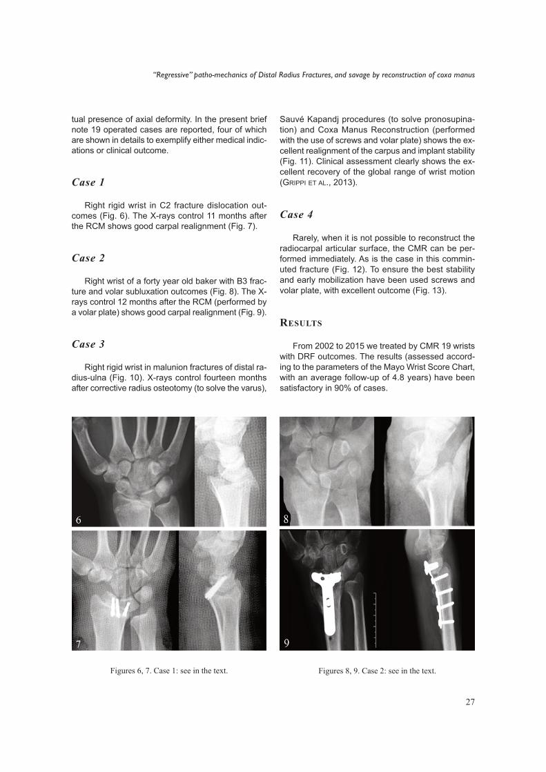
Right rigid wrist in C2 fracture dislocation out-comes (Fig. 6). The X-rays control 11 months afterthe RCM shows good carpal realignment (Fig. 7).
Case 2
Right wrist of a forty year old baker with B3 frac-ture and volar subluxation outcomes (Fig. 8). The X-rays control 12 months after the RCM (performed bya volar plate) shows good carpal realignment (Fig. 9).
Case 3
Right rigid wrist in malunion fractures of distal ra-dius-ulna (Fig. 10). X-rays control fourteen monthsafter corrective radius osteotomy (to solve the varus),
Figures 6, 7. Case 1: see in the text.
Sauvé Kapandj procedures (to solve pronosupina-tion) and Coxa Manus Reconstruction (performedwith the use of screws and volar plate) shows the ex-cellent realignment of the carpus and implant stability(Fig. 11). Clinical assessment clearly shows the ex-cellent recovery of the global range of wrist motion(GRIPPI ET AL., 2013).
Case 4
Rarely, when it is not possible to reconstruct theradiocarpal articular surface, the CMR can be per-formed immediately. As is the case in this commin-uted fracture (Fig. 12). To ensure the best stabilityand early mobilization have been used screws andvolar plate, with excellent outcome (Fig. 13).
RESULTS
From 2002 to 2015 we treated by CMR 19 wristswith DRF outcomes. The results (assessed accord-ing to the parameters of the Mayo Wrist Score Chart,with an average follow-up of 4.8 years) have beensatisfactory in 90% of cases.
Figures 8, 9. Case 2: see in the text.

CONCLUSIONS
In suffering post-DRF wrist, the CMR has provedto be a valid savage operation, able to perfectNature’s carpus adaptation in the trauma, with reli-able and satisfactory results.
REFERENCES
ALLIEU Y., BRAHIN B. & ASCENCIO G., 1982. Carpalinstabilities: Radiological and clinic-pathologicalclassification. Annales de Radiologie, 25: 275–287.
GRIPPI G.M., 1997. Cinematica del condilo carpale conintroduzione al Modello Carpale BiarticolareConcentrico (MBC) e sua applicazione al problemadell’instabilità carpale. Rivista di Chirurgia e Ri-abilitazione della Mano e Arto Superiore, 34: 389–401.
GRIPPI G.M., 2003. La ricostruzione della “Coxa Manus”Indicazioni e tecnica chirurgica. Rivista di Chirurgiadella Mano, 40 (3).
GRIPPI G.M., 2008. Patomeccanica “regressiva” delle
fratture articolari del radio distale e salvataggiocon l’intervento di Ricostruzione della Coxa Manus.Minerva Ortopedica e Traumatologica, 59: 283–298.
GRIPPI G.M., 2015. Biarticular Concentric Carpal Mech-anics and Coxa Manus Surgery. Proceedings of the9th Triennial Hand and Wrist Biomechanics Interna-tional (HWBI) Symposium Milan, Italy, June 16–17.
GRIPPI G.M. & CUGOLA L., 2011. Carpo adattativo e trat-tamento con la chirurgia della Coxa Manus. Rivistadi Chirurgia della Mano, 48: 93–97.
GRIPPI G.M. & POMPILIO D., 2002. Surgery in the Out-comes of Traumatic Wrist: Coxa Manus Surgery:Proceedings of 8 th Congress of the Federation of theEuropean Societies for Surgery of the Ha nd. Amster-dam, May 22–25, 2002. Editor Steven Hovius byMonduzzi Bologna, 57–64.
GRIPPI G.M., CUGOLA L., D’ARIENZO M. & PASSARETTIU., 2013. Prassi di Chirurgia della mano. Rivista diChirurgia della Mano, 50: 91–100.
LINSCHEID R.L., DOBYNS J.H., BEABOUT J.W. & BRYANR.S., 1972. Traumatic Instability of the wrist. Diag-nosis, Classification, and Pathomechanics. Journalof Bone and Joint Surgery, 54-A: 1612–1632.
28
GAETANO MAURIZIO GRIPPI
Figures 10, 11. Case 3: see in the text. Figures 12, 13. Case 4: see in the text.