AOCD PPT Presentation - fcm 020317 · 2018-04-01 · Relative Value Units and Related Informatio n...
22
3/7/2017 1 2017 Dermatology Coding Updates Faith C M McNicholas, RHIT, CPC, CPCD, PCS, CDC Manager, Coding and Reimbursement American Academy of Dermatology [email protected] 2017 Spring Current Concepts in Dermatology Seminar American Osteopathic College of Dermatology March 31, 2017 Practice tools is created by the American Academy of Dermatology Association. Copyright © 2016 American Academy of Dermatology. All rights reserved. 2017 CPT News CPT Code/Modifier Code Descriptor Telemedicine: Appendix P *99201 - *99215 Lists current CPT codes with evidence of some payer approval for use when provided via integrative audio and video telecommunication system *E/M codes included in this list Modifier 95 *Check local carrier policy on coverage Indicates a service generally reported as a face-to-face service was performed via a real-time interactive audio and video telecommunication system 0419T (will sunset January 2022) Destruction of neurofibroma, extensive (cutaneous, dermal extending into subcutaneous); face, head and neck, greater than 50 neurofibromas 04120T (will sunset January 2022) trunk and extremities, extensive, greater than 100 neurofibromas Practice tools is created by the American Academy of Dermatology Association. Copyright © 2016 American Academy of Dermatology. All rights reserved. 2017 Medicare Part B Premium and Deductible • 2017 Part B premium standard is $134 • Average is $109, up from $104 in 2016 • Low Social Security cost of living adjustment of 0.3%, • “Hold harmless” provision designed to protect seniors prevented a larger increase • 2017 Part B Deductible is $183, up from $166 in 2016 • Co-insurance remains 20% of Medicare Physician Fee Schedule (MPFS) allowed amount • File Medicaid claim prior to billing patient
Transcript of AOCD PPT Presentation - fcm 020317 · 2018-04-01 · Relative Value Units and Related Informatio n...

3/7/2017
1
2017 Dermatology Coding Updates
Faith C M McNicholas, RHIT, CPC, CPCD, PCS, CDCManager, Coding and Reimbursement
American Academy of Dermatology
2017 Spring Current Concepts in Dermatology Seminar
American Osteopathic College of Dermatology
March 31, 2017
Practice tools is created by the American Academy of Dermatology Association.
Copyright © 2016 American Academy of Dermatology. All rights reserved.
2017 CPT News
CPT Code/Modifier Code Descriptor
Telemedicine:Appendix P
*99201 - *99215
Lists current CPT codes with evidence of some payer approval for use when provided via integrative audio and video telecommunication system
*E/M codes included in this list
Modifier 95*Check local carrier policy on coverage
Indicates a service generally reported as a face-to-face service was performed via a real-time interactive audio and video telecommunication system
0419T(will sunset January 2022)
Destruction of neurofibroma, extensive (cutaneous, dermal extending into subcutaneous); face, head and neck, greater than 50 neurofibromas
04120T(will sunset January 2022)
trunk and extremities, extensive, greater than 100 neurofibromas
Practice tools is created by the American Academy of Dermatology Association.
Copyright © 2016 American Academy of Dermatology. All rights reserved.
2017 Medicare Part B Premium and Deductible
• 2017 Part B premium standard is $134
• Average is $109, up from $104 in 2016
• Low Social Security cost of living adjustment of 0.3%,
• “Hold harmless” provision designed to protect seniors preventeda larger increase
• 2017 Part B Deductible is $183, up from $166 in 2016
• Co-insurance remains 20% of Medicare Physician Fee Schedule (MPFS) allowed amount
• File Medicaid claim prior to billing patient

3/7/2017
2
Practice tools is created by the American Academy of Dermatology Association.
Copyright © 2016 American Academy of Dermatology. All rights reserved.
Medicare Beneficiary Identifier Revision
By April 2019 Medicare beneficiaries, Social Security ID numberswill be replaced with random identifiers
CMS requests provider’s assistance
• Check your patient’s address
• Ask the patient to update the Social Security Office directlyif any changes
https://faq.ssa.gov/ics/support/KBAnswer.asp?questionID=3704
Practice tools is created by the American Academy of Dermatology Association.
Copyright © 2016 American Academy of Dermatology. All rights reserved.
Administrative Law Judge (ALJ) and Federal Claim Appeal Increase
ALJ and Federal District Court amount incontroversy increase for 2017
*Total may encompass a single claim or the accumulation of claims
Appeal Claim Total Required
Administrative Law Judge Federal District Court
2016 $150 $1,500
2017 $160 $1,560
Practice tools is created by the American Academy of Dermatology Association.
Copyright © 2016 American Academy of Dermatology. All rights reserved.
Medicare Drug Waste Modifier
Medicare Change Request CR 9603
JW - Drug amount discarded/not administered to any patient, Documentation must be available if requested
• Beginning January 1, 2017
• Reporting unused drug and biological portions require JW modifier to identify discarded drugs and biologicals when processing Part B claims.
• Do not use JW modifier to report Competitive Acquisition Program (CAP) drugs and biologicals.

3/7/2017
3
Practice tools is created by the American Academy of Dermatology Association.
Copyright © 2016 American Academy of Dermatology. All rights reserved.
Medicare Drug Waste Modifier
Documentation must include• Date and time the drug was discarded• Amount discarded • Reason for wastage • Who wasted the drug
• Check local Medicare contractor for provider requirements for reporting of drug wastage
For more information see: • CR 9603: www.cms.gov/Outreach-and-Education/Medicare-Learning-Network-
MLN/MLNMattersArticles/Downloads/MM9603.pdf
• Derm Coding Consult, Spring 2015, Billing for Drugs and Biologicals in your Practice
Practice tools is created by the American Academy of Dermatology Association.
Copyright © 2017 American Academy of Dermatology. All rights reserved.
Medicare Payment Revision
Non-face-to-face prolonged E/M service
• Must be• Tied to an E/M service• Time spent by billing physician only
• Not within the scope of clinical staff• No other CPT to report this work
• Document • Time• Summary of non-face-to-face work
Practice tools is created by the American Academy of Dermatology Association.
Copyright © 2016 American Academy of Dermatology. All rights reserved.
Prolonged E/M Service
CPT Code Global CMS work RVU
99358 XXX 2.10
99359 XXX 1.00
3/7/2017
4
Practice tools is created by the American Academy of Dermatology Association.
Copyright © 2016 American Academy of Dermatology. All rights reserved.
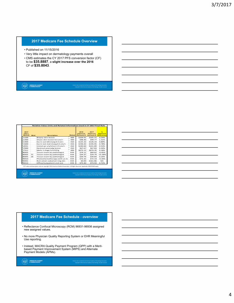
2017 Medicare Fee Schedule Overview
• Published on 11/15/2016
• Very little impact on dermatology payments overall
• CMS estimates the CY 2017 PFS conversion factor (CF)to be $35.8887, a slight increase over the 2016CF of $35.8043.
Practice tools is created by the American Academy of Dermatology Association.
Copyright © 2016 American Academy of Dermatology. All rights reserved.
1 CPT codes and descriptors only are copyright 2016 American Medical Association. All Rights Reserved. Applicable FARS/DFARS apply.
Relative Value Units and Related Information Used in CY 2017 Final Rule
2017 CPT1/
HCPCS Mod Description Global
2016 payment Amount $
2017 Payment Amount $
% Payment
Difference
11100 Biopsy skin lesion 000 $104.91 $105.15 0.24%
11300 Shave skin lesion 0.5 cm/< 000 $98.46 $99.77 1.31%
11400 Exc tr‐ext b9+marg 0.5 cm< 010 $125.32 $126.33 0.80%
11600 Exc tr‐ext mal+marg 0.5 cm/< 010 $194.42 $195.95 0.78%
12031 Intmd rpr s/a/t/ext 2.5 cm/< 010 $240.60 $241.89 0.53%
17000 Destruct premalg lesion 010 $67.67 $67.83 0.24%
17311 Mohs 1 stage h/n/hf/g 000 $673.12 $674.35 0.18%
88305 Tissue exam by pathologist XXX $74.11 $69.62 ‐6.45%
88305 TC Tissue exam by pathologist XXX $34.37 $29.79 ‐15.39%
88305 26 Tissue exam by pathologist XXX $39.74 $39.84 0.24%
96910 Photochemotherapy with uv‐b XXX $72.32 $72.14 ‐0.26%
96931 Rcm celulr subcelulr img skn XXX $0.00 $161.86 NA
99213 Office/outpatient visit est XXX $73.40 $73.93 0.72%
Practice tools is created by the American Academy of Dermatology Association.
Copyright © 2016 American Academy of Dermatology. All rights reserved.
2017 Medicare Fee Schedule - overview
• Reflectance Confocal Microscopy (RCM) 96931-96936 assigned new assigned values.
• No more Physician Quality Reporting System or EHR Meaningful Use reporting.
• Instead, MACRA Quality Payment Program (QPP) with a Merit-based Payment Improvement System (MIPS) and Alternate Payment Models (APMs).
3/7/2017
5
Practice tools is created by the American Academy of Dermatology Association.
Copyright © 2016 American Academy of Dermatology. All rights reserved.
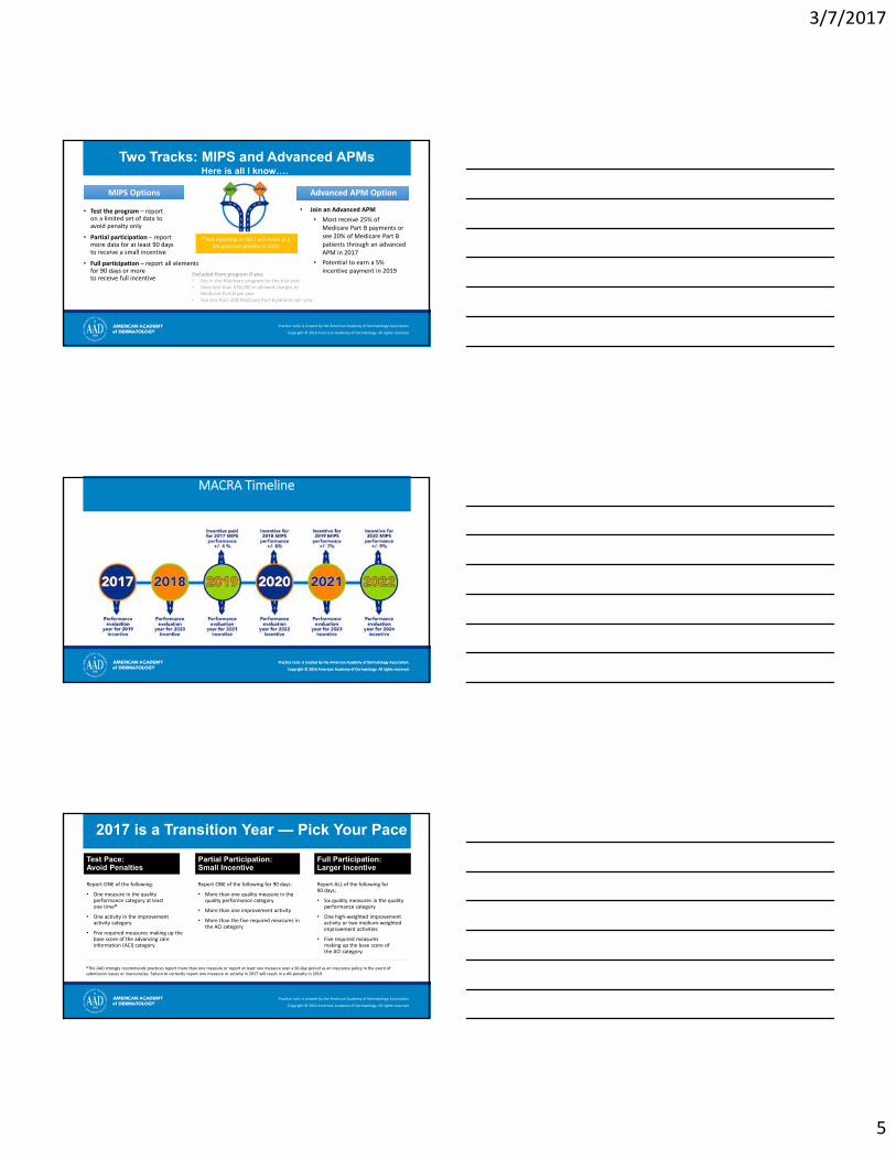
Two Tracks: MIPS and Advanced APMs
• Test the program – reporton a limited set of data toavoid penalty only
• Partial participation – reportmore data for at least 90 daysto receive a small incentive
• Full participation – report all elements for 90 days or moreto receive full incentive
• Join an Advanced APM
• Most receive 25% of Medicare Part B payments or see 20% of Medicare Part B patients through an advanced APM in 2017
• Potential to earn a 5% incentive payment in 2019
MIPS OptionsMIPS Options Advanced APM OptionAdvanced APM Option
Excluded from program if you:• Are in the Medicare program for the first year• Have less than $30,000 in allowed charges to
Medicare Part B per year• See less than 100 Medicare Part B patients per year
* Not reporting in 2017 will result in a 4% payment penalty in 2019
Here is all I know….
Practice tools is created by the American Academy of Dermatology Association.
Copyright © 2016 American Academy of Dermatology. All rights reserved.
Practice tools is created by the American Academy of Dermatology Association.
Copyright © 2016 American Academy of Dermatology. All rights reserved.
MACRA Timeline
Practice tools is created by the American Academy of Dermatology Association.
Copyright © 2016 American Academy of Dermatology. All rights reserved.
Test Pace:Avoid Penalties
Report ONE of the following:
• One measure in the quality performance category at leastone time*
• One activity in the improvement activity category
• Five required measures making up the base score of the advancing care information (ACI) category
Partial Participation:Small Incentive
Report ONE of the following for 90 days:
• More than one quality measure in the quality performance category
• More than one improvement activity
• More than the five required measures in the ACI category
2017 is a Transition Year — Pick Your Pace
Full Participation:Larger Incentive
Report ALL of the following for90 days:
• Six quality measures in the quality performance category
• One high‐weighted improvement activity or two medium‐weighted improvement activities
• Five required measuresmaking up the base score ofthe ACI category.
*The AAD strongly recommends practices report more than one measure or report at least one measure over a 90‐day period as an insurance policy in the event of submission issues or inaccuracies. Failure to correctly report one measure or activity in 2017 will result in a 4% penalty in 2019.
3/7/2017
6
Practice tools is created by the American Academy of Dermatology Association.
Copyright © 2016 American Academy of Dermatology. All rights reserved.
Got questions about MACRA?
http://community.aad.org/home
Practice tools is created by the American Academy of Dermatology Association.
Copyright © 2016 American Academy of Dermatology. All rights reserved.
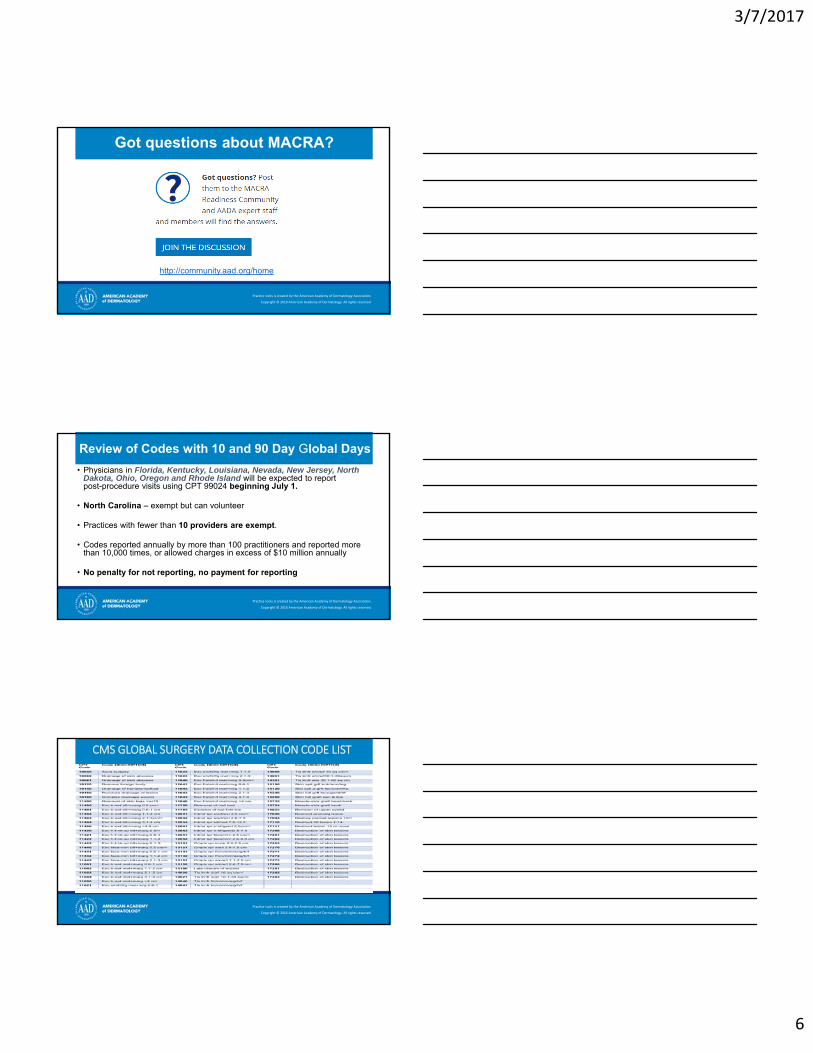
Review of Codes with 10 and 90 Day Global Days
• Physicians in Florida, Kentucky, Louisiana, Nevada, New Jersey, North Dakota, Ohio, Oregon and Rhode Island will be expected to reportpost-procedure visits using CPT 99024 beginning July 1.
• North Carolina – exempt but can volunteer
• Practices with fewer than 10 providers are exempt.
• Codes reported annually by more than 100 practitioners and reported more than 10,000 times, or allowed charges in excess of $10 million annually
• No penalty for not reporting, no payment for reporting
Practice tools is created by the American Academy of Dermatology Association.
Copyright © 2016 American Academy of Dermatology. All rights reserved.
CMS GLOBAL SURGERY DATA COLLECTION CODE LIST

3/7/2017
7
Practice tools is created by the American Academy of Dermatology Association.
Copyright © 2016 American Academy of Dermatology. All rights reserved.
Review of Codes With 10 and 90 Day Global Periods
• CMS survey practitioners to gain information regarding post-op visits may occur in mid-2017
• CMS will collect data from Pioneer and Next Gen ACOs on the “activities and resources involved in and around surgical events”
Practice tools is created by the American Academy of Dermatology Association.
Copyright © 2016 American Academy of Dermatology. All rights reserved.
ICD 10 Updates
• Code specificity required as of October 1, 2016
• No specific dermatology code changes. However, 2017 Guideline changes may
impact your practice
• Review Derm Coding Consult for more information
Nothing lasts forever: ICD‐10 code freeze and specificity safe harbor ends
No dermatology specific ICD-10 CMcode changes for 2017
FALL 2016 WINTER 2016
Practice tools is created by the American Academy of Dermatology Association.
Copyright © 2017 American Academy of Dermatology. All rights reserved.
2017 ICD-10 Guidelines
• Clinical Criteria and Code Assignment
• Excludes1
• Bilateral Conditions
• Etiology/Manifestation
• Terms – With, Use additional code
• Episode of Care
• Complications of Care

3/7/2017
8
Practice tools is created by the American Academy of Dermatology Association.
Copyright © 2017 American Academy of Dermatology. All rights reserved.
Clinical Criteria / Code Assignment
• Diagnosis codes reported are NOT selected basedon clinical criteria used to establish the diagnosis
• Physician’s statement alone that the condition ispresent supports reporting of the ICD-10 code.
Practice tools is created by the American Academy of Dermatology Association.
Copyright © 2017 American Academy of Dermatology. All rights reserved.
Excludes1
• Listed with mutually exclusive codes: • Two conditions cannot be reported together
• Clinically• Coding rule
• Clarification provided by CMS in October 2015 • If unrelated, report both conditions
Practice tools is created by the American Academy of Dermatology Association.
Copyright © 2017 American Academy of Dermatology. All rights reserved.
Example
Pathology Report
#1 specimen from abdomen –Lentigo maligna
#2 specimen from back –Malignant melanoma
ICD codes
D03.59 Melanoma in situ of otherparts of trunk
C43.59 Malignant melanoma of other parts of trunk
Excludes1 melanoma in situ (D03.-)
? Report both codesDocumentation must reflect separate sites withinsame anatomic location – per ICD grouping

3/7/2017
9
Practice tools is created by the American Academy of Dermatology Association.
Copyright © 2017 American Academy of Dermatology. All rights reserved.
Bilateral conditions
Bilateral Condition Is: Treatment is: Code as:
Present bilaterally Unilateral Bilateral
Present bilaterally Bilateral Bilateral
Present unilaterally –Previously treated side ishealed or no longer present
Unilateral
Unilateral – list side treatedIf applicable; History of code forpreviously treated side
• When available, report bilateral conditions with bilateral codes• Even when only unilateral treatment is provided
Practice tools is created by the American Academy of Dermatology Association.
Copyright © 2017 American Academy of Dermatology. All rights reserved.
Bilateral Conditions
Patient presents with swelling and pain of both ears. On exam, manipulation of auricle causes pain and a furuncle is present at left pre-auricle area.
Gentle debridement of both ears and neomycin applied.
Assessment: otitis externa cellulitis
Rx: hydrocortisone drops.
ICD-10-CM: H60.13 Cellulitis of external ear, bilateral
Patient returns in follow-up of otitis externa cellulitis. Patient states right side is better,left still hurts.
Exam shows condition is resolved on right.Left side ear canal is red, swollen, andlittered with moist, purulent debris.
RX: Continue hydrocortisone drops
Ciprofloxacin 500 mg po bid for 10 days
ICD-10-CM: H60.12 Cellulitis of leftexternal ear
Practice tools is created by the American Academy of Dermatology Association.
Copyright © 2017 American Academy of Dermatology. All rights reserved.
Etiology / Manifestation
Condition with underlying etiology and manifestations
• Sequencing rules apply• Underlying condition or cause is listed first• Manifestation of this condition or cause is listed second
• Code first/Use additional code• Links cause or etiology to condition being treated• May help to show complexity of care needed

3/7/2017
10
Practice tools is created by the American Academy of Dermatology Association.
Copyright © 2017 American Academy of Dermatology. All rights reserved.
Directional Terms - Updated
• Watch for “code first” and “use additional code” guidance
L51 – Erythema multiforme
Use additional code for adverse effect, if applicable,to identify drug
• “if applicable” was added to guidance for 2017
Practice tools is created by the American Academy of Dermatology Association.
Copyright © 2017 American Academy of Dermatology. All rights reserved.
Example
Combination Code
75 year-old Type 1 diabetic female patient presents with small raised, yellow and somewhat waxy lesions on lower part of her legs.
Final Diagnosis: Necrobiosis lipoidica diabeticoruma
ICD-10-CM code: E10.620 Type 1 DM with diabetic dermatitis
Practice tools is created by the American Academy of Dermatology Association.
Copyright © 2017 American Academy of Dermatology. All rights reserved.
Example
One Condition / Two CodesPatient presents for follow-up of non-pressure chronic ulcer of right ankle secondary to Type 1 diabetes. Breakdown of ulcer is limited to skin. Diabetes is followed by primary care.
Final diagnosis: non-pressure ulcer, DM type 1
ICD-10-CM codes:E10.621 Type 1 Diabetes mellitus with foot ulcer
Use additional code to identify site of ulcer
L97.311 Non-pressure chronic ulcer of right ankle limited to breakdown of skinCode first any associated underlying condition
3/7/2017
11
Practice tools is created by the American Academy of Dermatology Association.
Copyright © 2017 American Academy of Dermatology. All rights reserved.
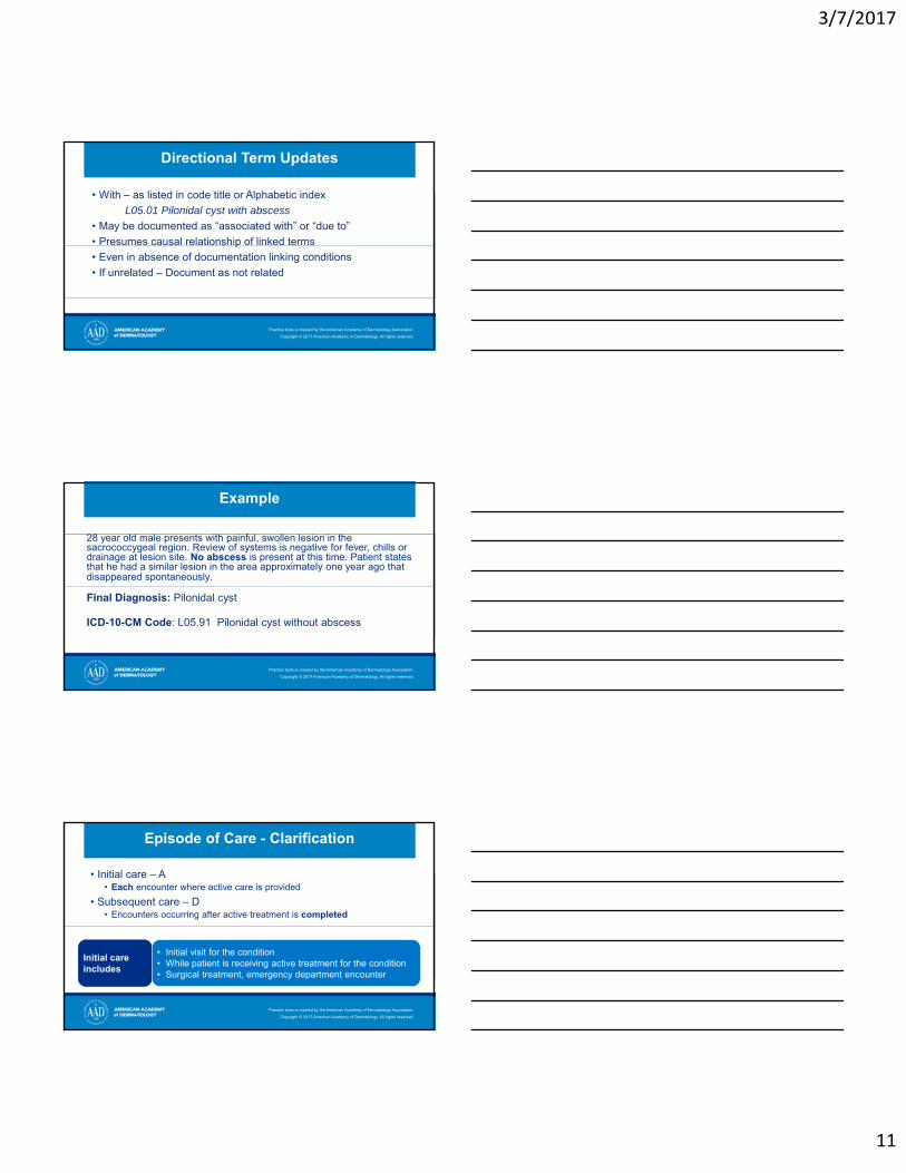
Directional Term Updates
• With – as listed in code title or Alphabetic index
L05.01 Pilonidal cyst with abscess
• May be documented as “associated with” or “due to”
• Presumes causal relationship of linked terms
• Even in absence of documentation linking conditions
• If unrelated – Document as not related
Practice tools is created by the American Academy of Dermatology Association.
Copyright © 2017 American Academy of Dermatology. All rights reserved.
Example
28 year old male presents with painful, swollen lesion in the sacrococcygeal region. Review of systems is negative for fever, chills or drainage at lesion site. No abscess is present at this time. Patient states that he had a similar lesion in the area approximately one year ago that disappeared spontaneously.
Final Diagnosis: Pilonidal cyst
ICD-10-CM Code: L05.91 Pilonidal cyst without abscess
Practice tools is created by the American Academy of Dermatology Association.
Copyright © 2017 American Academy of Dermatology. All rights reserved.
Episode of Care - Clarification
• Initial care – A• Each encounter where active care is provided
• Subsequent care – D• Encounters occurring after active treatment is completed
Initial careincludes
• Initial visit for the condition• While patient is receiving active treatment for the condition• Surgical treatment, emergency department encounter

3/7/2017
12
Practice tools is created by the American Academy of Dermatology Association.
Copyright © 2017 American Academy of Dermatology. All rights reserved.
Documentation Should Reflect Episode
Follow up patient for second degree caustic burns on back of right hand from drain cleaner. Doing well on steroids and topical medication. No signs of infection. Continue on current medications. Dressing changed, patient to return in two days for dressing change.
ICD-10-CM Codes: T23.661D Corrosion of second degree of back of hand, right
T54.3X1D Toxic effect of corrosive alkalis and alkali-like substances, accidental
Practice tools is created by the American Academy of Dermatology Association.
Copyright © 2017 American Academy of Dermatology. All rights reserved.
Complications of Care
• Based on documented relationship between condition and previous care or procedure
• Not all conditions occurring during or after procedures or treatment are considered complications
• Documentation must reflect cause-and-effect relationship• Complication codes may reflect episode of care
• Active treatment – treatment of the condition described by the code
Practice tools is created by the American Academy of Dermatology Association.
Copyright © 2017 American Academy of Dermatology. All rights reserved.
Example
Patient returns 5 days after punch biopsy withinfection at biopsy site.
ICD-10-CM Code: T81.4XXA Infection following a procedure, initial encounter

3/7/2017
13
CMS Guidelines
Practice tools is created by the American Academy of Dermatology Association.
Copyright © 2016 American Academy of Dermatology. All rights reserved.
Practice tools is created by the American Academy of Dermatology Association.
Copyright © 2016 American Academy of Dermatology. All rights reserved.
Active Local Coverage Determination (LCDs)
• Allergy Testing• Application of Bioengineered Skin Substitutes to Lower Extremity
Chronic Non-Healing Wounds • Debridement of Mycotic Nails • Moh’s Micrographic Surgery• Removal of Benign Skin Lesions• Surgical Treatment of Nails• Treatment of Varicose Veins and Venous Stasis Disease of the
Lower Extremities• Wound Care
List is not conclusive – based on MAC

3/7/2017
14
Practice tools is created by the American Academy of Dermatology Association.
Copyright © 2017 American Academy of Dermatology. All rights reserved.
LCD Utilization and Limitations Guidelines
• Guidelines set out by Medicare• Discuss the conditions affected by the policy• List the conditions covered under the policy• Describe what constitutes medical necessity• Describe what can cause claim payment denial
• Usually found on the first page of the LCD• Under Coverage Guidance
Practice tools is created by the American Academy of Dermatology Association.
Copyright © 2017 American Academy of Dermatology. All rights reserved.
Novitas Solutions Inc. (MAC) Local Coverage Determination (LCD): Removal of Benign Skin Lesion) (L34938)
Policy applies toReasons for non-coverage
Reasons for Coverage
SKs, Skin Tags, Milia, Molluscumcontagiosum, Seb. (epidermoid) cysts, Moles (nevi), Acquired hyperkeratosis (keratoderma) and Viral warts (excluding condyloma acuminatum)
Skin lesions that do not pose a threat to health or function is considered cosmeticand as such is not covered by the Medicare program.
Consider skin lesions as medically necessary, and not cosmetic, if one or more of the following conditions is present and clearly documentedin the medical record:
• Lesion has one or more of these characteristics: bleeding, intense itching, pain;
• Physical evidence of inflammation, e.g., purulence, oozing, edema, erythema;
• Obstructs an orifice or clinically restricts vision;• In an anatomical region subject to recurrent physical trauma
and there is documentation that such trauma has in fact occurred;
• Wart removals will be covered under all the above• In addition, if periocular wart, must be associated with
chronic recurrent conjunctivitis• Evidence of spread from one body area to another,
particularly inmmunocompromised patient
Practice tools is created by the American Academy of Dermatology Association.
Copyright © 2017 American Academy of Dermatology. All rights reserved.
Documentation
• Documentation in the medical record is critical in coding to ensure that every aspect of the patient condition and care is captured
• Use of the utilization and limitations guidelines can help improve your MR documentation
• Consider including words like:• Bleeding • Intense itching • Pain • Physical evidence of inflammation, e.g., purulence, oozing, edema, erythema• Obstructs an orifice or clinically restricts vision• Anatomical region subject to recurrent physical trauma

3/7/2017
15
Practice tools is created by the American Academy of Dermatology Association.
Copyright © 2017 American Academy of Dermatology. All rights reserved.
Documentation Example 1
A 17 y/o girl has a 1.1 cm raised brown nevus on her mid back that rubs on her bra. You remove it using a shave technique. Pathology report shows a benign compound nevus, and the lateral and underlying dermal margins are clear, confirming complete removal of the nevus.
You report:11302 – shaving of epidermal or dermal lesion, single lesion, trunk, lesion diameter 1.1 – 2.0cm
• Complete removal of this lesion does not make this an excision.
Practice tools is created by the American Academy of Dermatology Association.
Copyright © 2017 American Academy of Dermatology. All rights reserved.
Documentation Example 2
82 yo female patient presents with linear, splayed, vertical patterns of lesions on her chest. States over time, they have increased, get caught in neck chain, are inflamed and causing pain. They started off light tan in color, but have progressed to becoming dark brown.
You report:17110 – Destruction (eg, laser surgery, electrosurgery, cryosurgery, chemosurgery, surgical curettement), of benign lesions other than skin tags or cutaneous vascular proliferative lesions; up to 14 lesions
Practice tools is created by the American Academy of Dermatology Association.
Copyright © 2017 American Academy of Dermatology. All rights reserved.
Documentation Example 3
A 1.2 cm flesh-colored polypoid nodule on the upper thigh of a 45 y/o man is irritated by his clothing. It is removed at the base with scissors, exposing underlying fat, and hemostasis is achieved with electrocautery. Pathology confirms a benign fibrofatty polyp.
You report:11402 - Excision, benign lesion including margins, except skin tag, excised diameter 1.1 – 2.0 cm

3/7/2017
16
Practice tools is created by the American Academy of Dermatology Association.
Copyright © 2017 American Academy of Dermatology. All rights reserved.
Removal of Benign Lesion LCD
The LCD, further states the following will allow coverage
• If clinical diagnosis is uncertain, particularly where malignancy is a realistic consideration based on lesional appearance(e.g. non-response to conventional treatment, or changein appearance).
• Prior biopsy suggests or is indicative of lesion malignancy or premalignancy.
Practice tools is created by the American Academy of Dermatology Association.
Copyright © 2017 American Academy of Dermatology. All rights reserved.
Documentation Example 4
50 yo boater has discreet but irregular 8mm shiny red flat lesion on his back. Clinical diagnosis is probable superficial BCC and a specimen is obtained for pathology with curettage as a definitive procedure with therapeutic intent to cure. Pathology confirms a benign diagnosis.
You report:17110 – Destruction (eg, laser surgery, electrosurgery, cryosurgery, chemosurgery, surgical curettement), of benign lesions other than skin tags or cutaneous vascular proliferative lesions; up to 14 lesions
• If pt presented with more than 15 lesions, you would report 17111 –Destruction of benign lesions; 15 or more lesions
Practice tools is created by the American Academy of Dermatology Association.
Copyright © 2017 American Academy of Dermatology. All rights reserved.
Novitas Solutions, Inc. (MAC)Local Coverage Determination (LCD): MOHS Micrographic Surgery (MMS) (L34961)
Policy applies to
Reasons for non-coverage
Reasons for Coverage
MOHS Micrographic Surgery (MMS)
CPT Codes: 17311 - 17315
Recurrent and primary AK, with focal SCC in situ; Bowenoid AK; SCC in situ (AK type) of any size in all areas in healthy or immunocompromised patients.BCC/SCC located inArea L
*See AAD Appropriate Use Criteria (AUC)
• Requires a single physician to act in 2 separate and distinct capacities: surgeon and pathologist;
• Qualifications of the physician and office/facility team;
• Characteristics of the lesion (per-procedure);
• Documentation of the Medical Necessity
• If either of these responsibilities is delegated to another physician or other qualified health care professional who reports the service(s) separately, the MMS codes should not be reported
• The qualifications of the performing physician must be verifiable if requested by the Contractor. • Providers of MOHS surgery a limited to MD or DO
• The AUC provide necessary consideration of MOHS micrographic surgical treatment of a lesion.;
• See LCD Documentation Requirements section

3/7/2017
17
Practice tools is created by the American Academy of Dermatology Association.
Copyright © 2017 American Academy of Dermatology. All rights reserved.
Medicare on Mohs
• After careful review, Medicare Jurisdictions adopted coverage for Mohs in accordance with the 2012 Appropriate Use Criteria (AUC) for Mohs Micrographic Surgery as published in JAAD Volume 67, Issue 4, pp 531-550, October 2012
• MMS appropriate only when:• Superficial (lateral) or deep margins of the cancer lesion are uncertain
clinically.• Likelihood of surgical cure and reconstruction would be compromised without
use of immediate microscopic examination of the surgical margins.
• The medical records should clearly show that MMS was chosen because of the complexity (e.g. poorly defined clinical borders, possible deep invasion, prior irradiation), size or location (e.g. maximum conservation of tumor-free tissue is important).
Practice tools is created by the American Academy of Dermatology Association.
Copyright © 2017 American Academy of Dermatology. All rights reserved.
CMS MLN Matters® Number: SE1318
Guidance To Reduce Mohs Surgery Reimbursement Issues
(Applies to all Medicare carriers)
• Documentation should support the medical necessity for Mohs procedure
• Operative notes and pathology documentation must clearly indicate:• That Mohs was performed using accepted MMS technique; • That physician acted in two integrated, but distinct, capacities as surgeon
and pathologist; • That the location, number, and size of the lesion(s) treated; • That the number of stages performed; and• That the the number of specimens per stage.
Practice tools is created by the American Academy of Dermatology Association.
Copyright © 2017 American Academy of Dermatology. All rights reserved.
MLN Matters SE1318 Cont’d
Medicare Documentation Requirements
• Describe histology of the specimens taken in the first stage. Description should include:
• Depth of invasion;• Pathological pattern;• Cell morphology; and, if present, • Perineural invasion or presence of scar tissue.
• Subsequent stages: note pattern and morphology of the tumor (if still seen) is as described for the first stage
• Or, if differences are found, note the changes
3/7/2017
18
Practice tools is created by the American Academy of Dermatology Association.
Copyright © 2017 American Academy of Dermatology. All rights reserved.
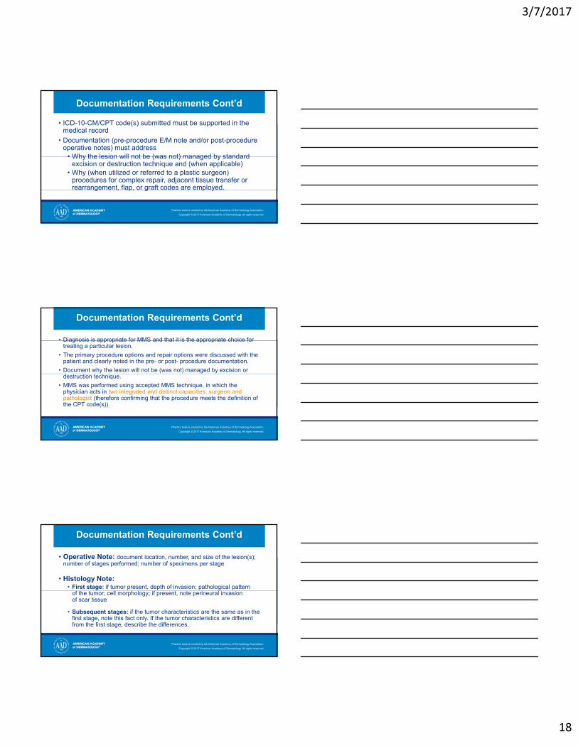
Documentation Requirements Cont’d
• ICD-10-CM/CPT code(s) submitted must be supported in the medical record
• Documentation (pre-procedure E/M note and/or post-procedure operative notes) must address
• Why the lesion will not be (was not) managed by standard excision or destruction technique and (when applicable)
• Why (when utilized or referred to a plastic surgeon) procedures for complex repair, adjacent tissue transfer or rearrangement, flap, or graft codes are employed.
Practice tools is created by the American Academy of Dermatology Association.
Copyright © 2017 American Academy of Dermatology. All rights reserved.
Documentation Requirements Cont’d
• Diagnosis is appropriate for MMS and that it is the appropriate choice for treating a particular lesion.
• The primary procedure options and repair options were discussed with the patient and clearly noted in the pre- or post- procedure documentation.
• Document why the lesion will not be (was not) managed by excision or destruction technique.
• MMS was performed using accepted MMS technique, in which the physician acts in two integrated and distinct capacities: surgeon and pathologist (therefore confirming that the procedure meets the definition of the CPT code(s)).
Practice tools is created by the American Academy of Dermatology Association.
Copyright © 2017 American Academy of Dermatology. All rights reserved.
Documentation Requirements Cont’d
• Operative Note: document location, number, and size of the lesion(s); number of stages performed; number of specimens per stage
• Histology Note: • First stage: if tumor present, depth of invasion; pathological pattern
of the tumor; cell morphology; if present, note perineural invasionof scar tissue
• Subsequent stages: if the tumor characteristics are the same as in the first stage, note this fact only. If the tumor characteristics are different from the first stage, describe the differences.
3/7/2017
19
Practice tools is created by the American Academy of Dermatology Association.
Copyright © 2017 American Academy of Dermatology. All rights reserved.
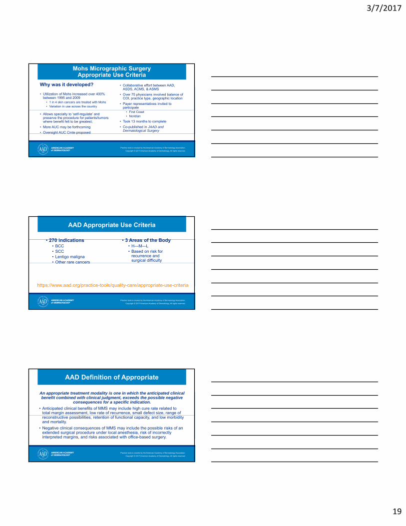
Mohs Micrographic SurgeryAppropriate Use Criteria
Why was it developed?
• Utilization of Mohs increased over 400% between 1995 and 2009
• 1 in 4 skin cancers are treated with Mohs• Variation in use across the country
• Allows specialty to ‘self-regulate’ and preserve the procedure for patients/tumors where benefit felt to be greatest;
• More AUC may be forthcoming
• Oversight AUC Cmte proposed
• Collaborative effort between AAD, ASDS, ACMS, & ASMS
• Over 75 physicians involved balance of COI, practice type, geographic location
• Payer representatives invited to participate
• First Coast• Noridian
• Took 13 months to complete
• Co-published in JAAD and Dermatological Surgery
Practice tools is created by the American Academy of Dermatology Association.
Copyright © 2017 American Academy of Dermatology. All rights reserved.
AAD Appropriate Use Criteria
• 270 indications• BCC• SCC• Lentigo maligna• Other rare cancers
• 3 Areas of the Body • H—M—L• Based on risk for
recurrence and surgical difficulty
https://www.aad.org/practice-tools/quality-care/appropriate-use-criteria
Practice tools is created by the American Academy of Dermatology Association.
Copyright © 2017 American Academy of Dermatology. All rights reserved.
AAD Definition of Appropriate
An appropriate treatment modality is one in which the anticipated clinical benefit combined with clinical judgment, exceeds the possible negative
consequences for a specific indication.
• Anticipated clinical benefits of MMS may include high cure rate related to total margin assessment, low rate of recurrence, small defect size, range of reconstructive possibilities, retention of functional capacity, and low morbidity and mortality.
• Negative clinical consequences of MMS may include the possible risks of an extended surgical procedure under local anesthesia, risk of incorrectly interpreted margins, and risks associated with office-based surgery.
3/7/2017
20
Practice tools is created by the American Academy of Dermatology Association.
Copyright © 2017 American Academy of Dermatology. All rights reserved.
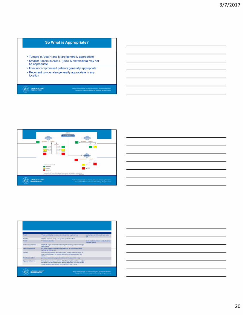
So What is Appropriate?
• Tumors in Area H and M are generally appropriate
• Smaller tumors in Area L (trunk & extremities) may not be appropriate
• Immunocompromised patients generally appropriate
• Recurrent tumors also generally appropriate in any location
Practice tools is created by the American Academy of Dermatology Association.
Copyright © 2017 American Academy of Dermatology. All rights reserved.
Practice tools is created by the American Academy of Dermatology Association.
Copyright © 2017 American Academy of Dermatology. All rights reserved.
Area H *Face, genitalia, hands, feet, nail units, ankles, nipples/areola *central face, eyelids, eyebrows, nose, lips
Area M Cheeks, forehead, scalp, neck, jawline, pretibial surface
Area L Trunk and extremities Excls. pretibial surfaces, hands, feet, nail units and ankles
Immunocompromised HIV/AIDS, organ transplant, hematologic malignancy or pharmacologic suppression
Genetic Syndromes BC nevus syndrome, xeroderma pigmentosa, or other syndromes at high risk for skin cancer
Healthy no immunosuppression, no prior radiation therapy to affected area, no chronic infections and no genetic syndromes that predispose to skin cancer
Prior Radiated Skin: previously received therapeutic radiation in this area of the body.
Aggressive features Skin cancers having one or more of the following features have a higher incidence of local recurrence and regional metastasis such that minimal margin excision may not be in the beneficiary's best interest
3/7/2017
21
Practice tools is created by the American Academy of Dermatology Association.
Copyright © 2017 American Academy of Dermatology. All rights reserved.
Mohs Takeaway• The physician (MD/DO) performing Mohs micrographic surgery must be specifically trained
and highly skilled in MMS techniques and pathologic identification.
• If a surgeon performs an excision using Mohs surgical techniques but does not personally provide the histologic evaluation of the specimen(s), the CPT codes for MMS shall not be used.
• Instead standard excision codes should be chosen for such medically necessary services (e.g., 11600 – 11646).
• Medicare is aware that a biopsy of the skin lesion for which Mohs surgery is planned may be necessary in order for the physician to determine the exact nature of the lesion(s) to be removed. Occasionally, that biopsy may need to be done on the same day that the Mohs surgery is planned.
• In order to allow separate payment for a biopsy and pathology on the same day as Mohs surgery, the -59 modifier is appropriate. The 59 modifier is also appropriate when a separate skin lesion, other than the lesion for which Mohs surgery is performed, is biopsied on the same day that the Mohs surgery is performed.
Practice tools is created by the American Academy of Dermatology Association.
Copyright © 2017 American Academy of Dermatology. All rights reserved.
Mohs Takeway
• If a prior biopsy of the site undergoing Mohs surgery has been previously performed within the last 60 days, the surgeon should make a reasonable effort to obtain those results rather than repeating the biopsy.
• Reporting both Mohs Micrographic Surgery CPT codes 17311-17315 and Surgical Pathology CPT® 88302-88309 on tissue used for margin evaluation during Mohs surgery is inappropriate and will indicate that true Mohs surgery was not done. Such claims for Mohs surgery (17311-17315) will be denied. There are occasional clinical situations in which tissue separate from the tissue examined during Mohs surgery is appropriately submitted for subsequent formalin fixed processing and histopathologic examination. The submitted tissue is not the same tissue that was processed during the Mohs surgery. It may constitute a tissue margin beyond that evaluated with Mohs surgery or it may involve a totally unrelated tissue specimen. In such situations both the Mohs surgery and the histopathology are subject to coverage. In such cases the clinical record must clearly show the reasoning for the histopathologic specimen and interpretation.
Practice tools is created by the American Academy of Dermatology Association.
Copyright © 2017 American Academy of Dermatology. All rights reserved.

3/7/2017
22
Practice tools is created by the American Academy of Dermatology Association.
Copyright © 2017 American Academy of Dermatology. All rights reserved.
2017 Coding Packs – NEW!
ORDER NOW!
AAD and AMACoding Resources
every practice needsin 2017!
PACKSSTARTING
AT $279VISIT STORE.AAD.ORG
Practice tools is created by the American Academy of Dermatology Association.
Copyright © 2017 American Academy of Dermatology. All rights reserved.
2017 Coding Webinar All-Access Pass
Practice tools is created by the American Academy of Dermatology Association.
Copyright © 2016 American Academy of Dermatology. All rights reserved.
Coding Questions
Peggy Eiden - [email protected]
Faith McNicholas - [email protected]
Cynthia Stewart – [email protected]