Antischistosomal activity of Mirazid in experimental schistosomiasis mansoni: exploring the...
7
-
Upload
mohammad-aziz -
Category
Health & Medicine
-
view
6 -
download
2
Transcript of Antischistosomal activity of Mirazid in experimental schistosomiasis mansoni: exploring the...
Parasitologists United Journal
Decem
ber 2014 Volum
e 7 • Num
ber 2
Antischistosomal activity of Mirazid in experimentalschistosomiasis mansoni: exploring the controversyMohammad A. Al-Kazzaz, Mona H. El-Sayad, Sahar A. Abu-Helw
Department of Parasitology, Medical Research
Institute, Alexandria University, Alexandria,
Egypt
Correspondence to Mohammad A. Al-kazzaz,
MSc, 27 Street, 15 Al-Awayed, Alexandria,
Egypt Tel: + +20 100 119 5791;
e-mail: [email protected]
Received 14 December 2015
Accepted 22 March 2016
Parasitologists United Journal2016, 9:31–36
BackgroundMirazid (MZD) was licensed in Egypt for treatment of schistosomiasis in the year2002; the drug gained much attention experimentally and clinically, with conflictingviews on its efficacy.ObjectiveThe study aimed to evaluate MZD anti-schistosomal activity in an animal model at aselected dose.Materials and methodsSwiss albino mice (n=36) were infected withSchistosomamansoni and divided intotwo equal groups of 18 mice each: group 1 was the nontreated infected controlgroup given only the vehicle; gourp 2was infected and treated withMZD at a dose of500mg/kg for 5 days per os 7 weeks postinfection. Efficacy of the drug wasassessed parasitologically with fecal egg counts evaluated every other day untilthe animal was euthanized at 1, 2, and 4 weeks post-treatment (WPT); wormburden, tissue egg count and oogram pattern were studied at 1, 2, and 4 WPT.ResultsMZD reduced fecal egg counts in infected mice (69.6%) and reduced total wormload (71.9%) and tissue egg counts in the intestine and the liver (66 and 77.4%,respectively) at 4 WPT. The drug changed oogram pattern with progress oftreatment.ConclusionMZD has moderate antischistosomal activity in animal models.
Keywords:mice, mirazid, oogram pattern, Schistosoma mansoni, tissue egg count, worm burden
Parasitologists United Journal 9:31–36
© 2016 Egyptian Parasitologists United Society
1687-7942
IntroductionMirazid (MZD) is a natural pharmaceuticalpreparation introduced into the Egyptian market byPharco Pharmaceuticals (Alexandria, Egypt) in theform of soft gelatin capsules since 2002. Eachcapsule contains 300mg of the purified oleoresinextract of Commiphora molmol (Myrrh) [1]. TheEgyptian drug authority in the Egyptian Ministry ofHealth registered this product for treatment ofschistosomiasis (Reg. No. 21655/2002) [2]. Theantischistosomal properties of MZD wereinvestigated, and reviewed both experimentally andclinically with controversy regarding its efficacy [3].
In humans, the efficacy of MZD as an anti-schistosomal drug was evaluated clinically withpositive results [4–9]. All in all, they enrolled 365schistosomiasis patients (adults and children) andtreated them with MZD at a dose of 10–11.5mg/kgfor 3–6 days. The cure rate, evaluated parasitologically(disappearance of fecal eggs) by Kato–Katz and rectalsnip techniques at 2–3 months after treatment, was80.7–100%. The drug proved to have nonsignificantside effects. The drug was tested by other researchers,
with disappointing results. It showed little or nobeneficial activity as reported by Barakat et al. [10],Botros et al. [11], and Osman et al. [12], whoaltogether enrolled 206 patients (adults or children)treated with MZD at a dose of 300–600mg daily for3–6 days. The drug showed a cure rate that ranged from3.7 to 15.6 % at 3–8 WPT.
Such controversy prompted us to study the efficacy ofMZD experimentally as there are many parasitologicalparameters other than fecal egg counts.
Materials and methodsExperimental studyThe study was conducted at the Department ofParasitology, Medical Research Institute (MRI),Alexandria University, Alexandria, Egypt, during theperiod from 21 July 2013 to 14 October 2014.
This is an open access article distributed under the terms of the Creative
Commons Attribution-NonCommercial-ShareAlike 3.0 License, which
allows others to remix, tweak, and build upon the work
noncommercially, as long as the author is credited and the new
creations are licensed under the identical terms.
Original article 31
© 2016 Parasitologists United Journal | Published by Wolters Kluwer - Medknow DOI: 10.4103/1687-7942.192993
MaterialsThe study included 36, 8-week-old female Swiss albinomice of the CD-1 strain weighing 20±2 g, obtained fromthe animal house of the MRI, Alexandria University,Alexandria, Egypt. Laboratory-bred Biomphalaria
alexandrina snails infected with miracidiae of theEgyptian (CD) strain of Schistosoma mansoni wereobtained from the Schistosome Biologic Supply Center,TheodoreBilharzResearchInstitute,Cairo,Egypt.MZDcapsules were obtained as free medical samples fromPharco Pharmaceuticals, Batch No: 296. Each capsule(300mg) was emptied in a flask containing 3ml of 4%CremophorELusedasthevehiclefordrugadministration.
MethodsCercarial shedding
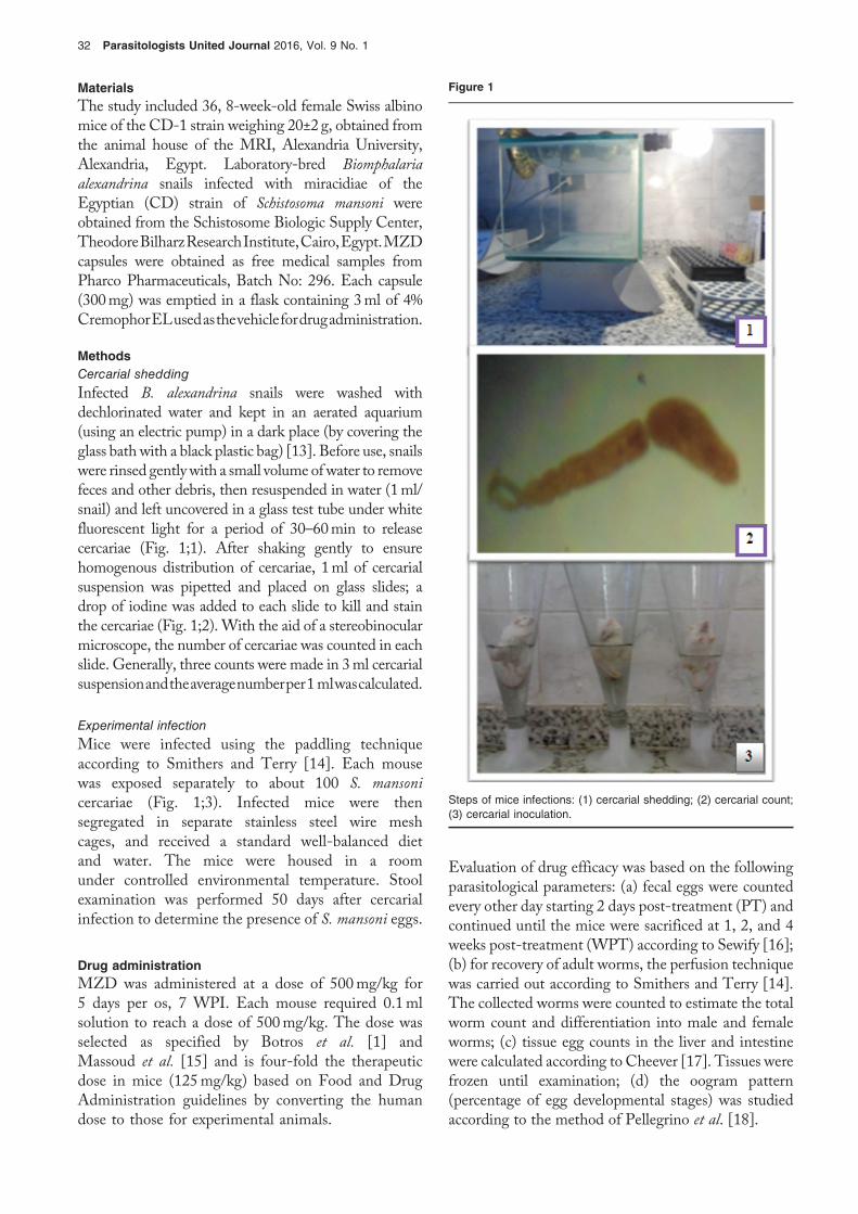
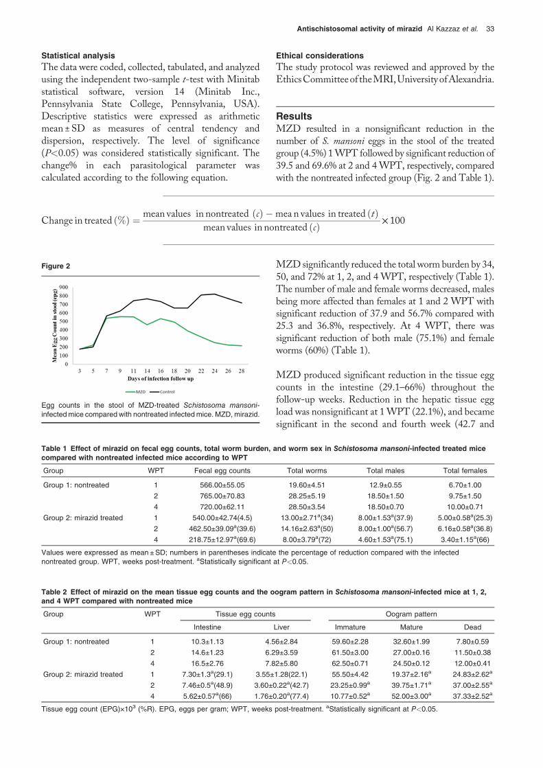
Infected B. alexandrina snails were washed withdechlorinated water and kept in an aerated aquarium(using an electric pump) in a dark place (by covering theglass bath with a black plastic bag) [13]. Before use, snailswere rinsed gentlywith a small volume ofwater to removefeces and other debris, then resuspended in water (1ml/snail) and left uncovered in a glass test tube under whitefluorescent light for a period of 30–60min to releasecercariae (Fig. 1;1). After shaking gently to ensurehomogenous distribution of cercariae, 1ml of cercarialsuspension was pipetted and placed on glass slides; adrop of iodine was added to each slide to kill and stainthe cercariae (Fig. 1;2). With the aid of a stereobinocularmicroscope, the number of cercariae was counted in eachslide. Generally, three counts were made in 3ml cercarialsuspensionandtheaveragenumberper1mlwascalculated.
Experimental infection
Mice were infected using the paddling techniqueaccording to Smithers and Terry [14]. Each mousewas exposed separately to about 100 S. mansoni
cercariae (Fig. 1;3). Infected mice were thensegregated in separate stainless steel wire meshcages, and received a standard well-balanced dietand water. The mice were housed in a roomunder controlled environmental temperature. Stoolexamination was performed 50 days after cercarialinfection to determine the presence of S. mansoni eggs.
Drug administrationMZD was administered at a dose of 500mg/kg for5 days per os, 7 WPI. Each mouse required 0.1mlsolution to reach a dose of 500mg/kg. The dose wasselected as specified by Botros et al. [1] andMassoud et al. [15] and is four-fold the therapeuticdose in mice (125mg/kg) based on Food and DrugAdministration guidelines by converting the humandose to those for experimental animals.
Evaluation of drug efficacy was based on the followingparasitological parameters: (a) fecal eggs were countedevery other day starting 2 days post-treatment (PT) andcontinued until the mice were sacrificed at 1, 2, and 4weeks post-treatment (WPT) according to Sewify [16];(b) for recovery of adult worms, the perfusion techniquewas carried out according to Smithers and Terry [14].The collected worms were counted to estimate the totalworm count and differentiation into male and femaleworms; (c) tissue egg counts in the liver and intestinewere calculated according to Cheever [17]. Tissues werefrozen until examination; (d) the oogram pattern(percentage of egg developmental stages) was studiedaccording to the method of Pellegrino et al. [18].
Figure 1
Steps of mice infections: (1) cercarial shedding; (2) cercarial count;(3) cercarial inoculation.
32 Parasitologists United Journal 2016, Vol. 9 No. 1
Statistical analysisThe data were coded, collected, tabulated, and analyzedusing the independent two-sample t-test with Minitabstatistical software, version 14 (Minitab Inc.,Pennsylvania State College, Pennsylvania, USA).Descriptive statistics were expressed as arithmeticmean ±SD as measures of central tendency anddispersion, respectively. The level of significance(P<0.05) was considered statistically significant. Thechange% in each parasitological parameter wascalculated according to the following equation.
Ethical considerationsThe study protocol was reviewed and approved by theEthicsCommitteeof theMRI,UniversityofAlexandria.
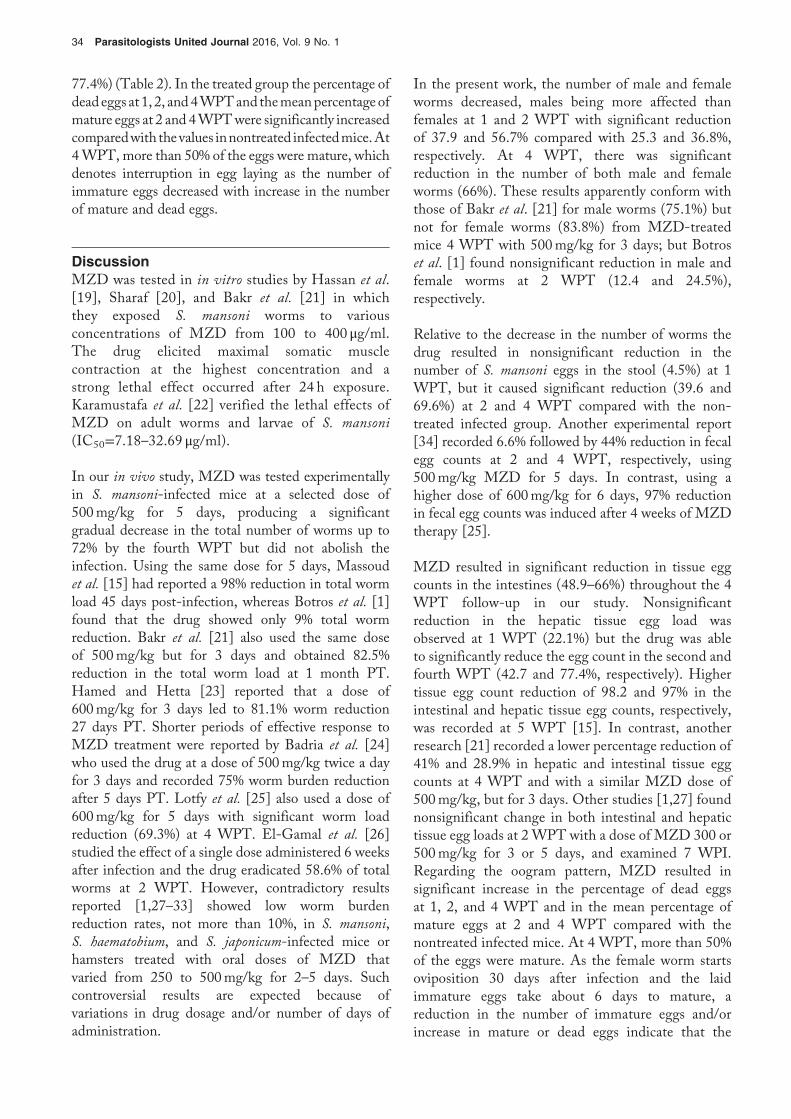
ResultsMZD resulted in a nonsignificant reduction in thenumber of S. mansoni eggs in the stool of the treatedgroup (4.5%) 1WPT followed by significant reduction of39.5 and 69.6% at 2 and 4WPT, respectively, comparedwith the nontreated infected group (Fig. 2 and Table 1).
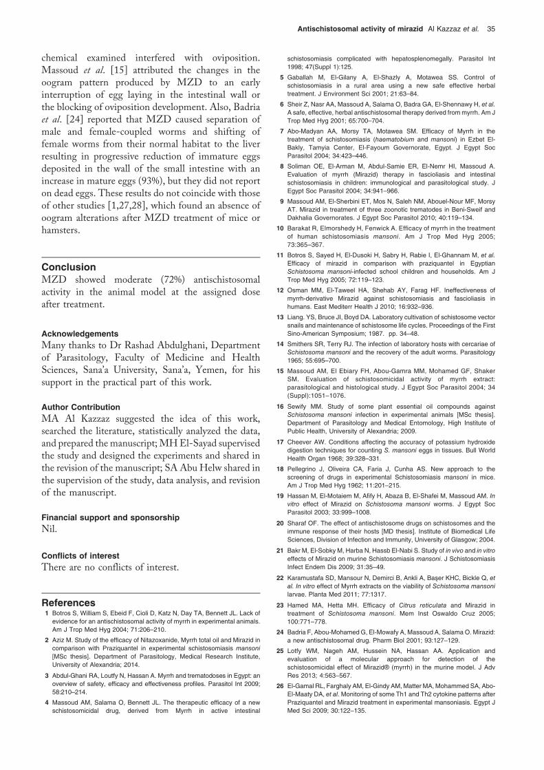
MZD significantly reduced the total worm burden by 34,50, and 72% at 1, 2, and 4 WPT, respectively (Table 1).The number of male and female worms decreased, malesbeing more affected than females at 1 and 2 WPT withsignificant reduction of 37.9 and 56.7% compared with25.3 and 36.8%, respectively. At 4 WPT, there wassignificant reduction of both male (75.1%) and femaleworms (60%) (Table 1).
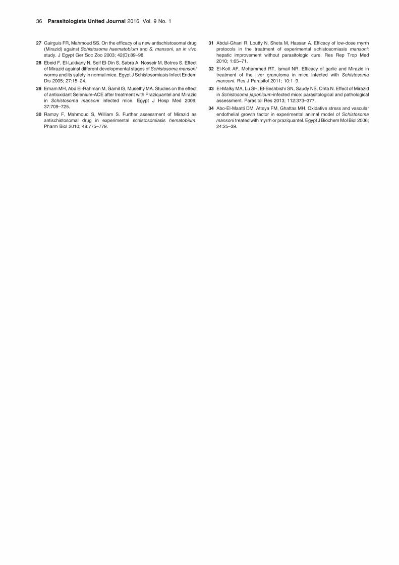
MZD produced significant reduction in the tissue eggcounts in the intestine (29.1–66%) throughout thefollow-up weeks. Reduction in the hepatic tissue eggload was nonsignificant at 1WPT (22.1%), and becamesignificant in the second and fourth week (42.7 and
Figure 2
Egg counts in the stool of MZD-treated Schistosoma mansoni-infectedmice compared with nontreated infectedmice. MZD,mirazid.
Table 1 Effect of mirazid on fecal egg counts, total worm burden, and worm sex in Schistosoma mansoni-infected treated micecompared with nontreated infected mice according to WPT
Group WPT Fecal egg counts Total worms Total males Total females
Group 1: nontreated 1 566.00±55.05 19.60±4.51 12.9±0.55 6.70±1.00
2 765.00±70.83 28.25±5.19 18.50±1.50 9.75±1.50
4 720.00±62.11 28.50±3.54 18.50±0.70 10.00±0.71
Group 2: mirazid treated 1 540.00±42.74(4.5) 13.00±2.71a(34) 8.00±1.53a(37.9) 5.00±0.58a(25.3)
2 462.50±39.09a(39.6) 14.16±2.63a(50) 8.00±1.00a(56.7) 6.16±0.58a(36.8)
4 218.75±12.97a(69.6) 8.00±3.79a(72) 4.60±1.53a(75.1) 3.40±1.15a(66)
Values were expressed as mean±SD; numbers in parentheses indicate the percentage of reduction compared with the infectednontreated group. WPT, weeks post-treatment. aStatistically significant at P<0.05.
Change in treated ð%Þ ¼ mean values in nontreated ðcÞ �mea n values in treated ðtÞmean values in nontreated ðcÞ × 100
Table 2 Effect of mirazid on the mean tissue egg counts and the oogram pattern in Schistosoma mansoni-infected mice at 1, 2,and 4 WPT compared with nontreated mice
Group WPT Tissue egg counts Oogram pattern
Intestine Liver Immature Mature Dead
Group 1: nontreated 1 10.3±1.13 4.56±2.84 59.60±2.28 32.60±1.99 7.80±0.59
2 14.6±1.23 6.29±3.59 61.50±3.00 27.00±0.16 11.50±0.38
4 16.5±2.76 7.82±5.80 62.50±0.71 24.50±0.12 12.00±0.41
Group 2: mirazid treated 1 7.30±1.3a(29.1) 3.55±1.28(22.1) 55.50±4.42 19.37±2.16a 24.83±2.62a
2 7.46±0.5a(48.9) 3.60±0.22a(42.7) 23.25±0.99a 39.75±1.71a 37.00±2.55a
4 5.62±0.57a(66) 1.76±0.20a(77.4) 10.77±0.52a 52.00±3.00a 37.33±2.52a
Tissue egg count (EPG)×103 (%R). EPG, eggs per gram; WPT, weeks post-treatment. aStatistically significant at P<0.05.
Antischistosomal activity of mirazid Al Kazzaz et al. 33
77.4%) (Table 2). In the treated group the percentage ofdeadeggs at 1, 2, and4WPTand themeanpercentage ofmature eggs at 2 and 4WPTwere significantly increasedcomparedwith thevalues innontreated infectedmice.At4WPT, more than 50% of the eggs were mature, whichdenotes interruption in egg laying as the number ofimmature eggs decreased with increase in the numberof mature and dead eggs.
DiscussionMZD was tested in in vitro studies by Hassan et al.[19], Sharaf [20], and Bakr et al. [21] in whichthey exposed S. mansoni worms to variousconcentrations of MZD from 100 to 400 μg/ml.The drug elicited maximal somatic musclecontraction at the highest concentration and astrong lethal effect occurred after 24 h exposure.Karamustafa et al. [22] verified the lethal effects ofMZD on adult worms and larvae of S. mansoni
(IC50=7.18–32.69 μg/ml).
In our in vivo study, MZD was tested experimentallyin S. mansoni-infected mice at a selected dose of500mg/kg for 5 days, producing a significantgradual decrease in the total number of worms up to72% by the fourth WPT but did not abolish theinfection. Using the same dose for 5 days, Massoudet al. [15] had reported a 98% reduction in total wormload 45 days post-infection, whereas Botros et al. [1]found that the drug showed only 9% total wormreduction. Bakr et al. [21] also used the same doseof 500mg/kg but for 3 days and obtained 82.5%reduction in the total worm load at 1 month PT.Hamed and Hetta [23] reported that a dose of600mg/kg for 3 days led to 81.1% worm reduction27 days PT. Shorter periods of effective response toMZD treatment were reported by Badria et al. [24]who used the drug at a dose of 500mg/kg twice a dayfor 3 days and recorded 75% worm burden reductionafter 5 days PT. Lotfy et al. [25] also used a dose of600mg/kg for 5 days with significant worm loadreduction (69.3%) at 4 WPT. El-Gamal et al. [26]studied the effect of a single dose administered 6 weeksafter infection and the drug eradicated 58.6% of totalworms at 2 WPT. However, contradictory resultsreported [1,27–33] showed low worm burdenreduction rates, not more than 10%, in S. mansoni,S. haematobium, and S. japonicum-infected mice orhamsters treated with oral doses of MZD thatvaried from 250 to 500mg/kg for 2–5 days. Suchcontroversial results are expected because ofvariations in drug dosage and/or number of days ofadministration.
In the present work, the number of male and femaleworms decreased, males being more affected thanfemales at 1 and 2 WPT with significant reductionof 37.9 and 56.7% compared with 25.3 and 36.8%,respectively. At 4 WPT, there was significantreduction in the number of both male and femaleworms (66%). These results apparently conform withthose of Bakr et al. [21] for male worms (75.1%) butnot for female worms (83.8%) from MZD-treatedmice 4 WPT with 500mg/kg for 3 days; but Botroset al. [1] found nonsignificant reduction in male andfemale worms at 2 WPT (12.4 and 24.5%),respectively.
Relative to the decrease in the number of worms thedrug resulted in nonsignificant reduction in thenumber of S. mansoni eggs in the stool (4.5%) at 1WPT, but it caused significant reduction (39.6 and69.6%) at 2 and 4 WPT compared with the non-treated infected group. Another experimental report[34] recorded 6.6% followed by 44% reduction in fecalegg counts at 2 and 4 WPT, respectively, using500mg/kg MZD for 5 days. In contrast, using ahigher dose of 600mg/kg for 6 days, 97% reductionin fecal egg counts was induced after 4 weeks of MZDtherapy [25].
MZD resulted in significant reduction in tissue eggcounts in the intestines (48.9–66%) throughout the 4WPT follow-up in our study. Nonsignificantreduction in the hepatic tissue egg load wasobserved at 1 WPT (22.1%) but the drug was ableto significantly reduce the egg count in the second andfourth WPT (42.7 and 77.4%, respectively). Highertissue egg count reduction of 98.2 and 97% in theintestinal and hepatic tissue egg counts, respectively,was recorded at 5 WPT [15]. In contrast, anotherresearch [21] recorded a lower percentage reduction of41% and 28.9% in hepatic and intestinal tissue eggcounts at 4 WPT and with a similar MZD dose of500mg/kg, but for 3 days. Other studies [1,27] foundnonsignificant change in both intestinal and hepatictissue egg loads at 2 WPT with a dose of MZD 300 or500mg/kg for 3 or 5 days, and examined 7 WPI.Regarding the oogram pattern, MZD resulted insignificant increase in the percentage of dead eggsat 1, 2, and 4 WPT and in the mean percentage ofmature eggs at 2 and 4 WPT compared with thenontreated infected mice. At 4 WPT, more than 50%of the eggs were mature. As the female worm startsoviposition 30 days after infection and the laidimmature eggs take about 6 days to mature, areduction in the number of immature eggs and/orincrease in mature or dead eggs indicate that the
34 Parasitologists United Journal 2016, Vol. 9 No. 1
chemical examined interfered with oviposition.Massoud et al. [15] attributed the changes in theoogram pattern produced by MZD to an earlyinterruption of egg laying in the intestinal wall orthe blocking of oviposition development. Also, Badriaet al. [24] reported that MZD caused separation ofmale and female-coupled worms and shifting offemale worms from their normal habitat to the liverresulting in progressive reduction of immature eggsdeposited in the wall of the small intestine with anincrease in mature eggs (93%), but they did not reporton dead eggs. These results do not coincide with thoseof other studies [1,27,28], which found an absence ofoogram alterations after MZD treatment of mice orhamsters.
ConclusionMZD showed moderate (72%) antischistosomalactivity in the animal model at the assigned doseafter treatment.
AcknowledgementsMany thanks to Dr Rashad Abdulghani, Departmentof Parasitology, Faculty of Medicine and HealthSciences, Sana’a University, Sana’a, Yemen, for hissupport in the practical part of this work.
Author ContributionMA Al Kazzaz suggested the idea of this work,searched the literature, statistically analyzed the data,and prepared themanuscript;MHEl-Sayad supervisedthe study and designed the experiments and shared inthe revision of the manuscript; SA AbuHelw shared inthe supervision of the study, data analysis, and revisionof the manuscript.
Financial support and sponsorshipNil.
Conflicts of interestThere are no conflicts of interest.
References1 Botros S, William S, Ebeid F, Cioli D, Katz N, Day TA, Bennett JL. Lack of
evidence for an antischistosomal activity of myrrh in experimental animals.Am J Trop Med Hyg 2004; 71:206–210.
2 Aziz M. Study of the efficacy of Nitazoxanide, Myrrh total oil and Mirazid incomparison with Praziquantel in experimental schistosomiasis mansoni[MSc thesis]. Department of Parasitology, Medical Research Institute,University of Alexandria; 2014.
3 Abdul-Ghani RA, Loutfy N, Hassan A. Myrrh and trematodoses in Egypt: anoverview of safety, efficacy and effectiveness profiles. Parasitol Int 2009;58:210–214.
4 Massoud AM, Salama O, Bennett JL. The therapeutic efficacy of a newschistosomicidal drug, derived from Myrrh in active intestinal
schistosomiasis complicated with hepatosplenomegally. Parasitol Int1998; 47(Suppl 1):125.
5 Gaballah M, El-Gilany A, El-Shazly A, Motawea SS. Control ofschistosomiasis in a rural area using a new safe effective herbaltreatment. J Environment Sci 2001; 21:63–84.
6 Sheir Z, Nasr AA, Massoud A, Salama O, Badra GA, El-Shennawy H, et al.A safe, effective, herbal antischistosomal therapy derived frommyrrh. Am JTrop Med Hyg 2001; 65:700–704.
7 Abo-Madyan AA, Morsy TA, Motawea SM. Efficacy of Myrrh in thetreatment of schistosomiasis (haematobium and mansoni) in Ezbet El-Bakly, Tamyia Center, El-Fayoum Governorate, Egypt. J Egypt SocParasitol 2004; 34:423–446.
8 Soliman OE, El-Arman M, Abdul-Samie ER, El-Nemr HI, Massoud A.Evaluation of myrrh (Mirazid) therapy in fascioliasis and intestinalschistosomiasis in children: immunological and parasitological study. JEgypt Soc Parasitol 2004; 34:941–966.
9 Massoud AM, El-Sherbini ET, Mos N, Saleh NM, Abouel-Nour MF, MorsyAT. Mirazid in treatment of three zoonotic trematodes in Beni-Sweif andDakhalia Governorates. J Egypt Soc Parasitol 2010; 40:119–134.
10 Barakat R, Elmorshedy H, Fenwick A. Efficacy of myrrh in the treatmentof human schistosomiasis mansoni. Am J Trop Med Hyg 2005;73:365–367.
11 Botros S, Sayed H, El-Dusoki H, Sabry H, Rabie I, El-Ghannam M, et al.Efficacy of mirazid in comparison with praziquantel in EgyptianSchistosoma mansoni-infected school children and households. Am JTrop Med Hyg 2005; 72:119–123.
12 Osman MM, El-Taweel HA, Shehab AY, Farag HF. Ineffectiveness ofmyrrh-derivative Mirazid against schistosomiasis and fascioliasis inhumans. East Mediterr Health J 2010; 16:932–936.
13 Liang. YS, Bruce JI, Boyd DA. Laboratory cultivation of schistosome vectorsnails and maintenance of schistosome life cycles. Proceedings of the FirstSino-American Symposium; 1987. pp. 34–48.
14 Smithers SR, Terry RJ. The infection of laboratory hosts with cercariae ofSchistosoma mansoni and the recovery of the adult worms. Parasitology1965; 55:695–700.
15 Massoud AM, El Ebiary FH, Abou-Gamra MM, Mohamed GF, ShakerSM. Evaluation of schistosomicidal activity of myrrh extract:parasitological and histological study. J Egypt Soc Parasitol 2004; 34(Suppl):1051–1076.
16 Sewify MM. Study of some plant essential oil compounds againstSchistosoma mansoni infection in experimental animals [MSc thesis].Department of Parasitology and Medical Entomology, High Institute ofPublic Health, University of Alexandria; 2009.
17 Cheever AW. Conditions affecting the accuracy of potassium hydroxidedigestion techniques for counting S. mansoni eggs in tissues. Bull WorldHealth Organ 1968; 39:328–331.
18 Pellegrino J, Oliveira CA, Faria J, Cunha AS. New approach to thescreening of drugs in experimental Schistosomiasis mansoni in mice.Am J Trop Med Hyg 1962; 11:201–215.
19 Hassan M, El-Motaiem M, Afify H, Abaza B, El-Shafei M, Massoud AM. Invitro effect of Mirazid on Schistosoma mansoni worms. J Egypt SocParasitol 2003; 33:999–1008.
20 Sharaf OF. The effect of antischistosome drugs on schistosomes and theimmune response of their hosts [MD thesis]. Institute of Biomedical LifeSciences, Division of Infection and Immunity, University of Glasgow; 2004.
21 Bakr M, El-Sobky M, Harba N, Hassb El-Nabi S. Study of in vivo and in vitroeffects of Mirazid on murine Schistosomiasis mansoni. J SchistosomiasisInfect Endem Dis 2009; 31:35–49.
22 Karamustafa SD, Mansour N, Demirci B, Ankli A, Baser KHC, Bickle Q, etal. In vitro effect of Myrrh extracts on the viability of Schistosoma mansonilarvae. Planta Med 2011; 77:1317.
23 Hamed MA, Hetta MH. Efficacy of Citrus reticulata and Mirazid intreatment of Schistosoma mansoni. Mem Inst Oswaldo Cruz 2005;100:771–778.
24 Badria F, Abou-MohamedG, El-Mowafy A, Massoud A, Salama O. Mirazid:a new antischistosomal drug. Pharm Biol 2001; 93:127–129.
25 Lotfy WM, Nageh AM, Hussein NA, Hassan AA. Application andevaluation of a molecular approach for detection of theschistosomicidal effect of Mirazid® (myrrh) in the murine model. J AdvRes 2013; 4:563–567.
26 El-Gamal RL, Farghaly AM, El-Gindy AM,Matter MA, Mohammed SA, Abo-El-Maaty DA, et al.Monitoring of some Th1 and Th2 cytokine patterns afterPraziquantel and Mirazid treatment in experimental mansoniasis. Egypt JMed Sci 2009; 30:122–135.
Antischistosomal activity of mirazid Al Kazzaz et al. 35
27 Guirguis FR, Mahmoud SS. On the efficacy of a new antischistosomal drug(Mirazid) against Schistosoma haematobium and S. mansoni, an in vivostudy. J Egypt Ger Soc Zoo 2003; 42(D):89–98.
28 Ebeid F, El-Lakkany N, Seif El-Din S, Sabra A, Nosseir M, Botros S. Effectof Mirazid against different developmental stages of Schistosoma mansoniworms and its safety in normal mice. Egypt J Schistosomiasis Infect EndemDis 2005; 27:15–24.
29 EmamMH, Abd El-RahmanM, Gamil IS, MuselhyMA. Studies on the effectof antioxidant Selenium-ACE after treatment with Praziquantel and Mirazidin Schistosoma mansoni infected mice. Egypt J Hosp Med 2009;37:709–725.
30 Ramzy F, Mahmoud S, William S. Further assessment of Mirazid asantischistosomal drug in experimental schistosomiasis hematobium.Pharm Biol 2010; 48:775–779.
31 Abdul-Ghani R, Loutfy N, Sheta M, Hassan A. Efficacy of low-dose myrrhprotocols in the treatment of experimental schistosomiasis mansoni:hepatic improvement without parasitologic cure. Res Rep Trop Med2010; 1:65–71.
32 El-Kott AF, Mohammed RT, Ismail NR. Efficacy of garlic and Mirazid intreatment of the liver granuloma in mice infected with Schistosomamansoni. Res J Parasitol 2011; 10:1–9.
33 El-Malky MA, Lu SH, El-Beshbishi SN, Saudy NS, Ohta N. Effect of Mirazidin Schistosoma japonicum-infected mice: parasitological and pathologicalassessment. Parasitol Res 2013; 112:373–377.
34 Abo-El-Maatti DM, Atteya FM, Ghattas MH. Oxidative stress and vascularendothelial growth factor in experimental animal model of Schistosomamansoni treated withmyrrh or praziquantel. Egypt J BiochemMol Biol 2006;24:25–39.
36 Parasitologists United Journal 2016, Vol. 9 No. 1