Antioxidant and anticancer activities of Trigonella foenum ...
12
RESEARCH ARTICLE Open Access Antioxidant and anticancer activities of Trigonella foenum-graecum, Cassia acutifolia and Rhazya stricta Bayan Al-Dabbagh 1* , Ismail A. Elhaty 1 , Ala’a Al Hrout 2 , Reem Al Sakkaf 1 , Raafat El-Awady 4 , S. Salman Ashraf 1 and Amr Amin 2,3* Abstract Background: Here, we determined in vitro antioxidant activity, total phenols and flavonoids and evaluated antiproliferative activity of three medicinal plant extracts: Trigonella foenum-graecum (Fenugreek), Cassia acutifolia (Senna) and Rhazya stricta (Harmal). Methods: The leaves of the three medicinal plants were extracted with 70% ethanol. Antioxidant activities of the extracts were determined by using DPPH (1,1-diphenyl-2-picrylhydrazyl) assay. Total flavonoid and phenolic contents were determined using colorimetric assays. MTT assay was used to estimate the antiproliferative activities of the extracts against human hepatoma (HepG2) cancer cell line. In addition, the effects of R. stricta extract on cell cycle, colony formation, and wound healing of HepG2 cells and tube formation of HUVEC cells were assessed. Results: Percentage inhibition of DPPH scavenging activity were dose-dependent and ranged between (89.9% ± 0.51) and (28.6% ± 2.07). Phenolic contents ranged between (11.5 ± 0.013) and (9.7 ± 0.008) mg GAE/g while flavonoid content ranged between (20.8 ± 0.40) and (0.12 ± 0.0.01) mg QE/g. Antiproliferative results of the extracts were found to be consistent with their antioxidant activity. Among the extracts evaluated, that of R. stricta showed the best antioxidant, antiproliferative and antimetastatic activities at low concentration. It also inhibited the colony-formation capacity of HepG2 cells and exhibited antiangiogenic activity. Cell cycle analysis showed significant arrest of cells at G2/M phase 12 and 48 h after treatment and significant arrest at G1/S phase after 24 h of treatment. Consistent data were observed in western blot analysis of protein levels of Cdc2 and its cyclin partners. Conclusions: These findings introduce R. stricta as a potentially useful anti-metastatic agent and a novel potential anti- tumour agent for hepatocellular carcinoma (HCC) treatment. Keywords: Antioxidants, Anticancer, Medicinal plants, Traditional medicine, Rhazya stricta Background Free radicals are mainly produced by oxidation processes and they have an important role in the processes of food spoilage and chemical materials degradation. They also contribute to human disorders such as aging-associated diseases, cardiovascular diseases, cancer and inflamma- tory diseases [1, 2]. Free radicals may also cause a deple- tion of the immune system antioxidants, a change in the gene expression and may induce the synthesis of abnormal proteins. About 5% or more of the inhaled oxygen (O 2 ) is converted to reactive oxygen species (ROS) such as O 2 - , H 2 O 2 , and OH radicals. ROS represents the major type of free radicals in any biological system. They are produced through the mitochondrial electrons transport chain [2, 3]. Antioxidants are used to neutralize the effects of free rad- icals. Thus, they protect humans against infection and de- generative diseases. Antioxidants can be classified into two major categories, natural and synthetic. Synthetic antioxi- dants include butylated hydroxy anisole (BHA), butylated hydroxyl toluenes (BHT), tertiary butylated hydroquinone and gallic acid esters. These antioxidants effectively inhibit * Correspondence: [email protected]; [email protected] 1 Department of Chemistry, College of Science, UAE University, PO Box 15551, Al Ain, UAE 2 Department of Biology, College of Science, UAE University, PO Box 15551, Al Ain, UAE Full list of author information is available at the end of the article © The Author(s). 2018 Open Access This article is distributed under the terms of the Creative Commons Attribution 4.0 International License (http://creativecommons.org/licenses/by/4.0/), which permits unrestricted use, distribution, and reproduction in any medium, provided you give appropriate credit to the original author(s) and the source, provide a link to the Creative Commons license, and indicate if changes were made. The Creative Commons Public Domain Dedication waiver (http://creativecommons.org/publicdomain/zero/1.0/) applies to the data made available in this article, unless otherwise stated. Al-Dabbagh et al. BMC Complementary and Alternative Medicine (2018) 18:240 https://doi.org/10.1186/s12906-018-2285-7
Transcript of Antioxidant and anticancer activities of Trigonella foenum ...
RESEARCH ARTICLE Open Access
Antioxidant and anticancer activities ofTrigonella foenum-graecum, Cassia acutifoliaand Rhazya strictaBayan Al-Dabbagh1*, Ismail A. Elhaty1, Ala’a Al Hrout2, Reem Al Sakkaf1, Raafat El-Awady4, S. Salman Ashraf1
and Amr Amin2,3*
Abstract
Background: Here, we determined in vitro antioxidant activity, total phenols and flavonoids and evaluatedantiproliferative activity of three medicinal plant extracts: Trigonella foenum-graecum (Fenugreek), Cassia acutifolia(Senna) and Rhazya stricta (Harmal).
Methods: The leaves of the three medicinal plants were extracted with 70% ethanol. Antioxidant activities of theextracts were determined by using DPPH (1,1-diphenyl-2-picrylhydrazyl) assay. Total flavonoid and phenolic contentswere determined using colorimetric assays. MTT assay was used to estimate the antiproliferative activities of theextracts against human hepatoma (HepG2) cancer cell line. In addition, the effects of R. stricta extract on cell cycle,colony formation, and wound healing of HepG2 cells and tube formation of HUVEC cells were assessed.
Results: Percentage inhibition of DPPH scavenging activity were dose-dependent and ranged between (89.9% ± 0.51)and (28.6% ± 2.07). Phenolic contents ranged between (11.5 ± 0.013) and (9.7 ± 0.008) mg GAE/g while flavonoid contentranged between (20.8 ± 0.40) and (0.12 ± 0.0.01) mg QE/g. Antiproliferative results of the extracts were found to beconsistent with their antioxidant activity. Among the extracts evaluated, that of R. stricta showed the best antioxidant,antiproliferative and antimetastatic activities at low concentration. It also inhibited the colony-formation capacity ofHepG2 cells and exhibited antiangiogenic activity. Cell cycle analysis showed significant arrest of cells at G2/M phase 12and 48 h after treatment and significant arrest at G1/S phase after 24 h of treatment. Consistent data were observed inwestern blot analysis of protein levels of Cdc2 and its cyclin partners.
Conclusions: These findings introduce R. stricta as a potentially useful anti-metastatic agent and a novel potential anti-tumour agent for hepatocellular carcinoma (HCC) treatment.
Keywords: Antioxidants, Anticancer, Medicinal plants, Traditional medicine, Rhazya stricta
BackgroundFree radicals are mainly produced by oxidation processesand they have an important role in the processes of foodspoilage and chemical materials degradation. They alsocontribute to human disorders such as aging-associateddiseases, cardiovascular diseases, cancer and inflamma-tory diseases [1, 2]. Free radicals may also cause a deple-tion of the immune system antioxidants, a change in the
gene expression and may induce the synthesis of abnormalproteins. About 5% or more of the inhaled oxygen (O2) isconverted to reactive oxygen species (ROS) such as O2
−,H2O2, and OH radicals. ROS represents the major type offree radicals in any biological system. They are producedthrough the mitochondrial electrons transport chain [2, 3].Antioxidants are used to neutralize the effects of free rad-
icals. Thus, they protect humans against infection and de-generative diseases. Antioxidants can be classified into twomajor categories, natural and synthetic. Synthetic antioxi-dants include butylated hydroxy anisole (BHA), butylatedhydroxyl toluenes (BHT), tertiary butylated hydroquinoneand gallic acid esters. These antioxidants effectively inhibit
* Correspondence: [email protected]; [email protected] of Chemistry, College of Science, UAE University, PO Box 15551,Al Ain, UAE2Department of Biology, College of Science, UAE University, PO Box 15551, AlAin, UAEFull list of author information is available at the end of the article
© The Author(s). 2018 Open Access This article is distributed under the terms of the Creative Commons Attribution 4.0International License (http://creativecommons.org/licenses/by/4.0/), which permits unrestricted use, distribution, andreproduction in any medium, provided you give appropriate credit to the original author(s) and the source, provide a link tothe Creative Commons license, and indicate if changes were made. The Creative Commons Public Domain Dedication waiver(http://creativecommons.org/publicdomain/zero/1.0/) applies to the data made available in this article, unless otherwise stated.
Al-Dabbagh et al. BMC Complementary and Alternative Medicine (2018) 18:240 https://doi.org/10.1186/s12906-018-2285-7
oxidation, may serve as chelating agents such as ethylenediamine tetra acetic acid (EDTA), and can bind metals re-ducing their contribution to the process [4]. However, anti-oxidants are thought to cause or promote negative healtheffects such as mutagenesis and carcinogenesis in humans[5]. Therefore, there is a strong trend to replace the syn-thetic with naturally occurring antioxidants that can pre-vent free radical-related diseases [4, 6].Natural antioxidants help in controlling the formation of
free radicals and activated oxygen species or they can inhibittheir reaction with biological structures [7]. These antioxi-dants include antioxidative enzymes, such as superoxide dis-mutase, catalase, and glutathione peroxidase, and smallnonenzymatic antioxidant molecules, such as glutathioneand vitamins C and E [8]. Many herbs and spices (rosemary,thyme, oregano, sage, basil, pepper, clove, cinnamon, nut-meg, and saffron), and plant extracts (tea, grapeseed, andlemon balm) contain antioxidant components [9–11].In this study, three hydroalcoholic extracts of tradition-
ally medicinal plants used in the United Arab Emirates(UAE) were evaluated for their antioxidant activities, phe-nol and flavonoid contents. These plants are, Trigonellafoenum-graecum, locally known as “Helba”, Cassia acutifo-lia, commonly known as “Holoul” and Rhazya strictawhich is locally called “Harmal”.T. foenum-graecum seeds are traditionally used as
herbal medicine for their carminative, tonic, aphrodisiacand anticancer effects [12–14]. The leaves of C. acutifo-lia is frequently used in folk medicine as a purgative fora long time [15]. The extracts of R. stricta leaves aretraditionally used for the treatment of various disorderssuch as diabetes, sore throat, helminthiasis, inflamma-tory conditions and rheumatism [16].Available treatment for hepatocellular carcinoma are
mainly limited to invasive hepatectomy or chemotherapy.However, the attention has shifted in recent years tonatural-based products for candidate anticancer therapeu-tics. In the present study, the antiproliferative effects ofTrigonella foenum-graecum, Cassia acutifolia, and Rhazyastricta on hepatoma cell line HepG2 were investigated.The use of HepG2 cells to test the cytotoxic effects of awide range of drugs has been well documented, due totheir wide availability, well-differentiation, and drug me-tabolizing activity [17].Despite playing a key role in cellular processes, free radi-
cals pose a threat to cells by damaging DNA, proteins,and cellular membranes, leading to onset of many diseasesincluding cancer [18, 19]. Thus, by decreasing free radicalsand oxidative stress, antioxidants play a role in ameliorat-ing DNA damage, reducing the rate of abnormal cell div-ision, and decreasing mutagenesis [20]. Therefore, manyantioxidant-rich plants possess anticancer activity [21–23].Vascular endothelial growth factor (VEGF) has been
recognized to be involved in several stages of angiogenesis
in malignant diseases by its multi-functional effects in ac-tivating and integrating signalling pathway networks [24].VEGF signalling blockade reduces new vessel growth andleads to endothelial cell apoptosis. Therefore, using tyro-sine kinase inhibitors or VEGF/VEGF receptor (VEGFR)antibodies to inhibit crucial angiogenic steps is a practicaltherapeutic strategy when treating neovascularisation dis-eases [25]. A potent angiogenesis inhibitor known as E7820,has been shown to reduce integrin α 2 mRNA expressionand inhibit basic fibroblast growth factor/VEGF-inducedHUVEC proliferation and tube formation [26, 27]. Integrinα 2β 1/α 1β 1 expression is reportedly regulated by VEGFand an inhibitory antibody against α 2β 1/α 1β 1 has beenshown to inhibit angiogenesis and tumour growth inVEGF-overexpressing tumour cells [28, 29]. Therefore, weinvestigated here the effect of R. stricta leaves extract onangiogenesis utilizing HUVEC tube formation assay; as itshowed the most promising antiproliferative activity. Inaddition, this work was set to determine the in vitro antioxi-dant activity, total phenols and flavonoids, anticancer activ-ities of tested plants with special interest in R. stricta.
MethodsChemicalsAll solvents were analytical grade. Agilent Cary 60UV-Vis Spectrophotometer was used in all spectro-photometric measurements. Ascorbic acid, ferric chlor-ide, aluminium chloride, potassium acetate, quercetin,DPPH reagent, Folin-Ciocalteau reagent, gallic acid, so-dium carbonate, methanol and ethanol were obtainedfrom Sigma Chemical Co. (St. Louis, MO, USA). Milli-pore deionized water was used throughout. ThiazolylBlue Tetrazolium Bromide (Sigma Aldrich, USA), Di-methyl Sulfoxide (Sigma Aldrich, USA).
Plant samplesDried leaves of C. acutifolia, R. stricta and T. foenum-graecum were purchased from the local market. Thetaxonomic authentication of all the plants was carriedout by Dr. Fatima Al-Ansari at the Biology Department,College of Science, United Arab Emirates University.Voucher specimens were deposited at the herbarium ofthe Biology Department (voucher reference numbers:BA2018–1, BA2018–2, BA2018–3).
Preparation of plant extractsThe leaves of the medicinal plants were crushed separ-ately in a grinder. A sample of 10 g of each plant was ex-tracted with 150 mL of 70% ethanol and 30% water as itshowed the best extraction yield [30]. The crushedplants were macerated for 48 h at 4 °C. The resultingmixture was then filtered under vacuum and concen-trated under reduced pressure in a rotary evaporator at40 °C. The extracts were further dried using a TELSTAR
Al-Dabbagh et al. BMC Complementary and Alternative Medicine (2018) 18:240 Page 2 of 12

CRYODOS freeze dryer machine then kept at − 20 °C forfurther analysis. A solution of 30 mg/mL of each plantwas prepared in 50% ethanol for the following tests.
Determination of total polyphenol contentThe total phenolic content (TPC) was determined byusing the Folin-Ciocalteau reagent [31]. A 10% solutionwas prepared from the stock solution (30 mg/mL) using50% ethanol. 100 μl of this solution was mixed with200 μl of the Folin-Ciocalteau reagent and 2 mL ofde-ionized water then incubated at room temperaturefor 3 min. A sample of 20% aqueous sodium carbonate(w/w, 1 mL) was then added to the mixture. The totalpolyphenols were determined after 1 h of incubation atroom temperature. A negative control sample was alsoprepared using the same procedure. The absorbance ofthe resulting blue colour was measured at 765 nm. Re-sults were expressed in mg gallic acid equivalents (GAE)per g dry weight of plant material using an equation ob-tained from gallic acid calibration curve. The sampleswere analyzed in triplicate.
Free-radical scavenging activityThe antioxidant activity of the extracts was assessedbased on their ability to scavenge the stable 1,1-diphe-nyl-2-picrylhydrazyl (DPPH) radical as described previ-ously [32]. Various concentrations of the three extractsin methanol were prepared (0.15 to 1.5 mg/mL). Amethanolic solution of DPPH (3.8 mL, 60 μg/mL) wasrapidly mixed with the plant extract (200 μl, 30 mg/mL)in a test tube, with methanol serving as the blank sampleand a control was also assayed simultaneously. The con-tents of the tubes were swirled then allowed to stand for30 min at room temperature in the dark. The absorb-ance was measured at 517 nm in a spectrophotometer.The scavenging ability of the plant extract was calculatedusing this equation: DPPH Scavenging activity (%) = [(Abscontrol–Abs sample)]/ (Abs control)] × 100, where Abscontrol is the absorbance of DPPH + methanol; Abs sam-ple is the absorbance of DPPH radical + sample (sampleor standard). The EC50 value (μg/mL), the effective con-centration at which DPPH· radicals are scavenged by 50%,was determined graphically. The total antioxidant activitywas expressed as ascorbic acid equivalent/g dry extract.The assay was done in triplicates.
Determination of total flavonoidsThe total flavonoids content in the extracts was deter-mined using the aluminium chloride colorimetricmethod [33]. A known concentration (600 μg/mL) ofeach extract in methanol was prepared. A 500 μl of theextracts were mixed separately with 0.1 mL of 10% (w/v)aluminium chloride solution, 0.1 mL of 1 M potassiumacetate solution, 1.5 mL of methanol and 2.8 mL of
distilled water. The solutions were thoroughly mixedand incubated at room temperature for 30 min. The ab-sorbance of the reaction mixture was measured at415 nm using a spectrophotometer. The total flavonoidscontent was determined using a standard curve withquercetin (1 to 25 μg/mL) as the standard. The mean ofthree readings was used and expressed as mg of quer-cetin equivalents (QE)/ g of the dry extract.
Cell cultureA human hepatocellular carcinoma (HCC)-derived cellline (HepG2) was cultured in RPMI 1640 medium con-taining 1% antibiotic cocktail and supplemented with10% fetal bovine serum. Cells were incubated at 37 °C in5% CO2 humidified incubator. Cells were passaged every2–3 days using 0.25% trypsin-EDTA.
Cytotoxicity assayHepG2 were seeded at a density of 5000 cells/well in a96-well plate, and were allowed to attach overnight. There-after, cells were treated with various concentrations of theplants extracts for 24 h. To assess the cytotoxic effect ofthe three plants extracts, MTT (3-[4,5-dimethylthiazo-l-2-yl]-2,5-diphenyltratrazolium bromide) assay was carriedout. Briefly, cells treated with the plant extracts were ex-posed to tetrazolium MTT at a concentration of 5 mg/mL.Viable active cells reduced yellow MTT salt to insolublepurple formazan, which was dissolved using DMSO. Theabsorbance of the coloured solution was measured at awavelength of 570 nm using Epoch microplate spectropho-tometer (BioTek). The obtained absorbance at 570 nm ofboth control and treated cells was used to calculate per-centage of cell viability. Assuming 100% viability in controlcells, percentage of treated cells viability will be calculatedaccordingly:
Percent of viable cells
¼ Abs: of treated cells=Abs: of control cellsð Þ � 100
Assessment of morphological changesHepG2 were seeded at a density of 0.25 × 10⁶ cells/ wellin a 6-well plate, and were allowed to attach overnight.After which, cells were treated without (0 μg, control) orwith increasing concentrations of R. stricta (10, 20, 30,50, 70 μg) for 24 h. The morphology of the cells wasassessed after being fixed and stained with 0.5% crystalviolet using bright-field microscopy (200 x magnifica-tion, scale = 200 μm).
Colony formation assayTo assess the effects of R. stricta on cell survival, thecolony formation assay was carried out in vitro. Briefly,HepG2 cells were seeded at a density of 1000 cells/ well
Al-Dabbagh et al. BMC Complementary and Alternative Medicine (2018) 18:240 Page 3 of 12

in a 6-well plate, and were incubate for 24 h to allow at-tachment. The second day, the cells were treated with-out (0 μg, control) or with increasing concentrations(10, 20, 30 μg) of the extract for 24 h. After which, themedia was replaced with fresh complete growth mediawithout the extract, and cells were left to incubate untilvisible colonies were formed; while changing the mediaevery 3–4 days. The experiment was carried out in tripli-cates. Colonies were fixed with absolute methanol, thenstained with 0.5% crystal violet. Results are representedas the percentage of the well area that is covered by col-onies (colony area percentage). Analysis has been carriedout using ImageJ plugin ColonyArea [34]. In addition,an absorption-based method was carried out to validatethe earlier results, by which the absorption of the crystalviolet dye in each well is measured after being dissolved.Briefly, the samples that had been analysed using ImageJwere subjected to 10% acetic acid solution, then wereplaced on an orbital shaker for 15 min. After which,100 μL of each triplicate sample was transferred to a96-well plate (in triplicates), and absorbance was measuredusing Epoch microplate spectrophotometer (BioTek).
Wound-healing assayTo assess the ability of HepG2 to migrate after the treat-ment with R. stricta, wound-healing assay was carried outin vitro. Cells were seeded at a density of 0.5 × 10⁶/ well in6-well plate, and were allowed to attach overnight. Ascratch in the cell monolayer was made using a sterile plas-tic pipetting tip, and then the monolayer was washed withPBS. The cells were treated without (0 μg) or with 20,30 μg of the extract. Images were taken at 0, 24, 48, 72 husing bright-field microscopy (40 x magnification). Analysiswas carried out using ImageJ, percent of open area was cal-culated according to the following formula [Tx = T24, T48,
or T72 (at time 24, 48, or 72 h, respectively]: Percent ofopen area = (open area at Tx/ open area at T0) X 100.Experiment was carried out in triplicates, data is represen-
tative of 3 random regions in each triplicate of each sample.
Cell cycle analysisEffect of R. stricta extract on cell cycle progression ofHepG2 cells was analysed as previously described [35].Cells were treated without or with 30 μg of R. stricta ex-tract at different time intervals (6 – 48 h), collected bytrypsinization, washed twice with PBS, fixed in 70% etha-nol, treated with RNase, stained with propidium iodideand then cell cycle distribution was analysed in BD AccuriC6 cytometer and software (BD Biosciences, USA).
Western blottingHepG2 were seeded at a density of 1 × 10⁶ in 60 mmdish and allowed to attach overnight. Cells were treatedwithout or with 30 μg of extract for 6, 12, 24, 48 h. Cells
were lysed and total protein was quantified using BCA.20 μg of total protein was separated on SDS-PAGE,transferred onto nitrocellulose membranes that wereblocked using 5% BSA TBST. Primary antibodies againstCdc2 (1:000; cell signalling), p-Cdc2 (1:1000; cell signal-ling), Cyclin B1 (1:1000; cell signalling), Cyclin A1 (1:000;Abcam) were used. GAPDH (1:15000; Abcam) was usedas loading control. Proteins were detected using LI-CORC-DiGit Chemiluminescence Western Blot Scanner.
Matrigel capillary tube formation96-well plate was coated with Matrigel matrix (Corning,NY, USA) at 50 ul/well and allowed to polymerize for60 min at 37 °C. HUVEC cells were then seeded on theMatrigel at a concentration of 2 × 10⁴ cells/well without(0 μg) or with (10, 20, 30 μg) R. stricta extract. After in-cubation for 18 h, tubules were imaged using an invertedmicroscope and analyzed with ImageJ software.
Statistical analysisAll data were expressed as mean ± standard deviation (SD)of three independent experiments. Correlation analysis ofantioxidants versus the total phenolic and flavonoid con-tents were carried out using the regression analysis, withGraphPad Prism 6.0 and Microsoft Excel 2016. P < 0.05was considered to indicate a significant difference.
Results and discussionMedicinal plants have been of great interest as a sourceof natural antioxidants used for health promotion. Thetherapeutic activity of plants is mostly due to their bio-logically active polyphenolic substances, mostly flavo-noids and phenolic acids. These substances exhibitantioxidant, anti-lipoxygenase and anticancer activities.The present study elaborates on the antioxidant activity,polyphenolic and flavonoid contents of three folk plantsfrom the UAE; T. foenum-graecum, C. acutifolia and R.stricta. The antiprolifrative effect of such plants wasstudied against human cancer cells HepG2 in an attemptto find a correlation with the antioxidant activity ofthose extracts that are based on their phenolic and fla-vonoid contents.
Plant extractionDifferent solvents have been used in the literature forthe preparation of plant extracts [36]. In this study, weused 70% ethanol as the extraction solvent. The amorph-ous solid of the leave extracts under investigation wasobtained by complete evaporation of ethanol/water. Theyield of each extract was calculated as w/w percent yield.The yields of T. foenum-graecum, C. acutifolia and R.stricta extracts were 25, 23 and 30% respectively.
Al-Dabbagh et al. BMC Complementary and Alternative Medicine (2018) 18:240 Page 4 of 12
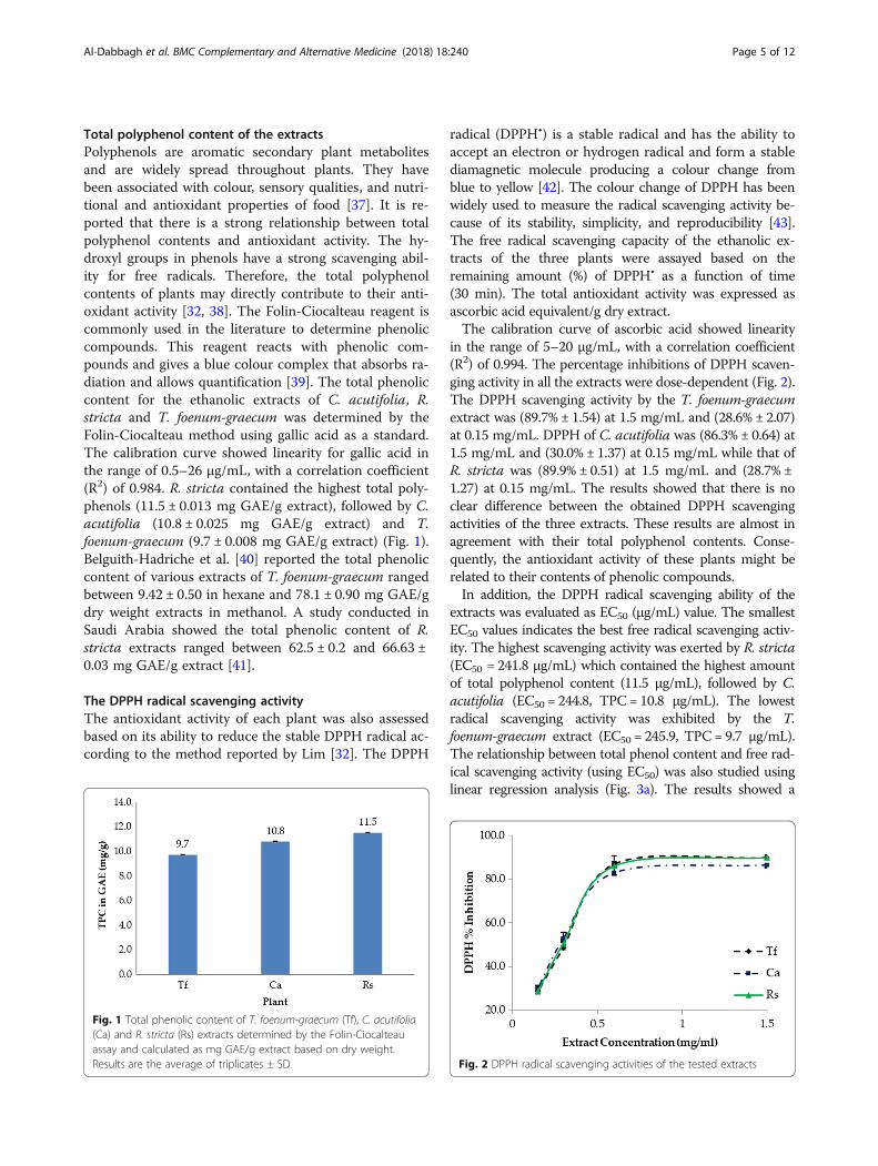
Total polyphenol content of the extractsPolyphenols are aromatic secondary plant metabolitesand are widely spread throughout plants. They havebeen associated with colour, sensory qualities, and nutri-tional and antioxidant properties of food [37]. It is re-ported that there is a strong relationship between totalpolyphenol contents and antioxidant activity. The hy-droxyl groups in phenols have a strong scavenging abil-ity for free radicals. Therefore, the total polyphenolcontents of plants may directly contribute to their anti-oxidant activity [32, 38]. The Folin-Ciocalteau reagent iscommonly used in the literature to determine phenoliccompounds. This reagent reacts with phenolic com-pounds and gives a blue colour complex that absorbs ra-diation and allows quantification [39]. The total phenoliccontent for the ethanolic extracts of C. acutifolia, R.stricta and T. foenum-graecum was determined by theFolin-Ciocalteau method using gallic acid as a standard.The calibration curve showed linearity for gallic acid inthe range of 0.5–26 μg/mL, with a correlation coefficient(R2) of 0.984. R. stricta contained the highest total poly-phenols (11.5 ± 0.013 mg GAE/g extract), followed by C.acutifolia (10.8 ± 0.025 mg GAE/g extract) and T.foenum-graecum (9.7 ± 0.008 mg GAE/g extract) (Fig. 1).Belguith-Hadriche et al. [40] reported the total phenoliccontent of various extracts of T. foenum-graecum rangedbetween 9.42 ± 0.50 in hexane and 78.1 ± 0.90 mg GAE/gdry weight extracts in methanol. A study conducted inSaudi Arabia showed the total phenolic content of R.stricta extracts ranged between 62.5 ± 0.2 and 66.63 ±0.03 mg GAE/g extract [41].
The DPPH radical scavenging activityThe antioxidant activity of each plant was also assessedbased on its ability to reduce the stable DPPH radical ac-cording to the method reported by Lim [32]. The DPPH
radical (DPPH•) is a stable radical and has the ability toaccept an electron or hydrogen radical and form a stablediamagnetic molecule producing a colour change fromblue to yellow [42]. The colour change of DPPH has beenwidely used to measure the radical scavenging activity be-cause of its stability, simplicity, and reproducibility [43].The free radical scavenging capacity of the ethanolic ex-tracts of the three plants were assayed based on theremaining amount (%) of DPPH• as a function of time(30 min). The total antioxidant activity was expressed asascorbic acid equivalent/g dry extract.The calibration curve of ascorbic acid showed linearity
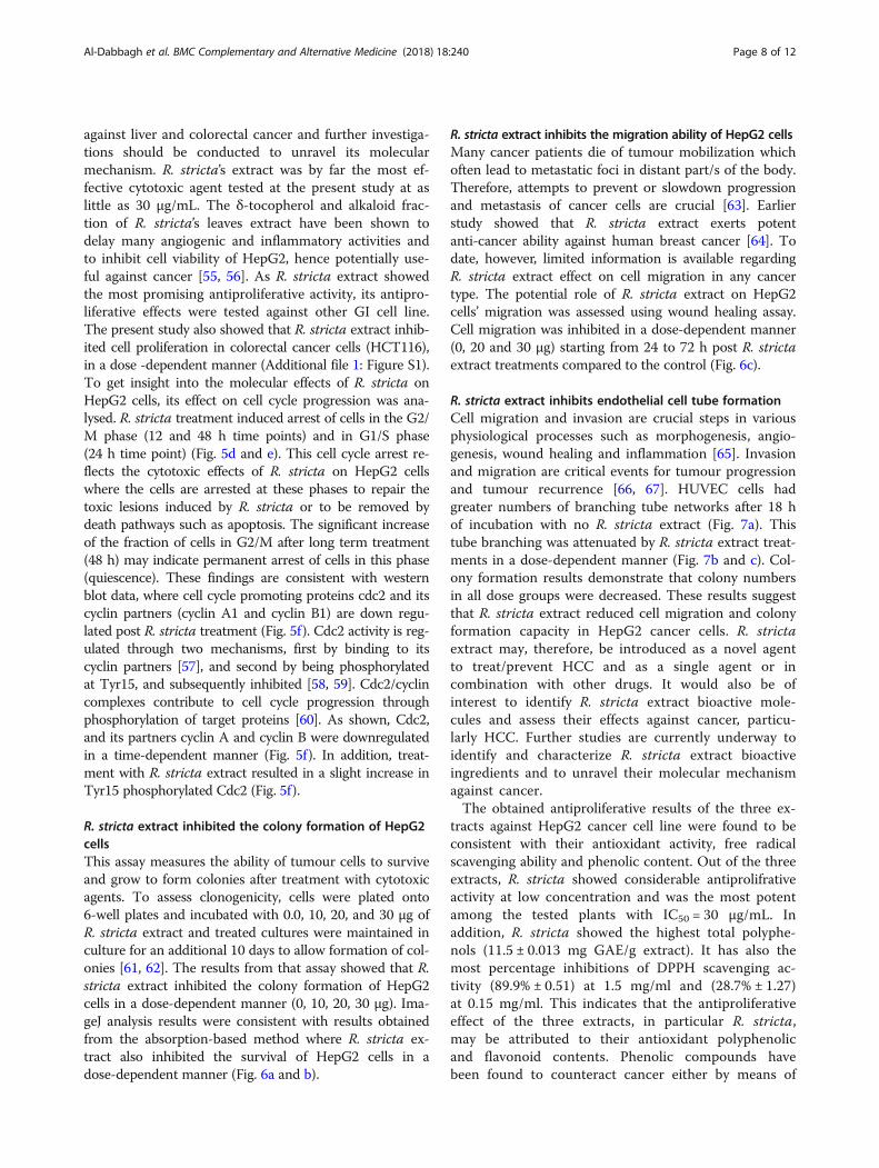
in the range of 5–20 μg/mL, with a correlation coefficient(R2) of 0.994. The percentage inhibitions of DPPH scaven-ging activity in all the extracts were dose-dependent (Fig. 2).The DPPH scavenging activity by the T. foenum-graecumextract was (89.7% ± 1.54) at 1.5 mg/mL and (28.6% ± 2.07)at 0.15 mg/mL. DPPH of C. acutifolia was (86.3% ± 0.64) at1.5 mg/mL and (30.0% ± 1.37) at 0.15 mg/mL while that ofR. stricta was (89.9% ± 0.51) at 1.5 mg/mL and (28.7% ±1.27) at 0.15 mg/mL. The results showed that there is noclear difference between the obtained DPPH scavengingactivities of the three extracts. These results are almost inagreement with their total polyphenol contents. Conse-quently, the antioxidant activity of these plants might berelated to their contents of phenolic compounds.In addition, the DPPH radical scavenging ability of the
extracts was evaluated as EC50 (μg/mL) value. The smallestEC50 values indicates the best free radical scavenging activ-ity. The highest scavenging activity was exerted by R. stricta(EC50 = 241.8 μg/mL) which contained the highest amountof total polyphenol content (11.5 μg/mL), followed by C.acutifolia (EC50 = 244.8, TPC= 10.8 μg/mL). The lowestradical scavenging activity was exhibited by the T.foenum-graecum extract (EC50 = 245.9, TPC= 9.7 μg/mL).The relationship between total phenol content and free rad-ical scavenging activity (using EC50) was also studied usinglinear regression analysis (Fig. 3a). The results showed a
Fig. 1 Total phenolic content of T. foenum-graecum (Tf), C. acutifolia(Ca) and R. stricta (Rs) extracts determined by the Folin-Ciocalteauassay and calculated as mg GAE/g extract based on dry weight.Results are the average of triplicates ± SD Fig. 2 DPPH radical scavenging activities of the tested extracts
Al-Dabbagh et al. BMC Complementary and Alternative Medicine (2018) 18:240 Page 5 of 12

significant negative correlation (R2 = − 0.856, P-value < 0.05)between EC50 (DPPH· scavenging) and total phenolic con-tent suggesting that the presence of the phenolic com-pounds contributed significantly to the antioxidant activityof the tested plants. These results are consistent with previ-ous works that showed a liner correlation between the totalphenolic content and the reducing antioxidant capacity ofsome plant extracts [32, 38, 44].
Total flavonoid contentsFlavonoids are a class of secondary plant phenolics. Flavo-noids and their derivatives have a wide range of biologicalactions including anticancer activity. The anticancer activ-ity of flavonoids is attributed to their potent antioxidanteffects which include metal chelation and free-radicalscavenging activities [45]. Flavonoids present in herbswere found to significantly contribute to their antioxidantproperties [44]. The flavonoid content was obtained usingaluminium chloride assay which based on the formationof a complex between the aluminium ion, Al (III), and thecarbonyl and hydroxyl groups of flavones and flavonolsthat produce a yellow colour [46]. Flavonoid content wascalculated from the regression equation of quercetin cali-bration curve and was expressed as quercetin equivalents.The calibration curve showed linearity in the range of 1–25 μg/mL, with a correlation coefficient (R2) of 0.999.Figure 4 shows the flavonoids contents in all the ex-
tracts. Using the standard curve generated by quercetin,the total flavonoids content of the T. foenum-graecumextract was (14.6 ± 0.21 mg QE/g), whereas, the C. actu-tifolia extract was (20.8 ± 0.40 mg QE/g) and finally thatof R. stricta was (9.2 ± 0.22 mg QE/g).The relationship between the total flavonoids content
and the free radical scavenging activity (using EC50) wasstudied using linear regression analysis (Fig. 3b). The re-sults showed a positive correlation (R2 = 0.460, P-value< 0.05) between EC50 (DPPH scavenging) and total flavo-noids content. The obtained correlation was moderate
suggesting that other compounds maybe participating inthe radical scavenging activity of these plant extracts.
Effects of extracts on cell viability in HepG2 cellsHCC remains among the leading cause of cancer-relateddeath worldwide [47, 48]. Although therapeutic approachesfor advanced HCC are limited to the use of multikinase in-hibitors, such as sorafenib, only modest survival benefitshave clinically been reported. Thus, identifying new com-pounds with promise antitumor activity against HCC is ex-ceedingly needed [48, 49]. Natural products, especiallyfrom plants, are often better tolerated than their syntheticanalogs used in cancer treatments [50]. They contain awide spectrum of bioactive secondary metabolites that arethe foundation of the recently introduced notion ofbroad-spectrum integrative approach for cancer preventionand treatment [51].Effects of tested extracts were investigated against human
hepatoma (HepG2) cancer cell line. A dose-dependent re-duction in cell viability was reported in cells treated with alltested extracts (Fig. 5). The IC50 values of extracts rangedfrom 30 μg/mL to 200 μg/mL. Treatment with R. strictasignificantly enhanced the mortality of cancer cells at thelowest concentration (30 μg/mL). T. foenum-graecum was
Fig. 3 Linear correlations between the amount of total phenols and DPPH· radical scavenging activity (a) and between the flavonoid content andDPPH· radical scavenging activity (b)
Fig. 4 Flavonoids content of the tested extracts
Al-Dabbagh et al. BMC Complementary and Alternative Medicine (2018) 18:240 Page 6 of 12

however less potent with IC50 of 200 μg/mL where 50% ofHepG2 cancer cells were eradicated at 200 μg/mL. Afenugreek-enriched diet decreased colon tumour incidenceand hepatic lipid peroxidation in liver cancer-induced ratsin addition to increasing the endogenous antioxidant activ-ities in liver [52]. Li et al. [53] showed that diosgenin, fenu-greek’s main active ingredient, down regulated theexpression of various STAT3-regulated genes, inhibitedproliferation and potentiated the apoptotic effects of pacli-taxel and doxorubicin, suggesting that diosgenin could be anovel and potential treatment option for HCC and othercancers. Therefore, the role of fenugreek extract and its ac-tive principals as supplements in diet-based preventive/therapeutic strategies to improve health care continues tobe a fast growing field of research. The correlation betweenthe antioxidant activities (free radical scavenging activity)and the anticancer activities (cell viability) of C. acutifolia,
R. stricta and T. foenum-graecum extracts was studiedusing linear regression analysis. The results showed a sig-nificant positive correlation (R2 = − 0.933, 0.997 and 0.797,P-value < 0.05) for C. acutifolia, R. stricta and T.foenum-graecum extracts respectively.At a concentration range of (100–200 μg/mL) Cassia
acutifolia’s extract was similarly cytotoxic to HepG2cells. Alkaloids extracted from Senna species reducedcell viability in a concentration-dependent manner ofdifferent tumour cell lines including HepG2 [49]. Sennaalkaloids showed important antiproliferative activity onHepG2 cells that was mediated by ERK inactivation anddown-regulation of cyclin D1 expression. Similarly, ex-tracts of different cassia species were able to inhibitgrowth of colorectal (DLD1), among other, human can-cer cell lines [54]. Thus, Senna’s extract may represent apotential new antitumor and/or adjuvant treatment
Fig. 5 Assessment of the cytotoxic effects of Trigonella foenum-graecum (Helba), Cassia acutifolia (Holoul), and Rhazya stricta (Harmal) extracts onHepG2 in vitro. a MTT assay results of HepG2 cells viability after treatment with increasing concentrations of Helba and Holoul for 24 h. *P < 0.05,**P < 0.001, ***P < 0.0001 b MTT assay results of HepG2 cells viability after treatment with increasing concentrations of R. stricta extract for 24 h.*P < 0.05, **P < 0.001, ***P < 0.0001 c Assessment of morphological changes of HepG2 cells after treatment with increasing concentrations of R.stricta extract for 24 h. Cells were fixed and stained with crystal violet (scale bar = 200 μm). d Cell cycle progression of HepG2 cells after treatmentwith R. stricta extract at a dose of 30 μg over a period of 48 h. e Quantitative distribution of HepG2 cells in different phases of the cell cycle atdifferent time intervals (*P < 0.05) f Immunoblot analysis of cell cycle regulatory proteins in HepG2 cells after treatment with R. stricta extract at adose of 30 μg over a period of 48 h
Al-Dabbagh et al. BMC Complementary and Alternative Medicine (2018) 18:240 Page 7 of 12
against liver and colorectal cancer and further investiga-tions should be conducted to unravel its molecularmechanism. R. stricta’s extract was by far the most ef-fective cytotoxic agent tested at the present study at aslittle as 30 μg/mL. The δ-tocopherol and alkaloid frac-tion of R. stricta’s leaves extract have been shown todelay many angiogenic and inflammatory activities andto inhibit cell viability of HepG2, hence potentially use-ful against cancer [55, 56]. As R. stricta extract showedthe most promising antiproliferative activity, its antipro-liferative effects were tested against other GI cell line.The present study also showed that R. stricta extract inhib-ited cell proliferation in colorectal cancer cells (HCT116),in a dose -dependent manner (Additional file 1: Figure S1).To get insight into the molecular effects of R. stricta onHepG2 cells, its effect on cell cycle progression was ana-lysed. R. stricta treatment induced arrest of cells in the G2/M phase (12 and 48 h time points) and in G1/S phase(24 h time point) (Fig. 5d and e). This cell cycle arrest re-flects the cytotoxic effects of R. stricta on HepG2 cellswhere the cells are arrested at these phases to repair thetoxic lesions induced by R. stricta or to be removed bydeath pathways such as apoptosis. The significant increaseof the fraction of cells in G2/M after long term treatment(48 h) may indicate permanent arrest of cells in this phase(quiescence). These findings are consistent with westernblot data, where cell cycle promoting proteins cdc2 and itscyclin partners (cyclin A1 and cyclin B1) are down regu-lated post R. stricta treatment (Fig. 5f). Cdc2 activity is reg-ulated through two mechanisms, first by binding to itscyclin partners [57], and second by being phosphorylatedat Tyr15, and subsequently inhibited [58, 59]. Cdc2/cyclincomplexes contribute to cell cycle progression throughphosphorylation of target proteins [60]. As shown, Cdc2,and its partners cyclin A and cyclin B were downregulatedin a time-dependent manner (Fig. 5f). In addition, treat-ment with R. stricta extract resulted in a slight increase inTyr15 phosphorylated Cdc2 (Fig. 5f).
R. stricta extract inhibited the colony formation of HepG2cellsThis assay measures the ability of tumour cells to surviveand grow to form colonies after treatment with cytotoxicagents. To assess clonogenicity, cells were plated onto6-well plates and incubated with 0.0, 10, 20, and 30 μg ofR. stricta extract and treated cultures were maintained inculture for an additional 10 days to allow formation of col-onies [61, 62]. The results from that assay showed that R.stricta extract inhibited the colony formation of HepG2cells in a dose-dependent manner (0, 10, 20, 30 μg). Ima-geJ analysis results were consistent with results obtainedfrom the absorption-based method where R. stricta ex-tract also inhibited the survival of HepG2 cells in adose-dependent manner (Fig. 6a and b).
R. stricta extract inhibits the migration ability of HepG2 cellsMany cancer patients die of tumour mobilization whichoften lead to metastatic foci in distant part/s of the body.Therefore, attempts to prevent or slowdown progressionand metastasis of cancer cells are crucial [63]. Earlierstudy showed that R. stricta extract exerts potentanti-cancer ability against human breast cancer [64]. Todate, however, limited information is available regardingR. stricta extract effect on cell migration in any cancertype. The potential role of R. stricta extract on HepG2cells’ migration was assessed using wound healing assay.Cell migration was inhibited in a dose-dependent manner(0, 20 and 30 μg) starting from 24 to 72 h post R. strictaextract treatments compared to the control (Fig. 6c).
R. stricta extract inhibits endothelial cell tube formationCell migration and invasion are crucial steps in variousphysiological processes such as morphogenesis, angio-genesis, wound healing and inflammation [65]. Invasionand migration are critical events for tumour progressionand tumour recurrence [66, 67]. HUVEC cells hadgreater numbers of branching tube networks after 18 hof incubation with no R. stricta extract (Fig. 7a). Thistube branching was attenuated by R. stricta extract treat-ments in a dose-dependent manner (Fig. 7b and c). Col-ony formation results demonstrate that colony numbersin all dose groups were decreased. These results suggestthat R. stricta extract reduced cell migration and colonyformation capacity in HepG2 cancer cells. R. strictaextract may, therefore, be introduced as a novel agentto treat/prevent HCC and as a single agent or incombination with other drugs. It would also be ofinterest to identify R. stricta extract bioactive mole-cules and assess their effects against cancer, particu-larly HCC. Further studies are currently underway toidentify and characterize R. stricta extract bioactiveingredients and to unravel their molecular mechanismagainst cancer.The obtained antiproliferative results of the three ex-
tracts against HepG2 cancer cell line were found to beconsistent with their antioxidant activity, free radicalscavenging ability and phenolic content. Out of the threeextracts, R. stricta showed considerable antiprolifrativeactivity at low concentration and was the most potentamong the tested plants with IC50 = 30 μg/mL. Inaddition, R. stricta showed the highest total polyphe-nols (11.5 ± 0.013 mg GAE/g extract). It has also themost percentage inhibitions of DPPH scavenging ac-tivity (89.9% ± 0.51) at 1.5 mg/ml and (28.7% ± 1.27)at 0.15 mg/ml. This indicates that the antiproliferativeeffect of the three extracts, in particular R. stricta,may be attributed to their antioxidant polyphenolicand flavonoid contents. Phenolic compounds havebeen found to counteract cancer either by means of
Al-Dabbagh et al. BMC Complementary and Alternative Medicine (2018) 18:240 Page 8 of 12

Fig. 6 Rhazya stricta extract inhibits colony forming ability and wound healing of HepG2 cells in a dose-dependent manner. a Representative images ofHepG2 colonies after treatment with increasing concentrations of R. stricta extract. b Percent of area occupied by colonies in treated and non-treatedwells (representative of triplicate samples; *P< 0.05, **P< 0.005, ***P< 0.0001) and absorbance of each treated and non-treated wells (representative of 3biological triplicates, each in triplicate; *P< 0.05, **P< 0.001, ***P< 0.0001). c Representative images and quantification (three regions of three biologicaltriplicates; *P< 0.05, **P< 0.001, ***P< 0.0001) of wound-healing assay results of HepG2 cells treated without or with 20 and 30 μg of R. stricta extract
Al-Dabbagh et al. BMC Complementary and Alternative Medicine (2018) 18:240 Page 9 of 12

antioxidant effect or by inhibiting the formation ofcarcinogenic metabolites that damage the vital bio-molecules [68].Finally, the investigated UAE plants have shown a
strong reducing antioxidant capacity and free radicalscavenging ability. These findings validate the use ofthese plants in folk medicine for the treatment of cer-tain diseases. Oxidative cell damage events are fre-quently correlated with the oxidative stress. We showhere that both of those properties are present in theethanolic extract of R. stricta, which has significantantiproliferative activity and a great antioxidant activ-ity. The results reported here are very promising indi-cators for the potential application of this plant forpreventive and therapeutic purposes.
ConclusionsIn conclusion, natural antioxidants have many importantapplications in health promotion, food preservation,food flavouring and cosmetics. They are preferred oversynthetic antioxidants because they are safer for con-sumption and more environmentally friendly. Thepresent study investigates the antioxidant activity andpolyphenolic content of three medicinal plants from theUAE. We found extracts rich in antioxidants and inpolyphenols, which merit further investigations. Ofthe extracts evaluated, that of R. stricta (Harmal)showed the greatest antioxidant, antiangiogenic andantiproliferative activities, a discovery that makes thisspecies a promising source of anticancer agent devel-opment especially for colon and liver cancers, and
hence worthy of further investigation. Isolation of ac-tive compounds and exploring their mode of actionagainst tumours by using in vivo experimental modelswould make an important future study.
Additional file
Additional file 1: Figure S1. Assessment of the cytotoxic effects ofTrigonella foenum-graecum (Helba), Cassia acutifolia (Holoul), and Rhazyastricta (Harmal) extracts on HCT116 in vitro. MTT assay results of9cHCT116 cells viability after treatment with increasing concentrations ofHelba (a) Holoul (b) and Harmal (c) for 24 h. *P < 0.05, **P < 0.005, ***P <0.0001. (DOCX 99 kb)
AcknowledgmentsThis work was supported by a start-up grant (Grant 31S 215), Division of Researchand Graduate Studies, UAEU and an individual research grant (Grant 31S 188),College of Science, UAEU to the PI: Dr. Bayan Al-Dabbagh and by Zayed BinSultan Centre for Health Sciences (ZCHS) grant number 31R050 for Dr. AmrAmin.
FundingThis work was supported by a start-up grant (Grant 31S 215), Division of Researchand Graduate Studies, UAEU and an individual research grant (Grant 31S 188),College of Science, UAEU to the PI: Dr. Bayan Al-Dabbagh and by Zayed BinSultan Centre for Health Sciences (ZCHS) grant number 31R050 for Dr. AmrAmin.
Availability of data and materials“The datasets used and/or analyzed during the current study are availablefrom the corresponding authors upon reasonable request.”
Authors’ contributionsBA and AA designed the research. BA, IAE, AAH, RA and RE performed theexperiments. SSA participated in the interpretation of the results. BA and AAanalyzed the data and wrote the paper. All authors read and approved thefinal manuscript.
Fig. 7 Rhazya stricta extract inhibits tube formation in HUVECs on Matrigel in a dose-dependent manner. Photographs of tube formation in HUVECson Matrigel after incubation with or without R. stricta extract at 18 h. Cells were treated with R. stricta extract at a series of concentrations (10 μg; b,20 μg; c, 30 μg; d) or DMSO vehicle (control; a) for 18 h
Al-Dabbagh et al. BMC Complementary and Alternative Medicine (2018) 18:240 Page 10 of 12
Ethics approval and consent to participateNot applicable
Consent for publicationNot applicable
Competing interestsThe authors declare that they have no competing interests.
Publisher’s NoteSpringer Nature remains neutral with regard to jurisdictional claims inpublished maps and institutional affiliations.
Author details1Department of Chemistry, College of Science, UAE University, PO Box 15551,Al Ain, UAE. 2Department of Biology, College of Science, UAE University, POBox 15551, Al Ain, UAE. 3Zoology Department, Cairo University, Giza, Egypt.4Department of Pharmacy Practice and Pharmacotherapeutics, SharjahInstitute for Medical Research and College of Pharmacy, University of Sharjah,Sharjah, UAE.
Received: 28 March 2018 Accepted: 11 July 2018
References1. Aruoma OI. Free radicals, antioxidants and international nutrition. Asia Pac J
Clin Nutr. 1999;8:53–63.2. Gupta VK, Sharma SK. Plants as natural antioxidants. Nat Prod Rad. 2006;5:326–34.3. Magder S. Reactive oxygen species: toxic molecules or spark of life? Crit
Care. 2006;10:208.4. Brewer MS. Natural antioxidants: sources, compounds, mechanisms of action,
and potential applications. Compr Rev Food Sci Food Saf. 2011;10:221–47.5. Lu LY, Ou N, Lu Q-B. Antioxidant induces DNA damage, cell death and
mutagenicity in human lung and skin normal cells. Sci Rep. 2013;3:3169.6. Soobrattee MA, Neergheen VS, Luximon-Ramma A, Aruoma OI, Bahorun T.
Phenolics as potential antioxidant therapeutic agents: mechanism andactions. Mutat Res. 2005;579:200–13.
7. Chaudière J, Ferrari-Iliou R. Intracellular antioxidants: from chemical tobiochemical mechanisms. Food Chem Toxicol. 1999;37:949–62.
8. Fridovich I. Fundamental aspects of reactive oxygen species, or what's thematter with oxygen? Ann N Y Acad Sci. 1999;893:13–8.
9. Hinneburg I, Damien Dorman HJ, Hiltunen R. Antioxidant activities ofextracts from selected culinary herbs and spices. Food Chem. 2006;97:122–9.
10. Amin A, Hamza AA, Bajbouj K, Ashraf SS, Daoud S. Saffron: a potentialcandidate for a novel anticancer drug against hepatocellular carcinoma.Hepatology. 2011;54:857–67.
11. Hamza AA, Ahmed MM, Elwey HM, Amin A. Melissa officinalis protectsagainst doxorubicin-induced cardiotoxicity in rats and potentiates itsanticancer activity on MCF-7 cells. PLoS One. 2016;11:e0167049.
12. Xue WL, Li XS, Zhang J, Liu YH, Wang ZL, Zhang RJ. Effect of Trigonellafoenum-graecum (fenugreek) extract on blood glucose, blood lipid andhemorheological properties in streptozotocin-induced diabetic rats. Asia PacJ Clin Nutr. 2007;16(Suppl 1):422–6.
13. Amin A, Alkaabi A, Al-Falasi S, Daoud SA. Chemopreventive activities ofTrigonella foenum graecum (fenugreek) against breast cancer. Cell Biol Int.2005;29:687–94.
14. El Bairi K, Ouzir M, Agnieszka N, Khalki L. Anticancer potential of Trigonellafoenum graecum: cellular and molecular targets. Biomed Pharmacother.2017;90:479–91.
15. Seethapathy GS, Ganesh D, Kumar JUS, Senthilkumar U, Newmaster SG,Ragupathy S, et al. Assessing product adulteration in natural healthproducts for laxative yielding plants, Cassia, Senna, and Chamaecrista, insouthern India using DNA barcoding. Int J Legal Med. 2015;129:693–700.
16. Elkady AI. Crude alkaloid extract of Rhazya stricta inhibits cell growth andsensitizes human lung cancer cells to cisplatin through induction ofapoptosis. Genet Mol Biol. 2013;36:12–21.
17. Faqi AS. A comprehensive guide to toxicology in preclinical drugdevelopment: Academic Press; 2013.
18. Diplock AT, Charuleux JL, Crozier-Willi G, Kok FJ, Rice-Evans C, Roberfroid M,et al. Functional food science and defence against reactive oxidativespecies. Br J Nutr. 1998;80:S77–S112.
19. Valko M, Leibfritz D, Moncol J, Cronin MT, Mazur M, Telser J. Free radicalsand antioxidants in normal physiological functions and human disease. Int JBiochem Cell Biol. 2007;39:44–84.
20. Shigenaga MK, Ames BN. Oxidants and mitogenesis as causes of mutationand cancer: the influence of diet. Basic Life Sci. 1993;61:419–36.
21. Greenwell M, Rahman P. Medicinal plants: their use in anticancer treatment.Int J Pharm Sci Res. 2015;6:4103.
22. Alam AHMK, Hossain ASMS, Khan MA, Kabir SR, Reza MA, Rahman MM, et al.The Antioxidative Fraction of White Mulberry Induces Apoptosis throughRegulation of p53 and NFκB in EAC Cells. PLoS ONE. 2016;11:e0167536.
23. Islam S, Nasrin S, Khan MA, Hossain ASMS, Islam F, Khandokhar P, et al.Evaluation of antioxidant and anticancer properties of the seed extracts ofSyzygium fruticosum Roxb. growing in Rajshahi, Bangladesh. BMCComplement Altern Med. 2013;13:142.
24. Carmeliet P. Angiogenesis in life, disease and medicine. Nature. 2005;438:932.25. Bar J, Goss GD. Tumor vasculature as a therapeutic target in non-small cell
lung cancer. J Thorac Oncol. 2012;7:609–20.26. Keizer RJ, Funahashi Y, Semba T, Wanders J, Beijnen J, Schellens J, et al.
Evaluation of α 2-integrin expression as a biomarker for tumor growthinhibition for the investigational integrin inhibitor E7820 in preclinical andclinical studies. AAPS J. 2011;13:230–9.
27. Mita M, Kelly KR, Mita A, Ricart AD, Romero O, Tolcher A, et al. Phase I study ofE7820, an oral inhibitor of integrin α-2 expression with antiangiogenic properties,in patients with advanced malignancies. Clin Cancer Res. 2011;17:193–200.
28. Senger DR, Perruzzi CA, Streit M, Koteliansky VE, de Fougerolles AR, DetmarM. The α1β1 and α2β1 integrins provide critical support for vascularendothelial growth factor signaling, endothelial cell migration, and tumorangiogenesis. Am J Pathol. 2002;160:195–204.
29. Senger DR, Claffey KP, Benes JE, Perruzzi CA, Sergiou AP, Detmar M.Angiogenesis promoted by vascular endothelial growth factor: regulationthrough α1β1 and α2β1 integrins. Proc Natl Acad Sci U S A. 1997;94:13612–7.
30. Amzad Hossain M, Shah MD. A study on the total phenols content andantioxidant activity of essential oil and different solvent extracts of endemicplant Merremia borneensis. Arab J Chem. 2015;8:66–71.
31. Singleton VL, Orthofer R, Lamuela-Raventós RM. Analysis of total phenolsand other oxidation substrates and antioxidants by means of folin-ciocalteureagent: Methods Enzymol Academic Press; 1999. p. 152–78.
32. Lim YY, Quah EPL. Antioxidant properties of different cultivars of Portulacaoleracea. Food Chem. 2007;103:734–40.
33. Chang C-C, Yang M-H, Wen H-M, Chern J-C. Estimation of total flavonoidcontent in propolis by two complementary colorimetric methods. J FoodDrug Anal. 2002;10:178–82.
34. Guzmán C, Bagga M, Kaur A, Westermarck J, Abankwa D. ColonyArea: anImageJ plugin to automatically quantify Colony formation in Clonogenicassays. PLoS One. 2014;9:e92444.
35. Saleh E, El-Awady R, Anis N. Predictive markers for the response to 5-fluorouracil therapy in cancer cells: constant-field gel electrophoresis as atool for prediction of response to 5-fluorouracil-based chemotherapy. OncolLett. 2013;5:321–7.
36. Khan MA, Rahman AA, Islam S, Khandokhar P, Parvin S, Islam MB, et al. Acomparative study on the antioxidant activity of methanolic extracts fromdifferent parts of Morus alba L. (Moraceae). BMC Res Notes. 2013;6:24.
37. Robbins RJ. Phenolic acids in foods: an overview of analytical methodology.J Agric Food Chem. 2003;51:2866–87.
38. Wojdyło A, Oszmiański J, Czemerys R. Antioxidant activity and phenoliccompounds in 32 selected herbs. Food Chem. 2007;105:940–9.
39. Pontis JA, LAMAd C, SJRd S, Flach A. Color, phenolic and flavonoid content,and antioxidant activity of honey from Roraima, Brazil. Food Sci. Technol(Campinas). 2014;34:69–73.
40. Belguith-Hadriche O, Bouaziz M, Jamoussi K, Simmonds MS, El Feki A, Makni-Ayedi F. Comparative study on hypocholesterolemic and antioxidant activitiesof various extracts of fenugreek seeds. Food Chem. 2013;138:1448–53.
41. Bukhari NA, Al-Otaibi RA, Ibhrahim MM. Phytochemical and taxonomicevaluation of Rhazya stricta in Saudi Arabia. Saudi J Biol Sci. 2017;24:1513–21.
42. Robards K, Prenzler PD, Tucker G, Swatsitang P, Glover W. Phenolic compoundsand their role in oxidative processes in fruits. Food Chem. 1999;66:401–36.
43. Kitts DD, Wijewickreme AN, Hu C. Antioxidant properties of a northAmerican ginseng extract. Mol Cell Biochem. 2000;203:1–10.
44. Shan B, Cai YZ, Sun M, Corke H. Antioxidant capacity of 26 spice extractsand characterization of their phenolic constituents. J Agric Food Chem.2005;53:7749–59.
Al-Dabbagh et al. BMC Complementary and Alternative Medicine (2018) 18:240 Page 11 of 12
45. Amin A, Mousa M. Merits of anti-cancer plants from the Arabian gulf region.Cancer Ther. 2007;5:55–66.
46. Popova M, Bankova V, Butovska D, Petkov V, Nikolova-Damyanova B, SabatiniAG, et al. Validated methods for the quantification of biologically activeconstituents of poplar-type propolis. Phytochem Anal. 2004;15:235–40.
47. Amin A, Hamza AA, Daoud S, Khazanehdari K, Hrout AA, Baig B, et al.Saffron-based Crocin prevents early lesions of liver Cancer: in vivo, in vitroand network analyses. Recent Pat Anticancer Drug Discov. 2016;11:121–33.
48. Wu C-T, Tsai Y-T, Lai J-N. Demographic and medication characteristics oftraditional Chinese medicine users among colorectal cancer survivors: anationwide database study in Taiwan. J Tradit Complement Med. 2016;7:188–94.
49. Pereira RM, Ferreira-Silva GA, Pivatto M, Santos Lde A, Bolzani Vda S, Chagasde Paula DA, et al. Alkaloids derived from flowers of Senna spectabilis,(−)-cassine and (−)-spectaline, have antiproliferative activity on HepG2 cellsfor inducing cell cycle arrest in G1/S transition through ERK inactivation anddownregulation of cyclin D1 expression. Toxicol In Vitro. 2016;31:86–92.
50. Newman DJ, Cragg GM. Natural products as sources of new drugs from1981 to 2014. J Nat Prod. 2016;79:629–61.
51. Block KI, Gyllenhaal C, Lowe L, Amedei A, Amin ARMR, Amin A, et al.Designing a broad-spectrum integrative approach for cancer preventionand treatment. Semin Cancer Biol. 2015;35(Supplement):S276–304.
52. Devasena T, Venugopal MP. Fenugreek seeds modulate 1,2-dimethylhydrazine-induced hepatic oxidative stress during coloncarcinogenesis. Ital J Biochem. 2007;56:28–34.
53. Li F, Fernandez PP, Rajendran P, Hui KM, Sethi G. Diosgenin: a steroidalsaponin, inhibits STAT3 signaling pathway leading to suppression ofproliferation and chemosensitization of human hepatocellular carcinomacells. Cancer Lett. 2010;292:197–207.
54. Chandra P, Pandey R, Kumar B, Srivastva M, Pandey P, Sarkar J, et al.Quantification of multianalyte by UPLC–QqQLIT–MS/MS and in-vitro anti-proliferative screening in Cassia species. Ind Crop Prod. 2015;76:1133–41.
55. Nehdi IA, Sbihi HM, Tan CP, Al-Resayes SI. Seed oil from Harmal (Rhazyastricta Decne) grown in Riyadh (Saudi Arabia): a potential source of δ-tocopherol. J Saudi Chem Soc. 2016;20:107–13.
56. El Gendy MAM, Ali BH, Michail K, Siraki AG, El-Kadi AOS. Induction ofquinone oxidoreductase 1 enzyme by Rhazya stricta through Nrf2-dependent mechanism. J Ethnopharmacol. 2012;144:416–24.
57. Brown NR, Noble ME, Endicott JA, Johnson LN. The structural basis forspecificity of substrate and recruitment peptides for cyclin-dependentkinases. Nat Cell Biol. 1999;1:438–43.
58. McGowan CH, Russell P. Human Wee1 kinase inhibits cell division byphosphorylating p34cdc2 exclusively on Tyr15. EMBO J. 1993;12:75.
59. Wells NJ, Watanabe N, Tokusumi T, Jiang W, Verdecia MA, Hunter T. The C-terminal domain of the Cdc2 inhibitory kinase Myt1 interacts with Cdc2complexes and is required for inhibition of G (2)/M progression. J Cell Sci.1999;112:3361–71.
60. Enserink JM, Kolodner RD. An overview of Cdk1-controlled targets andprocesses. Cell Div. 2010;5:11–52.
61. Gach K, Grądzka I, Wasyk I, Męczyńska-Wielgosz S, Iwaneńko T, Szymański J,et al. Anticancer activity and radiosensitization effect ofmethyleneisoxazolidin-5-ones in hepatocellular carcinoma HepG2 cells.Chem Biol Interact. 2016;248:68–73.
62. Rafehi H, Orlowski C, Georgiadis GT, Ververis K, El-Osta A, Karagiannis TC.Clonogenic assay: adherent cells. J Vis Exp. 2011;49:2573.
63. Jiang WG, Sanders AJ, Katoh M, Ungefroren H, Gieseler F, Prince M, et al.Molecular, biological and clinical perspectives. Semin Cancer Biol. 2015;35(Supplement):S244–75.
64. Baeshen NA, Elkady AI, Abuzinadah OA, Mutwakil MH. Potential anticanceractivity of the medicinal herb, Rhazya stricta, against human breast cancer.Afr J Biotechnol. 2012;11:8960–72.
65. Bozzuto G, Ruggieri P, Molinari A. Molecular aspects of tumor cell migrationand invasion. Ann Ist Super Sanita. 2010;46:66–80.
66. Alur I, Dodurga Y, Secme M, Elmas L, Bagci G, Goksin I, et al. Anti-tumoreffects of bemiparin in HepG2 and MIA PaCa-2 cells. Gene. 2016;585:241–6.
67. Lavictoire SJ, Parolin DA, Klimowicz AC, Kelly JF, Lorimer IA. Interaction ofHsp90 with the nascent form of the mutant epidermal growth factorreceptor EGFRvIII. J Biol Chem. 2003;278:5292–9.
68. Hocman G. Chemoprevention of cancer: phenolic antioxidants (BHT, BHA).Int J BioChemiPhysics. 1988;20:639–51.
Al-Dabbagh et al. BMC Complementary and Alternative Medicine (2018) 18:240 Page 12 of 12