Antigen Detection by MS -...
53
Antigen Detection by MS Antigen Detection by MS Clinical Protein Chemistry/Proteomics Unit Biomedicum Helsinki E-Mail: [email protected] (http://research.med.helsinki.fi/corefacilities/proteinchem)
Transcript of Antigen Detection by MS -...

Antigen Detection by MSAntigen Detection by MS
Clinical Protein Chemistry/Proteomics UnitBiomedicum Helsinki
E-Mail: [email protected]
(http://research.med.helsinki.fi/corefacilities/proteinchem)


Specialized towards Clinical ProteomicsSpecialized towards Clinical Proteomics
Clinical sample collection expertize
Cinical sample treatment expertize
Clinical sample analysis expertize
Bridging the clinics to the results

Services offered:Services offered:
- gel-based and gel-free proteome analysis (proteomics)- protein identification
- protein & peptide sequencing- protein characterization
- label free and multiplex quantification- PTM analysis by MS
- MALDI Imaging (IMS)- glycoproteomics (HI)
- Laser Capture Microdissection MS (LCM-MS)- peptide arrays (HI)
- pepspot epitope mapping (HI)- structural 3D modeling services
- training by lab courses, lectures and seminars
Definition of MS
MS is an analysis technique to determine by means of a mass spectrometer the molecule
mass of free ions in high vacuum


InstrumentationInstrumentation

MALDI TOF/TOF

HR Quadrupole TOF/TOFWith MALDI and ETD
Ion Mobility

Ion Trap MS n

Antigen?
From Wikipedia, the free encyclopedia
An antigen is a substance/molecule that, whenintroduced into the body, triggers theproduction of an antibody by the immunesystem, which will then kill or neutralize theantigen that is recognized as a foreign andpotentially harmful invader.

Proteineous material

Detection of antigens by basic technologies
gel electrophoresis
or
liquid chromatogram

We need a technology to find changes in We need a technology to find changes in ProteomeProteome
A protein Array

Two-dimensional gel electrophoresis (2D) could do it?
• 1st dimension, IEF, Proteins are separated
• according to their isoelectric point (IP)
• 2nd dimension, SDS-PAGE, Proteins are separated according to their molecular mass
• Efficient: More than a thousand proteins resolved in E-Coli cell lysates and ~8000 in brain lysates
pH 10
SDS-
PAG
E
pH 3

1st Dimension - Isoelectric Focusing
pH4 pH7
+ -+
pH5.5neutral
pI - isoelectric point
ready made Gel-strips
gelsurface

2DEMw
2nd Dimension – SDS-PAGE
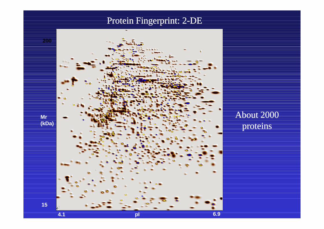
Protein Fingerprint: 2Protein Fingerprint: 2--DEDE
pI 6.94.1
Mr(kDa)
200
15
AboutAbout 20002000proteinsproteins

2D2D--PAGE Direct InPAGE Direct In--situ Digestsitu Digest
To identify the separated proteinsTo identify the separated proteins
Basic (+) Acidic (-)
#7
100 pmolrunning the gel- staining each spot of interest ( )- excise- in-gel digestion
PEPTIDE ANALYSIS
Trypsin digest

A 384 position MALDI-TOF sample target plate. To each position 0.5-2µl of sample together with matrix solution is pipetted and allowed to dry.
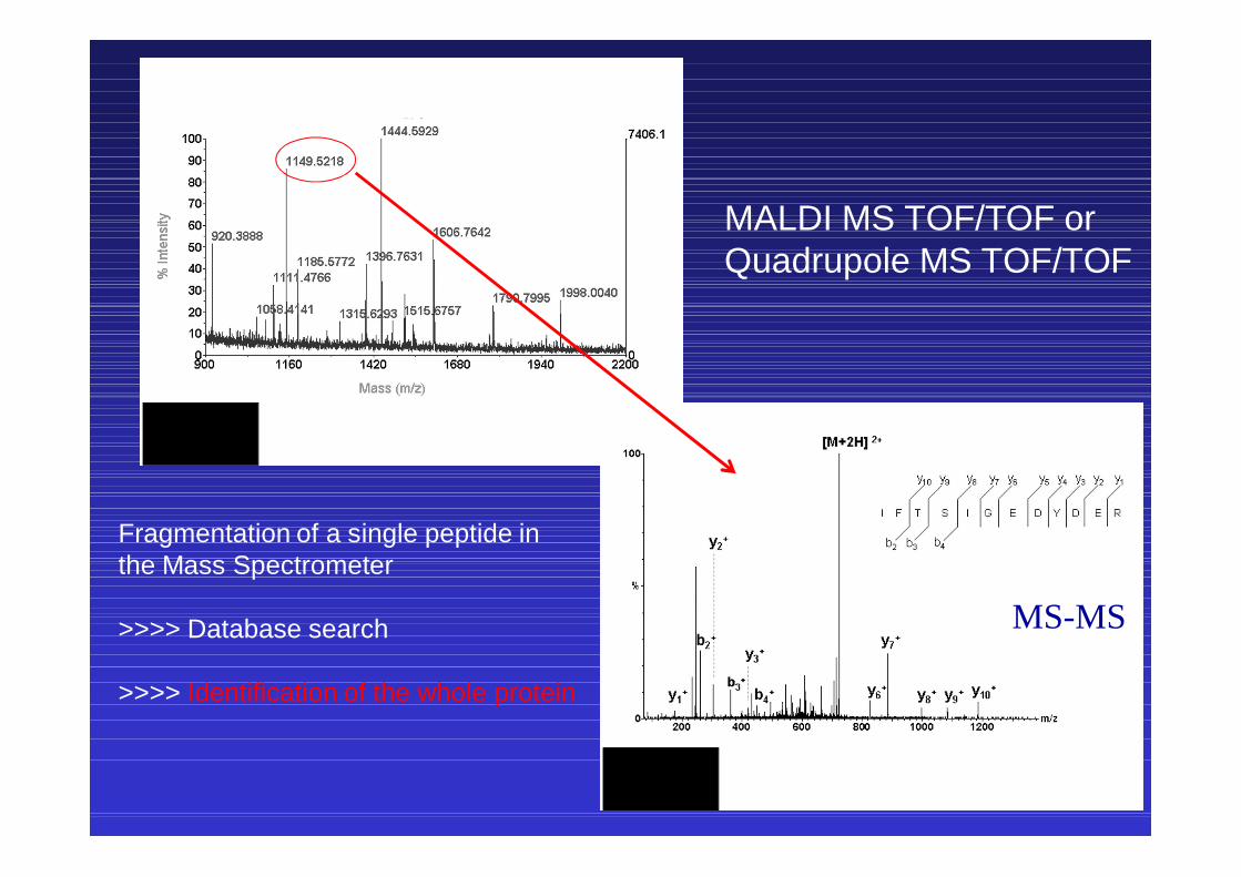
MALDI MS TOF/TOF or Quadrupole MS TOF/TOF
MS-MS
Fragmentation of a single peptide inthe Mass Spectrometer
>>>> Database search
>>>> Identification of the whole protein
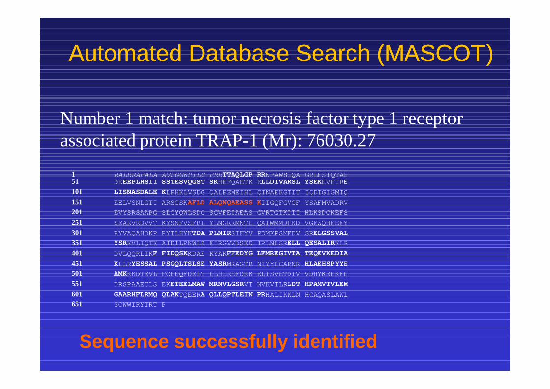
Number 1 match: tumor necrosis factor type 1 receptor associated protein TRAP-1 (Mr): 76030.27
Automated Database Search (MASCOT)Automated Database Search (MASCOT)
Sequence successfully identified
1 RALRRAPALA AVPGGKPILC PRRTTAQLGP RRNPAWSLQA GRLFSTQTAE51 DKEEPLHSII SSTESVQGST SKHEFQAETK KLLDIVARSL YSEKEVFIRE101 LISNASDALE KLRHKLVSDG QALPEMEIHL QTNAEKGTIT IQDTGIGMTQ151 EELVSNLGTI ARSGSKAFLD ALQNQAEASS KIIGQFGVGF YSAFMVADRV201 EVYSRSAAPG SLGYQWLSDG SGVFEIAEAS GVRTGTKIII HLKSDCKEFS 251 SEARVRDVVT KYSNFVSFPL YLNGRRMNTL QAIWMMDPKD VGEWQHEEFY 301 RYVAQAHDKP RYTLHYKTDA PLNIRSIFYV PDMKPSMFDV SRELGSSVAL351 YSRKVLIQTK ATDILPKWLR FIRGVVDSED IPLNLSRELL QESALIRKLR401 DVLQQRLIKF FIDQSKKDAE KYAKFFEDYG LFMREGIVTA TEQEVKEDIA451 KLLRYESSAL PSGQLTSLSE YASRMRAGTR NIYYLCAPNR HLAEHSPYYE501 AMKKKDTEVL FCFEQFDELT LLHLREFDKK KLISVETDIV VDHYKEEKFE551 DRSPAAECLS EKETEELMAW MRNVLGSRVT NVKVTLRLDT HPAMVTVLEM601 GAARHFLRMQ QLAKTQEERA QLLQPTLEIN PRHALIKKLN HCAQASLAWL651 SCWWIRYTRT P
2D2D--LC/MS/MSLC/MS/MS
(2(2--dimensional liquid chromatography mass dimensional liquid chromatography mass spectrometry)spectrometry)
Faster Protein Identification by MS/MSFaster Protein Identification by MS/MS

HR Quadrupole TOF/TOFWith MALDI and ETD
Ion Mobility and 2D-LC
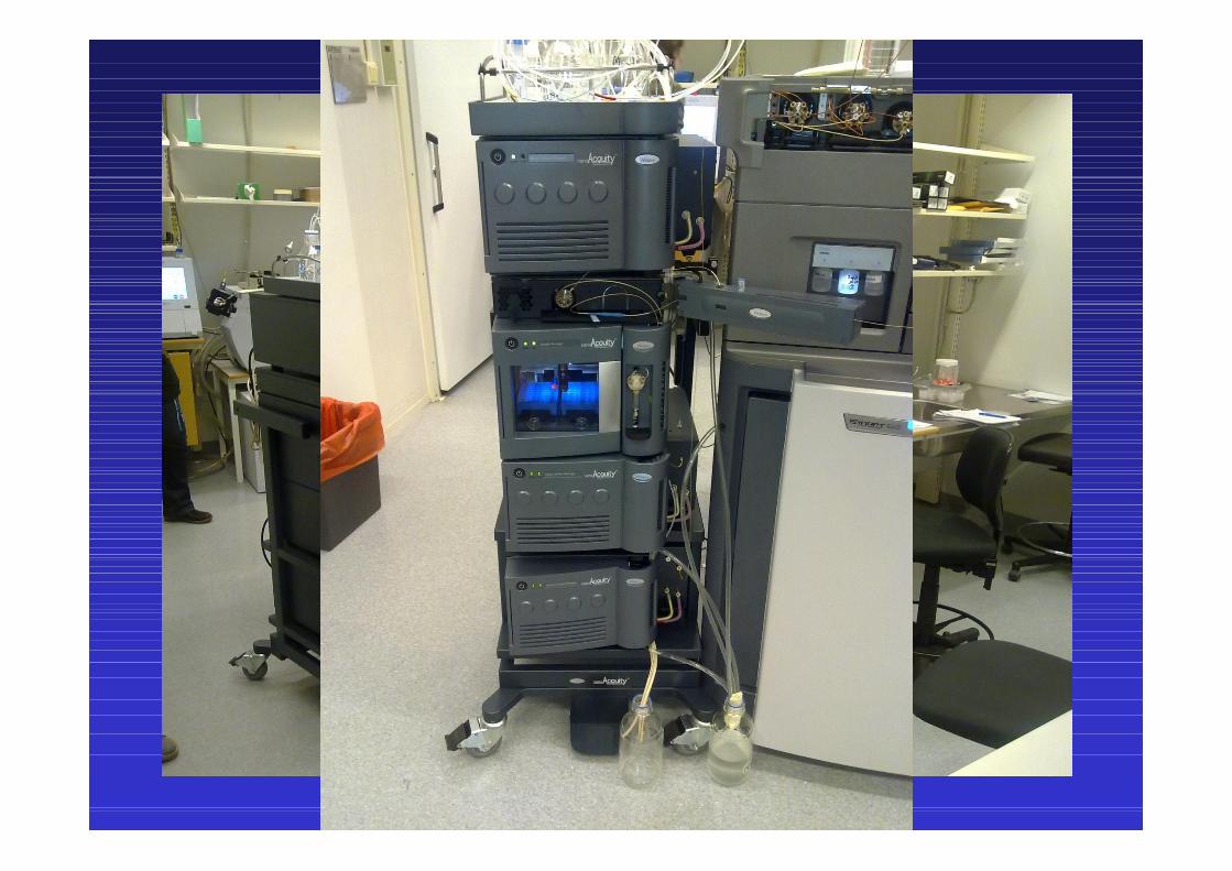

Quadrupole TOF/TOF
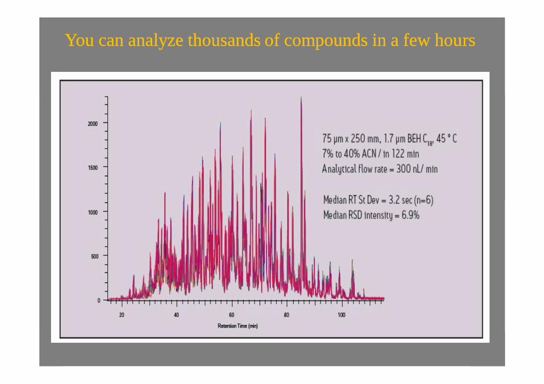
You can analyze thousands of compounds in a few hoursYou can analyze thousands of compounds in a few hours

You can combine several analyses in a dayYou can combine several analyses in a day

MALDI MS TOF/TOF or Quadrupole MS TOF/TOF
MS-MS
Fragmentation of a single peptide inthe Mass Spectrometer
>>>> Database search
>>>> Identification of the whole protein

Number 1 match: tumor necrosis factor type 1 receptor associated protein TRAP-1 (Mr): 76030.27
Automated Database Search (MASCOT)Automated Database Search (MASCOT)
Sequence successfully identified
1 RALRRAPALA AVPGGKPILC PRRTTAQLGP RRNPAWSLQA GRLFSTQTAE51 DKEEPLHSII SSTESVQGST SKHEFQAETK KLLDIVARSL YSEKEVFIRE101 LISNASDALE KLRHKLVSDG QALPEMEIHL QTNAEKGTIT IQDTGIGMTQ151 EELVSNLGTI ARSGSKAFLD ALQNQAEASS KIIGQFGVGF YSAFMVADRV201 EVYSRSAAPG SLGYQWLSDG SGVFEIAEAS GVRTGTKIII HLKSDCKEFS 251 SEARVRDVVT KYSNFVSFPL YLNGRRMNTL QAIWMMDPKD VGEWQHEEFY 301 RYVAQAHDKP RYTLHYKTDA PLNIRSIFYV PDMKPSMFDV SRELGSSVAL351 YSRKVLIQTK ATDILPKWLR FIRGVVDSED IPLNLSRELL QESALIRKLR401 DVLQQRLIKF FIDQSKKDAE KYAKFFEDYG LFMREGIVTA TEQEVKEDIA451 KLLRYESSAL PSGQLTSLSE YASRMRAGTR NIYYLCAPNR HLAEHSPYYE501 AMKKKDTEVL FCFEQFDELT LLHLREFDKK KLISVETDIV VDHYKEEKFE551 DRSPAAECLS EKETEELMAW MRNVLGSRVT NVKVTLRLDT HPAMVTVLEM601 GAARHFLRMQ QLAKTQEERA QLLQPTLEIN PRHALIKKLN HCAQASLAWL651 SCWWIRYTRT P


Image MS (tissue, cell, bacteria, virus...) Scanning
antigens directly from tissue with a mass spectrometer

Tissue slide for IMS-MS


100µm raster step

• Definitions:
• MALDI Imaging (MALDI-IMS)(MS-IMS)
• A technique for analyzing the spatial arrangement of proteins, peptides, lipids, and small molecules in biological tissues
• A protein profiling technique which enables the direct sampling of histological section
• A technology that utilizes MALDI MS to map molecules of interest in thin tissue sections
• Potentially can deliver highly parallel, multiplexed data on the specific localization of molecular ions in tissue samples directly, and to measure and map the variations of these ions during development and disease progression or treatment

PrinciplesTissue section (mouse brain)
2,000 15,000 30,000
Ion
inte
nsity
Acquisition x
Acq
uisi
tion
ym/z
12mm
• A laser is rastered over a defined area while acquiring a complete mass spectrum from each position, resulting in molecular images for multiple analytesCornett, et al., Nature Methods 2007

IMS workflow
Stoeckli, M, et al., Analytical Biochemistry 2002

Benefits of MALDI-MSI
• Analysis of entire sample in one reading
• Previous knowledge of molecular composition is not necessary
• Allows for investigation of disease formation, progression, and treatment
www.maldi-msi.org

MS imaging advantages
•No labeling requiredBiomolecules are functionally unmodified
•Image biomolecular modificationsPTM’s, Metabolites
•Detailed information on molecular identity•Large scope of different elements and molecules
Targeted labeling
Label free imaging

IMS vs. histochemical stain


Reconstruction of the Carbohydrate moietyOf WHEAT (Triticum aestivum)

Special Applications

Imaging of regions immunoreactive with anti-synaptophysin Ab in healthy human pancreas.
(A) Localization of synaptophysin positive cells by TAMSIM. The monoclonal rabbit anti-synaptophysin is conjugated with the tag El 307 (498 m/z). The false color green points in the section show the presence of the tag El 307 and thus synaptophysin positive cells.
(B) Classical IHC image with the anti-insulin Ab. The dark pink spots correspond to Langerhans islets and so the synaptophysin-positive cells. The distribution of synaptophysin positive cells in (A) is very similar to that in (B).
Immuno-MS-IMS
Thiery, G., et al., Proteomics 2008

Multiplex Targeted Secondary Detection (MTSD)
• Targeting molecule of interest with specific antibodies which carry a photocleavable mass tag sensor
• The mass tag sensor is a small molecule of known mass easily detectable by MALDI MS (indirect detection)
• The tag is released just before detection step due to irradiation with MALDI laser (photolabile linker)
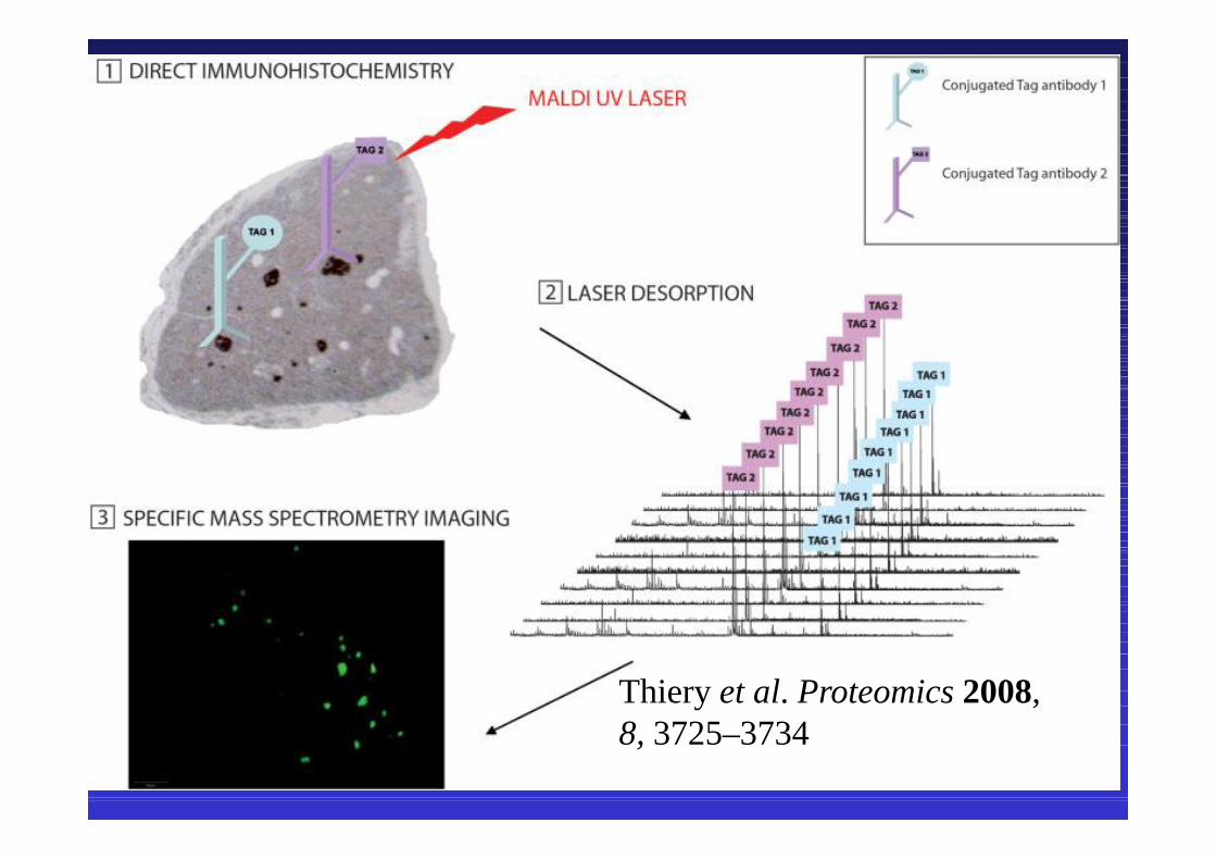
Thiery et al. Proteomics 2008, 8, 3725–3734

„normal“ MTSD Arrays
• Samples will be spotted on chip surface and immobilized
• Chip will be treated with mass tagged antibodies
• Matrix application• Under MS conditions: sensor tags will be
released and detected via TOF

Inverse MTSD Arrays
h.h.h.
• Antibodies are attached to chip surface• Sample is applied on chip, specific substrates
bind to their antibodies• Matrix application• Substrate-antibody complex gets released and
detected via TOF

Printing proteins as microarrays for high-throughput function determination
A single slide holding 10,800 spots. Protein G was printed 10,799 times
A single spot of FRB was printed in row 27, column 109. The slide was probed with MS-IMS

Clinical Proteomics UnitClinical Proteomics UnitProtein Chemistry/Proteomics and Array Unit
Institute of Biomedicine/Biochemistry
(http://research.med.helsinki.fi/corefacilities/proteinchem)


![%REP]XMGEP QIXLSHW MR 4VSXISQMGWresearch.med.helsinki.fi/corefacilities/proteinchem/Aalto 070512 Part1a.pdf · gsyph hs mx# wx hmqirwmsr -)* 4vsximrw evi witevexih eggsvhmrk xs xlimv](https://static.fdocuments.in/doc/165x107/5e6a6d7486783478684a5dbc/repxmgep-qixlshw-mr-070512-part1apdf-gsyph-hs-mx-wx-hmqirwmsr-4vsximrw.jpg)