Antidyskinetic Treatment with MTEP Affects Multiple Molecular Pathways...
9
Research Article Antidyskinetic Treatment with MTEP Affects Multiple Molecular Pathways in the Parkinsonian Striatum Jing-ya Lin, Zhen-guo Liu, Cheng-long Xie, Lu Song, and Ai-juan Yan Department of Neurology, Xin Hua Hospital Affiliated to Shanghai Jiao Tong University School of Medicine, 1665 Kongjiang Road, Shanghai 200092, China Correspondence should be addressed to Zhen-guo Liu; [email protected] Received 17 February 2017; Revised 8 May 2017; Accepted 17 September 2017; Published 30 October 2017 Academic Editor: Francisco Grandas Copyright © 2017 Jing-ya Lin et al. is is an open access article distributed under the Creative Commons Attribution License, which permits unrestricted use, distribution, and reproduction in any medium, provided the original work is properly cited. Parkinson’s disease is characterized by dopaminergic neuron loss and dopamine (DA) depletion in the striatum. Standard treatment is still focused on the restoration of dopamine with exogenous L-Dopa, which however causes L-Dopa-induced dyskinesia (LID). Several studies have shown that antagonism of the metabotropic glutamate receptor 5 alleviates LID, but the underlying mechanisms have remained unclear. We set out to determine where this alleviation may depend on restoring the equilibrium between the two main striatofugal pathways. For this purpose, we examined molecular markers of direct and indirect pathway involvement (prodynorphin and proenkephalin, resp.) in a rat model of LID treated with the mGluR5 antagonist MTEP. Our results show that MTEP cotreatment significantly attenuates the upregulation of prodynorphin mRNA induced by L-Dopa while also decreasing the expression levels of proenkephalin mRNA. We also examined markers of the mGluR5-related PKC/MEK/ERK1/2 signaling pathway, finding that both the expression of PKC epsilon and the phosphorylation of MEK and ERK1/2 had decreased significantly in the MTEP-treated group. Taken together, our results show that pharmacological antagonism of mGluR5 normalizes several abnormal molecular responses in the striatum in this experimental model of LID. 1. Introduction e loss of dopaminergic neurons in the substantia nigra and the depletion of dopamine are main neuropathological features of Parkinson’s disease (PD) [1, 2]. Current standard treatment for PD still focuses on the dopamine replacement therapy with L-Dopa [2]. However, long-term use of this drug causes a decrease in the efficacy and disabling abnormal involuntary movement (AIM), which was known as L-Dopa- induced dyskinesia (LID) [3]. e striatum is deeply involved in handling the motor information that comes from the cortex. In the classic model of striatal connectivity, there are direct and indirect pathways in the striatum [4]. Both direct and indirect pathways originate from medium spiny efferent neurons (MSNs). In the indirect pathway, the MSNs express enkephalin, while in the direct pathway the MSNs express dynorphin [5]. e first report of a direct correlation between prodynorphin level and LID was provided by Cenci et al. [6]; furthermore, they also proposed that an imbalance between the direct and indirect pathways leads to the appearance of the motor signs of parkinsonism [7]. In a recent research, it was also reported that both the dynorphin and enkephalin expression levels were involved in the development of motor complications while being treated with L-Dopa. Sgroi et al. pointed out that, on the one hand, the preproenkephalin level was increased before the use of L-Dopa aſter 6-OHDA lesion, and it remained high aſter L-Dopa washout; on the other hand, there is a correlation between the rotational AIM and preproenkephalin level in the on state [8]. All these phenomenons suggested that the increased proenkephalin mRNA level may be a prerequisite to the locomotor sensiti- zation before L-Dopa treatment [8]. On the other hand, the association of prodynorphin with LID is clearer than that with proenkephalin, because it has been consistently proven in several studies [5, 8–10]. We also know that overactivation of glutamatergic signal- ing and the hypersensitivity of the glutamatergic system in the basal ganglia play an important role in the pathophysiology of LID. Several groups of researchers reported that the blockade of the metabotropic glutamate receptor 5 (mGluR5) could attenuate the LID [11–16]; emerging evidence has come to Hindawi Parkinson’s Disease Volume 2017, Article ID 5798734, 8 pages https://doi.org/10.1155/2017/5798734
Transcript of Antidyskinetic Treatment with MTEP Affects Multiple Molecular Pathways...

Research ArticleAntidyskinetic Treatment with MTEP Affects Multiple MolecularPathways in the Parkinsonian Striatum
Jing-ya Lin, Zhen-guo Liu, Cheng-long Xie, Lu Song, and Ai-juan Yan
Department of Neurology, Xin Hua Hospital Affiliated to Shanghai Jiao Tong University School of Medicine,1665 Kongjiang Road, Shanghai 200092, China
Correspondence should be addressed to Zhen-guo Liu; [email protected]
Received 17 February 2017; Revised 8 May 2017; Accepted 17 September 2017; Published 30 October 2017
Academic Editor: Francisco Grandas
Copyright © 2017 Jing-ya Lin et al. This is an open access article distributed under the Creative Commons Attribution License,which permits unrestricted use, distribution, and reproduction in any medium, provided the original work is properly cited.
Parkinson’s disease is characterized by dopaminergic neuron loss and dopamine (DA) depletion in the striatum. Standard treatmentis still focused on the restoration of dopamine with exogenous L-Dopa, which however causes L-Dopa-induced dyskinesia (LID).Several studies have shown that antagonismof themetabotropic glutamate receptor 5 alleviates LID, but the underlyingmechanismshave remained unclear. We set out to determine where this alleviation may depend on restoring the equilibrium between thetwo main striatofugal pathways. For this purpose, we examined molecular markers of direct and indirect pathway involvement(prodynorphin and proenkephalin, resp.) in a rat model of LID treated with the mGluR5 antagonist MTEP. Our results show thatMTEP cotreatment significantly attenuates the upregulation of prodynorphin mRNA induced by L-Dopa while also decreasingthe expression levels of proenkephalin mRNA. We also examined markers of the mGluR5-related PKC/MEK/ERK1/2 signalingpathway, finding that both the expression of PKC epsilon and the phosphorylation of MEK and ERK1/2 had decreased significantlyin the MTEP-treated group. Taken together, our results show that pharmacological antagonism of mGluR5 normalizes severalabnormal molecular responses in the striatum in this experimental model of LID.
1. Introduction
The loss of dopaminergic neurons in the substantia nigraand the depletion of dopamine are main neuropathologicalfeatures of Parkinson’s disease (PD) [1, 2]. Current standardtreatment for PD still focuses on the dopamine replacementtherapy with L-Dopa [2]. However, long-term use of thisdrug causes a decrease in the efficacy and disabling abnormalinvoluntary movement (AIM), which was known as L-Dopa-induced dyskinesia (LID) [3].
The striatum is deeply involved in handling the motorinformation that comes from the cortex. In the classic modelof striatal connectivity, there are direct and indirect pathwaysin the striatum [4]. Both direct and indirect pathwaysoriginate from medium spiny efferent neurons (MSNs). Inthe indirect pathway, the MSNs express enkephalin, whilein the direct pathway the MSNs express dynorphin [5]. Thefirst report of a direct correlation between prodynorphinlevel and LID was provided by Cenci et al. [6]; furthermore,they also proposed that an imbalance between the direct andindirect pathways leads to the appearance of the motor signs
of parkinsonism [7]. In a recent research, it was also reportedthat both the dynorphin and enkephalin expression levelswere involved in the development of motor complicationswhile being treated with L-Dopa. Sgroi et al. pointed outthat, on the one hand, the preproenkephalin level wasincreased before the use of L-Dopa after 6-OHDA lesion,and it remained high after L-Dopa washout; on the otherhand, there is a correlation between the rotational AIMand preproenkephalin level in the on state [8]. All thesephenomenons suggested that the increased proenkephalinmRNA level may be a prerequisite to the locomotor sensiti-zation before L-Dopa treatment [8]. On the other hand, theassociation of prodynorphin with LID is clearer than thatwith proenkephalin, because it has been consistently provenin several studies [5, 8–10].
We also know that overactivation of glutamatergic signal-ing and the hypersensitivity of the glutamatergic system in thebasal ganglia play an important role in the pathophysiology ofLID. Several groups of researchers reported that the blockadeof the metabotropic glutamate receptor 5 (mGluR5) couldattenuate the LID [11–16]; emerging evidence has come to
HindawiParkinson’s DiseaseVolume 2017, Article ID 5798734, 8 pageshttps://doi.org/10.1155/2017/5798734

2 Parkinson’s Disease
6-OHDA/salinelesioned
AIM test//cyber test
Treatment phase
Day: 214
125
Producing model
Western blot
Apomorphineinduction Sacri�ce
Q-PCR
Figure 1: The protocol of the experiment. Dopamine depletion was induced by 6-OHDA injections in the medial forebrain bundle (MFB)while the sham groupwas injectedwith saline in theMFB.Threeweeks later, the rats that exhibited apomorphine-induced rotations exceeding7 turns/min in the apomorphine induction test were put to the subsequent experiment. The sham and PD groups were injected with salineonce daily; the LID group received L-Dopa (25mg/kg) and benserazide (6.25mg/kg) cocktail once daily, while the MTEP group receivedMTEP (5mg/kg) 30min before the injection of L-Dopa and benserazide. All treatments were performed for 14 days; behavior tests like AIMtest, open field test, and cylinder test were performed on days 2, 5, 12, and 14. Two hours after the last injection on the 14th day, all groupswere sacrificed for western blot and Q-PCR.
support the important role of mGluR5 in the developmentof LID [17]. But the mechanism behind the alleviation effectis unclear [18]. It is of great importance to investigate theextent to which the blockade of the metabotropic receptor 5affects the imbalance between direct and indirect pathwaysand to investigate what the molecular alterations in themGluR5-related signaling pathway are, in order to interpretthe antidyskinesia effect of the antagonists of mGluR5. Inorder to address these questions, we tested the proteinlevel of protein kinase C (PKC), MEK, and extracellularsignal-regulated kinase 1/2 (ERK1/2) in the mGluR5 medi-ated PKC/MEK/ERK1/2 signaling pathway; we also testedthe mRNA expression level of prodynorphin (PDyn) andproenkephalin (PEnk) in order to verify the effect of theblockade of mGluR5 on the direct and indirect pathways.
2. Materials and Methods
2.1. Experimental Design. As shown in Figure 1, Sprague-Dawley rats were given 6-OHDA injections in the medialforebrain bundle (MFB) in the right side of the brain, whilefive SD rats were given saline injections instead as shamgroup. Contralateral turning behavior was tested on the 6-OHDA rats after the apomorphine injection. The rats whoseapomorphine-induced rotations are more than 7 turns/minwere enrolled in the follow-up experiments as the Parkinsondisease model animals. The selected rats were distributedinto 3 groups randomly. The first group was given L-Dopa(25mg/kg, i.p.) plus benserazide (6.25mg/kg, i.p.) once dailyfor 14 days, labeled as LID group; the second group was givensaline once daily for 14 days, labeled as PD group; and thethird group was given MTEP (5mg/kg, i.p.) 20mins beforethe injection of L-Dopa plus benserazide for 14 days, labeledasMTEP group. During this period, AIM and open field testswere conducted in all the groups on days 2, 5, 8, 12, and 14 bya new assigned observer who did not know the details of eachgroup. The animals were sacrificed 2 h after the last injectionfor western blot and Q-PCR.
2.2. Animals. The study was conducted on adult femaleSprague-Dawley rats (Sprague-Dawley, 180–220 g, Sippr-BK
Ltd., Shanghai, China). The maintenance of the animalsfollowed the guidelines of the National Institutes of Healthfor the care and use of laboratory animals. All experimentalprotocols involving animals were approved by the EthicalCommittee of the Medical School of Shanghai JiaotongUniversity.
2.3. Drugs. L-Dopa and benserazide were purchased fromSigma-Aldrich (Spain), and MTEP was purchased fromAbcam (UK). All drugs were freshly prepared in 0.9%saline before use. L-Dopa (Sigma-Aldrich) plus benserazide(Sigma-Aldrich) was administrated once daily. MTEP (3-[(2-methyl-1,3-thiazol-4-yl)ethynyl]-pyridine, Abcam, UK)preceded the L-Dopa cocktail 20 minutes earlier once dailyfor 2 weeks.
2.4. 6-OHDA Lesions and Treatment. For the stereotaxic pro-cedure, the rats (weighing 180∼220 g) were anesthetizedwith 10% chloral hydrate (0.5ml/100 g) deeply. As previouslydescribed [19, 20], the surgery was performed on the rightside medial forebrain bundle (MFB) by unilateral injectionof 6-OHDA (20mmol/L, containing 0.02% ascorbic acid;Sigma-Aldrich, Spain) at the coordination of MFB. Sham-operated rats received the vehicle at the same spot. A volumeof 4 𝜇l was injected in each spot. 21 days later, all therats were tested with 0.05mg/kg subcutaneous injection ofapomorphine (i.p. WOKO, Japan). Contralateral rotationtest was performed and the animals exhibiting full bodyturns of over 7 turns/min towards the unlesioned side wereenrolled and started on a 2-week course of daily i.p. injectionsof MTEP (5mg/kg) followed by L-Dopa (25mg/kg) plusbenserazide (6.25mg/kg) 20min later.
2.5. Behavior Assessment. To evaluate LID, we used thecombined “time ∗ amplitude” scale which was first appliedby Rylander et al. [21]; mice were observed in a clear-glass cylinder and were observed and evaluated by a trainedexperimenter. Rat abnormal involuntary movements (AIMs)were classified into three subtypes: axial, limb, and orolingualdyskinesia. Each individual dyskinesia subtype scores from 0to 4. During a period of 120min following levodopa injection,

Parkinson’s Disease 3
the severity of AIM was assessed at a 20min interval (20, 40,60, 80, 100, and 120min). The ALO AIM scores were ratedat 2, 5, 12, and 14 days during levodopa treatment. Motorcoordination was evaluated with the cylinder test at 2, 5, 12,and 14 days and the locomotor activities were tested by theopen field test on the 2nd and 14th days during levodopatreatment. The open field test and cylinder test were theindex of Parkinsonian disability. In the cylinder test, the ratswere placed in a glass cylinder with a diameter of 22 cmand a height of 35 cm to record forelimb use during verticalexploration for 60min. During a period of 60min beforelevodopa treatment, the forelimb functional test was assessedevery 15min (3min monitoring period for each). The finalvalue was expressed in terms of the percentage use of theimpaired forelimb compared with the total number of limbuse movements. All the behavioral experiments were carriedout with the observer blinded to the groups and treatment.
2.6. Western Blot. Striatum tissue of rats was harvested 2hours after the last injection of L-Dopa and homogenizedin RIPA lysis buffer (Beyotime Institute of Biotechnology)and fresh-added protease inhibitor cocktail and phosphataseinhibitor (Roche Diagnostics, Switzerland). And then thecytosol was prepared by centrifugation at 12000𝑔 for 10minat 4∘C. An equal amount of protein (40 ug) from each samplewas added to 10% SDS-PAGE and separated by electrophore-sis and transferred to polyvinylidene difluoride membranesin a Tris-glycine transfer buffer. Each sample was heatedat 95∘C previously for 5min. The membrane was blockedfor half an hour at room temperature (26∘C) in 5% instantnonfat milk and then incubated with primary antibodiescorresponding to epsilon PKC (1 : 1000, Abcam, UK), p-MEK andMEK (1 : 1000, Abcam, UK), p-ERK1/2 and ERK1/2(1 : 1000, Cell Signaling Technology (CST), USA), and 𝛽-actinIgG (diluted 1 : 1000; Beyotime Institute of Biotechnology),respectively, at 4∘C overnight (14–16 hours). The membraneswere subsequently washed with TBST (50mM Tris-HCl(pH 7.5), 150mM NaCl, and 0.05% Tween 20) and thenincubated with horseradish peroxidase conjugated secondaryanti-rabbit and anti-mouse IgG (diluted 1 : 1000; BeyotimeInstitute of Biotechnology) for one hour at room temperature.The signal was visualized by ECL (A : B = 1 : 1; Millipore)and quantified using Quantity One software (Image Lab). Allindividual protein bands were compared with their internalcontrol actin values in order to provide relative proteinabundance. All the procedures were repeated 3 times.
2.7. Real-Time PCR. Striatal tissues of rats were homogenizedand total ribonucleic acid (RNA) was extracted by TRIzolreagent (Invitrogen, USA). cDNA was generated from totalRNA samples using the Revert Aid First Strand cDNASynthesis Kit (Takara, Japan). Q-PCR was performed usingthe ABI 7500 Real-Time PCR System (Life Technologies,USA) according to the supplier’s instructions. The primersequences used in this study were as follows:
5-CTTGTGTTCCCTGTGTGCAGTG-3 (forward)3-AGCAACCTCATTCTCCAAGTCA-5 (reverse) forPDyn mRNA
5-GAAGATGGATGAGCTTTACCCC-3 (forward)3-CAAGGTGTCTCCCTCATCTGC-5 (reverse) forproenkephalin mRNA
Amplification was performed with 40 cycles of denat-uration at 95∘C for 15 s, annealing at 60∘C for 60 s, andextension at 75∘C for 20 s using the ABI 7300 Real-TimePCR System (Applied Biosystems, CA, USA). Results wereexpressed as relative expression corrected to the GAPDHgene. The detector used in real-time PCR reaction is SYBRGreen.
2.8. Statistical Analysis. Data were expressed as the mean ±standard deviation (SD) unless stated otherwise. Behavioraldata were analyzed using Kruskal-Wallis test followed byDunn’s test formultiple comparisons in the case of comparingdata overmultiple days, or aMann–Whitney𝑈 test.Thewest-ern blot and Q-PCR conformed to normal distribution, andanalyses of their data were performed using one-way analysisof variance (ANOVA) followed by LSD post hoc comparisonswhen appropriate as indicated in the figure legends. 𝑝 values< 0.05 were considered statistically significant. Analysis wasperformed with GraphPad Prism 5.
3. Result
3.1. MTEP Prevented the Development of L-Dopa-InducedDyskinesia. The PD group received saline injection over14 days, while the LID group was injected with L-Dopa(25mg/kg) plus benserazide (6.25mg/kg). The MTEP groupwas given MTEP (5mg/kg, i.p.) 20min before the L-Dopacocktail injection.The evaluation of the AIM scores included3 subtypes: axial, limb, and orolingual AIMs. The scoresdemonstrated a rat dyskinesia scale.We found that 2 weeks ofL-Dopa treatment induced full development of LID features,as demonstrated by the increased ALO AIM scores in theLID rats (𝑝 < 0.05 for treatment effect, 𝑝 < 0.05 for timeeffect, and 𝑝 < 0.01 for treatment and time interaction,Figure 2). This result is in line with our previous study.AIM scores decreased in the MTEP group. MTEP treatmentfor 14 days significantly reduced the total dyskinesia scoreswhile the rats of PD group that received saline for 14 daysdid not develop dyskinesia. Furthermore, the MTEP groupdemonstrated a reduction in all testing sessions. These dataindicate that treatment withMTEP significantly inhibited thedevelopment of LID.
3.2. MTEP Did Not Compromise the Anti-Parkinsonian Effectof L-Dopa. We then sought to determine whether theadministration of antagonists of mGluR5 ameliorated LIDcompromised the therapeutic response to L-Dopa in PD rats.The cylinder test was used to assess spontaneous forelimb use.We conducted the cylinder test on the 2nd, 5th, 12th, and14th days. We observed that 6-OHDA-lesioned rats treatedwith L-Dopa prefer to use the contralateral forelimb to touchthe inner wall of the cylinder compared with the 6-OHDA-lesioned rats treated with saline, but the preferential useof the contralateral forelimb was lower compared with thesham group rats (Figure 3, ∗𝑝 < 0.01). Data showed that

4 Parkinson’s Disease
∗∗
∗∗
∗ ∗
∗ ∗
LIDPD
MTEP (5 mg/kg)
5 12 142Day of L-Dopa treatment
0
20
40
60G
loba
l ALO
AIM
scor
e
(a)
∗ ∗
∗∗
∗∗∗
∗
LIDPD
MTEP (5 mg/kg)
0
5
10
15
20
25
Axi
al A
LO A
IM sc
ore
5 12 142Day of L-Dopa treatment
(b)
∗
∗
∗∗
∗∗
∗ ∗
LIDPD
MTEP (5 mg/kg)
0
5
10
15
Lim
b A
LO A
IM sc
ore
5 12 142Day of L-Dopa treatment
(c)
∗∗
∗
∗
∗ ∗
∗ ∗
LIDPD
MTEP (5 mg/kg)
0
5
10
15
20
Oro
lingu
al A
LO A
IM sc
ore
5 12 142Day of L-Dopa treatment
(d)
Figure 2: Effect of MTEP on the AIM scores. 14 days’ use of MTEP significantly reduced AIM scores. At the 2nd, 5th, 12th, and 14th days, atotal AIM score was calculated as the sum of the basic scores multiplied by the amplitude of the score for each AIM subtype: limb, orolingual,and axial, excluding the rotation subtype. (a) Time course of the total scores; sum of the axial, limb, and orolingual subtype scores; (b) timecourse of changes in the axial scores; (c) time course of changes in the limb score; and (d) time course of changes in the orolingual score. Ineach testing session, the AIM scores were rated following the administration of the drugs. Data are presented as the mean ± SD. ∗𝑝 < 0.01versus the LID group (Kruskal-Wallis test followed by Dunn’s test for multiple comparisons or Mann–Whitney 𝑈 test).
the coinjection of MTEP with L-Dopa did not impact thepreferential use of the contralateral forelimb (Figure 3, #𝑝 <0.05). There is no significant difference between the LIDgroup and the MTEP group.
3.3. Blockade of mGluR5 Prevents the Expression of PKC andPhosphorylation of MEK and ERK1/2 Protein Level. Proteinkinase C was reported to contribute to the development ofLID; in particular, the expression level of the novel PKCisoform, PKC epsilon, ipsilateral to the lesion side of thestriatum, was increased after chronic L-Dopa treatment [22].Here, we confirmed that intermittent administration of L-Dopa in hemi-Parkinsonian animals greatly increased theexpression level of epsilon PKC, but this enhancement was
reversed by the injection of MTEP (#𝑝 < 0.05, Figure 4(a))compared with the LID group. It was documented that L-Dopa produces pronounced activation of ERK1/2 signalingin the dopamine-denervated striatum through aD1-receptor-dependent mechanism. This effect is associated with thedevelopment of dyskinesia [23]. In this study, we found thatthis elevation in p-MEK and p-ERK1/2 level was reduced intheMTEP group (#𝑝 < 0.05, Figures 4(b) and 4(c)) comparedwith the LID group. The MTEP group also showed a minorbut significant reduction compared with PD in the PKCexpression level and phosphorylation of MEK (∗𝑝 < 0.05,Figures 4(b) and 4(c)), but there is no significant differencein the phosphorylation level of ERK1/2 between the PD andthe MTEP groups (𝑝 > 0.05).

Parkinson’s Disease 5
PDLIDMTEP
Sham
#
##
##
∗#∗#∗#
∗#
∗#∗#
∗#∗#
∗
∗
∗
∗
0.0
0.2
0.4
0.6
0.8
L/(L
+ R
)
5 12 142(Day)
Figure 3: Spontaneous forelimb use of the rat in various experimen-tal groups. Cylinder test. ∗ indicates a significant decrease relativeto the sham group (∗𝑝 < 0.01), and # indicates a significant increasefrom PD group (#𝑝 < 0.05). Columns indicate the mean, and barsindicate the SD; the cylinder test was performed on the 2nd, 5th,12th, and 14th days, using one-way ANOVA followed by Bonferronipost hoc tests.
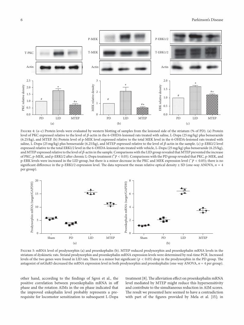
3.4. MTEP Reduces the Expression of Prodynorphin andProenkephalin in Parkinsonian Rats with LID. The mRNAexpression levels of prodynorphin and proenkephalin weremeasured by real-time PCR. We found that prodynorphin(Figure 5(a)) expression has a minor reduction in thePD group, but it increased in the LID group; however,MTEP treatment significantly reversed the tendency. Theproenkephalin (Figure 5(b)) levels were increased in PD ratsprimed with L-Dopa. However, MTEP significantly reducedthe expression level of proenkephalin in PD rats primed withL-Dopa.
4. Discussion
Hypersensitivity and overactivation of glutamatergic signal-ing in the basal ganglia play a key role in the developmentof LID [24]. We used a hemi-Parkinsonian rat model ofdyskinesia based on unilateral 6-OHDA striatal injection inthe MFB, followed with the intraperitoneal administrationof chronic L-Dopa daily at a dose of 25mg/kg in the LIDgroup and saline in the PD group and the injection of MTEP20min before the L-Dopa cocktail in the MTEP group. Inaccord with our previous work [20, 25, 26], these 6-OHDA-lesioned Parkinsonian rats developed progressive dyskinesiafollowing the chronic use of L-Dopa (Figures 2(a)–2(d)). Wealso confirmed that antagonizing mGluR5 reduced the AIMscores in the rat model animals without ablating the anti-Parkinsonian effect of L-Dopa (Figure 3). In this presentstudy, we explored the possible mechanism of the alleviationeffect mediated by the antagonist of mGluR5. We found thatPKC level increased in the LID group, which was consistentwith the findings of Smith et al.; they also found that theantagonization of PKC could reduce the motor symptoms
of LID [22]. We also come up with the result that thephosphorylation of ERK1/2 and MEK was reduced in theMTEP group (Figures 4(a)–4(c)).
Numerous researches had already shown that the PKAsignaling pathway in the striatum is closely related to the acti-vation of D1R, which was deeply involved in the expressionof LID [19, 20, 27]. After the enhancement of PKA signaling,many downstream molecules like ERK1/2 were upregulated.The phosphorylation of ERK1/2 was also closely correlatedwith the appearance of L-Dopa-induced dyskinesia [27–29].
It was well documented that there is a close functionalinteraction between the D1R and the mGluR5; with the long-term use of L-Dopa, the PKC/MEK/ERK1/2 pathway wasactivated [30], while blockade of the mGluR5 might call backthe overactivated signal pathways.
It is well established that the opioidergic neuropeptidesdynorphin and enkephalin are also involved in the striatalcontrol of motor and behavioral function [8]. Changes inthe striatal expression of proenkephalin and prodynorphinmRNA level have been reported in Parkinsonian rats with L-Dopa-induced dyskinesia. It is known that increased PDynmRNA is attributed to increased activity in the direct path-way; the expression level of PDyn mRNA has also beenreported to be closely related to the genesis of LID [31].Enkephalin is an important striatal marker in the indirectpathway. In the classic model of the basal ganglia circuit, theenhancement of activities in the direct pathway increases theexpression level of prodynorphin mRNA, and the inhibitionof the indirect pathway was also reinforced [8, 32]. All thesemolecular changes lead to further asymmetrical pathologicalchanges and further asymmetrical dysfunction of the neuralcircuit in the basal ganglia. To our knowledge, it is nowestablished that 6-OHDA-lesioned Parkinsonian rats havean abnormal increase in the mRNA level of the direct(prodynorphin) and indirect (proenkephalin) markers [5, 7,8]; in this present study, we found that striatal mRNA ofproenkephalin was increased in PD rats and continued toincrease after intermittent use of L-Dopa; as for the mRNAlevel of prodynorphin, it exhibited aminor drop in 6-OHDA-lesioned PD rats and this tendency reversed (increased)significantly after priming with L-Dopa in the followingdays (Figures 5(a) and 5(b)); these were consistent with thefindings of Sgroi et al. [8]. After the use of the antagonistof mGluR5, as is shown in Figure 2, coadministration ofMTEP (5mg/kg, i.p.) with L-Dopa (25mg/kg, i.p.) did notdevelop severe dyskinesia over the 14-day treatment; theantagonization of the mGluR5 downregulated the overacti-vated phosphorylation of the ERK1/2 via PKC/MEK/ERK1/2pathways. On the other hand, the overactivation of the PKApathway induced the enhancement in both prodynorphinand proenkephalin mRNA expression level after L-Dopapriming. In this study, we demonstrated that, with theantagonizing of themGluR5, the L-Dopa induced an increaseof the expression level of the phosphorylation of ERK1/2,and the mRNA expression levels of prodynorphin andproenkephalin were reduced in the 6-OHDA lesioned rats.On the one hand, the downregulation of both prodynorphinand proenkephalin helps to restore the imbalance of the basalganglia circuit in the direct and indirect pathways. On the
6 Parkinson’s Disease
0.0
0.5
1.0
1.5
2.0
2.5
PKC
relat
ive d
ensit
y
LIDPD MTEP
T-PKC
Actin
##∗
∗
(a)
Actin
#∗#
∗
P-MEK
T-MEK
0
1
2
3
MEK
relat
ive d
ensit
y
LID MTEPPD(b)
#∗#
∗
Actin
P-ERK1/2
T-ERK1/2
0.0
0.5
1.0
1.5
2.0
ERK
relat
ive d
ensit
y
LID MTEPPD(c)
Figure 4: (a–c) Protein levels were evaluated by western blotting of samples from the lesioned side of the striatum (% of PD). (a) Proteinlevel of PKC expressed relative to the level of 𝛽-actin in the 6-OHDA-lesioned rats treated with saline, L-Dopa (25mg/kg) plus benserazide(6.25/kg), and MTEP. (b) Protein level of p-MEK level expressed relative to the total MEK level in the 6-OHDA-lesioned rats treated withsaline, L-Dopa (25mg/kg) plus benserazide (6.25/kg), and MTEP expressed relative to the level of 𝛽-actin in the sample. (c) p-ERK1/2 levelexpressed relative to the total ERK1/2 level in the 6-OHDA-lesioned rats treated with vehicle, L-Dopa (25mg/kg) plus benserazide (6.25/kg),andMTEP expressed relative to the level of𝛽-actin in the sample. Comparisonswith the LID group revealed thatMTEPprevented the increaseof PKC, p-MEK, and p-ERK1/2 after chronic L-Dopa treatment (#𝑝 < 0.05). Comparisons with the PD group revealed that PKC, p-MEK, andp-ERK levels were increased in the LID group, but there is a minor decrease in the PKC and MEK expression level (∗𝑝 < 0.05); there is nosignificant difference in the p-ERK1/2 expression level. The data represent the mean relative optical density ± SD (one-way ANOVA, 𝑛 = 4per group).
0
5
10
15
20
Prod
ynor
phin
mRN
A (t
arge
t/GA
PDH
)
PD LIDSham MTEP(a)
0
2
4
6
8
Proe
nkep
halin
mRN
A (t
arge
t/GA
PDH
)
PD LIDSham MTEP(b)
Figure 5: mRNA level of prodynorphin (a) and proenkephalin (b). MTEP reduced prodynorphin and proenkephalin mRNA levels in thestriatum of dyskinetic rats. Striatal prodynorphin and proenkephalin mRNA expression levels were determined by real-time PCR. Increasedlevels of the two genes were found in LID rats. There is a minor but significant (𝑝 < 0.05) drop in the prodynorphin in the PD group. Theantagonist of mGluR5 decreased the mRNA expression level in both prodynorphin and proenkephalin (one-way ANOVA, 𝑛 = 4 per group).
other hand, according to the findings of Sgroi et al., thepositive correlation between proenkephalin mRNA in offphase and the rotation AIMs in the on phase indicated thatthe improved enkephalin level probably represents a pre-requisite for locomotor sensitization to subsequent L-Dopa
treatment [8].The alleviation effect on proenkephalinmRNAlevel mediated by MTEP might reduce this hypersensitivityand contribute to the simultaneous reduction in AIM scores.The result we presented here seemed to have a contradictionwith part of the figures provided by Mela et al. [15]; in
Parkinson’s Disease 7
their paper, they found that the acute injection of L-Dopatogether with MTEP did not modify the upregulation ofthe proenkephalin mRNA induced by DA degeneration, butMela et al. also confirmed in that very paper that, during thechronic L-Dopa treatment, the mGluR5 antagonism partiallyblocked the additional upregulation of both prodynorphinand proenkephalin, which was consistent with our presenteddata. Furthermore, on the one hand, it was reported that theDA can exert its effect on the D2 receptor through a non-cAMP-dependent way [33]; it upregulated the phosphory-lation of Akt/GSK3𝛽𝛽 pathway to affect the DA-dependentbehavior; on the other hand, recent research shows that theantagonism of mGluR5 could inhibit the phosphorylation ofAkt/GSK3𝛽𝛽 [34]; this might contribute to the restoration ofthe activity of the indirect pathway and might be followed bya decrease of PPE.
5. Conclusion
Hypersensitivity and overactivation of glutamatergic signal-ing in the basal ganglia play a key role in the development ofLID. Antagonizing mGluR5 could reduce the AIM scores inthe rodent and primate PD model animals. While the antag-onist downregulates the signaling on the PKC/MEK/ERK1/2pathways, it also reduced the expression level of prodynor-phin and proenkephalin mRNA significantly. Antidyskinetictreatment withMTEP affects multiple molecular pathways inthe Parkinsonian striatum.
Conflicts of Interest
The authors declare that this research was conducted in theabsence of any commercial or financial relationships thatcould be construed as potential conflicts of interest.
Authors’ Contributions
Jing-ya Lin conceived the study and participated in the designof the manuscript and extracted data and helped draft themanuscript. Cheng-long Xie and Lu Song prepared figuresand offered technical support during the whole process. Ai-juan Yan carried out the statistical analysis and interpretationof data. Zhen-guo Liu participated in the conceptualizationand design of the experiment and data extraction and analysisand drafted the manuscript. All authors read and approvedthe final manuscript.
Acknowledgments
This study was supported by projects of the National NaturalScience Foundation of China (81400925, 81471148, 81671273,81771211, and 81703852), projects of the Shanghai Committeeof Science and Technology (17401901000), National Key R&DProgram of China (2017YFC1310300), and SHSMU-IONResearch Center for Brain Disorders (2015NKX007).
References
[1] M. Feyder, A. Bonito-Oliva, and G. Fisone, “L-DOPA-induceddyskinesia and abnormal signaling in striatal medium spiny
neurons: Focus on dopamine D1 receptor-mediated transmis-sion,” Frontiers in Behavioral Neuroscience, no.OCTOBER, 2011.
[2] P. Calabresi, M. D. Filippo, V. Ghiglieri, N. Tambasco, andB. Picconi, “Levodopa-induced dyskinesias in patients withParkinson’s disease: filling the bench-to-bedside gap,” TheLancet Neurology, vol. 9, no. 11, pp. 1106–1117, 2010.
[3] F. Niccolini, L. Rocchi, and M. Politis, “Molecular imagingof levodopa-induced dyskinesias,” Cellular and Molecular LifeSciences, vol. 72, no. 11, pp. 2107–2117, 2015.
[4] C.Winkler, D. Kirik, A. Bjorklund, andM. A. Cenci, “L-DOPA-induced dyskinesia in the intrastriatal 6-hydroxydopaminemodel of Parkinson’s disease: Relation to motor and cellularparameters of nigrostriatal function,” Neurobiology of Disease,vol. 10, no. 2, pp. 165–186, 2002.
[5] C. Marin, M. Bonastre, G. Mengod, R. Cortes, and M. C.Rodrıguez-Oroz, “From unilateral to bilateral parkinsonism:Effects of lateralization on dyskinesias and associatedmolecularmechanisms,” Neuropharmacology, vol. 97, pp. 365–375, 2015.
[6] M. A. Cenci, C. S. Lee, and A. Bjorklund, “L-DOPA-induceddyskinesia in the rat is associated with striatal overexpressionof prodynorphin- and glutamic acid decarboxylase mRNA,”European Journal of Neuroscience, vol. 10, no. 8, pp. 2694–2706,1998.
[7] M. A. Cenci, “Dopamine dysregulation of movement control inl-DOPA-induced dyskinesia,” Trends in Neurosciences, vol. 30,no. 5, pp. 236–243, 2007.
[8] S. Sgroi, C. Capper-Loup, P. Paganetti, and A. Kaelin-Lang,“Enkephalin and dynorphin neuropeptides are differently cor-related with locomotor hypersensitivity and levodopa-induceddyskinesia in parkinsonian rats,” Experimental Neurology, vol.280, pp. 80–88, 2016.
[9] D. Rylander, A. Recchia, F. Mela, A. Dekundy, W. Danysz, andM. A. Cenci Nilsson, “Pharmacological modulation of glu-tamate transmission in a rat model of L-DOPA-induced dyski-nesia: Effects on motor behavior and striatal nuclear signaling,”The Journal of Pharmacology and Experimental Therapeutics,vol. 330, no. 1, pp. 227–235, 2009.
[10] X.-B. Cao, Q. Guan, Y. Xu, L.Wang, and S.-G. Sun, “Mechanismof over-activation in direct pathway mediated by dopamineD1 receptor in rats with levodopa-induced dyskinesias,” Neu-roscience Bulletin, vol. 22, no. 3, pp. 159–164, 2006.
[11] K. A. Johnson, P. J. Conn, and C. M. Niswender, “Glutamatereceptors as therapeutic targets for Parkinson’s disease,” CNS &Neurological Disorders—Drug Targets, vol. 8, no. 6, pp. 475–491,2009.
[12] F. Tison, C. Keywood, M. Wakefield et al., “A Phase 2A Trialof the Novel mGluR5-Negative Allosteric Modulator Dipraglu-rant for Levodopa-Induced Dyskinesia in Parkinson’s Disease,”Movement Disorders, vol. 31, no. 9, pp. 1373–1380, 2016.
[13] A. R. Salomons, N. E. Pinzon, H. Boleij et al., “Differentialeffects of diazepam and MPEP on habituation and neuro-behavioural processes in inbred mice,” Behavioral and BrainFunctions, vol. 8, article 30, 2012.
[14] O. Rascol, S. Perez-Lloret, and J. J. Ferreira, “New treatments forlevodopa-induced motor complications,” Movement Disorders,vol. 30, no. 11, pp. 1451–1460, 2015.
[15] F. Mela, M. Marti, A. Dekundy, W. Danysz, M. Morari, and M.A. Cenci, “Antagonism ofmetabotropic glutamate receptor type5 attenuates L-DOPA-induced dyskinesia and its molecular andneurochemical correlates in a rat model of Parkinson’s disease,”Journal of Neurochemistry, vol. 101, no. 2, pp. 483–497, 2007.

8 Parkinson’s Disease
[16] A. Dekundy, M. Pietraszek, D. Schaefer, M. A. Cenci, and W.Danysz, “Effects of group I metabotropic glutamate receptorsblockade in experimental models of Parkinson’s disease,” BrainResearch Bulletin, vol. 69, no. 3, pp. 318–326, 2006.
[17] V. Sgambato-Faure and M. A. Cenci, “Glutamatergic mecha-nisms in the dyskinesias induced by pharmacological dopaminereplacement and deep brain stimulation for the treatment ofParkinson’s disease,” Progress in Neurobiology, vol. 96, no. 1, pp.69–86, 2012.
[18] F. Nicoletti, J. Bockaert, G. L. Collingridge et al., “Metabotropicglutamate receptors: from the workbench to the bedside,”Neuropharmacology, vol. 60, no. 7-8, pp. 1017–1041, 2011.
[19] L. Song, Z. Zhang, R. Hu et al., “Targeting the D1-N-methyl-D-aspartate receptor complex reduces L-dopa-induced dyskinesiain 6-hydroxydopamine-lesioned Parkinson’s rats,”Drug Design,Development andTherapy, vol. 10, pp. 547–555, 2016.
[20] C.-L. Xie, J.-Y. Lin, M.-H. Wang et al., “Inhibition of GlycogenSynthase Kinase-3𝛽 (GSK-3𝛽) as potent therapeutic strategy toameliorates L-dopa-induced dyskinesia in 6-OHDA parkinso-nian rats,” Scientific Reports, vol. 6, Article ID 23527, 2016.
[21] D. Rylander, H. Iderberg, Q. Li et al., “A mGluR5 antago-nist under clinical development improves L-DOPA-induceddyskinesia in parkinsonian rats and monkeys,” Neurobiology ofDisease, vol. 39, no. 3, pp. 352–361, 2010.
[22] C. P. S. Smith, J. D. Oh, F. Bibbiani, M. A. Collins, I. Avila,and T. N. Chase, “Tamoxifen effect on L-DOPA inducedresponse complications in parkinsonian rats and primates,”Neuropharmacology, vol. 52, no. 2, pp. 515–526, 2007.
[23] J. E. Westin, L. Vercammen, E. M. Strome, C. Konradi, and M.A. Cenci, “Spatiotemporal pattern of striatal ERK1/2 phospho-rylation in a rat model of L-DOPA-induced dyskinesia and therole of dopamineD1 receptors,”Biological Psychiatry, vol. 62, no.7, pp. 800–810, 2007.
[24] H. Awad, G. W. Hubert, Y. Smith, A. I. Levey, and P. J. Conn,“Activation of metabotropic glutamate receptor 5 has directexcitatory effects and potentiates NMDA receptor currents inneurons of the subthalamic nucleus,” The Journal of Neuro-science, vol. 20, no. 21, pp. 7871–7879, 2000.
[25] S. Zhang, C. Xie, Q. Wang, and Z. Liu, “Interactions ofCaMKII with dopamine D2 receptors: roles in levodopa-induced dyskinesia in 6-hydroxydopamine lesioned Parkinson’srats,” Scientific Reports, vol. 4, article 6811, 2014.
[26] H. S. Lindgren, D. R. Andersson, S. Lagerkvist, H. Nissbrandt,and M. A. Cenci, “L-DOPA-induced dopamine efflux in thestriatum and the substantia nigra in a rat model of Parkinson’sdisease: Temporal and quantitative relationship to the expres-sion of dyskinesia,” Journal of Neurochemistry, vol. 112, no. 6, pp.1465–1476, 2010.
[27] X. Yang, H. Zhao, H. Shi et al., “Intranigral administration ofsubstance P receptor antagonist attenuated levodopa-induceddyskinesia in a rat model of Parkinson’s disease,” ExperimentalNeurology, vol. 271, pp. 168–174, 2015.
[28] E. Santini, E. Valjent, and G. Fisone, “Parkinson’s disease:levodopa-induced dyskinesia and signal transduction,” FEBSJournal, vol. 275, no. 7, pp. 1392–1399, 2008.
[29] N. Pavon, A. B. Martın, A. Mendialdua, and R. Moratalla,“ERK phosphorylation and FosB expression are associatedwith L-DOPA-induced dyskinesia in hemiparkinsonian mice,”Biological Psychiatry, vol. 59, no. 1, pp. 64–74, 2006.
[30] T. Fieblinger, I. Sebastianutto, C. Alcacer et al., “Mechanismsof dopamine D1 receptor-mediated ERK1/2 activation in the
parkinsonian striatum and their modulation by metabotropicglutamate receptor type 5,”The Journal of Neuroscience, vol. 34,no. 13, pp. 4728–4740, 2014.
[31] N. Yamamoto and J.-J. Soghomonian, “Metabotropic gluta-matemGluR5 receptor blockade opposes abnormal involuntarymovements and the increases in glutamic acid decarboxylasemRNA levels induced by L-DOPA in striatal neurons of 6-hydroxydopamine-lesioned rats,” Neuroscience, vol. 163, no. 4,pp. 1171–1180, 2009.
[32] B. Henry, S. Duty, S. H. Fox, A. R. Crossman, and J. M. Brotchie,“Increased striatal pre-proenkephalin B expression is associatedwith dyskinesia in Parkinson’s disease,”ExperimentalNeurology,vol. 183, no. 2, pp. 458–468, 2003.
[33] J. M. Beaulieu, T. D. Sotnikova, S. Marion, R. J. Lefkowitz, R.R. Gainetdinov, and M. G. Caron, “An Akt/𝛽-arrestin 2/PP2Asignaling complex mediates dopaminergic neurotransmissionand behavior,” Cell, vol. 122, no. 2, pp. 261–273, 2005.
[34] M. Morissette, P. Samadi, A. H. Tahar, N. Belanger, and T.Di Paolo, “Striatal Akt/GSK3 signaling pathway in the devel-opment of L-Dopa-induced dyskinesias in MPTP monkeys,”Progress in Neuro-Psychopharmacology & Biological Psychiatry,vol. 34, no. 3, pp. 446–454, 2010.

Submit your manuscripts athttps://www.hindawi.com
Stem CellsInternational
Hindawi Publishing Corporationhttp://www.hindawi.com Volume 2014
Hindawi Publishing Corporationhttp://www.hindawi.com Volume 2014
MEDIATORSINFLAMMATION
of
Hindawi Publishing Corporationhttp://www.hindawi.com Volume 2014
Behavioural Neurology
EndocrinologyInternational Journal of
Hindawi Publishing Corporationhttp://www.hindawi.com Volume 2014
Hindawi Publishing Corporationhttp://www.hindawi.com Volume 2014
Disease Markers
Hindawi Publishing Corporationhttp://www.hindawi.com Volume 2014
BioMed Research International
OncologyJournal of
Hindawi Publishing Corporationhttp://www.hindawi.com Volume 2014
Hindawi Publishing Corporationhttp://www.hindawi.com Volume 2014
Oxidative Medicine and Cellular Longevity
Hindawi Publishing Corporationhttp://www.hindawi.com Volume 2014
PPAR Research
The Scientific World JournalHindawi Publishing Corporation http://www.hindawi.com Volume 2014
Immunology ResearchHindawi Publishing Corporationhttp://www.hindawi.com Volume 2014
Journal of
ObesityJournal of
Hindawi Publishing Corporationhttp://www.hindawi.com Volume 2014
Hindawi Publishing Corporationhttp://www.hindawi.com Volume 2014
Computational and Mathematical Methods in Medicine
OphthalmologyJournal of
Hindawi Publishing Corporationhttp://www.hindawi.com Volume 2014
Diabetes ResearchJournal of
Hindawi Publishing Corporationhttp://www.hindawi.com Volume 2014
Hindawi Publishing Corporationhttp://www.hindawi.com Volume 2014
Research and TreatmentAIDS
Hindawi Publishing Corporationhttp://www.hindawi.com Volume 2014
Gastroenterology Research and Practice
Hindawi Publishing Corporationhttp://www.hindawi.com Volume 2014
Parkinson’s Disease
Evidence-Based Complementary and Alternative Medicine
Volume 2014Hindawi Publishing Corporationhttp://www.hindawi.com