Phytochemicals and Biogenic Metallic Nanoparticles as Anticancer ...

Journal of Genetic Engineering and Biotechnology (2016) 14, 195–202
HO ST E D BY
Academy of Scientific Research & Technology andNational Research Center, Egypt
Journal of Genetic Engineering and Biotechnology
www.elsevier.com/locate/jgeb
ORIGINAL ARTICLE
Anticancer activity of eco-friendly gold
nanoparticles against lung and liver cancer cells
* Address: School of Bio-Sciences and Technology, VIT University,
Vellore 632014, TN, India.
E-mail addresses: [email protected], ssrajeshkumar@hotmail.
com.
Peer review under responsibility of National Research Center, Egypt.
http://dx.doi.org/10.1016/j.jgeb.2016.05.0071687-157X � 2016 Production and hosting by Elsevier B.V. on behalf of Academy of Scientific Research & Technology.This is an open access article under the CC BY-NC-ND license (http://creativecommons.org/licenses/by-nc-nd/4.0/).
S. Rajeshkumar *
School of Bio-Sciences and Technology, VIT University, Vellore, TN, India
Received 17 December 2015; revised 1 April 2016; accepted 22 May 2016
Available online 9 June 2016
KEYWORDS
Biosynthesis;
Gold nanoparticles;
Characterization;
Anticancer;
Sequencing
Abstract Gold nanoparticles have many applications in biomedical field. Improving delivery of
anticancer agents to tumors using nanoparticles is one of the most promising research arenas in
the field of nanotechnology. Eco-friendly gold nanoparticles synthesis was studied using marine
bacteria Enterococcus sp. The nanoparticle synthesis started at 2 h of incubation time was identified
by the formation of ruby red in the reaction mixture and SPR band centered at 545 nm. XRD shows
that the strong four intense peaks indicate crystalline nature of nanoparticles. Morphology of
nanoparticles analyzed by TEM shows that they are mostly spherical in shape with size ranging
from 6 to 13 nm. EDX supports the presence of gold in the synthesized nanoparticles. FTIR reveals
the active functional groups in the culture supernatant interaction with gold nanoparticles. As a
result synthesized stable gold nanoparticles show more significant anticancer activity against
HepG2 and A549 cells at 100 lg concentration of nanoparticles. This synthesis approach is simple,
large scaled up a new door for development of targeted anticancer activity using gold nanoparticles
and is novel in biomedical applications.� 2016 Production and hosting by Elsevier B.V. on behalf of Academy of Scientific Research &
Technology. This is an open access article under the CC BY-NC-ND license (http://creativecommons.org/
licenses/by-nc-nd/4.0/).
1. Introduction
Noble metal nanoparticles are having distinct propertiescompared to other metallic nanoparticles due to their optical,
electronic and molecular recognition properties. Noblenanomaterials have large optical field enhancements due tothe resonant oscillation of their free electrons in the presence
of light. Because of these properties noble metal nanoparticles
are greatly utilized in optical, imaging, sensors, cosmeticscancer therapy, and drug delivery [40,28]. Among these noblemetals, gold and gold nanoparticles are precious, inert and
not easily oxidized when exposing to oxygen or highly acid envi-ronments [3]. Gold nanoparticles exhibit different colors such asred, blue or other colors depending their size, shape and amountof aggregation [3,4]. These visible colors reflect the oscillations
of conduction band electrons at appropriate wavelengths [29].Gold nanoparticles are highly stable, sensitive and have higherlevels of consistency. Due to these properties they are greatly
admired and utilized for biomedical applications such as drugdelivery, imaging, photothermal therapy and immunochro-matographic detection of pathogen in food and clinical
specimen [17]. Recently they are used in applications such as

196 S. Rajeshkumar
detection of heavy metal ions [47] and target delivery of thera-peutic agent [13]. Various methods are adopted for synthesisand assembly of gold nanoparticles such as physical, chemical
and biological methods. Among these methods, using biologicalsystems is an eco-benign, less toxic, clean, and less time consum-ing called as green chemistry compared with other synthesis
methods [8]. The biological systems for nanoparticle synthesis,microorganisms play an important role and act as living nanofactory for the production of functional biomolecules [19,25].
Microorganisms have been successfully implemented for thesynthesis, nucleation, and assembly of nanomaterials. Manymicroorganisms, both unicellular andmulticellular can producenanoparticles either intracellular or extracellular routes. In
extracellular synthesis, the cell filtrate could be used to achiev-ing better control over size and polydispersity of nanoparticles.This synthesis method of nanoparticles using cell filtrate was
more beneficial than the intracellular synthesis method [27].Gold nanoparticles synthesized by several bacteria arePseudomonas aeruginosa [18], Cyanobacteria [21,22],
Rhodopseudomonas capsulata [15], Escherichia coli DH5a [8],Bacillus subtilis [41], and Stenotrophomonas maltophilia [31].
Cancer is an abnormal growth of tissue or cells exhibiting
uncontrolled division autonomously resulting in a progressiveincrease in the number of cell divisions [20]. It causes signifi-cant morbidity and mortality and is a major health problemworldwide [5] and there are increasing demands for anticancer
therapy [44]. The fight against cancer is difficult particularly inthe development of therapies for severely multiplying tumors.Chemotherapy is available for treatment of cancer but still it
exhibits low specificity and is restricted by dose limitingtoxicity. It is a challenge to find the therapy and drugs forthe treatments of various types of cancer. So, conventional
methods require the combination of controlled releasedtechnology and targeted drug delivery which is more effectiveand less harmful. Nanomaterials are expected hopefully to rev-
olutionize cancer diagnosis and therapy [37]. Recently silvernanoparticles used in anticancer therapy for several types ofcancer are Hep2 cell line [37,7], HT-29 cell lines [6], Vero cellline [35] and breast cancer line MCF-7 [43]. In the present
study we investigate gold nanoparticle synthesis using bacteriaby extracellular route and their anticancer activity against livercancer cell lines (HepG2) and lung cancer cell lines (A549).
2. Materials and methods
Chemicals such as Chloroauric acid (HAuCl4), 3-(4,5-di-met
hyl-2-thiazolyl)-2,5-diphenyl-2H-tetrazolium bromide (MTT),and Dimethyl sulfoxide (DMSO) were purchased from SigmaAldrich.
2.1. Isolation and identification of marine bacteria
The marine water sample was collected from Kanyakumari
coastal area, Kanyakumari district, South Tamilnadu, Indiain a clean sample container. Isolation of marine bacteria wascarried using the ‘‘Serial Dilution Method”. The isolated bac-terium is stored in nutrient agar plates. The morphological
and physiological characterization of the marine bacterialisolate was performed according to the methods described inBergey’s manual of determinative bacteriology. The 16S rRNA
sequencing was performed for the identification of the bacteria.
2.2. Extracellular biosynthesis of gold nanoparticles
The extracellular production of gold nanoparticles has widerapplications in various fields. In this method, 100 ml ofnutrient broth was prepared inoculated with the Enterococcus
sp. and then incubated for 24–48 h at room temperature in theorbital shaker. After that the culture solution was centrifugedat 7500 rpm for 15 min. Then the supernatant was taken into aclean 250 ml conical flask and 1 mM gold chloride was added
to 100 ml culture of supernatant. Then the mixture wasincubated in the orbital shaker for the synthesis of goldnanoparticles. The color change was observed visually and
photographs were taken. After that culture solution was cen-trifuged for 7500 rpm for 15 min. Then the pellet was collectedand it was dried in a hot air oven the scrap and made into a
powder form, it is used for the characterization of the particu-lar nanoparticles. The powder was used for further character-ization studies.
2.3. Characterization of gold nanoparticles
Extracellularly bioreduced gold ions by the culture super-natant of Enterococcus sp. were preliminarily analyzed using
Double beam UV–vis spectrophotometer (Perkin Elmer,Singapore) at different wavelengths. Upon further characteri-zation studies the synthesized gold nanoparticles were
obtained by repeated centrifugation at 8000 rpm for 15 minand dried at room temperature. Crystalline structure of driednanoparticles was characterized by XRD (Bruker, Jermany,
model: D8Advance) and morphological characters such assize, shape and distribution were analyzed using the Transmis-sion Electron Microscope (Hitachi, Model: S-3400N) withSAED. Presence of elemental gold was confirmed by EDX.
The functional biomolecules associated with gold nanoparti-cles were characterized by FT-IR (BrukerOptik GmbH ModelNo – Tensor 27). The dried nanoparticle sample was ground
with KBr pellets and measured at the wavelength ranging from4000 to 400 cm�1.
2.4. Anticancer activity of gold nanoparticles against HepG2 andA549 cell lines
The HepG-2 and A549 cells were plated separately in 96 well
plates at a concentration of 1 � 104 cells/well. After 24 h, cellswere washed twice with 100 ll of serum-free medium andstarved for an hour at 37 �C. After starvation, cells were trea-ted with different concentrations of gold nanoparticles (1, 10,
25, 50, 100 lg/ml) for 24 h. At the end of the treatment periodthe medium was aspirated and serum free medium containingMTT (0.05 mg/ml) was added and incubated for 4 h at 37 �Cin a CO2 incubator. The inhibitory concentration value (IC)of the gold nanoparticles was identified for normal untreatedcell line. The MTT containing medium was then discarded
and the cells were washed with PBS (200 ll). The crystals werethen dissolved by adding 100 ll of DMSO and this was mixedproperly. Spectrophotometrical absorbance of the purple blue
formazan dye was measured in a microplate reader at 570 nm(Biorad 680). Cytotoxicity was determined using Graph padprism5 software. The assay is based on the reduction of solubleyellow tetrazolium salt to insoluble purple formazan crystals
by metabolically active cells. Only live cells are able to take

Anticancer activity of gold nanoparticles 197
up the tetrazolium salt. The enzyme (succinate dehydrogenase)present in the mitochondria of the live cells is able to convertinternalized tetrazolium salt to formazan crystals, which are
purple in color.
3. Results and discussion
Pure colonies were obtained and characterized as Enterococcussp. based on the results described in Bergey’s manual of deter-minative bacteriology [16].
3.1. Phylogenetic analysis
In this study, 16S rRNA genes of different species coming
under Enterococcus genus (different strains of a species) wereobtained from GenBank; however totally 20 species were con-sidered as homologous sequences and selected on the basis of
high sequence identity (%) for phylogenetic analysis. Thecumulative results from a limited number of studies to datesuggest that 16S rRNA gene sequencing provides genusidentification in most cases (>90%) but less so with regard
to species (65–83%), with regard to species from 1% to 14%of the isolates remaining unidentified after testing. Fukushimaet al. [12] reported phylogenetic analysis using the 16S rRNA
gene sequence will be able to classify some bacteria. For ana-lyzing this hypothesis and results we have performed the phy-logenetic analysis on the basis of 16S rRNA genes. Randomly,
totally 20 different strains which come under the genus ofEnterococcus were used for tree reconstruction. Fig. 1 showsclearly that our strain RMAA had an own branch withEnterococcus sp. with 96% bootstrap value. It is supported
by the maximum sequence identity 99%. Interestingly, thestrain RMAA had an own branch with Enterococcus sp. with88% bootstrap support. It is supported by the maximum
sequence identity 97%. It’s assuring that strains RMAA had
Figure 1 Phylogenetic tree is based on th
a maximum identity and phylogenetically cluster withEnterococcus sp. RMAA. It confirms that RMAA belongs toEnterococcus sp.
3.2. Biosynthesis of gold nanoparticles using Enterococcus sp.
3.2.1. Visual inspection
The visual inspection is the preliminary analysis process toconfirm the synthesis of gold nanoparticles using the
Enterococcus sp. Fig. 2a shows the culture supernatant ofEnterococcus sp. Fig. 2b shows the 1 h incubation of gold chlo-ride with Enterococcus sp. Fig. 2c exhibits the 24 h incubation
of gold chloride with Enterococcus sp. The formation of rubyred color observed after 1 h reveals the synthesis of goldnanoparticles using the Enterococcus sp. The intensity of rubyred color was increased with increased incubation time from 2
to 12 h. After that the appearance of dark ruby red color indi-cates the gold nanoparticle synthesis process is completed. Thecolor of the solution is changed from yellow to ruby red for
gold due to the excitation of surface plasmon vibrations inthe gold nanoparticles [33]. Several hydroquinones with excel-lent redox properties could act as electron shuttle in metal
reductions [9]. Thus, it was evident that electron shuttles orother reducing agents released by the bacterial strains werecapable to reduce the ions to nanoparticles [1].
3.2.2. UV–vis spectroscopy
The collection of excitation present in the surface of thenanoparticles known as surface plasmon resonance was used
for the confirmation of synthesis, size and shape of thenanoparticles [10]. The UV–vis spectra recorded from theEnterococcus sp. with gold ion reaction mixture at differentreaction times are plotted in Fig. 3. The spectra show a well-
defined surface plasmon band centered at around 545 nm forgold nanoparticles, which is the characteristic absorbance of
e 16S rRNA gene sequence of bacteria.

Figure 2 Biosynthesis of gold nanoparticles using Enterococcus
sp. (a) Bacterial culture (b) HAuCl4 with bacterial culture initial at
1 h incubation (c) final color 24 h incubation.
Figure 3 Absorbance spectra of biosynthesis of gold nanopar-
ticles using marine bacteria Enterococcus sp.
Figure 4 Representative XRD pattern of gold nanoparticles
formed after reaction of Enterococcus sp. with HAuCl4.
198 S. Rajeshkumar
gold nanoparticles [14]. The color intensity of gold is raisedwhile increasing the incubation time and it reveals the forma-tion of an increased number of nanoparticles. Observation of
this peak, assigned to a surface plasmon is well-documentedfor various metal nanoparticles with sizes ranging from 2 nmto 100 nm [38,39]. The stability of nanoparticles is mainlybased on the capping agents like enzymes and proteins present
in the bacteria [11].
3.2.3. X-ray diffraction
The shape, size, stability (high and low), visible aggregationand precipitation of the nanoparticles synthesized frombacterial isolate were identified through XRD analysis [39].The XRD pattern of gold nanoparticles is shown in Fig. 4.
The XRD pattern shows intense peaks in the whole spectrumof 2h values, ranging from 20� to 80�. It is important to knowthe exact nature of the formed gold nanoparticles can be
deduced from the XRD spectrum of the sample. XRD spectraof pure crystalline gold structures have been published by theJoint Committee on Powder Diffraction Standards (file nos.
04-0784). A comparison of our XRD spectrum with the stan-dard sample confirmed that the gold nanoparticles had been
formed in the form of crystalline. The strong peaks at 2h valuesof 38.21�, 44.44�, 64.61� and 77.63� correspond to (111),(200), (220) and (311) set of planes for fcc gold nanoparticles.
Thus, the XRD pattern clearly demonstrates that the goldnanoparticles synthesized by the bio-method are crystallinein nature.
3.2.4. Transmission Electron Microscope
The structure and size distribution of gold nanoparticles wereanalyzed using TEM (Fig. 5a). In general, the gold nanoparti-
cles were mostly spherical in shape with size ranging from6–13 nm. Most of the nanoparticles were aggregates with onlya few of them scattered, as observed under this microscope.
The gold nanoparticles are uniformly distributed with spheri-cal in shape and smaller nanoparticles were found in the aver-age size of 10 nm. This falls nearer to the range of goldnanoparticles produced using other microbes like Shewanella
algae, Bacillus sp., Aspergillus flavus [46,32,45]. The differencein size and shape of nanoparticles synthesized by bio-method isdue to the different growth phases of particles. SAED pattern
shows (Fig. 5b) four circular rings corresponding to (111),(200), (220) and (311) were documented to confirm thesynthesized gold NPs are crystalline in nature.
3.2.5. Energy dispersive X-ray analysis
The Fig. 6 shows the energy dispersive X-ray analysis (EDX)of Enterococcus sp. derived gold NPs reveal the strongest sig-
nal in the gold region and confirm the formation of Aunanoparticles. Metallic gold nanocrystals generally show typi-cal strong optical absorption peak was observed due to surface
plasmon resonance [24].
3.2.6. Fourier Transmittance Infrared Spectroscopy
FTIR spectrum of gold nanoparticles suggests that the biolog-
ical molecules like proteins and enzymes could possibly executethe function for the formation and stabilization of the goldnanoparticles using the Enterococcus sp. [11]. The gold
nanoparticles synthesized from Enterococcus sp. (Fig. 7) hav-ing the peak at 3621 cm�1 and 3429 cm�1 correspond toOAH stretching group of phenols and alcohol. The band at3549 cm�1 shows OAH stretching of carboxylic groups, the

Figure 5 HR-TEM images of gold nanoparticles formed by bacteria (a) 50 nm scale and (b) selected area diffraction pattern.
Figure 6 EDX spectrum for gold nanoparticles synthesized from marine bacteria Enterococcus sp.
Anticancer activity of gold nanoparticles 199
band at 2932 cm�1 indicates CAH stretching of alkanes. Thestrong peak at 1652 cm�1 shows C‚O stretching of amides.
The intense peak at 1398 cm�1 corresponds to CAH in planebend of alkenes. The weak band at 1248 cm�1 and1089 cm�1 shows that CAN stretching of aliphatic amines
and CAO stretching of carboxylic, amines respectively. Linet al. previously reported that the hydroxyl groups (OAH)have a stronger ability to bind with gold ions [23].
The exact mechanism leading to the reduction of metal ionsis yet to be elucidated for marine bacteria Enterococcus sp. Theprevious reports suggest that the variations in the peak posi-tions indicate that the proteins responsible for synthesis of
gold nanoparticles and they can bind to gold nanoparticlesthrough free amine groups in the proteins [33]. That the aminegroup of protein can bind to nano gold reveals that the amine
group might be involved in reduction and stabilization ofsynthesized nanoparticles.
3.2.7. Anticancer activity of gold nanoparticles
In vitro cytotoxic activity against Hep-G2 and A549 cell line atdifferent concentrations was evaluated and compared with thestandard drug cyclophosphamide. The anticancer activities of
the gold nanoparticles were performed with different concen-trations such as 1 lg, 10 lg, 25 lg, 50 lg and 100 lg. The anti-cancer activity of gold nanoparticles against HepG2 increased
while in the concentration of gold nanoparticles (Fig. 8). Goldnanoparticles exhibit good results when compare with thestandard cyclophosphamide. Previously, the anticancer activ-ity of Silver and gold nanoparticles has been studied against
Dalton’s lymphoma ascite (DLA) cell lines, Human laryngealHep-2 cell lines and human leukemic monocyte lymphomarespectively [42,7,36].
The anticancer effect of gold nanoparticles againstHepG-2 and lung cancer cell (A549) lines was performed.The results show the good cytotoxic activity against the
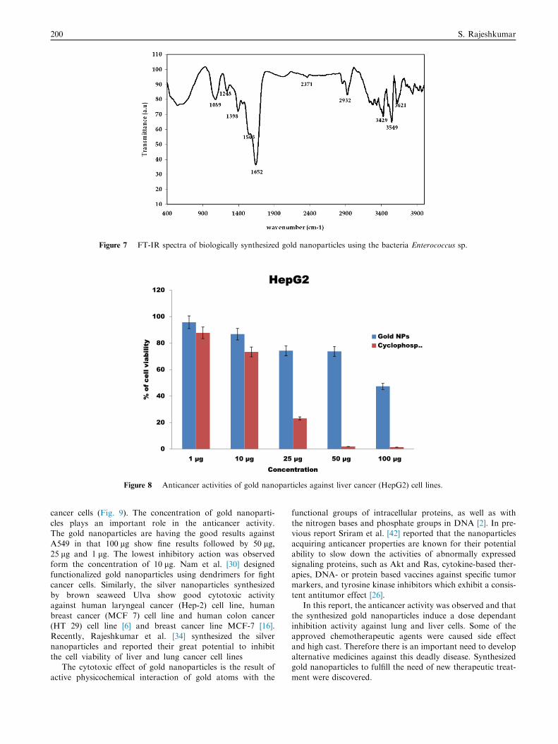
Figure 7 FT-IR spectra of biologically synthesized gold nanoparticles using the bacteria Enterococcus sp.
Figure 8 Anticancer activities of gold nanoparticles against liver cancer (HepG2) cell lines.
200 S. Rajeshkumar
cancer cells (Fig. 9). The concentration of gold nanoparti-cles plays an important role in the anticancer activity.The gold nanoparticles are having the good results against
A549 in that 100 lg show fine results followed by 50 lg,25 lg and 1 lg. The lowest inhibitory action was observedform the concentration of 10 lg. Nam et al. [30] designed
functionalized gold nanoparticles using dendrimers for fightcancer cells. Similarly, the silver nanoparticles synthesizedby brown seaweed Ulva show good cytotoxic activityagainst human laryngeal cancer (Hep-2) cell line, human
breast cancer (MCF 7) cell line and human colon cancer(HT 29) cell line [6] and breast cancer line MCF-7 [16].Recently, Rajeshkumar et al. [34] synthesized the silver
nanoparticles and reported their great potential to inhibitthe cell viability of liver and lung cancer cell lines
The cytotoxic effect of gold nanoparticles is the result of
active physicochemical interaction of gold atoms with the
functional groups of intracellular proteins, as well as withthe nitrogen bases and phosphate groups in DNA [2]. In pre-vious report Sriram et al. [42] reported that the nanoparticles
acquiring anticancer properties are known for their potentialability to slow down the activities of abnormally expressedsignaling proteins, such as Akt and Ras, cytokine-based ther-
apies, DNA- or protein based vaccines against specific tumormarkers, and tyrosine kinase inhibitors which exhibit a consis-tent antitumor effect [26].
In this report, the anticancer activity was observed and that
the synthesized gold nanoparticles induce a dose dependantinhibition activity against lung and liver cells. Some of theapproved chemotherapeutic agents were caused side effect
and high cast. Therefore there is an important need to developalternative medicines against this deadly disease. Synthesizedgold nanoparticles to fulfill the need of new therapeutic treat-
ment were discovered.

Figure 9 Anticancer activity of gold nanoparticles against lung cancer (A549) cells.
Anticancer activity of gold nanoparticles 201
4. Conclusion
Extracellular synthesis of gold nanoparticles was achieved
using culture supernatant of Enterococcus sp. Synthesized goldnanoparticles were initially identified by a formation of rubyred color and UV–vis spectrophotometer analysis shows the
surface plasmon resonance band at 545 nm. Crystallinestructure of gold nanoparticles was confirmed by XRD andSAED. TEM image shows the size, shape and distribution ofnanoparticles. Carboxylic and amine groups are the functional
groups present in the gold nanoparticles identified by FTIR.Anticancer activity of extracellularly synthesized goldnanoparticles was carried out by MTT assay against Hep-G2
and lung cancer cell (A549) lines. In this study the increasedanticancer activity was found to be at an increased concentra-tion of gold nanoparticles. This biosynthesis approach was
easy, large scaled up and eco-friendly. Thus synthesizednanoparticles were more efficient in the biomedical applica-tions in cancer treatment for their high anticancer activity.
Acknowledgements
Authors gratefully acknowledge STIC, Cochin for providing
SEM and EDX, VIT University, Vellore for XRD, IITBombay for TEM facility.
References
[1] R.S. Ahmad, M. Sara, R.S. Hamid, J. Hossein, N. Ashraf-
Asadat, Proc. Biochem. 42 (2007) 919–923.
[2] P. Blagoi Yu, V.L. Galkin, G.O. Gladchenko, S.V. Kornilova,
V.A. Sorokin, A.G. Shkorbatov, Naukova Dumka, Kiev, 1991,
p. 272.
[3] M.C. Daniel, D. Astruc, Chem. Rev. 104 (2004) 293.
[4] S.S. Dash, B.G. Bag, Appl. Nanosci. (2012), http://dx.doi.org/
10.1007/s13204-012-0179-4.
[5] J.S. Devi, B.V. Bhimba, Asian Pac. J. Trop. Dis. (2012) S87–
S93.
[6] J.S. Devi, B.V. Bhimba, Sci. Rep. 1 (4) (2012) 242.
[7] J.S. Devi, B.V. Bhimba, K. Ratnam, Int. J. Pharm. Pharm. Sci. 4
(4) (2012) 710–715.
[8] L. Du, H. Jiang, X. Liu, E. Wang, Electrochem. Commun. 9 (5)
(2007) 1165–1170.
[9] N. Duran, P.D. Marcato, O.L. Alves, G. D’Souza, E. Esposito,
J. Nanobiotechnol. 3 (2005) 8–14.
[10] M.A. Fayaz, C.S. Tiwary, P.T. Kalaichelvan, R. Venkatesan,
Colloid. Surf B Biointerf. 75 (2009) 175–178.
[11] M.A. Fayaz, M. Girilal, R. Venkatesan, P.T. Kalaichelvan,
Colloid. Surf B Biointerf. 88 (1) (2011) 287–291.
[12] M. Fukushima, K. Kakinuma, R. Kawaguchi, J. Clinic.
Microbiol. 40 (2002) 2779–2785.
[13] P. Ghosha, G. Hana, M. Dea, C.K. Kima, V.M. Rotello, Adv.
Drug. Del. Rev. 60 (2008) 1307–1315.
[14] K. Govindaraju, S.K. Basha, V.G. Kumar, G. Singaravelu, J.
Mater. Sci. 43 (2008) 5115–5122.
[15] S. He, Y. Zhang, Z. Guo, N. Gu, Biotechnol. Program. 24
(2008) 476–480.
[16] J.G. Holt, R.N. Krieg, P.H.A. Sneath, J.T. Staley, S.T.
Williams, Bergey’s Manual of Determinative Bacteriology,
ninth ed., Williams and Wilkins Baltimore, 1994.
[17] X. Huang, P.K. Jain, I.H. El-Sayed, Photochem. Photobiol. 82
(2006) 412–417.
[18] I.M. Husseiny, A.M. El-Aziz, Y. Badr, A.M. Mahmoud,
Spectrochimica. Acta Part A 67 (2007) 1003–1006.
[19] T.K. Joerger, R. Joerger, E. Olsson, C.G. Granqvist, Trends
Biotechnol. 19 (2001) 15.
[20] A. Kanchana, M. Balakrishna, Int. J. Pharm. Pharm. Sci. 3 (4)
(2011) 356–364.
[21] M. Lengke, M.E. Fleet, G. Southam, Langmuir 22 (2006) 2780–
2787.
[22] M. Lengke, B. Ravel, M.E. Fleet, G. Wanger, R.A. Gordon, G.
Southam, Environ. Sci. Technol. 40 (2006) 6304–6309.
[23] Z. Lin, J. Wu, R. Xue, Y. Yang, Spectrochim. Acta A 61 (2005)
761–765.
[24] P. Magudapatty, B.K. Gangopadhyayrans, K.G.M. Panigrahi
Nair, S. Dhara, Phys. B 299 (2001) 142–146.
[25] D. Mandal, M.E. Bolander, D. Mukhopadhyay, G. Sarkar, P.
Mukherjee, Appl. Microbiol. Bioechnol. 69 (2006) 485–492.
[26] D. Martins, L. Frungillo, M.C. Anazzetti, P.S. Melo, N. Duran,
Int. J. Nanomed. 5 (2010) 77–85.
[27] P. Mohanpuria, N.K. Rana, S.K. Yadav, J. Nanopart. Res. 10
(2008) 507–517.
[28] P. Mukherjee, A. Ahmad, D. Mandal, S. Senapati, S.R. Sainkar,
M.I. Khan, R. Ramani, R. Parischa, P.A.V. Kumar, M. Alam,
M. Sastry, R. Kumar, Angew. Chem. Int. Ed. 40 (2001) 3585–
3588.

202 S. Rajeshkumar
[29] C.J. Murphy, A.M. Gole, J.W. Stone, P.N. Sisco, A.M.
Alkilany, E.C. Goldsmith, S.C. Baxter, Acc. Chem. Res. 41
(2008) 1721–1730.
[30] J. Nam, N. Won, H. Jin, H. Chung, S. Kim, J. Am. Chem. Soc.
131 (2009) 13639–13645.
[31] Y. Nangia, N. Wangoo, N. Goyal, G. Shekhawat, C.R. Suri,
Microb. Cell Fact. 8 (2009) 39.
[32] N. Pugazhenthiran, S. Anandan, G. Kathiravan, N.K.U.
Prakash, S. Crawford, M. Ashokkumar, J. Nanopart. Res. 11
(2009) 1811–1815.
[33] P. Rajasekharreddy, P.U. Rani, B. Sreedhar, J. Nanopart. Res.
12 (2010) 1711–1721.
[34] S. Rajeshkumar, C. Malarkodi, M. Vanaja, G. Annadurai, J.
Mol. Struct. 1116 (2016) 165–173.
[35] K. Renugadevi, D. Inbakandan, M. Bavanilatha, V. Poornima,
Int. J. Pharm. Bio. Sci. 3 (3) (2012) 437–445.
[36] L.S. Rieznichenko, S.M. Dybkova, T.G. Gruzina, Z.R. Ulberg,
I.N. Todor, N.Y. Lukyanova, S.I. Shpyleva, V.F. Chekhun,
Exp. Oncol. 34 (2012) 25–28.
[37] F.S. Rosarin, V. Arulmozhi, S. Nagarajan, S. Mirunalini, Asian
Pac. J. Trop. Med. (2012) 1–10.
[38] M. Sastry, K.S. Mayya, K. Bandyopadhyay, Colloid. Surf A
Physicochem. Eng. Aspects 127 (1997) 221–228.
[39] M. Sastry, V. Patil, S.R. Sainkar, J. Phy, Chem. B 102 (1998)
1404–1410.
[40] S.S. Shankar, A. Rai, A. Ahmad, M. Sastry, J. Colloid. Interf.
Sci. 275 (2004) 496–502.
[41] G. Southam, T.J. Beveridge, Geochim. Cosmochim. Acta 58
(1994) 4527–4530.
[42] M.I. Sriram, S.B. ManiKanth, K. Kalishwaralal, S.
Gurunathan, Int. J. Nanomed. 5 (2010) 753–762.
[43] N. Thangaraju, R.P. Venkatalakshmi, A. Chinnasamy, K.
Pandian, Nano Biomed. Eng. 4 (2) (2012) 89–94.
[44] Y. Unno, Y. Shino, F. Kondo, N. Igarashi, G. Wang, R.
Shimura, T. Yamaguchi, T. Asano, H. Saisho, S. Sekiya, H.
Shirasawa, Clin. Cancer Res. 11 (12) (2005) 4553–4560.
[45] N. Vigeshwaran, M. Ashtaputre, R.P. Nachane, K.M.
Paralikar, H. Balasubramanya, Mater. Lett. 61 (2007) 1413–
1418.
[46] K. Yasuhiro, O. Kaori, S. Norizoh, N. Toshiyuki, N. Shinsuke,
H. Hajime, T. Yoshio, U. Tomoya, J. Biotechnol. 128 (2007)
648–653.
[47] K. Yungjin, R.C. Johnson, J.T. Hupp, Nano. Lett. 1 (2001) 165–
167.