32.4 Excretory System Hand-in worksheet on the Excretory/Urinary System.

Antibody Quantum Dot Conjugates Developed via Copper-Free ClickChemistry for Rapid Analysis of Biological Samples Using aMicrofluidic Microsphere Array SystemNalinikanth Kotagiri,† Zhenyu Li,‡ Xiaoxiao Xu,§ Suman Mondal,† Arye Nehorai,§ and Samuel Achilefu*,†
†Department of Radiology, Washington University School of Medicine, St. Louis, Missouri 63110, United States‡Department of Electrical and Computer Engineering, The George Washington University, Washington District of Columbia 20052,United States§Department of Electrical and Systems Engineering, Washington University in St. Louis, St. Louis, Missouri 63130, United States
*S Supporting Information
ABSTRACT: Antibody-based proteomics is an enabling technology that hassignificant implications for cancer biomarker discovery, diagnostic screening,prognostic and pharmacodynamic evaluation of disease state, and targetedtherapeutics. Quantum dot based fluoro-immunoconjugates possess promisingfeatures toward realization of this goal such as high photostability, brightness,and multispectral tunability. However, current strategies to generate suchconjugates are riddled with complications such as improper orientation ofantigen binding sites of the antibody, aggregation, and stability issues. Wereport a facile yet effective strategy to conjugate anti-epidermal growth factorreceptor (EGFR) antibody to quantum dots using copper-free click reaction,and compared them to similar constructs prepared using traditional strategiessuch as succinimidyl-4-(N-maleimidomethyl) cyclohexane-1-carboxylate(SMCC) and biotin−streptavidin schemes. The Fc and Fab regions of theconjugates retain their binding potential, compared to those generated throughthe traditional schemes. We further applied the conjugates in testing a novelmicrosphere array device designed to carry out sensitive detection of cancerbiomarkers through fluoroimmunoassays. Using purified EGFR, wedetermined the limit of detection of the microscopy centric system to be12.5 ng/mL. The biological assay, in silico, was successfully tested andvalidated by using tumor cell lysates, as well as human serum from breastcancer patients, and the results were compared to normal serum. A pattern consistent with established clinical data was observed,which further validates the effectiveness of the developed conjugates and its successful implementation both in vitro as well as insilico fluoroimmunoassays. The results suggest the potential development of a high throughput in silico paradigm for predictingthe class of patient cancer based on EGFR expression levels relative to normal reference levels in blood.
■ INTRODUCTION
Fluoroimmunoassays are sensitive platforms to achieve anti-body (Ab)-based detection of tumor biomarkers. The perform-ance of these assays is dependent on the reliable functioning ofthe molecular recognition and binding probes. Although Ab-fluorophore conjugates are popular and several conjugationstrategies are available, the low binding efficiency andnonspecific labeling is predominant, often leading to erroneousinterpretations.1,2 Therefore, careful optimization of conjuga-tion and binding conditions is critical for the proper evaluationof the biological labeling. Because of their excellent photo-stability, high quantum yield, and the potential for multiplexinginformation based on single excitation and multiple emissionwavelengths, quantum dots (QDs) are ideal fluorophores for amicroscopy centric system design.3 However, the dispropor-tionate dimensions of QD and Ab need careful consideration.
Unlike organic fluorophores and Ab conjugates, where multipledyes can be conjugated to a single Ab without interference withthe Ab binding sites, QD-Ab conjugates can possess multipleAbs per nanoparticle.4 This molecular orientation could lead toimproper orientation of the biomolecule’s binding sites,consequently attenuating the binding potential of the Ab-QDconjugate.4
Several strategies have been used to conjugate Ab to QD,5,6
but retention of the biological function of ligands such as Ab inthese QD conjugates remains a challenge. For example,previous reports have shown that succinimidyl-4-(N-maleimi-domethyl) cyclohexane-1-carboxylate (SMCC)-based Ab-QD
Received: March 31, 2014Revised: June 8, 2014
Article
pubs.acs.org/bc
© XXXX American Chemical Society A dx.doi.org/10.1021/bc500139u | Bioconjugate Chem. XXXX, XXX, XXX−XXX
conjugates demonstrated poor stability in aqueous aeratedsolutions, resulting in low binding and staining efficiency.4,7
Although biotin−streptavidin based Ab-QD conjugates havedemonstrated relatively better performance, they suffer frompoor biospecificity because of the low number of functional Ab.Several factors can mediate this inefficiency, including the largedimensions of the functional groups, overall size of the probe,aggregation caused by Ab cross-linking to multiple QDs, andrandom orientation of the Ab.6
Here, we report the development of Ab-QD conjugatesemploying copper-free “click” chemistry reaction. Copper(Cu)-free cycloaddition reactions are highly favored over Cucatalyzed reactions because of the fluorescence quenchingpotential of Cu ions on dyes and QDs.8 The rapid, specific,efficient, stable, facile, modular, and aqueous-phase conjugationstrategy of “click” reaction has proven to be a reliable andpowerful technique that is employed widely.9 While thisstrategy has been used to conjugate transferrin to QDs in thepast,10 we have adapted it to conjugating antibodies, bothbivalent (whole) and monovalent (half) Abs, with suitablemodifications such as the selection of appropriate cross-linkersto ensure a highly modular assembly process. Certainapplications and immunochemical techniques require the Abin its smaller sized analogue, which offers several advantagessuch as specific binding to thiol (SH) groups forbioconjugation, lower stearic hindrance, higher tissue pene-tration, and lower immunogenicity.11,12 The versatile nature ofthe conjugation strategy is applied to generate stable buildingblocks from both whole and half Ab, which enhanced theefficiency and yield of the Ab-QD constructs. In addition, weevaluated the Ab-QD conjugates further by assessing theirbinding efficiency and biospecificity both in cellulo as well as in
fluoroimmunoassays and found that the Ab on the Ab-QDconstructs indeed retains its Fc and Fab binding characteristics.We also compared the Ab-QD construct developed using clickreaction with similar constructs prepared through traditionalconjugation methods such as SMCC-based amine-thiol andbiotin−streptavidin affinity reactions and found superiorlabeling efficiency of EGFR expressing cancer cells using theconstructs developed using click reaction in comparison to thetraditional strategies.Finally, we demonstrate the application of the constructs in
silico using a novel microsphere array (3D MSA) designed forhighly sensitive detection of cancer biomarkers in serum andother biological fluids. The 3D MSA device has some obviousadvantages over traditional arrays such as ordered placement ofmicrospheres for increased sensitivity, simplification of imageprocessing, and controlled binding conditions through amicrofluidic setup.13,14 We have previously demonstratedcontrolled trapping of polystyrene microspheres and simulta-neously applied advanced signal and image processingtechniques to achieve a highly optimized device for performingfluoroimmunoassays.15 Herein, we have implemented thebiological protocol of the immunoassay to the device andtested its performance and sensitivity. The versatile andefficient conjugation and analysis used in this study creates aplatform for high throughput screening of biological samples.
■ RESULTS AND DISCUSSION
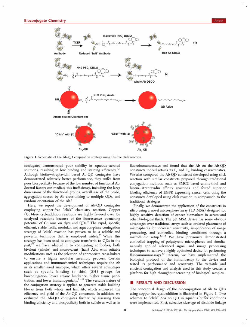
The conceptual design of the bioconjugation of Ab to QDsusing copper-free cycloaddition is illustrated in Figure 1. Twoschemes to “click” Abs on QD in aqueous buffer conditionswere implemented. First, selective cleavage of disulfide linkage
Figure 1. Schematic of the Ab-QD conjugation strategy using Cu-free click reaction.
Bioconjugate Chemistry Article
dx.doi.org/10.1021/bc500139u | Bioconjugate Chem. XXXX, XXX, XXX−XXXB

of whole Ab to obtain half Ab was performed using tris (2-carboxyethyl) phosphine (TCEP). Cleaved, “half”-Ab present-ing thiol groups was functionalized with dibenzocyclooctyne(DBCO) via a maleimide-PEG-DBCO cross-linker. Second,“whole” Ab was functionalized with DBCO using an N-hydroxysuccinimide (NHS)-based cross-linker, NHS-PEG-DBCO. Mouse anti-human EGFR monoclonal Ab was usedthroughout the study. Concurrently, QD-azide adducts wereprepared by reacting amine coated QD with NHS-PEG-Azidecross-linker. The whole Ab or half Ab, and QD adducts weresubsequently “clicked” to obtain Abw-QD or Abh-QDconjugates, respectively, with high yield (88%). We alsoprepared Ab-QD conjugates by using traditional strategies forcomparative analysis with the click reaction. Thus, Abs-QD wasprepared by reacting SMCC with the PEG linker on half Ab,and Abb-QD was obtained by mixing biotinylated whole Abwith streptavidin coated QDs, which forms strong noncovalentintermolecular binding.6
The degree of DBCO incorporation on the half and wholeAb was determined using UV−vis absorption scans. Based onthe relative absorbance values of DBCO (λmax = 307 nm) andAb (λmax = 280 nm) (Figure 2a), the number of DBCO per halfAb and whole Ab was estimated to be 3.4 and 13.5, respectively(Figure 2b). Incorporation of the azide groups on QDs wasdetermined by zeta potential and the hydrodynamic size of thenanoparticles was determined by dynamic light scattering(DLS) analysis (Table 1). The successful conjugation ofQD525 (λmax = 515 nm) to Ab, after purification to removeunbound Ab, was confirmed using UV−vis absorption data(Figure 2c). In addition, the hydrodynamic diameter and zetapotential values increased from 23.9 ± 1.8 nm to 32.4 ± 2.1 nmand from −16.7 mV to −5.7 mV, respectively. The yield of Ab-QD conjugates was high, which can be attributed to the stabilityof the DBCO and azide groups in aqueous solution and the
efficiency of the reaction. Using Bradford protein assay, thenumber of Ab per QD were estimated to be approximately 8.4for Abh-QD conjugates and 7.2 for Abw-QD (Figure 2d). Incomparison, we observed that the overall yield of Abs-QD wasmodest after purification (∼60%), with 3.9 Abs per QD; andthat of Abb-QD was appreciably higher (∼82%), with 1.6 Absper QD. This suggests that during the two-step conjugation,some of the maleimide groups on SMCC could haveundergone hydrolysis, thus rendering the functional groupinactive for reaction with the SH groups on Ab.16 However,using cross-linkers such as DBCO and azide allowed highlyselective reaction, resulting in high yields with minimalpurification steps. Due to the unique nature of Ab and QDinteraction, the various Ab-QD constructs would still have asingle dedicated QD, irrespective of the loading number.However, Ab-QD conjugates with high loading number such asAbh-QD and Abw-QD, could possess a higher number of Abswith the optimal orientation for functional binding than thosewith low loading numbers. Essentially, this means thatsuccessful binding of any Ab-QD conjugate to its target,irrespective of the loading number, will yield similar brightness.This suggests that the performance of a particular Ab-QDconstruct will depend on the number of optimally oriented andfunctional Ab decorated on QD surface, which to a certaindegree is dependent on the loading number and efficiency ofthe conjugation strategy. Additionally, variables such as cleaved
Figure 2. (a) Absorption spectra of Ab functionalized with DBCO groups, compared to nonfunctionalized Ab. (b) Comparison of degree of DBCOincorporation to Abh and Abw estimated using Bradford assay. (c) Absorption spectra of Ab-QD construct compared to QD alone. (d) Comparisonof the number of anti-EGFR Ab conjugated per QD among the four Ab-QD conjugates.
Table 1. Physical Characterization of the Ab-QD Constructs
sample size (nm) zeta potential (mV)
QD-Amine 23.9 ± 1.8 −16.7QD-Azide 27.8 ± 1.4 −23.7Abw-QD 32.4 ± 2.1 −5.7ABh-QD 31.1 ± 1.8 −6.1
Bioconjugate Chemistry Article
dx.doi.org/10.1021/bc500139u | Bioconjugate Chem. XXXX, XXX, XXX−XXXC

Ab with half the number of antigen binding sites as whole Ab,also could significantly impact the functionality of the Ab-QDconjugates.To determine if the binding potential of the Fc region of the
Ab-QD conjugates was retained, we employed Protein Gcoated PM beads (PGPM), which have an average diameter of15 μm. Protein G is an immunoglobulin binding protein ofbacterial origin with very high specificity and affinity to the Fcregion of Ab.17 Successful binding of Ab-QD to protein G wasdetermined by evaluating the fluorescence of the PM beads(see Supporting Information) by using confocal laser scanningmicroscopy (CLSM). For quantification of fluorescenceintensity and to prevent subjective data analysis, we havedeveloped MATLAB-based algorithms for signal and image
processing of the five constructs, including the control product,PGPM alone (Figure 3a). Our results show that Abh-QD, Abw-QD, and Abs-QD constructs exhibit appreciable binding to thePGPM surface relative to Abb-QD and the control product(Figure 3b). This suggests that the biotin−streptavidininteraction might have blocked the binding domains in the Fcregion, thus decreasing the binding efficiency. Given the largesize of streptavidin, it is likely that proximity and stearic effectsinterfered with the availability of the binding domains forligand−receptor molecular interaction. To ensure that thebinding of Ab to Protein G is through the Fc region and not bynonspecific binding, we also employed nonfunctionalized PMbeads and Protein A coated PM beads. Protein A is a subtype ofProtein G, but with lower binding specificity and affinity for Ab,
Figure 3. (a) MATLAB generated fluorescence intensity images showing the binding of the Fc region of the four Ab-QD constructs to Protein Gcoated PM. Scale bar represents 100 μm and is unchanged across all images. (b) Quantitation of the relative fluorescence intensity of Ab-QD-PMassembly. (c) CLSM images showing binding characteristics of the Fab region of Ab-QD constructs to BxPC-3 and MDA-MB-231 cells after 4 h. Thereflectance and fluorescence channels were merged. Scale bar represents 30 μm and is unchanged across all images.
Bioconjugate Chemistry Article
dx.doi.org/10.1021/bc500139u | Bioconjugate Chem. XXXX, XXX, XXX−XXXD
than Protein G.18 None of the constructs demonstrated anyappreciable binding to the plain PM beads as well as Protein Acoated PM beads, confirming the specific binding of the Ab tothe PGPM beads through the Fc region.Next, we sought to determine retention of antigen
recognition and binding of the Fab region of the Ab-QDconstructs. The anti-human EGFR Ab based constructs wereevaluated for EGFR binding in cellulo, using MDA-MB-231(EGFR positive) and BxPc-3 (EGFR positive) cancer cell lines.MDA-MB-231 cells are known to internalize the Ab-receptorcomplex after binding of the ligands to the receptors, whereasBxPc-3 resists such internalization.19,20 We investigated if theconstructs retain the same pattern of internalization for therespective cell lines. This strategy was chosen over using EGFRnegative cell lines because most cell lines express a basal level ofEGFR, which could confound data analysis. Moreover, ourapproach will also facilitate the delineation of specific binding ofthe constructs to EGFR from cross reactions to analogoussurface receptors. Click conjugated Ab-QD constructs,particularly Abw-QD, showed a high degree of binding to thecells and conformed to the internalization characteristics of therespective cell lines (Figure 3c). In contrast, Abs-QD and Abb-QD displayed low binding affinity and poor correlation with theexpected internalization pattern, suggesting improper orienta-tion of Ab on the QD surface. Interestingly, Abh-QDdemonstrated good binding affinity to BxPc-3 cells and poorinternalization in MDA-MB-231 cells. Typically, internalization
of EGF receptor−ligand complex is preceded by dimerizationof the surface receptors, essentially by bivalent antibodies(whole Ab with both Fab regions intact).
21 Monovalent Ab (halfAb with single Fab region) can prevent receptor dimerizationand activation of EGFR.22 Although Abh-QD is capable ofsuccessfully binding to EGFR on the cell surface, it is likely thatthis conjugate is not able to initiate receptor dimerization, thuspreventing internalization of the construct. It is well-known thatnanoparticles can show a high degree of nonspecific uptake bycells. To assess this potential effect, we performed blockingstudies with the anti-EGFR Ab. The results demonstrateminimal binding and internalization of Abh-QD and Abw-QD inthe respective cell lines (see Supporting Information).Motivated by the performance of Abw-QD, we carried out
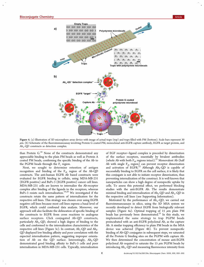
fluoroimmunoassays in silico, using the 3D MSA system werecently developed to detect EGFR from biologically relevantsamples (Figure 4a). Optimal trapping of 15 μm plain PMbeads has previously been demonstrated.23 In this study, weimplemented the same strategy to trap PGPM beadspreincubated with an anti-EGFR polyclonal Ab, as the captureAb. A similar trapping efficiency to plain PM beads in the MSAdevice was achieved (Figure 4b). To prevent nonspecificbinding of Ab-QD conjugates in subsequent steps, we saturatedall the Protein G binding sites on the PGPM with capture Ab.We then determined the concentration of mouse anti-EGFRpolyclonal Ab required to saturate the 15 μm PGPM beads byintroducing Abw-QD and measuring fluorescence intensity from
Figure 4. (a) Illustration of 3D microsphere array device with image of actual traps (top) and traps filled with PM (bottom). Scale bars represent 30μm. (b) Schematic of the fluoroimmunoassay involving Protein G coated PM, monoclonal anti-EGFR capture antibody, EGFR as target protein, andAbw-QD constructs as detection complex.
Bioconjugate Chemistry Article
dx.doi.org/10.1021/bc500139u | Bioconjugate Chem. XXXX, XXX, XXX−XXXE

Abw-QD to detect potential false positives (Figure 5a). We didnot observe any fluorescence signal after adding 2 μg of captureAb (∼1.5 × 10⁵ IgG/bead), suggesting successful saturation
and blocking of Fc binding sites on PGPM. We then tested thecapture and proper functioning of the fluoroimmunoassay insolution using purified sample of EGFR at various titrations.
Figure 5. (a) Estimation of the binding capacity of anti-EGFR Ab on PGPM to ensure saturation of all Fc binding sites on Protein G by capture Ab.(b) Binding studies with Abw-QD showing the response of the assay to increasing concentrations of EGFR on a Protein G based PM platform insolution phase. (b) Sensitivity of the fluoroimmunoassay using purified EGFR in solution phase, defining the limit of detection.
Figure 6. (a) Sensitivity of the fluoroimmunoassay using purified EGFR in silico, defining the limit of detection. (b) Plot showing the assay responseto EGFR receptor density from cell culture lysates of A431 and MDA-MB-435 cell lines. (c) CLSM image of single PM after assay performed usingheterogeneous protein samples such as culture lysates and human serum. Scale bar represents 7.5 μm. (d) CLSM image of single PM after assayperformed using purified EGFR. Scale bar represents 7.5 μm. (e) Plot showing the assay response to serum EGFR levels from breast cancer patientand compared to normal serum.
Bioconjugate Chemistry Article
dx.doi.org/10.1021/bc500139u | Bioconjugate Chem. XXXX, XXX, XXX−XXXF

The capture and detection Abs used in this assay bind todifferent epitopes of the extracellular domain of EGFR. Asshown in Figure 5b, we observed a strong positive nonlinearcorrelation between fluorescence intensity and EGFR concen-tration, in solution phase. Saturation limit of the capture Ab-PGPM complex can simply be extended by addition of morebeads, thus improving the detection range of the target protein.Because the in silico fluoroimmunoassay is designed for
CLSM based readout, we carried out sensitivity studies first insolution phase to determine the limit of detection (LOD) ofthe assay using CLSM. The intensity of fluorescent halosurrounding the bead was used as a measure of varying EGFRconcentration. There was no detectable fluorescence at <12.5ng/mL EGFR concentration (Figure 5c). For reference, normalserum EGFR level in healthy individuals is 75.3 ng/mL andthose with cancer can have either an elevated or lower basalserum EGFR values depending on the cancer type.24 Therefore,it is envisaged that the implementation of such assays will betoward determining the treatment response and prognosisrather than diagnosis of primary cancer.Implementation of the fluoroimmunoassay in silico was
carried out by sequential injection of the PGPM-capture Abcomplex, followed by injection of EGFR and finally Abw-QD,allowing an incubation time of 5 min each. The sequentialloading and flowthrough design facilitated effective washing ofunreacted components (Figure 4a). The system also enabledrapid binding of reactants within the confined volume. We didnot observe significant changes in fluorescence readout uponlonger incubation times, suggesting optimal binding of EGFRand detection complex at shorter incubation times (seeSupporting Information). A MATLAB algorithm (see Support-ing Information) was generated to process the CLSM images.This will provide accurate quantitation of fluorescence intensityfrom the PM surface. The algorithm takes into account imagingartifacts such as nonspecific fluorescence from sources otherthan the PM beads, provides accurate and unbiased informationon a huge number of beads and images, and enables automatedsignal processing, thus simplifying data interpretation. Weobserved similar sensitivity (12.5 ng/mL) in silico (Figure 6a)relative to the solution phase method. Correlation of thesensitivities of these two methods suggests successful trans-lation and implementation of the fluoroimmunoassay in theMSA device. While efforts to improve the LOD and sensitivityof the in silico method is an ongoing study, the currentsensitivity is sufficient to investigate patterns in differentialexpression of serum EGFR in various tumor models.For final validation of the microfluidic protocol, the challenge
was to determine the selectivity of the assay for detecting EGFRin a heterogeneous mixture of proteins such as in vitro tumorcell lysates, and in more clinically relevant samples such ashuman serum derived from breast cancer patients. As in vitromodels of biomarker expression, EGFR overexpressing tumorcell line A431 and lower EGFR expressing tumor cell lineMDA-MB-435 were compared. MDA-MB-435 expresses 2 ×10⁴ EGFR receptors per cell compared to 2 × 10⁶ receptors percell expressed by A431.25,26 Therefore, MDA-MB-435 cell linecan be classified as a representative model of “normal” cells.Cell lysates for each cell type were prepared using standardprotocols and their total protein concentration of 78 ± 3 mg/mL was determined. Different dilutions of the lysates were theninjected into the MSA and assayed to determine the relativecontribution of EGFR from the protein mixture to thefluorescence intensity. We observed a positive nonlinear
correlation between fluorescence intensity and EGFR densityas well as EGFR concentration across the two cell types (Figure6b). This suggests the involvement of EGFR selectiverecognition and binding to Ab-based PM and QD constructsand also demonstrates successful signal collection andprocessing. CLSM images revealed dense aggregations on thePM surface (Figure 6c). Similar images of PM taken withpurified EGFR do not exhibit these dense surface deposits(Figure 6d). This is not surprising because the complex andheterogeneous solutions are more likely to contain proteinsthat have a high tendency for nonspecific interactions on thebead surface. Thus, attempts to quantitate the proteins ofinterest from such mixtures are complicated and remain achallenge. However, the dense aggregates did not influence thefluorescence signal output and the LOD remained unchanged.Finally, we evaluated human serum samples to confirm the
selectivity of the assay in a more complex and clinically relevantheterogeneous protein mixture than the preceding study.Clinical serum samples from breast cancer patients and normalindividuals were used for comparison. A total proteinconcentration of 107 ± 4 mg/mL was estimated for bothsamples. Diluted mixtures were injected into the MSA andassayed. We observed a positive nonlinear correlation betweenfluorescence intensity and EGFR concentration. However,serum samples from breast cancer patients exhibited 2-foldlower EGFR levels compared to normal serum (Figure 6e).This result is consistent with previous studies that havereported significantly lower serum levels of EGFR amongpatients with breast and ovarian cancer.27,28 The median valuesare 56.3 ng/mL and 30.9 ng/mL at time of primary diagnosisand metastasis, respectively, when compared to normal EGFRlevels of 75.3 ng/mL.24 In contrast, cancers of brain and lungare known to have elevated serum EGFR.29 These data providefurther validation of the accuracy and selectivity of the assay indetermining the relative levels of EGFR in representative tumormodels and clinical samples.
■ CONCLUSIONWe have demonstrated a facile and effective strategy toconjugate whole and half Ab to QDs using Cu-free clickreaction. In doing so, we have demonstrated that functional andbinding sites, Fc and Fab regions, of the Ab remains unaffected.We have compared the Ab-QD constructs prepared herein withsimilar constructs using traditional conjugation strategiesthrough EGFR-specific cell binding studies. The efficientbinding of the constructs, particularly Abw-QD, promptedtheir use to evaluate a novel microsphere array microfluidicdevice. Fluoroimmunoassays were carried out on the devicewith CLSM based readouts and MATLAB-based algorithms forsignal and image processing. Using EGFR as model biomarker,we estimated the sensitivity and limit of detection of the systemto be 12.5 ng/mL. The robustness of the device and the assaywas further tested on heterogeneous protein mixtures derivedfrom cell culture lysates of EGFR overexpressing andunderexpressing cell lines, which showed a positive correlationbetween EGFR receptor density and fluorescence intensity.Similarly, clinically relevant human serum samples obtainedfrom breast cancer patients were tested and compared tonormal serum for selective and sensitive detection of EGFR.Interestingly, lower EGFR levels were found in serum derivedfrom breast cancer patients, which is consistent with recentclinical studies. Therefore, this highly versatile and efficientconjugation strategy can enable molecular imaging of disease
Bioconjugate Chemistry Article
dx.doi.org/10.1021/bc500139u | Bioconjugate Chem. XXXX, XXX, XXX−XXXG

biomarkers both in cellulo and in silico as demonstrated here,with the potential for translation to in vivo systems forantibody-based imaging and biomarker characterization.
■ EXPERIMENTAL METHODSI. Conjugation of Antibodies to Quantum Dots. Anti-
human EGFR monoclonal antibodies (Ab) were purchasedfrom Sigma-Aldrich Inc. (St. Louis, MO). The Ab were cleavedusing a reducing agent, TCEP from Thermo Scientific Inc.(Rockford, IL), to generate reduced “half” Ab. Briefly, to 20 μMof Ab in 100 μM of phosphate buffered saline (PBS) with a pHof 7.2, 50 μM of TCEP was added and incubated at roomtemperature (RT) for 1 h. Unlike other disulfide bond reducingagents such as dithiothreitol (DTT), TCEP does not needremoval before performing the cross-linking reactions. Imme-diately afterwards, 100 μM of DBCO-PEG4-maleimidepurchased from Click Chemistry Tools (Scottsdale, AZ) wasadded and incubated at RT for 2 h. The Abh-DBCO conjugatesthus formed are purified to remove unreacted reagents andexcess TCEP, using Slide-A-Lyzer MINI Dialysis Device, 10kMWCO from Thermo Scientific Inc. (Rockford, IL) overnight.Removal of TCEP is necessary before performing click reactionsince azide groups are reduced by TCEP. Similarly, 20 μM ofwhole Ab were reacted with 100 μM of DBCO-PEG4-NHScross-linker from Click Chemistry Tools, in 50 mM boratebuffer pH 8.5 at RT for 2 h to generate Abw-DBCO. Theconjugates were purified using dialysis as mentioned above. Toprepare QD-Azide conjugates, 300 nM of amine functionalizedQD525, with an excitation and emission wavelength of 515 and525 nm, respectively, from Life Technologies Inc. (Carlsbad,CA) in borate buffer was incubated with 3 μM of Azide-PEG4-NHS from Click Chemistry Tools, at RT for 2 h. The QD-Azide conjugates were purified overnight using dialysis. Clickreaction was performed using the conjugates by incubating Abh-DBCO and QD-Azide at 37 °C for 6 h in a water bath togenerate Abh-QD. Similarly, Abw-DBCO and QD-Azide wereclicked to generate Abw-QD constructs. Conjugation of Ab toQD using SMCC and biotin−streptavidin strategies wasadopted from previously well characterized protocols.6
II. Physicochemical Characterization. The absorptionspectra to determine DBCO incorporation and successfulconjugation of Ab to QDs was recorded on a Beckman CoulterDU 640 UV−visible spectrophotometer (Brea, CA) andanalyzed using Graphpad Prism statistical software. At λ =307 nm, the characteristic DBCO peak can be distinguishedfrom the protein peak at λ = 280 nm. The respectiveabsorbance readings for Abh-DBCO and Abw-DBCO wererecorded. The number of DBCO per IgG was calculated usingthe formula MolarityDBCO/MolarityIgG, where MolarityDBCO =AbsorbanceDBCO/extinction coefficientDBCO and MolarityIgG =AbsorbanceIgG/extinction coefficientIgG. The extinction coef-ficient of DBCO and IgG are 12 000 and 204 000 M−1 L cm−1,respectively. The number of IgG per QD was calculated usingBio-Rad protein assay dye (Hercules, CA) according to themanufacturer’s protocol, where the extinction coefficient ofQD525 was taken as 130 000 M−1 L cm−1. The absorbancevalues were corrected for background using unconjugated QDs,including QD525-strepatavidin. Dynamic light scatteringmeasurements were taken using a Malvern Zetasizer NanoZS (Westborough, MA) instrument at 25 °C. Two measure-ments were conducted for each sample with at least 10 runs andeach run lasting 10 s. All sizes reported were based on intensityaverage.
III. Fc and Fab Binding Assays. For Fc binding studies,Protein G coated polystyrene beads from Spherotech Inc.(Lake Forest, IL) with an average diameter of 15 μm was used.As control, Protein A coated PM beads purchased fromPolysciences Inc. (Warrington, PA), with an average diameterof 10 μm was used. Equimolar solutions of the Ab-QDconstructs and PGPM were incubated at 25 °C for 15 m in ashaker and fluorescence images were acquired using anOlympus FV1000 confocal laser scanning microscope (CenterValley, PA). Fluorescence/reflectance images were taken with a20× objective using He:Ne 488 nm excitation laser andemission range of dichroic mirrors set to 455−675 nm.MATLAB from MathWorks (Natick, MA) was used to processthe images using standard algorithms to quantitate the data. Anaverage of 100 particles were counted for each group.MDA-MB-231 and BxPc-3 cell lines from American Type
Culture Collection − ATCC (Manassas, VA) were culturedunder recommended standard conditions. MDA-MB-231 werecultured in Dulbecco’s Modified Eagle’s Medium containing10% fetal bovine serum (FBS), L-glutamine (2 mM), penicillin(100 units/mL), and streptomycin (100 μg/mL), incubated at37 °C in a humidified atmosphere of 5% CO2 and 95% air.BxPc-3 cells were cultured in RPMI-1640 under similarconditions. 5 × 10⁵ cells per well were grown in in an 8 wellchamber culture slide from BD Biosciences (San Jose, CA) towhich 100 nM of Ab-QD was added and incubated at 37 °C ina humidified, 5% CO2 atmosphere for 4 h. The slide substrateswere gently washed 2× with PBS before imaging with CLSM.Fluorescence/reflectance images were taken with a 60×objective using He:Ne 488 nm excitation laser and emissionrange of dichroic mirrors set to 455−575 nm. Fluorescence andreflectance image overlay with false color was performed usingFluoview FV10-ASW software from Olympus (Center Valley,PA).
IV. 3D Microsphere Array Studies. The binding capacityand concentration of mouse anti-human EGFR polyclonal Abfrom Sigma-Aldrich (St. Louis, MO) as capture Ab on PGPMwas first determined using different titrations of the Ab (0.1,0.25, 0.5, 1, 2, and 3 μg) on 0.25 mg/mL of PGPM. Afterincubation for 15 m at RT, the beads were centrifuged at 10000 rpm for 5 m to remove unbound Ab. Abw-QD (0.25 mM)was added and incubated for 15 m at RT. After centrifugationto remove unbound Abw-QD, the final mixture was added to96-well plates and end point fluorescence readings wererecorded using Synergy HT multimode plate reader fromBioTek Instruments Inc. (Winooski, VT). The values werenormalized relative to the highest fluorescence reading. Basedon the binding capacity, number of PGPM beads per unit mass(∼5.2 × 10⁷/mg) and the number of Protein G per bead (∼5 ×10⁶), the number of Ab/bead was calculated. The microfluidictrap-based array device was fabricated in polydimethylsiloxane(PDMS) using soft lithography techniques as shownpreviously.23 The device was mounted on the CLSM withthe inlet connected to a syringe pump from Harvard Apparatus(Holliston, MA) set to an infusion rate of 100 μL/min. PGPMbeads (0.5% w/v) were diluted 50× and 2 μg of anti-humanEGFR polyclonal Ab from Sigma-Aldrich (St. Louis, MO) wasadded to a final volume of 100 μL and incubated for 15 m at 25°C. The PGPM-Ab complex was injected into the device andtrapping process was monitored in real time using CLSM set toa fast scan rate in reflectance mode. A wash cycle with PBS wasinitiated to remove unbound Ab using the microfluidic-syringepump setup. Subsequent cycles consisted of injecting purified
Bioconjugate Chemistry Article
dx.doi.org/10.1021/bc500139u | Bioconjugate Chem. XXXX, XXX, XXX−XXXH
EGFR at various dilutions from 0 to 250 ng/mL into thesystem and performing wash steps to remove unbound protein,after allowing an incubation time of 5 m. Finally, Abw-QD wasinjected and incubated for an additional 5 m and followed by awash step to remove unreacted components. Fluorescence/reflectance images were recorded using CLSM using the 488nm laser with emission set to 455−575 nm. A uniqueMATLAB algorithm (in Supporting Information) was used toextract data such as number of beads, number of fluorescentbeads, intensity of fluorescence, background fluorescence, andgraphical representation of the accumulated data. Similarly,fluoroimmunoassays using lysed tumor cells and human serumwere performed after dilutions to the order of 500−1000×, toprevent clogging of the microfluidic channels. Lysis of A431and MDA-MB-435 cells, grown in DMEM, was performedusing NP40 cell lysis buffer from Life Technologies Inc.(Carlsbad, CA) following the manufacturer’s instructions.Human serum from breast cancer patients and normal serumwere purchased from Bioreclamation Inc. (Liverpool, NY).Dilutions were performed in PBS before injection.Solution phase fluoroimmunoassays were performed by first
mixing PGPM-Ab and purified EGFR and incubating for 5 m at25 °C followed by centrifugation at 10 000 rpm for 2 m. Thesupernatant was carefully decanted and the process repeated2×. Subsequently, Abw-QD was added to the mixture andincubated for 5 m and similarly washed using PBS to removeexcess reactants. The final mixture was added to 96-well platesand end point fluorescence readings were recorded using aplate reader and analyzed using Graphpad Prism statisticalsoftware.
■ ASSOCIATED CONTENT*S Supporting InformationDetails on MATLAB algorithm, fluorescence images, CLSMimages. This material is available free of charge via the Internetat http://pubs.acs.org.
■ AUTHOR INFORMATIONCorresponding Author*Phone: +1-314-362-8599. Fax: +1-314-747-5191. E-mail:[email protected] authors declare no competing financial interest.
■ ACKNOWLEDGMENTSThis study was supported by grant from National ScienceFoundation (CCF0963742). We thank Dr. Pinaki Sarder forhelpful discussions and assistance with data analysis.
■ REFERENCES(1) McCormack, T., O’Keeffe, G., Mac Craith, B., and O’Kennedy, R.(1996) Assessment of the effect of increased fluorophore labelling onthe binding ability of an antibody. Anal. Lett. 29, 953−968.(2) Vira, S., Mekhedov, E., Humphrey, G., and Blank, P. S. (2010)Fluorescent-labeled antibodies: Balancing functionality and degree oflabeling. Anal. Biochem. 402, 146−150.(3) Michalet, X., Pinaud, F. F., Bentolila, L. A., Tsay, J. M., Doose, S.,Li, J. J., Sundaresan, G., Wu, A. M., Gambhir, S. S., and Weiss, S.(2005) Quantum dots for live cells, in vivo imaging, and diagnostics.Science 307, 538−544.(4) Pathak, S., Davidson, M. C., and Silva, G. A. (2007)Characterization of the functional binding properties of antibodyconjugated quantum dots. Nano Lett. 7, 1839−1845.
(5) Medintz, I. L., Uyeda, H. T., Goldman, E. R., and Mattoussi, H.(2005) Quantum dot bioconjugates for imaging, labelling and sensing.Nat. Mater. 4, 435−446.(6) Xing, Y., Chaudry, Q., Shen, C., Kong, K. Y., Zhau, H. E., Chung,L. W., Petros, J. A., O’Regan, R. M., Yezhelyev, M. V., Simons, J. W.,Wang, M. D., and Nie, S. (2007) Bioconjugated quantum dots formultiplexed and quantitative immunohistochemistry. Nat. Protoc. 2,1152−1165.(7) Lee, J., Choi, Y., Kim, K., Hong, S., Park, H. Y., Lee, T., Cheon, G.J., and Song, R. (2010) Characterization and cancer cell specificbinding properties of anti-EGFR antibody conjugated quantum dots.Bioconjugate Chem. 21, 940−946.(8) Kotagiri, N., Niedzwiedzki, D. M., Ohara, K., and Achilefu, S.(2013) Activatable probes based on distance-dependent luminescenceassociated with Cerenkov radiation. Angew. Chem., Int. Ed. Engl. 52,7756−7760.(9) Best, M. D. (2009) Click chemistry and bioorthogonal reactions:unprecedented selectivity in the labeling of biological molecules.Biochemistry 48, 6571−6584.(10) Schieber, C., Bestetti, A., Lim, J. P., Ryan, A. D., Nguyen, T. L.,Eldridge, R., White, A. R., Gleeson, P. A., Donnelly, P. S., Williams, S.J., and Mulvaney, P. (2012) Conjugation of transferrin to azide-modified CdSe/ZnS core-shell quantum dots using cyclooctyne clickchemistry. Angew. Chem., Int. Ed. Engl. 51, 10523−10527.(11) Jain, R. K. (1990) Physiological barriers to delivery ofmonoclonal antibodies and other macromolecules in tumors. CancerRes. 50, 814s−819s.(12) Yokota, T., Milenic, D. E., Whitlow, M., and Schlom, J. (1992)Rapid tumor penetration of a single-chain Fv and comparison withother immunoglobulin forms. Cancer Res. 52, 3402−3408.(13) Walt, D. R. (2000) Techview: molecular biology. Bead-basedfiber-optic arrays. Science 287, 451−452.(14) Meza, M. B. (2000) Bead-based HTS applications in drugdiscovery. Drug Discovery Today 5 (Supplement1), 38−41.(15) Xu, X., Sarder, P., Kotagiri, N., Achilefu, S., and Nehorai, A.(2013) Performance analysis and design of position-encoded micro-sphere arrays using the Ziv-Zakai bound. IEEE Trans. Nanobioscience12, 29−40.(16) Sharpless, N. E., and Flavin, M. (1966) The reactions of aminesand amino acids with maleimides. Structure of the reaction productsdeduced from infrared and nuclear magnetic resonance spectroscopy.Biochemistry 5, 2963−2971.(17) Kato, K., Lian, L. Y., Barsukov, I. L., Derrick, J. P., Kim, H.,Tanaka, R., Yoshino, A., Shiraishi, M., Shimada, I., Arata, Y., et al.(1995) Model for the complex between protein G and an antibody Fcfragment in solution. Structure 3, 79−85.(18) Akerstrom, B., Brodin, T., Reis, K., and Bjorck, L. (1985)Protein G: a powerful tool for binding and detection of monoclonaland polyclonal antibodies. J. Immunol. 135, 2589−2592.(19) Patel, D., Lahiji, A., Patel, S., Franklin, M., Jimenez, X., Hicklin,D. J., and Kang, X. (2007) Monoclonal antibody cetuximab binds toand down-regulates constitutively activated epidermal growth factorreceptor vIII on the cell surface. Anticancer Res. 27, 3355−3366.(20) Arnoletti, J. P., Buchsbaum, D. J., Huang, Z. Q., Hawkins, A. E.,Khazaeli, M. B., Kraus, M. H., and Vickers, S. M. (2004) Mechanismsof resistance to Erbitux (anti-epidermal growth factor receptor)combination therapy in pancreatic adenocarcinoma cells. J. Gastro-intest. Surg. 8, 960−969.(21) Schlessinger, J. (2002) Ligand-induced, receptor-mediateddimerization and activation of EGF receptor. Cell 110, 669−672.(22) Spaargaren, M., Defize, L. H., Boonstra, J., and de Laat, S. W.(1991) Antibody-induced dimerization activates the epidermal growthfactor receptor tyrosine kinase. J. Biol. Chem. 266, 1733−1739.(23) Xu, X., Sarder, P., Li, Z., andNehorai, A. (2013) Optimization ofmicrofluidic microsphere-trap arrays, Biomicrofluidics 7.(24) Asgeirsson, K. S., Agrawal, A., Allen, C., Hitch, A., Ellis, I. O.,Chapman, C., Cheung, K. L., and Robertson, J. F. (2007) Serumepidermal growth factor receptor and HER2 expression in primary andmetastatic breast cancer patients, Breast Cancer Res. 9.
Bioconjugate Chemistry Article
dx.doi.org/10.1021/bc500139u | Bioconjugate Chem. XXXX, XXX, XXX−XXXI

(25) Buss, J. E., Kudlow, J. E., Lazar, C. S., and Gill, G. N. (1982)Altered epidermal growth factor (EGF)-stimulated protein kinaseactivity in variant A431 cells with altered growth responses to EGF.Proc. Natl. Acad. Sci. U. S. A. 79, 2574−2578.(26) Weigum, S. E., Floriano, P. N., Christodoulides, N., andMcDevitt, J. T. (2007) Cell-based sensor for analysis of EGFRbiomarker expression in oral cancer. Lab Chip 7, 995−1003.(27) Baron, A. T., Cora, E. M., Lafky, J. M., Boardman, C. H.,Buenafe, M. C., Rademaker, A., Liu, D., Fishman, D. A., Podratz, K. C.,and Maihle, N. J. (2003) Soluble epidermal growth factor receptor(sEGFR/sErbB1) as a potential risk, screening, and diagnostic serumbiomarker of epithelial ovarian cancer. Cancer Epidemiol. BiomarkersPrev. 12, 103−113.(28) Sandri, M. T., Johansson, H. A., Zorzino, L., Salvatici, M.,Passerini, R., Maisonneuve, P., Rocca, A., Peruzzotti, G., and Colleoni,M. (2007) Serum EGFR and serum HER-2/neu are useful predictiveand prognostic markers in metastatic breast cancer patients treatedwith metronomic chemotherapy. Cancer 110, 509−517.(29) Quaranta, M., Divella, R., Daniele, A., Di Tardo, S., Venneri, M.T., Lolli, I., and Troccoli, G. (2007) Epidermal growth factor receptorserum levels and prognostic value in malignant gliomas. Tumori 93,275−280.
Bioconjugate Chemistry Article
dx.doi.org/10.1021/bc500139u | Bioconjugate Chem. XXXX, XXX, XXX−XXXJ