Antibacterial and Anti-Inflammatory activities of ... 1/52-RNP-1307-381.pdf · 2Laboratório de...
13
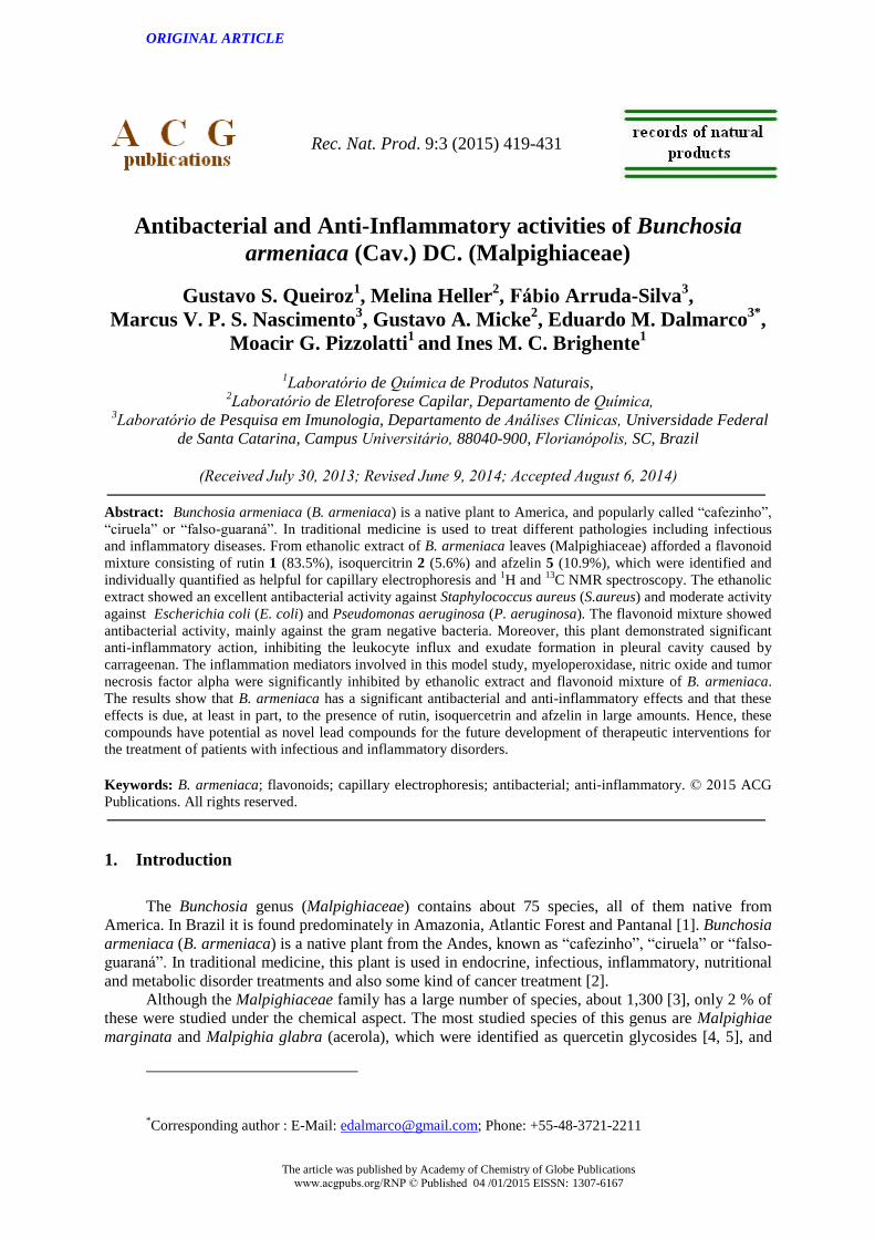
ORIGINAL ARTICLE The article was published by Academy of Chemistry of Globe Publications www.acgpubs.org/RNP © Published 04 /01/2015 EISSN: 1307-6167 Rec. Nat. Prod. 9:3 (2015) 419-431 Antibacterial and Anti-Inflammatory activities of Bunchosia armeniaca (Cav.) DC. (Malpighiaceae) Gustavo S. Queiroz 1 , Melina Heller 2 , Fábio Arruda-Silva 3 , Marcus V. P. S. Nascimento 3 , Gustavo A. Micke 2 , Eduardo M. Dalmarco 3* , Moacir G. Pizzolatti 1 and Ines M. C. Brighente 1 1 Laboratório de Química de Produtos Naturais, 2 Laboratório de Eletroforese Capilar, Departamento de Química, 3 Laboratório de Pesquisa em Imunologia, Departamento de Análises Clínicas, Universidade Federal de Santa Catarina, Campus Universitário, 88040-900, Florianópolis, SC, Brazil (Received July 30, 2013; Revised June 9, 2014; Accepted August 6, 2014) Abstract: Bunchosia armeniaca (B. armeniaca) is a native plant to America, and popularly called “cafezinho”, “ciruela” or “falso-guaraná”. In traditional medicine is used to treat different pathologies including infectious and inflammatory diseases. From ethanolic extract of B. armeniaca leaves (Malpighiaceae) afforded a flavonoid mixture consisting of rutin 1 (83.5%), isoquercitrin 2 (5.6%) and afzelin 5 (10.9%), which were identified and individually quantified as helpful for capillary electrophoresis and 1 H and 13 C NMR spectroscopy. The ethanolic extract showed an excellent antibacterial activity against Staphylococcus aureus (S.aureus) and moderate activity against Escherichia coli (E. coli) and Pseudomonas aeruginosa (P. aeruginosa). The flavonoid mixture showed antibacterial activity, mainly against the gram negative bacteria. Moreover, this plant demonstrated significant anti-inflammatory action, inhibiting the leukocyte influx and exudate formation in pleural cavity caused by carrageenan. The inflammation mediators involved in this model study, myeloperoxidase, nitric oxide and tumor necrosis factor alpha were significantly inhibited by ethanolic extract and flavonoid mixture of B. armeniaca. The results show that B. armeniaca has a significant antibacterial and anti-inflammatory effects and that these effects is due, at least in part, to the presence of rutin, isoquercetrin and afzelin in large amounts. Hence, these compounds have potential as novel lead compounds for the future development of therapeutic interventions for the treatment of patients with infectious and inflammatory disorders. Keywords: B. armeniaca; flavonoids; capillary electrophoresis; antibacterial; anti-inflammatory. © 2015 ACG Publications. All rights reserved. 1. Introduction The Bunchosia genus (Malpighiaceae) contains about 75 species, all of them native from America. In Brazil it is found predominately in Amazonia, Atlantic Forest and Pantanal [1]. Bunchosia armeniaca (B. armeniaca) is a native plant from the Andes, known as “cafezinho”, “ciruela” or “falso- guaraná”. In traditional medicine, this plant is used in endocrine, infectious, inflammatory, nutritional and metabolic disorder treatments and also some kind of cancer treatment [2]. Although the Malpighiaceae family has a large number of species, about 1,300 [3], only 2 % of these were studied under the chemical aspect. The most studied species of this genus are Malpighiae marginata and Malpighia glabra (acerola), which were identified as quercetin glycosides [4, 5], and * Corresponding author : E-Mail: [email protected]; Phone: +55-48-3721-2211
Transcript of Antibacterial and Anti-Inflammatory activities of ... 1/52-RNP-1307-381.pdf · 2Laboratório de...
ORIGINAL ARTICLE
The article was published by Academy of Chemistry of Globe Publications
www.acgpubs.org/RNP © Published 04 /01/2015 EISSN: 1307-6167
Rec. Nat. Prod. 9:3 (2015) 419-431
Antibacterial and Anti-Inflammatory activities of Bunchosia
armeniaca (Cav.) DC. (Malpighiaceae)
Gustavo S. Queiroz1, Melina Heller
2, Fábio Arruda-Silva
3,
Marcus V. P. S. Nascimento3, Gustavo A. Micke
2, Eduardo M. Dalmarco
3*,
Moacir G. Pizzolatti1 and Ines M. C. Brighente
1
1Laboratório de Química de Produtos Naturais,
2Laboratório de Eletroforese Capilar, Departamento de Química,
3Laboratório de Pesquisa em Imunologia, Departamento de Análises Clínicas, Universidade Federal
de Santa Catarina, Campus Universitário, 88040-900, Florianópolis, SC, Brazil
(Received July 30, 2013; Revised June 9, 2014; Accepted August 6, 2014)
Abstract: Bunchosia armeniaca (B. armeniaca) is a native plant to America, and popularly called “cafezinho”,
“ciruela” or “falso-guaraná”. In traditional medicine is used to treat different pathologies including infectious
and inflammatory diseases. From ethanolic extract of B. armeniaca leaves (Malpighiaceae) afforded a flavonoid
mixture consisting of rutin 1 (83.5%), isoquercitrin 2 (5.6%) and afzelin 5 (10.9%), which were identified and
individually quantified as helpful for capillary electrophoresis and 1H and
13C NMR spectroscopy. The ethanolic
extract showed an excellent antibacterial activity against Staphylococcus aureus (S.aureus) and moderate activity
against Escherichia coli (E. coli) and Pseudomonas aeruginosa (P. aeruginosa). The flavonoid mixture showed
antibacterial activity, mainly against the gram negative bacteria. Moreover, this plant demonstrated significant
anti-inflammatory action, inhibiting the leukocyte influx and exudate formation in pleural cavity caused by
carrageenan. The inflammation mediators involved in this model study, myeloperoxidase, nitric oxide and tumor
necrosis factor alpha were significantly inhibited by ethanolic extract and flavonoid mixture of B. armeniaca.
The results show that B. armeniaca has a significant antibacterial and anti-inflammatory effects and that these
effects is due, at least in part, to the presence of rutin, isoquercetrin and afzelin in large amounts. Hence, these
compounds have potential as novel lead compounds for the future development of therapeutic interventions for
the treatment of patients with infectious and inflammatory disorders.
Keywords: B. armeniaca; flavonoids; capillary electrophoresis; antibacterial; anti-inflammatory. © 2015 ACG
Publications. All rights reserved.
1. Introduction
The Bunchosia genus (Malpighiaceae) contains about 75 species, all of them native from
America. In Brazil it is found predominately in Amazonia, Atlantic Forest and Pantanal [1]. Bunchosia
armeniaca (B. armeniaca) is a native plant from the Andes, known as “cafezinho”, “ciruela” or “falso-
guaraná”. In traditional medicine, this plant is used in endocrine, infectious, inflammatory, nutritional
and metabolic disorder treatments and also some kind of cancer treatment [2].
Although the Malpighiaceae family has a large number of species, about 1,300 [3], only 2 % of
these were studied under the chemical aspect. The most studied species of this genus are Malpighiae
marginata and Malpighia glabra (acerola), which were identified as quercetin glycosides [4, 5], and
*Corresponding author : E-Mail: [email protected]; Phone: +55-48-3721-2211

Antibacterial and anti-inflammatory activities of Bunchosia armeniaca 420
some flavonoids which have been assigned various biological effects [6]. Other specie Banisteropsis
caapi, known as “cipós da Amazonia” contains β-carboline alkaloids which exhibit hallucinogenic
properties [7].
From Byrsonima species have been isolated some flavonoid compounds [8], mostly catechins
and quercetin glycosides, although the triterpenes represent the most frequent natural substance classes
[9], especially those with oleanane skeleton [10]. In Camarea genus has been reported free aglycone
presences such as apigenin, crisoeriol, kaempferol and quercetin, as well as, 7-O-glycosides of
apigenin and luteolin, 3-O-glycosides of quercetin and kaempferol [11]. However, until today it does
not have scientific reports about the main constituents of B. armeniaca, moreover, also there is no
scientific evidence about their biological proprieties, while there are reports of their use in Brazilian
traditional medicine. Other species from Malpighiaceae family present several reports highlighting
their antibacterial and anti-inflammatory effects [12, 13].
The increasing indiscriminate use of antibacterial drugs is reflected in the significant increasing
of hospital bacterial resistance against traditional medicines. This fact has been led the researchers to
search for new compounds or substances with antibacterial proprieties including the ones from natural
products. Moreover, there is some inflammatory diseases that are still without effective treatment,
such as glomerulopathies [14], vasculitis [15], rheumatoid arthritis [16], and psoriasis [17] and the
searching for new medicines with the objective to increase the quality of life of affected patients
remains in evidence.
Due to these facts, we decided to study the phytochemical profile of leaf extract and most of the
compounds isolated from B. armeniaca, and also to evaluate its possible antibacterial and anti-
inflammatory activities.
2. Material and Methods
2.1. Plant Material
B. armeniaca (Cav.) DC leaves were collected in February/2010 in Palhoça, Santa Catarina,
Brazil (27.867°S, 48.604°W). A voucher specimen was identified by Prof. Dr. Daniel de Barcellos
Falkenberg and deposited at Federal University of Santa Catarina Herbarium under the number of
FLOR-41422.
2.2. General
Purchases happened as following: Muller Hinton broth and agar from Oxoid (Hampshire, UK);
gentamicine from Laboratório Chile (Santiago, Chile); 2,3,5-triphenyltetrazolium chloride TTC
fromVetec (São Paulo, SP, Brazil); organic solvents: acetone, chloroform, n-hexane, ethyl acetate, n-
butanol, methanol, and ethanol (all analytical grade) from Synth (Diadema, SP, Brazil); sheep’s blood
(Newprov, Curitiba, PR, Brazil); Dimethylsulfoxide – DMSO, Carrageenan (degree IV), Evans blue
dye, hidrogen peroxide, human neutrophil myeloperoxidase, vanadium (III) chloride, o-dianisidine
•2HCl, phenol, sodium azide from Sigma–Aldrich (St. Louis, MI, USA), sodium nitroprussiate,
naphthylethylendiamide dihydrochloride, sulphanilamide, phosphoric acid, dexamethasone (Ache
pharmaceutical laboratories S.A., São Paulo, SP, Brazil), Enzyme-linked immunosorbent assay
(ELISA) for quantitative determination of mouse TNF-α (BD - Biosciences Pharmingen, San Diego,
CA, USA; Cat. N. 559732) Other reagents used were of analytical grade and were obtained from
various commercial sources.
Nuclear magnetic resonance spectra were recorded at 400 MHz for 1H and 100 MHz for
13C on
a VARIAN NMR AS 400 spectrometer. Infrared (IR) spectra were recorded on ABB FTIR –
FTLA2000 spectrometer. Thin layer chromatography (TLC) was performed on a pre-coated silica gel
type-60 plate (Macherey-Nagel).
Queiroz et al., Rec. Nat. Prod. (2015) 9:3 419-431 421
2.3. Vegetal material obtained
B. armeniaca (580.0 g) dried leaves were powdered and extracted with 96 % ethanol by
maceration at room temperature for 7 days (three times). After filtration, the ethanol extract was
further evaporated to dryness at 45 ºC under reduced pressure, yielding the crude extract, CE (14.11g /
100 g of leaves). This extract was dissolved in an ethanol solution of 20 % and after, the soluble part
was partitioned by liquid-liquid extraction with solvents of increasing polarity to give hexane, ethyl
acetate, buthanol, and water fractions. During the partitioning procedure with buthanol was observed
a yellow precipitation formation as little grains (1.20 g).
2.4. Capillary electrophoresis analysis
The experiments were performed on an Agilent Technologies HP3D
CE apparatus (Palo Alto,
CA, USA), equipped with a diode array detector. The wavelength chosen for detection was 200 nm.
Data acquisition and treatment were performed with HP Chemstation software.
Electrophoretic measurements were performed at 25 °C on an uncoated fused-silica capillary
(Ltot 48.5 cm × Ldet 40.0 cm × 75 µm I.D. × 375 µm O.D.) obtained from Polymicro Technologies
(Phenix, USA). Prior to the first use, the capillary was conditioned with NaOH 1 mol/L and deionized
water for 30 min, respectively. Daily, capillaries were conditioned with NaOH 1 mol/L, deionized
water and BGE (background electrolyte, separation buffer) for 5 min, consecutively. The optimized
BGE was composed of 20 mmol/L sodium tetraborate and 10% methanol, pH 9.3. Between the runs
the capillary was flushed for 1.0 min with BGE. Standard solutions and samples were introduced from
the inlet capillary extremity and injected hydro-dynamically at 50 mbar for 6 s (50 mbar = 4996.2 Pa).
The applied separation voltage was 30 kV, positive polarity on the injection side.
The sample (12.5 mg) was prepared by dissolving 10 mL of methanol. Hydrolyzed and non-
hydrolyzed samples were diluted two times with MeOH:H2O (1:1, v/v) mixture before the injection in
the capillary electrophoresis equipment.
In order to increase the analysis reliability it was performed standard additional tests on the
samples. The proportion of each component in the sample was calculated from the respective peak
areas shown in electropherograms, since this is proportional to the concentration. To minimize
instrumental errors benzenesulfonic acid at concentration of 40 mg/L was used as internal standard.
2.5. Acid hydrolysis of the flavonoid mixtures
The yellow precipitation (FLV) was dissolved in 10 mL of HCl 2 mol/L in methanol and left
under stirring and reflux for three hours. The reaction mixture was extracted with ethyl acetate and the
solvent was evaporated, yielding a yellow solid.
For the capillary electrophoresis analysis, the acid hydrolysis was performed as following: 500
µL of the extract methanolic solution was added to 1.0 mL of H2O and transferred to a test tube with
cap. It was added to the sample 200 µL HCl 8 mol/L and the tube was placed in a stove at 100 °C for 1
h for hydrolysis. After cooling, it was added 2 mL H2O and 2 mL of EtOAc for liquid-liquid
extraction, twice. The organic phase was dried under a flow of N2 and heated to 70 °C. The residue
was dissolved in 200 µL of MeOH:H2O (1:1, v/v) solution mixture for analysis.
2.6. Antibacterial assays
The microorganisms used were acquired from American Type Collection Culture (ATCC),
Staphylococcus aureus ATCC 25923 (S. aureus), Escherichia coli ATCC 25922 (E.coli) and
Pseudomonas aeruginosa ATCC 27853 (P. aeruginosa). The strain identifications were confirmed by
their biochemical profiles, according to the recommendation of the Clinical Microbiology Manual
[18].
Antibacterial activity of the vegetal samples were evaluated by the minimum inhibitory
concentration (MIC) determination [19, 20]. This test was performed in sterile 96-well micro-plates.
The crude extract and flavonoid mixtures were dissolved in DMSO:H2O (10 %, v/v) and transferred to

Antibacterial and anti-inflammatory activities of Bunchosia armeniaca 422
each micro-plate well in order to obtain a two-fold serial dilution on the original samples (crude
extract 10000 µg/mL and flavonoid mixtures 1000 µg/mL). After that, each well was inoculated with 5
µL of suspension containing 108 CFU/mL of each tested bacteria. The antibiotics gentamicin and
penicillin were included in the assays, as a positive control. The plates were incubated at 24 h at 37 ºC.
Bacterial growth was performed by adding 10 μL of 2,3,5-triphenyltetrazolium chloride 5 mg/mL in
sterile water, to each well. The plates were incubated again at 37 ºC for 1 h [20]. Bacterial growth in
the wells was indicated by a red color, whereas clear wells indicated growth inhibition by the sample.
MIC values were recorded as the lowest sample concentration showing clear wells. To crude extract, a
MIC below 100 µg/mL was considered as an excellent effect, from 100 to 500 µg/mL as moderate,
from 500 to 1000 µg/mL as weak, and over 1000 µg/mL as inactive. For isolated compounds a MIC
below 10 µg/mL was excellent, 10 to 100 µg/mL was good, and over 100 µg/mL was inactive.
2.7. Anti-inflammatory assays
2.7.1. Animals
Swiss mice weighing 18 to 25 g were housed under standardized conditions (room maintained
at 20 ± 2 °C, with alternating 12 h periods of light and dark) and were allowed free access to a
standard mouse chow and water before use. The procedure was approved by the Committee for Ethics
in Animal Research at University (protocol PP00757), and the experiments were performed according
to Brazilian College norms of Animal Experimentation (COBEA).
2.7.2. Pleurisy Induction and analysis
The pleurisy was induced by a single intra-pleural (ipl.) injection of 0.1 mL of sterile saline
(NaCl 0.95 %) plus carrageenan (Cg, 1%). Every inflammatory parameter was evaluated 4 h after
pleurisy induction. After sacrificing the animals with a pentobarbital overdose (120 mg/kg, i.p.), the
thorax was opened and the pleural cavity was washed with 1.0 mL of sterile phosphate buffered saline
(PBS) (pH 7.6) containing NaCl (130 mmol/L), Na2HPO4 (5 mmol/L), and KH2PO4 (1 mmol/L) in
distilled water containing heparin (20 IU/mL). Several pleural fluid samples were collected for further
total and differential leukocytes determination, exudate concentrations, myeloperoxidase activity
(MPO), nitric oxide products (NOx), as well as tumor necrosis factor alpha levels (TNF-α).
The dose-response curves analyzed different animal groups and were treated with different
crude extract (CE) (50, 100 and 200 mg/kg, i.p.) doses, and the flavonoid mixtures (0.1, 1.0 and 10
mg/kg, i.p.) 0.5 h before carrageenan administration. In these experiments, total leukocyte count and
exudation levels in pleural fluid were analyzed. According to the results obtained in the experiments
above, we chose CE lower doses and flavonoid mixtures that inhibited total leukocytes, as well as
exudate concentrations. The CE (200 mg/kg) doses and flavonoid mixtures (1.0 mg/kg) were chosen to
determine the other inflammatory parameters, such as MPO activity, NOx and TNF-α levels. Further,
the crude extract and flavonoid mixtures were only effective at inhibiting inflammatory parameters
when it was administered 0.5 h before Cg injection (Data not shown). In parallel, different animal
groups were treated with: 1) only with 0.1 mL of Cg (1 %, ipl.), considered the positive control group;
2) only with 0.1 mL of sterile saline or vehicle (NaCl 0.95%, ipl.), considered the negative control
group; 3) treated dexamethasone (0.5 mg/kg, i.p., 0.5 h before) plus Cg (1%, ipl.), considered a
reference anti-inflammatory treatment.
2.7.3. Quantification of leukocytes and exudate levels
Total and differential leukocyte counts were determined in a veterinarian automatic counter
adjusted for mouse-specific parameters (MINDRAY, BC-2800 Vet, Nanshan, Shenzhen, China). All
animals, except in the experiments that analyzed the MPO activity, NOx and TNF-α levels, were
previously challenged (0.5 h) with an Evans blue dye (25 mg/kg, i.v.) solution to evaluate the
exudation extent into the pleural space. A fluid sample (500 L) was collected from the pleural cavity,
and the dye amount was estimated by colorimeter using an enzyme-linked immune-sorbent assay
Queiroz et al., Rec. Nat. Prod. (2015) 9:3 419-431 423
(ELISA) plate reader (Organon Tecknica, Roseland, NJ, USA) at 620 nm by interpolation from
Evans blue dye standard curve ranging from 0.01 to 50 g/mL.
2.7.4. Quantification of myeloperoxidase activity (MPO)
In-house assays of MPO was used according to the methods developed by Rao et al. (1993)
[21]. By using conventional reagents, the concentration of each enzyme was estimated in the fluid
leakage of the pleural cavity by means of colorimetric measurements (absorbance of 450 nm) in an
ELISA plate reader (Organon Tecknica, Roseland, NJ, USA). The results were expressed as mU/mL
(MPO).
2.7.5. Quantification of nitric oxide products (NOx)
Nitric oxide was measured as a breakdown of nitrite (NO2−) and nitrate (NO3
−) products using
Griess method [22]. The fluid leakage samples of the pleural cavity obtained from controls and treated
animals were collected, separated, and stored at -70 ºC, and nitrate/nitrite levels were determined
reducing as previously described [23]. The results were expressed in µM.
2.7.6. Quantification of tumor necrosis factor (TNF-α)
To analyze TNF-α levels, fluid leakage samples of pleural cavity were collected and
immediately prepared for cytokine level analyses. This protocol used a commercially available kit
with monoclonal specific antibodies against TNF-α. The cytokine levels were measured with an
enzyme linked immune-sorbent assay Kit according to the manufacturer's instructions. The results
were expressed in pg/mL.
2.7.7. Data analysis of anti-inflammatory tests
The data were reported as the mean ± SEM and the parameter comparisons between groups
were performed by one-way variance analyses (ANOVA) followed by Dunnett’s and/or Student’s t-
tests for post hoc analysis, as necessary. P values of less than 0.05 were considered statistically
significant.
3. Results and Discussion
3.1. Phytochemical study
The yellow precipitation (FLV) obtained during the liquid-liquid partitioning (14.7 mg/g CE),
was recrystallized in methanol, showing m.p. 167.7–175.0 ºC. The Shinoda’s test (HCl/Mg) showed
the red color appearance, indicating flavonoid presences. The TLC analysis (EtOAc/HCO2/AcOH/H2O
100:11:11:26) and revelation with FeCl3 5% (EtOH) showed three spots with Rf: 0.36, 0.43 and 0.64.
The IR analysis showed absorption bands in 3428 cm-1
(O-H), 1655 cm-1
(C=O) 1602 cm-1
(C=C),
1297 cm-1
(C-O) and 2909 cm-1
(C-H).
The electropherogram of this precipitation shows three peaks and, by the standard UV-VIS
comparison was possible to identify two glycoside flavonoids as rutin 1 (83.5%) and isoquercitrin 2
(5.6%), and a third flavonoid (10.95 %), that remained unknown (Figure 1a). The precipitation was
submitted at acid hydrolysis and analyzed by capillary electrophoresis again in which only two peaks
was obtained corresponding to quercetin 3 (87.0%) and kaempferol 4 (13.0%) (Figure 1b). From these
analyses were possible to suggest that the flavonoids are of O-glycosides type [24], two of them
composed by quercetin (rutin 1 and isoquercitrin 2) and the other with kaempferol aglycone. The non-
hydrolyzed sample was analyzed again adding standards to confirm the glycosyl flavonoids identity
(Figure 2).

Antibacterial and anti-inflammatory activities of Bunchosia armeniaca 424
Figure 1. Flavonoid mixtures electropherograms before and after acid hydrolysis
Figure 2. Flavonoid mixtures electropherograms with and without standard addition and UV-
VIS spectra overlapping
The hydrolyzed precipitation was also analyzed by 1H and
13C NMR spectroscopy. The
1H
NMR spectrum showed two peak sets with different signal integrations (ratio about 1:7). The signals
in the main ratio presented two doublets at H 6.17 and 6.38 (J = 2.0 Hz, meta correlated) assigned to

Queiroz et al., Rec. Nat. Prod. (2015) 9:3 419-431 425
H-6 and H-8, respectively, in the A ring of flavonoids. It was observed two doublets at H 6.87 (J = 8.6
Hz, ortho correlated) and 7.73 (J = 2.1 Hz, meta correlated), and a doublet of doublets at H 7.62 (J =
8.6 and 2.1 Hz), an ortho-meta coupling pattern corresponding to tri substituted aromatic ring. These
peaks are assigned to H-5’, H-2’ and H-6’ of B ring, respectively. These data are coherent with the
flavonoid quercetin 3 [25, 26].
The minor integration signals showed two doublets at H 6.12 and 6.32 (J = 1.8 Hz) assigned to
H-6 and H-8, respectively, in the A ring of flavonoids and two doublets at H 6.89 and 8.08 (J = 9.0
Hz), an ortho coupling pattern corresponding to para irreplaceable aromatic ring. These peaks are
assigned both to H-3’/5’ and H-2’/6’ of B ring, respectively. These data correspond to kaempferol
structure 4 [27, 28]. The 13
C confirmed 30 carbon presences for these two aglycones (Table 1).
After the capillary electrophoresis analysis was possible to confirm the corresponding peaks of
quercetin3-O-α-L-rhamnopyranosyl-(1→6)-β-D-glycopyranoside or rutin 1 and quercetin 3-O-β-D-
glycopyranoside or isoquercitrin 2 in the 1H and
13C NMR non-hydrolyzed sample spectra. Both
compounds showed the same quercetin signal patterns as aglycone part. The 1H spectrum of rutin 1,
the main compound, can be highlighted two anomeric hydrogen doublets at H 5.10 (J = 7.6 Hz, β-
anomer) and H 4.51(J = 1.6 Hz, α-anomer). The anomeric carbons in 13
C NMR at C 102.37 and
102.25 were assigned, respectively to glucose and rhamnose of rutinose. Additionally, can be cited the
doublet at H 1.11 (J = 6.2 Hz) and C 17.87 assigned to rhamnose methy l group. The NMR
isoquercitrin 2 spectra, the minor compound, can be cited the anomeric hydrogen doublet at H 5.24 (J
= 7.4 Hz, -anomer), and at C 102.28 assigned to glucose anomeric carbon (Table 1) [29].
Table 1. NMR spectral data of flavonoids from Bunchosia armeniaca before acid hydrolysis
rutin 1 isoquercitrin 2 afzelin 5
C H C H C H C-2 158.39 - * - 159.3 -
C-3 135.59 - * - 135.54 -
C-4 179.34 - * - 179.31 -
C-5 162.87 - * - * -
C-6 99.89 6.20 (d, 2.1) 99.88 6.25 (d, 2.0) 99.95 6.15 (d, 2.0)
C-7 165.97 - * - * -
C-8 94.84 6.39 (d, 2.1) 94.79 6.44 (d, 2.0) 94.91 6.34 (d, 2.0)
C-9 158.43 - * 158.39 -
C-10 104.64 - 104.63 105.57 -
C-1’ 123.06 - * 123.02 -
C-2’ 115.99 7.66 (d, 2.1) * 7.58 (d, 2.3) 132.36 8.05 (d, 9.0)
C-3’ 145.78 - * 116.11 6.88 (d, 9.0)
C-4’ 149.76 - 149.71 161.42 -
C-5’ 116.04 6.87 (d, 8.4) * 6.82 (d, 8.6) 116.11 6.88 (d, 9.0)
C-6’ 123.06 7.62 (dd, 8.4, 2.1) * 7.57 (dd, 2.3, 8.6) 132.36 8.05 (d, 9.0)
Glc Glc Rha
C-1” 102.37 5.10 (d, 7.6) 102.28 5.24 (d, 7.4) 102.32 4.46 (d, 1.6)
C-2” 75.68 * 75.63 * 71.39 *
C-3” 78.11 * 78.06 * 72.06 *
C-4” 71.34 * 69.65 * 73.86 *
C-5” 78.07 * 77.06 * 71.27 *
C-6” 68.5 3.80 (dd, 1.0, 10.7)
3.38 (dd, 6.1, 10.7) * * 17.83 1.06 (d, 6.2)
Rha
C-1’” 102.25 4.51 (d, 1.6)
C-2’” 72.03 *
C-3’” 72.19 *
C-4’” 73.89 *
C-5’” 69.68 *
C-6’” 17.87 1.11 (d, 6.2)

Antibacterial and anti-inflammatory activities of Bunchosia armeniaca 426
13C NMR 100 MHz;
1H NMR 400 MHz; methanol-D. Data: chemical shift / ppm (multiplicity – d=doublet, dd=
doublet of doublets; coupling – J / Hz). * difficult to assign because of overlapping peaks. Glc = glucose, Rha =
rhamnose.
Finally, it was possible to identify the third compound, by analyzing medium peak integrations
as kaempferol 3-O--L-rhamnopyranoside or afzelin 5. The 1H NMR spectrum of this compound
showed the same kaempferol signal patterns. In the sugar part can be highlighted the anomeric
hydrogen at H4.46 (J = 1.6 Hz) doublet, and the anomeric carbon at C 102.32. Additionally, it can be
cited the doublet at H 1.06 (J = 6.2 Hz) and C 17.83 assigned to rhamnose methyl group (Table 1)
[30].
Therefore, from ethanolic leaf extract of B. armeniaca was obtained flavonoid mixtures formed
to rutin 1 (83.5%), isoquercitrin 2 (5.6%) and afzelin 5 (10.9%), with a yielding of 1.47 % of leaf
extract. This was the first report of afzelin presence in Malpighiaceae family species.
Figure 3. Flavonoids isolated from Bunchosia armeniaca. Compound 01: Rutin; compound 02:
isoquercitrin; compound 03: quercetin; compound 04: kaempferol and compound 05: afzelin.
3.2. Antibacterial activity
Regarding the antibacterial activity, Table 2 shows that B. armeniaca ethanolic extract has a
moderate to excellent antibacterial activities against all microorganisms tested. The flavonoid mixtures
showed an excellent effect against all tested bacteria. The ethanolic extract showed more active against
S. aureus (MIC = 87.5 µg/mL), as expected, due to natural products to be more active against gram
positive bacteria. This is based on different characteristics observed among gram positive and gram
negative cell-wall, fact that limits the natural product effects against gram negative bacteria, that
present outer-membrane permeability barrier which limits the compound accesses to their targets [31].
Table 2. Minimal inhibitory concentrations (MICs: µg/mL) of crude extract and flavonoid mixtures
isolated from B. armeniaca
S. aureus ATCC25923
(µg/mL)
E. coli ATCC 25922
(µg/mL)
P. aeruginosa ATCC 27853
(µg/mL)
Gentamicin - 1.25 0.625
Penicillin 0.625 - -
CE 87.5 175 350 FLV 3.0 1.5 1.5
CE = Ethanolic extract of leaves; FLV = Flavonoid mixtures. ATCC – American type collection culture (Data
from three experiments).
Queiroz et al., Rec. Nat. Prod. (2015) 9:3 419-431 427
Although, ethanolic extract results of B. armeniaca against gram negative bacteria cannot be
neglected (MIC = 175 and 350 µg/mL), an unusual fact observed on natural products [32]. Some
authors also reported significantly antibacterial activity of other Malpighiaceae family genus, such as
Mascagnia macroptera hexane extract that had an activity against enterophatogenic bacteria [33], the
methanolic and chloroformic extracts of Byrsonima crassa inhibit H. pylori growth in vitro [34], and
ethyl acetate extract of Byrsonima crassifolia roots inhibit gram-positive (S. aureus) and gram-
negative (Salmonella typhi) bacteria [35].
The flavonoid mixtures showed also a great antibacterial activity against all three bacteria
studied, and these effects can be compared with the results obtained with the commercial antibiotics
gentamicin and penicillin (Table 2). These results were surprising as it is not common the natural
product to show excellent antibacterial activity against gram negative bacteria (E. coli and P.
aeruginosa, MIC = 1.5 µg/mL). This fact, lead us to hypothesize that the effects observed in ethanolic
extract studied is attributed, at least in part, to the flavonoids present in this extract. The flavonoids are
natural products with recognized antibacterial activity [36], including all three identified in B.
armeniaca mixture such as rutin [37], isoquercitrin [38] and afzelin [39].
The flavonoid antibacterial activities is mainly due to the DNA damage cased in the bacteria
[40].These effects seem to be via different action mechanisms such as: i) complex with the bacterial
cell wall and the decrease with microbial growth [41], ii) inhibiting DNA topoisomerase II (DNA
gyrase) activity [42], and iii) inhibiting FtsZ protein, the tubulin bacterial analog, which mediates
bacterial cell division [43].
3.3. Anti-inflammatory activity
3.3.1. Leukocyte influx and exudate
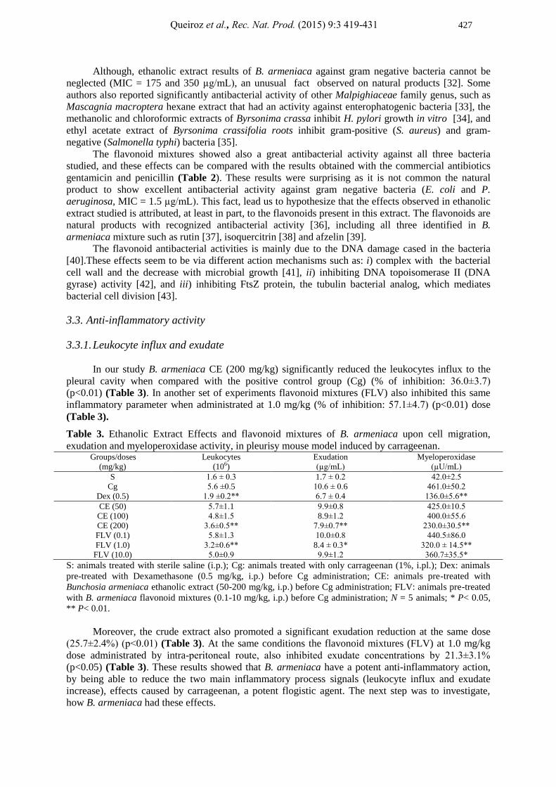
In our study B. armeniaca CE (200 mg/kg) significantly reduced the leukocytes influx to the
pleural cavity when compared with the positive control group (Cg) (% of inhibition: 36.0±3.7)
(p<0.01) (Table 3). In another set of experiments flavonoid mixtures (FLV) also inhibited this same
inflammatory parameter when administrated at 1.0 mg/kg (% of inhibition: 57.1±4.7) (p<0.01) dose
(Table 3).
Table 3. Ethanolic Extract Effects and flavonoid mixtures of B. armeniaca upon cell migration,
exudation and myeloperoxidase activity, in pleurisy mouse model induced by carrageenan. Groups/doses
(mg/kg)
Leukocytes
(106)
Exudation
(µg/mL)
Myeloperoxidase
(µU/mL)
S 1.6 ± 0.3 1.7 ± 0.2 42.0±2.5
Cg 5.6 ±0.5 10.6 ± 0.6 461.0±50.2
Dex (0.5) 1.9 ±0.2** 6.7 ± 0.4 136.0±5.6**
CE (50) 5.7±1.1 9.9±0.8 425.0±10.5
CE (100) 4.8±1.5 8.9±1.2 400.0±55.6
CE (200) 3.6±0.5** 7.9±0.7** 230.0±30.5**
FLV (0.1) 5.8±1.3 10.0±0.8 440.5±86.0
FLV (1.0) 3.2±0.6** 8.4 ± 0.3* 320.0 ± 14.5**
FLV (10.0) 5.0±0.9 9.9±1.2 360.7±35.5*
S: animals treated with sterile saline (i.p.); Cg: animals treated with only carrageenan (1%, i.pl.); Dex: animals
pre-treated with Dexamethasone (0.5 mg/kg, i.p.) before Cg administration; CE: animals pre-treated with
Bunchosia armeniaca ethanolic extract (50-200 mg/kg, i.p.) before Cg administration; FLV: animals pre-treated
with B. armeniaca flavonoid mixtures (0.1-10 mg/kg, i.p.) before Cg administration; N = 5 animals; * P< 0.05,
** P< 0.01.
Moreover, the crude extract also promoted a significant exudation reduction at the same dose
(25.7±2.4%) (p<0.01) (Table 3). At the same conditions the flavonoid mixtures (FLV) at 1.0 mg/kg
dose administrated by intra-peritoneal route, also inhibited exudate concentrations by 21.3±3.1%
(p<0.05) (Table 3). These results showed that B. armeniaca have a potent anti-inflammatory action,
by being able to reduce the two main inflammatory process signals (leukocyte influx and exudate
increase), effects caused by carrageenan, a potent flogistic agent. The next step was to investigate,
how B. armeniaca had these effects.

Antibacterial and anti-inflammatory activities of Bunchosia armeniaca 428
3.3.2. Myeloperoxidase activity
The inflammatory model used in our experiments (pleurisy induced by carrageenan) is
characterized by a large leukocyte migration to pleural cavity, and these migration is almost all
neutrophils [23].
The crude extract, CE, at the dose of 200 mg/kg reduced significantly the myeloperoxidase
activity (25.7±2.4%) (p<0.01). At the same conditions, the flavonoid mixtures, at the dose of 1.0
mg/kg, also reduced significantly the myeloperoxidase activity in pleural cavity by 30.6±2.0%
(p<0.01) (Table 3). Therefore, the inhibitory effect upon leukocytes observed in previous experiments
was associated decreasing myeloperoxidase activity. Abundantly expressed in primary neutrophils
(PMNs) granules, the myeloperoxidase plays a pivotal role in inflammatory process and the increase is
directly linked with PMNs activation [44]. With these results, we can concluded that the natural
product studied not only reduced the leukocyte influx to pleural cavity due to inhibition of PMNs
migration, as also inhibited their activation.
3.3.3. Nitrate/nitrite and TNF-α levels
The CE (200 mg/kg) and FLV (1.0 mg/kg) of B. armeniaca were able to decrease significantly
the NOx levels by 40.3±4.3% and 51.0±3.6% respectively (p<0.01) (Figure 4a). The NO is an
important relaxant factor derivate from endothelium which maintains the vascular tonus and micro-
vascular permeability. Moreover, it plays a pivotal function as inflammation mediator. The increase of
NO levels mainly causes an increasing vascular permeability [45]. Thus, NO also plays an oxidative
role in promoting edema formation mainly because of peroxynitrite formation [46].
This results suggest that the inhibition observed with animal treatments with B. armeniaca (CE
and FLV) upon exudate could be attributed at least in part to its ability to decrease NO formation by
inflammatory cells and endothelium [23].
In addition, the CE and FLV caused a significantly decrease in TNF-α levels. TNF-α inhibition
in each treatment group was as following: CE (200 mg/kg) 43.5±13.0 and FLV (1.0 mg/kg) 63.5±13.5
(p<0.05) (Figure 4b). TNF-α is a pleiotropic cytokine and plays a key role in the innate and adaptive
immune response, mainly released by resident immune cells. In the airway inflammatory diseases
TNF-α plays an important role as chemo-attractant for neutrophils and increases the release of other
cytokines and also, it is directly involved in T cell activations that are considered the inflammatory
process manager [47].
This statement corroborates with the findings about the significant reduction observed in
leukocyte migration to pleural cavity, fact that allows attributing the leukocyte influx reduction to the
ability of B. armeniaca to inhibit TNF-α releasing by resident immune cells present in pleural fluid.
4. Conclusions
B. armeniaca ethanolic extract and flavonoid mixtures that are the main compounds present in
this extract, displayed a significant antibacterial action against gram positive and negative bacteria.
Moreover, this species also showed an important anti-inflammatory action by inhibiting the leukocyte
influx and exudate increase and be able to reduce some inflammatory mediators, in the pleurisy
murine model. These results confirm the traditional use of this plant and showed that the flavonoid
mixture is the main responsible for antibacterial and anti-inflammatory effects attributed to Bunchosia
armeniaca. However additional studies are needed, these compounds have potential as novel lead
compounds for the future development of therapeutic interventions for the treatment of patients with
infectious and inflammatory disorders.

Queiroz et al., Rec. Nat. Prod. (2015) 9:3 419-431 429
Figure 4. Ethanolic Extract Effects and flavonoid mixtures of Bunchosia armeniaca upon NOx
(a) and TNF-α (b) mouse pleurisy model levels induced by carrageenan.
Acknowledgements
The authors thank CAPES and CNPq for financial support and research grants.
References
[1] A. Bonpland, A.V. Humboldt and K.S. Kunth (1821). Nova genera et species plantarum: quas in
peregrinatione ad plagam æquinoctialem orbis novi collegerunt, descripserunt, partim adumbraverunt.
Amat. Bonpland et Alex. de Humboldt ; ex schedis autographis Amati Bonplandi in ordinem digessit
Carol. Sigismund. Kunth. v. 5., Lutetiae Parisiorum: sumtibus Librariae Graeco-Latino-Germanico.
[2] M. Giraldi and N. Hanazaki (2010). Uso e conhecimento tradicional de plantas medicinais no Sertão do
Ribeirão, Florianópolis, SC, Brasil, Acta. Bot. Bras. 24(2), 395-406.
[3] W.R. Anderson (2014). Herbarium - University of Michigan - Malpighiaceae website. [cited 2014
March, 07]; Available from: http://herbarium.lsa.umich.edu/herb/malpigh/.
[4] A. Bonpland, A.V. Humboldt and K.S. Kunth (2008). Survey of medicinal plants used in the region
Northeast of Brazil, Rev. Bras. Farmacogn. 18(3), 472-508.
b
a
A
B

Antibacterial and anti-inflammatory activities of Bunchosia armeniaca 430
[5] T. Hanamura, T. Hagiwara and H. Kawagishi (2005). Structural and functional characterization of
polyphenols isolated from acerola (Malpighia emarginata DC.) fruit, Biosci. Biotech. Bioch. 69(2), 280-
286.
[6] M. Kawaguchi, H. Tanabe and K. Nagamine (2007). Isolation and characterization of a novel flavonoid
possessing a 4,2 ''-glycosidic linkage from green mature acerola (Malpighia emarginata DC.) fruit,
Biosci. Biotech. Bioch. 71(5), 1130-1135.
[7] M.C.M. Costa, M.C. Figueiredo and S.O.S. Cazenave (2005). Ayahuasca: uma abordagem toxicológica
do uso ritualístico, Rev. Psi. Clín. 32(6), 310-318.
[8] C. Dosseh, C, Moretti, A.M. Tessier and P. Delaveau (1980). Chemical study on the leaves of
Byrsonima verbascifolia Rich. ex Juss, Plan. Med. Phytother. 14(3), 136-142.
[9] O.R. Gottlieb, P. Henriques Mendes and M. Taveira Magalhaes (1975). Triterpenoids from Byrsonima
verbascifolia, Phytochem. 14(5-6), 1456-1456.
[10] F. Guilhon-Simplicio and M.D. Pereira (2011). Chemical and pharmacological aspects of Byrsonima
(Malpighiaceae), Quím. Nova. 34(6), 1032-1041.
[11] L.B. Motta, C. M. Furlan, A. Salatino and M.L.F. Salatino (2009). Flavonoids and the taxonomy of
Camarea (Malpighiaceae), Biochem. Syst. Ecol. 37(3), 201-205.
[12] F. Guilhon-Simplicio, C.C. de Souza Pinheiro, G.G. Conrado, G.S. Barbosa, P.A. dos Santos, M. M.
Pereira and E.S.Lima (2012). Anti-inflammatory, anti-hyperalgesic, antiplatelet and antiulcer activities
of Byrsonima japurensis A. Juss. (Malpighiaceae), J. Ethnopharmacol. 140(2), 282-286.
[13] R.C. Santos, H. Kushima, C.M. Rodrigues, M. Sannomiyac, L.R.M. Rocha, T. M. Bauab, J.
Tamashiroe, W. Vilegas and C.A. Hiruma-Lima (2012). Byrsonima intermedia A. Juss.: Gastric and
duodenal anti-ulcer, antimicrobial and antidiarrheal effects in experimental rodent models, J.
Ethnopharmacol 140(2), 203-212.
[14] A.S. Bomback and G.B. Appel (2012). Pathogenesis of the C3 glomerulopathies and reclassification of
MPGN, Nat. Rev. Nephrol. 8(11), 634-642.
[15] M.S. Genta, R.M. Genta and C. Gabay (2006). Systemic rheumatoid vasculitis: A review, Semin.
Arthritis. Rheu. 36(2), 88-98.
[16] M. Meeus, S. Vervish, L.S. De Clerck, G. Moorkens, G. Hans and J. Nijs (2012). Central Sensitization
in Patients with Rheumatoid Arthritis: A Systematic Literature Review, Sem. Arthritis. Rheu. 41(4),
556-567.
[17] L. Puig, J.M. Carrascosa, E. Daudén, J. Sánchez-Carazo, C. Ferrándiz, M. Sánchez-regaña, M. García-
Bustinduy, X. Bordas, J.C. Moreno, J.M. Hernanz, S. Laguarda and V. García-Patos (2009). Spanish
Evidence-Based Guidelines on the Treatment of Moderate-to-Severe Psoriasis With Biologic Agents,
Actas. Dermosifiliogr. 100(5), 386-413.
[18] P.R. Murray, et al. (2003). Algorithms for identification of aerobic Gram-negative and Gram-positive
bacteria, in Manual of clinical microbiology, E.J. Baron, Editor. ASM Press: Washington. p. 268 - 700
and 719 - 779.
[19] (CLSI), C.L.S.I., Performance standards for antimicrobial susceptibility testing : eighteenth
informational supplement M100-S18. 2008, Wayne, PA.: Clinical and Laboratory Standards Institute.
[20] J.N. Eloff (1998). A sensitive and quick microplate method to determine the minimal inhibitory
concentration of plant extracts for bacteria, Planta. Med. 64(8), 711-713.
[21] T.S. Rao, J.L. Currie, A.F. Shaffer and P.C. Isakson (1993). Comparative evaluation of arachidonic acid
(AA)- and tetradecanoylphorbol acetate (TPA)-induced dermal inflammation, Inflammation 17(6), 723-
741.
[22] L.C. Green, D.A. Wagner, J. Glogowski, P.L. Skipper, J.S. Wishnok and S.R. Tannenbaum (1982).
Analysis of nitrate, nitrite, and [15
N]nitrate in biological fluids, Anal. Biochem. 126(1), 131-138.
[23] E.M. Dalmarco, G. Astolfi, C.M. de Córdova and T.S. Fröde (2012). Modulatory effect of
mycophenolate mofetil on carrageenan-induced inflammation in the mouse air pouch model, Int.
Immunopharmacol 13(4), 476-482.
[24] K.R. Markham (1982). Techniques of flavonoid identification. ed: New York: Academic Press, London.
[25] M. Miyazawa and M. Hisama (2003). Antimutagenic activity of flavonoids from Chrysanthemum
morifolium, Biosci. Biotech. Biochem. 67(10), 2091-2099.
[26] P.K. Agrawal (1989). Carbon 13 NMR of flavonoids. Elsevier, Amsterdam
[27] T. Itoh, M. Ninomiya, M. Yasuda, K. Koshikawa, Y. Deyashiki, Y. Nozawa, Y. Akao and M. Koketsu
(2009). Inhibitory effects of flavonoids isolated from Fragaria ananassa Duch on IgE-mediated
degranulation in rat basophilic leukemia RBL-2H3, Bioorg. Med. Chem. 17(15), 5374-5379.
[28] T. Okuyama, K. Hosoyama, Y. Hiraga, G. Kurono and T. Takemoto (1978). The Constituents of
Osmunda spp. II. : A New Flavonol Glycoside of Osmunda asiatica, Chem. Pharm. Bull. 26(10), 3071-
3074.

Queiroz et al., Rec. Nat. Prod. (2015) 9:3 419-431 431
[29] K. Kazuma, N. Noda and M. Suzuki (2003). Malonylated flavonol glycosides from the petals of
Clitoria ternatea, Phytochemistry 62(2), 229-237.
[30] T. Fossen, et al. (1999). Flavonoids from blue flowers of Nymphaea caerulea, Phytochemistry 51(8),
1133-1137.
[31] J.B. Dalmarco, E.M. Dalmarco, J. Koelzer, M.G. Pizzolatti and T.S. Frode (2010). Isolation and
identification of bioactive compounds responsible for the anti-bacterial efficacy of Lotus corniculatus
var. São Gabriel, Int. J. Green Pharm. 4(2), 108-114.
[32] H.A. Kirst (2013). Developing new antibacterials through natural product research, Expert Opin. Drug.
Dis. 8(5), 479-493.
[33] S. F. M. Salazar, A. E. Verdugo, C. C. López, E. B. Martínez, T. M. Candelas and R. E. Robles-Zepeda
(2008). Activity of medicinal plants used by native populations from Sonora, Mexico, against
enteropathogenic bacteria, Pharm. Biol. 46(10-11), 732-737.
[34] C. Bonacorsi, M. S. G Raddi, I. Z. Carlos, M.Sannomiya and W. Vilegas (2009). Anti-Helicobacter
pylori activity and immunostimulatory effect of extracts from Byrsonima crassa Nied. (Malpighiaceae),
BMC. Complem. Altern. M. 9, 1-7.
[35] M. Martinez-Vazquez, et al. (1999). Antimicrobial activity of Byrsonima crassifolia (L.) HBK, J.
Ethnopharmacol 66, 79-82.
[36] M. Daglia (2012). Polyphenols as antimicrobial agents, Curr. Opin. Biotech. 23(2), 174-181.
[37] A.R. da Silva, S.M. de Morais, M.M. Marques, D.F. de Oliveira, C.C. Barros, R.R. de Almeida, I.G.
Vieira and M.I. Guedes (2012). Chemical composition, antioxidant and antibacterial activities of two
Spondias species from northeastern Brazil, Pharm. Biol. 50(6), 740-746.
[38] J.R. Soberón, M.A. Sgariglia, D.A. Sampietro, E.N. Quiroga, M.G. Sierra and M.A. Vattuone (2010).
Purification and identification of antibacterial phenolics from Tripodanthus acutifolius leaves, J. Appl.
Microbiol. 108(5), 1757-1768.
[39] L. Havyarimana, S.T. Ndendoung, J.D. Tamokou, A.T. Atchadé and J.M. Tany (2012). Chemical
constituents of Millettia barteri and their antimicrobial and antioxidant activities, Pharm. Biol. 50(2),
141-146.
[40] S. Kilani, M. B. Sghaier, I. Limem, I. Bouhlel, J. Boubaker, W. Bhouri, I. Skandrani, A. Neffatti, R. B.
Ammar, M. G. Dijoux-Franca, K. Ghedira and L. Chekir-Ghedira (2008). In vitro evaluation of
antibacterial, antioxidant, cytotoxic and apoptotic activities of the tubers infusion and extracts of
Cyperus rotundus, Bioresource. Technol. 99, 9004-9008.
[41] S. Urgaonkar , H. S. La Pierre , I. Meir, H. Lund , D. R. Chaudhuri and J. T. Shaw (2005). Synthesis of
antimicrobial natural products targeting FtsZ: (+/-)-Dichamanetin and (+/-)-2 '''-hydroxy-5 ''-
benzylisouvarinol-B, Org. Lett. 7(25), 5609-5612.
[42] T.P.T. Cushnie and A.J. Lamb (2005). Antimicrobial activity of flavonoids, Int. J. Antimicrob. Ag.
26(5), 343-356.
[43] W. Vollmer (2006). The prokaryotic cytoskeleton: a putative target for inhibitors and antibiotics, Appl.
Microbiol. Biot. 73(1), 37-47.
[44] J. Arnhold and J. Flemmig (2010). Human myeloperoxidase in innate and acquired immunity, Arch.
Biochem. Biophys. 500(1), 92-106.
[45] A. Knethen, M. Soller and B. Brüne (2007). Peroxisome proliferator-activated receptor γ (PPARγ) and
sepsis, Arch. Immunol. Ther. Ex. 55(1), 19-25.
[46] H. Yoshida, H. Yanai, Y. Namiki, K. Fukatsu-Sasaki,N. Furutani and N. Tada (2006). Neuroprotective
effects of edaravone: a novel free radical scavenger in cerebrovascular injury, CNS. Drug. Rev. 12(1), 9-
20.
[47] C. Brightling, M. Berry and Y. Amrani (2008). Targeting TNF-alpha: A novel therapeutic approach for
asthma, J. Allergy. Clin. Immun. 121(1), 5-10.
© 2015 ACG Publications