Agar Bacto® Agar . Agar Flake . Agar, Granulated . Agar Noble Agar Bacteriological Technical
Nutrition Research and Practice 2018;12(6):479-485ⓒ2018 The Korean Nutrition Society and the Korean Society of Community Nutrition
http://e-nrp.org
Anti-inflammatory effects of Agar free-Gelidium amansii (GA) extracts in high-fat diet-induced obese miceYunkyoung Lee1, Hyunhee Oh2 and Myoungsook Lee3§
1 Department of Food Science and Nutrition, Jeju National University, Jeju 63243, Korea 2 Cancer and Diabetes institute, Gachon University, Seongnam, Gyeonggi 13120, Korea3 Department of Food and Nutrition & Research Institute of Obesity Sciences, Sungshin Women’s University, 76ga-55, Dobong-ro, Gangbuk-gu, Seoul 01133, Korea
BACKGROUND/OBJECTIVES: Gelidium amansii (GA) contains plenty of agars and various biological substances, which make them a popular functional food to control body weight in previous studies. Unlike previous studies focused on agar in GA, objectives of this study were to investigate the effects of agar-free GA extract (AfGAE) on preventive and treatment models by using diets-induced obese (DIO) C57BL/6J mice. MATERIALS/METHODS: AfGAE were used to test their effects on the prevention (Exp-1) and treatment (Exp-2) against obesity after pilot study in DIO mice. The weight changes of the body and fat tissues and protein expression related to lipid metabolism and inflammation as well as plasma lipid profile and insulin were detected.RESULTS: Although AfGAE did not prevent long-term DIO, it did increase the levels of anti-inflammatory cytokine production and lipolysis protein. We further evaluated various doses of AfGAE in preventive and treatment models. As a result, our findings suggested that an AfGAE administration as a preventive model might be a better approach to achieve its anti-inflammatory and lipolysis-promoting effects in DIO mice. CONCLUSION: Although future studies to investigate the target materials such as polyphenols in AfGAE are required, the result suggests that GA without agar might be a therapeutic tool to improve health conditions related to inflammation.
Nutrition Research and Practice 2018;12(6):479-485; https://doi.org/10.4162/nrp.2018.12.6.479; pISSN 1976-1457 eISSN 2005-6168
Keywords: Gelidium amansii, agar-free, DIO, Inflammatory cytokine, IL-10
INTRODUCTION3)
Seaweeds are categorized as brown algae, red algae, and green algae, and domestic seaweeds contain 6-42% of proteins and 6-74% of carbohydrates with some variation [1,2]. Sea algae also contain a variety of bioactive substances such as polyphenols, polysaccharides, minerals and amino acids. Especially, red algae are well-known for their biological activities including anti- bacterial, anti-oxidant, and anti-asthmatic effects [3-6]. Red algae are composed of approximately 40-75% carbohydrate based on their dry weight, and the majority components are cellulose, xylan, mannan, and agar [7]. Among them, Gelidium amansii (GA) contains plenty of agars, which make them a popular functional food for reducing body weight. Also, GA has been reported to have various bioactive properties such as antioxidant, anti-inflammatory, anti-diabetic, anti-obesity and anti-atherogenic effects [8-10].
A recent study reported a decrease of paraepididymal and perirenal fat tissues, blood triglyceride, and inflammatory
cytokines such as adipocytokines, tumor necrosis factor (TNF)-α and interleukin (IL)-6 in rats fed high-fat diet (HFD) + GA for 11 wks [11]. Also, agar consumption to diabetic patients to reduce hunger sensation had a cholesterol-lowering effect as well as reducing blood glucose levels and fat mass [12]. GA extract reduced mRNA expression of NADPH oxidase 4 (NOX4) and a reactive oxygen species (ROS) marker while it increased antioxi-dant enzymes protein expressions such as superoxide dismutase (SOD) 1/2, glutathione peroxidase (GPx), and glutathione reductase (GR) in fully differentiated 3T3-L1 adipocytes [10].
Previously, other studies have demonstrated an anti-inflammatory effect of GA-ethanol extracts in in vitro model by using mouse preadipocytes or in vivo mouse model [8,13,14]. Collectively, various studies have shown that agar in GA may have anti-obesity effects; however, it is not clear yet whether the bioactive properties of GA extract (GAE) are due to agar itself or not. In this study, we investigated potential health-beneficial effects of GAE without agar, AfGAE, in diet-induced obese (DIO) C57BL/6J mouse model. Initially, anti-obesity effects of AfGAE
This research was a part of the project titled ‘Development of an Individual Case-Authorized Functional new biomaterial from marine organism resource’, which is funded by the Ministry of Oceans and Fisheries [20130280], the Ministry for Food, Agriculture, Forestry and Fisheries [811003033SU000], and a National Research Foundation grant [2014R1A2A1A11049611/3], Republic of Korea.§ Corresponding Author: Myoungsook Lee, Tel. 82-2-920-7211, Fax. 82-2-920-2078, Email. [email protected]: May 24, 2018, Revised: June 4, 2018, Accepted: August 8, 2018This is an Open Access article distributed under the terms of the Creative Commons Attribution Non-Commercial License (http://creativecommons.org/licenses/by-nc/3.0/) which permits unrestricted non-commercial use, distribution, and reproduction in any medium, provided the original work is properly cited.
480 Agar free-Gelidium amansii on anti-inflammation
Fig. 1. Design of in vivo experiments. Pilot: male 8-wk old C57BL/6J mice were stabilized for 1 wk, followed by 5 wks of DIO induction by HFD, and then were fed either HFD or HFD + AfGAE (250 mg/kg of body weight) for 8 wks (n = 5/group); Exp-1. Preventive model of AfGAE: male 8-wk old C57BL/6J mice were stabilized (S) and acclimatized (A) for oral administration, followed by oral administration of different doses of AfGAE (0, 250, 300, 500, 1,000 mg/kg of body weight, n = 5/group) with HFD for 8 wks; Exp-2. Treatment model of AfGAE: male 8-wk old C57BL/6J mice were stabilized for 1 wk, followed by 5 wks of DIO induction by HFD including the acclimation period of oral administration, and then fed different doses of AfGAE (0, 250, 300, 500, 1,000 mg/kg, n = 5/group) for 8 wks.
Ingredients Amounts per gram of GA
Carbohydrate 407.6 mg
Sugar 248.0 mg
Protein 195.5 mg
Fat 107.3 mg
Moist 38.6 mg
Ash 250.7 mg
Sodium 41.32 mg
Polyphenols 17.4 mg
Table 1. Composition of Gelidium amansii (GA) extracts (GAE)were examined in HFD-induced obese mouse model as a pilot study. With anti-inflammatory and lipolytic effects of GAE without agar observed in the pilot study, further studies were carried out to compare the preventive model vs treatable model with various doses of GAE without agar in C57BL/6J mice.
MATERIALS AND METHODS
Preparation of Gelidium amansii extracts (GAE)Gelidium amansii (GA) was obtained from Seojin Biotech Co
Ltd. (Yongin-si, Gyounggi-do, Republic of Korea). The dried Gelidium amansii (GA) powder (30 g) was extracted using 100 mL of 70% aqueous ethanol. Extracts were evaporated and dissolved in dimethyl sulfoxide (30 mg/mL). Extraction of GA was processed per a protocol published [15]. Components of GAE were analyzed in Korea Health Supplement Institute (KHSI, Seongnam-si, Gyeonggi-do, Republic of Korea) (Table 1). To eliminate a major high-molecular oligosaccharide, agar, from GAE, it was treated with 0.1 N HCL and then autoclaved at 120°C for 15 mins. Residual substance was removed by centrifugation at 150 rpm for 1 hr, and the prepared AfGAE was quantified [1,16].
Experimental designMale C57BL/6J mice (30-34 g) at 8 wks of age were purchased
from Orient Bio Co. (Seongnam-si, Gyeonggi-do, Republic of Korea) and housed individually at the Center of Animal Care and Use of the Cancer and Diabetes institute in Gachon University. Animals were maintained in a temperature (21 ± 2°C) and humidity (50 ± 20%) controlled room with a 12 hr dark-light cycle. Ethical treatment of animals was assured by the CACU Institutional Animal Care and Use Committee (#LCDI-2014-0071) in Gachon University. Design for animal experiments is shown in Fig. 1. In pilot experiments, mice were acclimated to oral administration and fed with 60% calories from fat, (HFD,
Research Diet D12492, NJ, USA) or HFD + agar-free GAE (AfGAE, 250 mg/kg of body weight) for 8 wks (n = 5/group). AfGAE was further studied in prevention and treatment (Fig. 1, Experiment (Exp)-1 and Exp-2). For Exp-1, a preventive model, male 8-wk old C57BL/6J mice were stabilized and acclimatized for oral administration, followed by oral administration of different doses of AfGAE (0, 250, 300, 500, 1,000 mg/kg of body weight, n = 5/group) with HFD for 8 wks. For Exp-2, a treatment model, male 8-wk old C57BL/6J mice were stabilized for 1 wk, followed by 5 wks of DIO induction by HFD including the acclimation period of oral administration, and then fed different doses of AfGAE (0, 250, 300, 500, 1,000 mg/kg of body weight, n =5/group) for 8 wks.
Plasma lipid and insulin levles Total triglyceride (TG) and cholesterol (TC) levels in plasma
were determined using commercial assay kits (Asan Pharm., Seoul, Republic of Korea) according to the manufacturer’s instructions. Mouse insulin in plasma was measured by mouse ELISA kits (Thermo Scientific, Fredrick, MD, USA) according to the manufacturer’s instructions. All samples and standards were measured in duplicate.
Yunkyoung Lee et al. 481
(A)
(B)
(C)
(D)
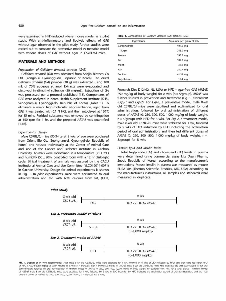
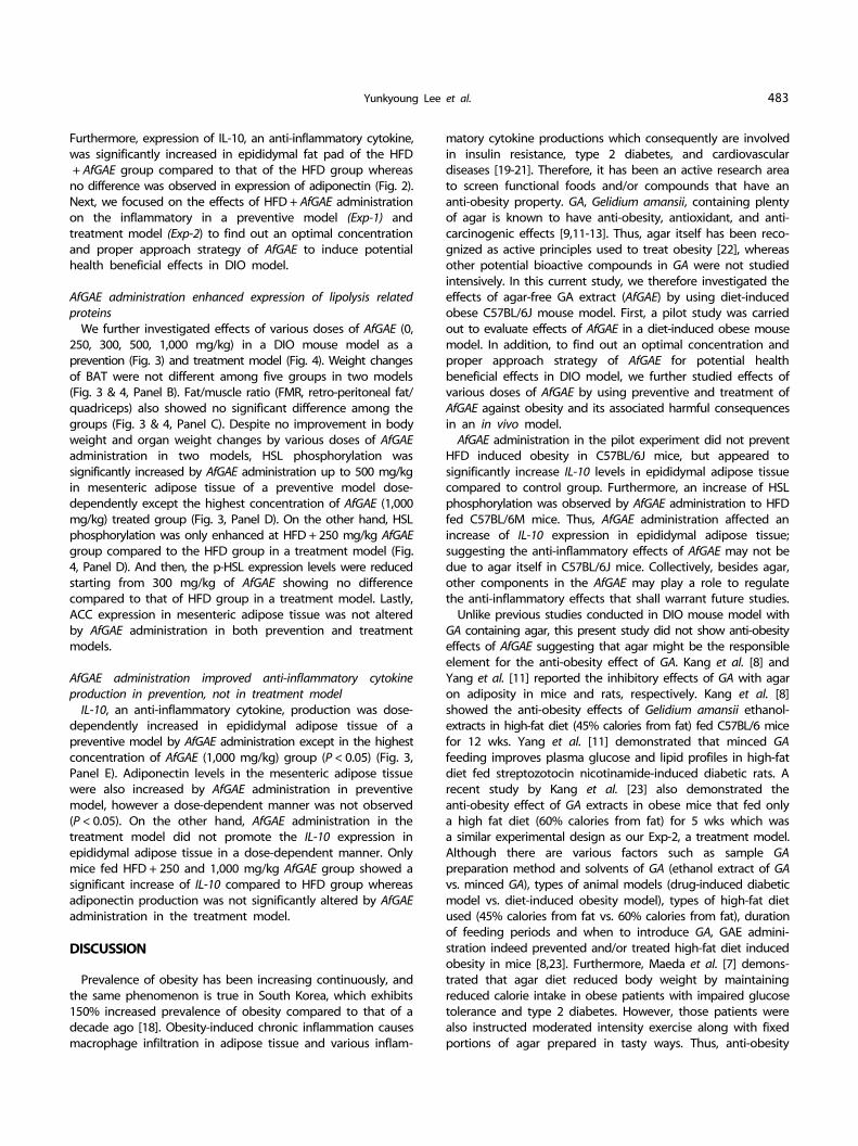
Fig. 2. Effects of Agar-free GAE (AfGAE) administration regarding inflammation and lipolysis related factors in HFD fed C57BL/6J mice. Data are represented as the Mean±SD(n = 5). Bars that do not share the same superscript are significantly different by t-test (P < 0.05). Panel A & B; representative immunoblot analysis of lipolysis related proteins, ACC and pHSL/total HSL of mesenteric fat pad, Panel C & D; representative immunoblot analysis of anti-inflammatory cytokines, IL-10 and adiponectin, in epididymal fat pad from mice fed HFD or HFD + AfGAE.
MeasurementsGroups
HFD HFD + AfGAE
Final body weight (g) 40.4 ± 1.4 44.4 ± 1.5
Feed intakes (g/wk) 2.50 ± 0.102) 2.70 ± 0.10
FER3) (%) 6.41 ± 1.06 8.04 ± 0.53*
Fat mass (g) Total fat 4.28 ± 0.26 5.09 ± 0.25*
Brown adipose tissue 0.10 ± 0.01 0.12 ± 0.02
Muscle mass (g) Total muscle 0.44 ± 0.01 0.42 ± 0.00
Quadriceps 0.18 ± 0.00 0.19 ± 0.01
Liver (g) 0.95 ± 0.04 1.09 ± 0.057*
Plasma Insulin (μU/mL) 21.8 ± 4.19 45.10 ± 13.80*
TG (mg/dL) 72.7 ± 5.9 71.30 ± 5.80
TC (mg/dL) 140.4 ± 8.7 152.10 ± 11.30
C57BL/6J mice were acclimated to oral administration and fed with HFD or HFD+ agar-free GAE (AfGAE, 250 mg/kg) for 8 wks (n = 5/group).
1) Data are presented as the Mean±SD (n = 5). 2) Means in the same row indicated with an asterisk are significantly different by
t-test (* P < 0.05, ** P < 0.01).3) FER (Food Efficiency Ratio) = body weight gain/feed intake during 8 wks of
feeding.
Table 2. Metabolic parameters in pilot experiment1)Western blotting analysisProtein from frozen epididymal and mesenteric fat tissues
were isolated by using lysis buffer (Cell signaling, Boston, MA, USA) and immediately homogenized as previously described [17]. Proteins (30-35 μg) were separated by 8% to 15% SDS- PAGE gel, and transferred to nitrocellulose membrane and incubated with the indicated antibody and horseradish peroxidase- coupled anti-species antibodies. Proteins were visualized by Bio-Rad chemiluminescence system. Concentrations of antibodies used in this study are as follows; p-HSL (1:1000), acetyl CoA carboxylase (ACC, 1:1000), Adiponectin (1:1000), IL-10 (1:1000), β-actin (1:5,000), GAPDH (1:5,000). Antibodies for hormone sensitive lipase (HSL), p-HSL, adiponectin, β-actin, and GAPDH were purchased from Cell Signaling (Danvers, MA, USA). The antibodies for total ACC and IL-10 were obtained from Abcam (Cambridge, MA, USA).
Statistical analysisData are presented as Mean±standard deviation (SD).
Statistical difference was determined by the t-test or one-way ANOVA (one-way analysis of variance) followed by Tukey’s multiple comparison test by using SPSS 12.0 (SPSS Inc, Chicago, IL, USA). Significance was set at a P-value < 0.05.
RESULTS
AfGAE improved HFD-induced inflammation and lipolysis related factors
Comparison of metabolic parameters in mice fed HFD or HFD + AfGAE for 8 wks in the pilot study are shown in Table 2. Mice fed HFD + AfGAE had a similar weight gain to the mice fed HFD suggesting no anti-obesity effects of AfGAE oral administration to HFD fed C57BL/6J mice. In addition, food efficiency ratio (FER) was significantly increased in mice fed HFD + AfGAE compared to mice fed HFD only. Total fat mass and brown adipose tissue (BAT) in mice fed HFD + AfGAE seemed to be slightly higher
than that in HFD group, but that did not reach statistical significance. Plasma analysis results showed insulin levels were significantly increased in mice fed HFD + AfGAE compared to mice fed HFD. However, there were no significant differences in TG and TC levels in plasma between HFD and HFD + AfGAE.
We further observed alterations of lipolysis related proteins such as total acetyl CoA carboxylase (ACC), total hormone- sensitive lipase (HSL) and p-HSL expression in adipose tissue from the HFD group and HFD + AfGAE group (Fig. 2). No significant difference was detected in ACC protein expression in the mesenteric fat pad between the HFD group and HFD + AfGAE group, meanwhile phosphorylation levels of HSL were significantly upregulated in mesenteric fat pad of mice fed HFD + AfGAE compared to that of mice fed HFD only (P < 0.01).
482 Agar free-Gelidium amansii on anti-inflammation
(A)
(D)
(E)
(B) (C)
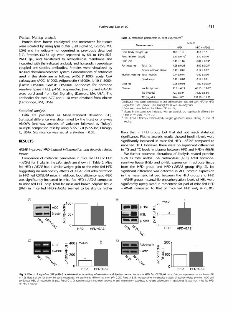
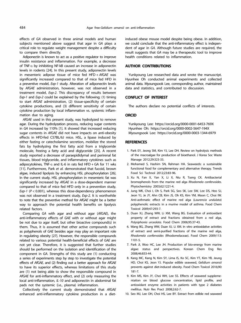
Fig. 3. Effects of AfGAE administration in a preventive model, Exp-1, on changes of metabolic parameters, lipolysis and inflammation-related proteins in mesenteric adipose tissue of HFD fed C57BL/6J mice. Data are represented as Mean±SD (n = 5). Bars that do not share the same superscript are significantly different by ANOVA (P < 0.05). Panel A-C: final body weight, brown adipose tissue (BAT) weight, and fat to muscle mass ratio, Panel D & E: Representative immunoblot analysis of lipolysis and inflammation-related proteins, phosphorylation levels of HSL, total ACC, IL-10, and adiponectin in mesenteric fat pad from mice fed HFD or HFD + various doses of AfGAE in a preventive model.
(A)
(D)
(E)
(B) (C)
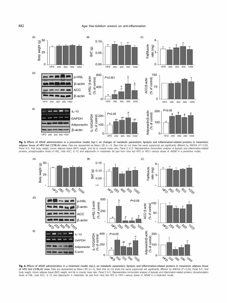
Fig. 4. Effects of AfGAE administration in a treatment model, Exp-2, on metabolic parameters, lipolysis and inflammation-related proteins in mesenteric adipose tissue of HFD fed C57BL/6J mice. Data are represented as Mean±SD (n = 5). Bars that do not share the same superscript are significantly different by ANOVA (P < 0.05). Panel A-C: final body weight, brown adipose tissue (BAT) weight, and fat to muscle mass ratio, Panel D & E: Representative immunoblot analysis of lipolysis and inflammation-related proteins, phosphorylation levels of HSL, total ACC, IL-10, and adiponectin in mesenteric fat pad from mice fed HFD or HFD + various doses of AfGAE in a treatment model.
Yunkyoung Lee et al. 483
Furthermore, expression of IL-10, an anti-inflammatory cytokine, was significantly increased in epididymal fat pad of the HFD+ AfGAE group compared to that of the HFD group whereas
no difference was observed in expression of adiponectin (Fig. 2). Next, we focused on the effects of HFD + AfGAE administration on the inflammatory in a preventive model (Exp-1) and treatment model (Exp-2) to find out an optimal concentration and proper approach strategy of AfGAE to induce potential health beneficial effects in DIO model.
AfGAE administration enhanced expression of lipolysis related proteins
We further investigated effects of various doses of AfGAE (0, 250, 300, 500, 1,000 mg/kg) in a DIO mouse model as a prevention (Fig. 3) and treatment model (Fig. 4). Weight changes of BAT were not different among five groups in two models (Fig. 3 & 4, Panel B). Fat/muscle ratio (FMR, retro-peritoneal fat/ quadriceps) also showed no significant difference among the groups (Fig. 3 & 4, Panel C). Despite no improvement in body weight and organ weight changes by various doses of AfGAE administration in two models, HSL phosphorylation was significantly increased by AfGAE administration up to 500 mg/kg in mesenteric adipose tissue of a preventive model dose- dependently except the highest concentration of AfGAE (1,000 mg/kg) treated group (Fig. 3, Panel D). On the other hand, HSL phosphorylation was only enhanced at HFD + 250 mg/kg AfGAE group compared to the HFD group in a treatment model (Fig. 4, Panel D). And then, the p-HSL expression levels were reduced starting from 300 mg/kg of AfGAE showing no difference compared to that of HFD group in a treatment model. Lastly, ACC expression in mesenteric adipose tissue was not altered by AfGAE administration in both prevention and treatment models.
AfGAE administration improved anti-inflammatory cytokine production in prevention, not in treatment model
IL-10, an anti-inflammatory cytokine, production was dose- dependently increased in epididymal adipose tissue of a preventive model by AfGAE administration except in the highest concentration of AfGAE (1,000 mg/kg) group (P < 0.05) (Fig. 3, Panel E). Adiponectin levels in the mesenteric adipose tissue were also increased by AfGAE administration in preventive model, however a dose-dependent manner was not observed (P < 0.05). On the other hand, AfGAE administration in the treatment model did not promote the IL-10 expression in epididymal adipose tissue in a dose-dependent manner. Only mice fed HFD + 250 and 1,000 mg/kg AfGAE group showed a significant increase of IL-10 compared to HFD group whereas adiponectin production was not significantly altered by AfGAE administration in the treatment model.
DISCUSSION
Prevalence of obesity has been increasing continuously, and the same phenomenon is true in South Korea, which exhibits 150% increased prevalence of obesity compared to that of a decade ago [18]. Obesity-induced chronic inflammation causes macrophage infiltration in adipose tissue and various inflam-
matory cytokine productions which consequently are involved in insulin resistance, type 2 diabetes, and cardiovascular diseases [19-21]. Therefore, it has been an active research area to screen functional foods and/or compounds that have an anti-obesity property. GA, Gelidium amansii, containing plenty of agar is known to have anti-obesity, antioxidant, and anti- carcinogenic effects [9,11-13]. Thus, agar itself has been reco-gnized as active principles used to treat obesity [22], whereas other potential bioactive compounds in GA were not studied intensively. In this current study, we therefore investigated the effects of agar-free GA extract (AfGAE) by using diet-induced obese C57BL/6J mouse model. First, a pilot study was carried out to evaluate effects of AfGAE in a diet-induced obese mouse model. In addition, to find out an optimal concentration and proper approach strategy of AfGAE for potential health beneficial effects in DIO model, we further studied effects of various doses of AfGAE by using preventive and treatment of AfGAE against obesity and its associated harmful consequences in an in vivo model.
AfGAE administration in the pilot experiment did not prevent HFD induced obesity in C57BL/6J mice, but appeared to significantly increase IL-10 levels in epididymal adipose tissue compared to control group. Furthermore, an increase of HSL phosphorylation was observed by AfGAE administration to HFD fed C57BL/6M mice. Thus, AfGAE administration affected an increase of IL-10 expression in epididymal adipose tissue; suggesting the anti-inflammatory effects of AfGAE may not be due to agar itself in C57BL/6J mice. Collectively, besides agar, other components in the AfGAE may play a role to regulate the anti-inflammatory effects that shall warrant future studies.
Unlike previous studies conducted in DIO mouse model with GA containing agar, this present study did not show anti-obesity effects of AfGAE suggesting that agar might be the responsible element for the anti-obesity effect of GA. Kang et al. [8] and Yang et al. [11] reported the inhibitory effects of GA with agar on adiposity in mice and rats, respectively. Kang et al. [8] showed the anti-obesity effects of Gelidium amansii ethanol- extracts in high-fat diet (45% calories from fat) fed C57BL/6 mice for 12 wks. Yang et al. [11] demonstrated that minced GA feeding improves plasma glucose and lipid profiles in high-fat diet fed streptozotocin nicotinamide-induced diabetic rats. A recent study by Kang et al. [23] also demonstrated the anti-obesity effect of GA extracts in obese mice that fed only a high fat diet (60% calories from fat) for 5 wks which was a similar experimental design as our Exp-2, a treatment model. Although there are various factors such as sample GA preparation method and solvents of GA (ethanol extract of GA vs. minced GA), types of animal models (drug-induced diabetic model vs. diet-induced obesity model), types of high-fat diet used (45% calories from fat vs. 60% calories from fat), duration of feeding periods and when to introduce GA, GAE admini-stration indeed prevented and/or treated high-fat diet induced obesity in mice [8,23]. Furthermore, Maeda et al. [7] demons-trated that agar diet reduced body weight by maintaining reduced calorie intake in obese patients with impaired glucose tolerance and type 2 diabetes. However, those patients were also instructed moderated intensity exercise along with fixed portions of agar prepared in tasty ways. Thus, anti-obesity
484 Agar free-Gelidium amansii on anti-inflammation
effects of GA observed in three animal models and human subjects mentioned above suggest that agar in GA plays a critical role to regulate weight management despite a difficulty to compare them directly.
Adiponectin is known to act as a positive regulator to improve insulin resistance and inflammation. For example, a decrease of TNF-α by inhibiting NF-kB caused an increase in adiponectin levels in rodents [24]. In this present study, adiponectin levels in mesenteric adipose tissue of mice fed HFD + AfGAE was significantly increased compared to that of mice fed HFD in a preventive model, Exp-1 study. Alteration of adiponectin levels by AfGAE administration, however, was not observed in a treatment model, Exp-2. This discrepancy of results between Exp-1 and Exp-2 could be explained by the following; (1) timing to start AfGAE administration, (2) tissue-specificity of certain cytokine productions, and (3) different sensitivity of certain cytokine production by local inflammation vs. systemic inflam-mation due to aging.
AfGAE used in this present study, was hydrolyzed to remove agar. During the hydrolyzation process, reducing sugar contents in GA increased by 110% [1]. It showed that increased reducing sugar contents in AfGAE did not have impacts on anti-obesity effects in HFD-fed C57BL/6J mice. HSL, a lipase induced by either fasting or catecholamine secretion, mobilize the stored fats by hydrolyzing the first fatty acid from a triglyceride molecule, freeing a fatty acid and diglyceride [25]. A recent study reported a decrease of paraepididymal and perirenal fat tissues, blood triglyceride, and inflammatory cytokines such as adipocytokines, TNF-α and IL-6 in rats fed HFD + GA for 11 wks [11]. Furthermore, Park et al. demonstrated that fucoid, brown algae, induced lipolysis by enhancing HSL phosphorylation [26].
In the current study, HSL phosphorylation in mesenteric fat was significantly increased by AfGAE in a dose-dependent manner compared to that of mice fed HFD only in a prevention study, Exp-1 (P < 0.001), whereas this dose-dependency phenomenon was not observed in a treatment study, Exp-2. It is worthwhile to note that the preventive method for AfGAE might be a better way to approach the potential health benefits on lipolysis related factors.
Comparing GA with agar and without agar (AfGAE), the anti-inflammatory effects of GAE with or without agar might be not due to agar itself, but other bioactive compound(s) in them. Thus, it is assumed that other active compounds such as polyphenols of GAE besides agar may play an important role in regulating obesity [27]. However, the responsible compounds related to various potential health-beneficial effects of GAE are not yet clear. Therefore, it is suggested that further studies should be performed on the isolation and identification of the component in GA. Strengths of this study are (1) conducting a series of experiments step by step to investigate the potential effects of AfGAE, and (2) finding out a better approach for AfGAE to have its superior effects, whereas limitations of this study are (1) not being able to show the responsible compound in AfGAE for anti-inflammatory effect, and (2) only measuring the local anti-inflammation, IL-10 and adiponectin in abdominal fat pads not the systemic (i.e., plasma) inflammation.
Collectively the current study demonstrated that AfGAE enhanced anti-inflammatory cytokine production in a diet-
induced obese mouse model despite being obese. In addition, we could conclude that the anti-inflammatory effect is indepen-dent of agar in GA. Although future studies are required, the result suggests that GA may be a therapeutic tool to improve health conditions related to inflammation.
AUTHOR CONTRIBUTIONS
Yunkyoung Lee researched data and wrote the manuscript. Hyunhee Oh conducted animal experiments and collected animal data. Myoungsook Lee, corresponding author, maintained data and statistics, and contributed to discussion.
CONFLICT OF INTEREST
The authors declare no potential conflicts of interests.
ORCID
Yunkyoung Lee: https://orcid.org/0000-0001-6453-769XHyunhee Oh: https://orcid.org/0000-0002-5647-194XMyoungsook Lee: https://orcid.org/0000-0003-1344-6979
REFERENCES
1. Park EY, Jeong SM, Kim YJ, Lee DH. Review on hydrolysis methods of the macroalgae for production of bioethanol. J Korea Soc Waste Manage 2012;29:323-33.
2. Mohamed S, Hashim SN, Rahman HA. Seaweeds: a sustainable functional food for complementary and alternative therapy. Trends Food Sci Technol 2012;23:83-96.
3. Xu N, Fan X, Yan X, Li X, Niu R, Tseng CK. Antibacterial bromophenols from the marine red alga Rhodomela confervoides. Phytochemistry 2003;62:1221-4.
4. Jung WK, Choi I, Oh S, Park SG, Seo SK, Lee SW, Lee DS, Heo SJ, Jeon YJ, Je JY, Ahn CB, Kim JS, Oh KS, Kim YM, Moon C, Choi IW. Anti-asthmatic effect of marine red alga (Laurencia undulata) polyphenolic extracts in a murine model of asthma. Food Chem Toxicol 2009;47:293-7.
5. Duan XJ, Zhang WW, Li XM, Wang BG. Evaluation of antioxidant property of extract and fractions obtained from a red alga, Polysiphonia urceolata. Food Chem 2006;95:37-43.
6. Wang BG, Zhang WW, Duan XJ, Li XM. In vitro antioxidative activities of extract and semi-purified fractions of the marine red alga, Rhodomela confervoides (Rhodomelaceae). Food Chem 2009;113: 1101-5.
7. Park JI, Woo HC, Lee JH. Production of bio-energy from marine algae: status and perspectives. Korean Chem Eng Res 2008;46:833-44.
8. Kang MC, Kang N, Kim SY, Lima IS, Ko SC, Kim YT, Kim YB, Jeung HD, Choi KS, Jeon YJ. Popular edible seaweed, Gelidium amansii prevents against diet-induced obesity. Food Chem Toxicol 2016;90: 181-7.
9. Kim MS, Kim JY, Choi WH, Lee SS. Effects of seaweed suppleme-ntation on blood glucose concentration, lipid profile, and antioxidant enzyme activities in patients with type 2 diabetes mellitus. Nutr Res Pract 2008;2:62-7.
10. Seo MJ, Lee OH, Choi HS, Lee BY. Extract from edible red seaweed

Yunkyoung Lee et al. 485
(Gelidium amansii) inhibits lipid accumulation and ROS production during differentiation in 3T3-L1 cells. Prev Nutr Food Sci 2012;17: 129-35.
11. Yang TH, Yao HT, Chiang MT. Red algae (Gelidium amansii) reduces adiposity via activation of lipolysis in rats with diabetes induced by streptozotocin-nicotinamide. J Food Drug Anal 2015;23:758-65.
12. Maeda H, Yamamoto R, Hirao K, Tochikubo O. Effects of agar (kanten) diet on obese patients with impaired glucose tolerance and type 2 diabetes. Diabetes Obes Metab 2005;7:40-6.
13. Kang MC, Kang N, Ko SC, Kim YB, Jeon YJ. Anti-obesity effects of seaweeds of Jeju Island on the differentiation of 3T3-L1 preadipocytes and obese mice fed a high-fat diet. Food Chem Toxicol 2016;90: 36-44.
14. Samarakoon KW, Elvitigala DAS, Lakmal HHC, Kim YM, Jeon YJ. Future prospects and health benefits of functional ingredients from marine bio-resources: a review. Fish Aquat Sci 2014;17:275-90.
15. Kim J, Kim HJ, Lee M. The suppressive effect of Gelidium amansi-EtOH extracts on the adipogenesis with MAPK signals in adipocytes with or without macrophages. Food Sci Biotechnol 2017;26:1715-23.
16. Lee SM, Yu BJ, Kim YM, Choi SJ, Ha JM, Lee JH. Production of bio-ethanol from agar using Saccharomyces cerevisiae. J Korean Ind Eng Chem 2009;20:290-5.
17. Kim SY, Wi HR, Choi S, Ha TJ, Lee BW, Lee M. Inhibitory effect of anthocyanin-rich black soybean testa (Glycine max (L.) Merr.) on the inflammation-induced adipogenesis in a DIO mouse model. J Funct Foods 2015;14:623-33.
18. Na SY, Myung SJ. Obesity and colorectal cancer. Korean J Gastroen-
terol 2012;59:16-26.19. Cinti S, Mitchell G, Barbatelli G, Murano I, Ceresi E, Faloia E, Wang
S, Fortier M, Greenberg AS, Obin MS. Adipocyte death defines macrophage localization and function in adipose tissue of obese mice and humans. J Lipid Res 2005;46:2347-55.
20. Weisberg SP, McCann D, Desai M, Rosenbaum M, Leibel RL, Ferrante AW Jr. Obesity is associated with macrophage accumulation in adipose tissue. J Clin Invest 2003;112:1796-808.
21. Xu H, Barnes GT, Yang Q, Tan G, Yang D, Chou CJ, Sole J, Nichols A, Ross JS, Tartaglia LA, Chen H. Chronic inflammation in fat plays a crucial role in the development of obesity-related insulin resistance. J Clin Invest 2003;112:1821-30.
22. Moro CO, Basile G. Obesity and medicinal plants. Fitoterapia 2000;71 Suppl 1:S73-82.
23. Kang JH, Lee HA, Kim HJ, Han JS. Gelidium amansii extract ameliorates obesity by down-regulating adipogenic transcription factors in diet-induced obese mice. Nutr Res Pract 2017;11:17-24.
24. Shoelson SE, Lee J, Yuan M. Inflammation and the IKK β/I κ B/NF-κ
B axis in obesity- and diet-induced insulin resistance. Int J Obes Relat Metab Disord 2003;27 Suppl 3:S49-52.
25. Okuda H, Morimoto C, Tsujita T. Effect of substrates on the cyclic AMP-dependent lipolytic reaction of hormone-sensitive lipase. J Lipid Res 1994;35:1267-73.
26. Park MK, Jung U, Roh C. Fucoidan from marine brown algae inhibits lipid accumulation. Mar Drugs 2011;9:1359-67.
27. Heo SJ, Cha SH, Lee KW, Jeon YJ. Antioxidant activities of red algae from Jeju Island. Algae 2006;21:149-56.