
Anti-Cancer Drug Loaded Iron–Gold Core–Shell …20... · RESEARCH ARTICLE Kayal and Ramanujan...
13
RESEARCH ARTICLE Copyright © 2010 American Scientific Publishers All rights reserved Printed in the United States of America Journal of Nanoscience and Nanotechnology Vol. 10, 1–13, 2010 Anti-Cancer Drug Loaded Iron–Gold Core–Shell Nanoparticles (Fe@Au) for Magnetic Drug Targeting Sibnath Kayal and Raju Vijayaraghavan Ramanujan ∗ School of Materials Science and Engineering, Nanyang Technological University, 639798, Singapore Magnetic drug targeting, using core–shell magnetic carrier particles loaded with anti-cancer drugs, is an emerging and significant method of cancer treatment. Gold shell-iron core nanoparticles (Fe@Au) were synthesized by the reverse micelle method with aqueous reactants, surfactant, co-surfactant and oil phase. XRD, XPS, TEM and magnetic property measurements were utilized to characterize these core–shell nanoparticles. Magnetic measurements showed that the particles were superparamagnetic at room temperature and that the saturation magnetization decreased with increasing gold concentration. The anti-cancer drug doxorubicin (DOX) was loaded onto these Fe@Au nanoparticle carriers and the drug release profiles showed that upto 25% of adsorbed drug was released in 80 h. It was found that the amine (–NH 2 ) group of DOX binds to the gold shell. An in vitro apparatus simulating the human circulatory system was used to determine the retention of these nanoparticle carriers when exposed to an external magnetic field. A high percentage of mag- netic carriers could be retained for physiologically relevant flow speeds of fluid. The present findings show that DOX loaded gold coated iron nanoparticles are promising for magnetically targeted drug delivery. Keywords: Core–Shell, Reverse Micelle Method, Superparamagnetic Nanoparticles, Magnetic Drug Targeting. 1. INTRODUCTION Magnetic nanoparticles have exciting biomedical applica- tions such as magnetic separation, 1–6 magnetic resonance imaging (MRI) contrast agents, 7–15 targeted drug delivery for cancer therapy, 16–30 as well as conventional applica- tions such as magnetic seals, printing, recording. 31–33 The main goal in cancer therapy is to destroy cancer cells with- out damaging normal cells. Conventional methods of can- cer therapy such as surgery, radiation and chemotherapy are either invasive or have undesirable side effects. Mag- netically directed drug delivery using magnetic nanopar- ticles, optionally combined with hyperthermia, is a very attractive technique to improve the performance of cur- rent methods of cancer treatment. 34–39 The strategy is to concentrate the drug loaded magnetic nanoparticle carri- ers at the tumor site by using an external magnetic field. The drug can then be released from the carriers either via enzymatic activity or changes in physiological conditions such as pH, osmolality, or temperature, 40 to be taken up by tumor cells. If the magnetic particles are coated with a gold shell, this shell can provide advantages such as improved computed tomography (CT) contrast, 41 it can be heated by ∗ Author to whom correspondence should be addressed. near infrared (NIR) light irradiation; the resulting heat can destroy tumor cells without damaging healthy tissues, this modality of treatment is known as hyperthermia resulting from this photothermal effect. 42–44 Nanoparticles can exhibit novel material properties that differ considerably from those of the bulk state. 45 Mag- netic nanoparticles smaller than about 30 nm can exhibit superparamagnetism, in superparamagnetic nanoparticles the magnetic moment can reorient in times less than 1 ns due to thermal agitation. 46 As the particle size decreases, the reactivity of the particles also increases and the mag- netic properties are influenced by surface effects. 47 Hence, a more robust structure for nanoparticles is a core/shell configuration where the magnetic core is coated by a shell layer. Such structures are useful for studying proximity effects and ideal for structure stabilization, as the shell can protect the core from oxidation. Additionally, the shell provides a platform for surface modification, functional- ization, tuning magnetic properties and biocompatibility. 48 The synthesis of gold coated magnetic nanoparticles and the study of their structural, magnetic properties have been the focus of recent experimental studies. 49–60 The synthesis of gold coated iron nanoparticles (Fe@Au) is of special interest since gold provides useful surface chemistry and J. Nanosci. Nanotechnol. 2010, Vol. 10, No. xx 1533-4880/2010/10/001/013 doi:10.1166/jnn.2010.2461 1
Transcript of Anti-Cancer Drug Loaded Iron–Gold Core–Shell …20... · RESEARCH ARTICLE Kayal and Ramanujan...

RESEARCH
ARTIC
LE
Copyright © 2010 American Scientific PublishersAll rights reservedPrinted in the United States of America
Journal ofNanoscience and Nanotechnology
Vol. 10, 1–13, 2010
Anti-Cancer Drug Loaded Iron–Gold Core–ShellNanoparticles (Fe@Au) for Magnetic Drug Targeting
Sibnath Kayal and Raju Vijayaraghavan Ramanujan∗
School of Materials Science and Engineering, Nanyang Technological University, 639798, Singapore
Magnetic drug targeting, using core–shell magnetic carrier particles loaded with anti-cancer drugs,is an emerging and significant method of cancer treatment. Gold shell-iron core nanoparticles(Fe@Au) were synthesized by the reverse micelle method with aqueous reactants, surfactant,co-surfactant and oil phase. XRD, XPS, TEM and magnetic property measurements were utilizedto characterize these core–shell nanoparticles. Magnetic measurements showed that the particleswere superparamagnetic at room temperature and that the saturation magnetization decreasedwith increasing gold concentration. The anti-cancer drug doxorubicin (DOX) was loaded onto theseFe@Au nanoparticle carriers and the drug release profiles showed that upto 25% of adsorbed drugwas released in 80 h. It was found that the amine (–NH2) group of DOX binds to the gold shell. Anin vitro apparatus simulating the human circulatory system was used to determine the retention ofthese nanoparticle carriers when exposed to an external magnetic field. A high percentage of mag-netic carriers could be retained for physiologically relevant flow speeds of fluid. The present findingsshow that DOX loaded gold coated iron nanoparticles are promising for magnetically targeted drugdelivery.
Keywords: Core–Shell, Reverse Micelle Method, Superparamagnetic Nanoparticles, MagneticDrug Targeting.
1. INTRODUCTION
Magnetic nanoparticles have exciting biomedical applica-tions such as magnetic separation,1–6 magnetic resonanceimaging (MRI) contrast agents,7–15 targeted drug deliveryfor cancer therapy,16–30 as well as conventional applica-tions such as magnetic seals, printing, recording.31–33 Themain goal in cancer therapy is to destroy cancer cells with-out damaging normal cells. Conventional methods of can-cer therapy such as surgery, radiation and chemotherapyare either invasive or have undesirable side effects. Mag-netically directed drug delivery using magnetic nanopar-ticles, optionally combined with hyperthermia, is a veryattractive technique to improve the performance of cur-rent methods of cancer treatment.34–39 The strategy is toconcentrate the drug loaded magnetic nanoparticle carri-ers at the tumor site by using an external magnetic field.The drug can then be released from the carriers either viaenzymatic activity or changes in physiological conditionssuch as pH, osmolality, or temperature,40 to be taken up bytumor cells. If the magnetic particles are coated with a goldshell, this shell can provide advantages such as improvedcomputed tomography (CT) contrast,41 it can be heated by
∗Author to whom correspondence should be addressed.
near infrared (NIR) light irradiation; the resulting heat candestroy tumor cells without damaging healthy tissues, thismodality of treatment is known as hyperthermia resultingfrom this photothermal effect.42–44
Nanoparticles can exhibit novel material properties thatdiffer considerably from those of the bulk state.45 Mag-netic nanoparticles smaller than about 30 nm can exhibitsuperparamagnetism, in superparamagnetic nanoparticlesthe magnetic moment can reorient in times less than 1 nsdue to thermal agitation.46 As the particle size decreases,the reactivity of the particles also increases and the mag-netic properties are influenced by surface effects.47 Hence,a more robust structure for nanoparticles is a core/shellconfiguration where the magnetic core is coated by a shelllayer. Such structures are useful for studying proximityeffects and ideal for structure stabilization, as the shellcan protect the core from oxidation. Additionally, the shellprovides a platform for surface modification, functional-ization, tuning magnetic properties and biocompatibility.48
The synthesis of gold coated magnetic nanoparticles andthe study of their structural, magnetic properties have beenthe focus of recent experimental studies.49–60 The synthesisof gold coated iron nanoparticles (Fe@Au) is of specialinterest since gold provides useful surface chemistry and
J. Nanosci. Nanotechnol. 2010, Vol. 10, No. xx 1533-4880/2010/10/001/013 doi:10.1166/jnn.2010.2461 1
RESEARCH
ARTIC
LE
Anti-Cancer Drug Loaded Iron–Gold Core–Shell Nanoparticles (Fe@Au) for Magnetic Drug Targeting Kayal and Ramanujan
biological reactivity.61–63 Bare iron nanoparticles can notbe directly used for drug delivery since(a) free iron induces the formation of dangerous freeradicals,(b) iron nanoparticles can aggregate resulting in the for-mation of thromboses and(c) free iron nanoparticles are easily oxidized.
Coating the iron nanoparticles with a stable noble metallike gold results in air-stable nanoparticles which are pro-tected from oxidation.56 Gold is a favoured coating mate-rial because of well known synthesis procedures as well asits chemical functionality.64�65 Surface derivatization withgold also helps to reduce particle agglomeration by stericor electronic repulsion and improves biocompatibility.66
Considerable interest has been directed towards the func-tionalization of gold nanoparticles by the use of thiolchemistry, which facilitates the attachment of biologicallyrelevant molecules using a variety of thiol linkers.67�68
Doxorubicin (DOX) is commonly used to treat diversecancers including ovarian, bladder, thyroid, lung, breast,liver cancer.69 Free DOX used in chemotherapy treatmentshowed a very narrow therapeutic index and its clinical usehas been hampered by side-effects like cardio toxicity,70
hence, selectively targeting DOX to cancer tissues canimprove its efficacy and safety.Several groups have studied the structural, optical and
magnetic properties of gold coated iron (Fe@Au) nanopat-icles. Lin et al. successfully synthesized gold-coated ironnanoparticles by the reverse micelle technique and showedthat no iron oxide was present in the X-ray diffraction(XRD) analysis.71 The aqueous reactants were introducedinto the micelle solution and allowed to react within theconfines of the micelles. By controlling the relative con-centrations of surfactant and water, synthesis of particlesin water-in-oil microemulsion allows better control oversize and shape of resulting nanoparticles.72 Cho and co-workers reported the synthesis of iron–gold core–shellparticles with large iron cores, such particles exhibited fer-romagnetism at room temperature.48 Gold coated acicu-lar and spherical shaped iron nanoparticles were preparedand their morphology and magnetic properties were char-acterized using transmission electron microscopy (TEM)and alternate gradient magnetometry.73 However, no drugloading and release kinetics have been reported with theseFe@Au nanoparticles.In this work, we report the synthesis, characterization
and magnetic behaviour of gold coated iron nanoparticles(Fe@Au). We have attached the anti-cancer drug doxoru-bicin (DOX) to Fe@Au nanoparticles and demonstratedthe binding mechanism of DOX with gold coating. Wehave studied DOX loading and release profile of Fe@Aunanoparticles and in vitro targeting of Fe@Au nanopar-ticles in a fluid, for flow velocities comparable to thatencountered in the capillary bed of tumor, for the first time.The novelty of this work is the loading of anti-cancer drug
DOX onto the superparamagnetic Fe@Au nanoparticles,the binding mechanism of DOX with the gold coating, thedetermination of DOX loading and release profile and thein vitro targeting of Fe@Au nanoparticles. Our results sug-gest that drug loaded Fe@Au nanoparticles are a promis-ing system for magnetically targeted drug delivery.
2. EXPERIMENTAL PROCEDURES
2.1. Materials
All chemicals were used without further purification.Chemicals used for the synthesis of gold coatediron nanoparticles were cetyltrimethylammonium bromide(CTAB) (C19H42BrN), iron (II) sulphate heptahydrate(FeSO4 · 7H2O), hydrogen tetrachloroaurate (HAuCl4),sodium borohydride (NaBH4), octane (C8H18) andn-butanol (C4H10O). Deionised water was used for all theexperiments. CTAB was obtained from Acros Organics,FeSO4 ·7H2O from Fischer Chemicals, NaBH4 from FlukaChemical, other chemicals were obtained from Aldrich.Doxorubicin hydrochloride (Aldrich) was used for the drugloading and release studies.
2.2. Synthesis of Gold Coated IronNanoparticles (Fe@Au)
The synthesis reaction was carried out in an oxygen freeglove box in two steps, in each step predetermined amountof aqueous reactants were mixed with CTAB, n-butanoland octane to form the reverse micelle solution. The pro-cedure and the quantity of components used for the syn-thesis are schematically shown in Figure 1. In a typicalexperiment, in a beaker (A), 0.333 g FeSO4 was dissolvedin 2.4 ml deionised water and 6 g CTAB, 5 g 1-butanoland 15 g octane were added. In another beaker (B), 0.9 gNaBH4 was dissolved in 2.4 ml water and mixed with6 g CTAB, 5 g 1-butanol and 15 g octane. Micellar solu-tions containing FeSO4 (beaker A) and NaBH4 (beaker B)were mixed together under stirring, color change frompale green to black was observed. NaBH4 reduces FeSO4
resulting in the formation of iron core material. Once theiron nanoparticles formed, a micellar solution containing0.0245 (M) HAuCl4 (aq), 3 g CTAB, 2.5 g 1-butanol and15 g octane was added followed by the addition of a micel-lar solution of NaBH4 (aq). Au (III) was reduced to Au (0)by NaBH4 and gold forms a coating on the outer sur-face of the iron particles.74 The volume of HAuCl4 wasvaried to alter the gold coating thickness. The iron con-taining nanoparticles were separated by placing the finalreaction solution in a magnetic field. The remaining sur-factants and byproducts were removed by repeated wash-ing with 1:1 methanol/water mixture. The particles weredried under vacuum and stored, the resulting powder wasblack. Following the above synthesis method, three samplesets were prepared by varying the concentration of gold
2 J. Nanosci. Nanotechnol. 10, 1–13, 2010

RESEARCH
ARTIC
LE
Kayal and Ramanujan Anti-Cancer Drug Loaded Iron–Gold Core–Shell Nanoparticles (Fe@Au) for Magnetic Drug Targeting
0.333 g FeSO4 in 2.4 mldeionised water
6 g CTAB5 g 1-Butanol15 g Octane
Mix
0.9 g NaBH4 in 2.4 mldeionised water
6 g CTAB5 g 1-Butanol15 g Octane
Fe nanoparticles
0.108 g NaBH4 in 1.8 mldeionised water
3 g CTAB2.5 g 1-Butanol
15 g Octane
Fe@Au nanoparticles
N2
N2
XmL (X = 0, 5, 10) of 0.0245 (M)HAuCl4 (aq)3 g CTAB
2.5 g 1-Butanol15 g Octane
Fig. 1. Schematic diagram showing a typical procedure for formation of Fe@Au nanoparticles by reverse micelle technique. FeSO4 and HAuCl4 arereduced by NaBH4, CTAB is surfactant, 1-butanol is co-surfactant and octane is oil phase.
(Table I). Increasing the gold concentration beyond therange studied in this work resulted in low saturation mag-netization (Ms), hence they were not examined further. Inthis work, the uncoated sample set is referred as S-1 andthose coated with 0.102 and 0.204 mmole of Au/mmole ofFe are referred as S-2 and S-3 respectively in subsequentresults and discussion.
2.3. Instrumentation
A Shimadzu XRD-6000 X-ray diffractometer with Cu-K�
radiation (wavelength= 0.154056 nm) was used for pow-der X-ray diffraction (XRD) measurement. The powderwas packed in the glass sample holder and the XRD pat-terns were collected between 10� < 2� < 90� with dwellingtime of 2 seconds and a step size of 0.02�. X-ray pho-toelectron spectroscopy (XPS) data were recorded on a
Kratos analytical XSAM 800 using an aluminium anoderadiation source. The size and morphological character-ization of the particles were carried out using a JEOL2010 transmission electron microscope (TEM) operatingat 200 kV. TEM samples were prepared by dispersingnanoparticles in acetone for 30 min by ultrasonic vibration.The aqueous dispersion was dropped on a carbon coatedcopper TEM grid with filter paper underneath to absorb theacetone and allowed to air-dry. Fourier transform infrared(FTIR) spectra were recorded on a Nicolet Magna-IR550 spectrometer in the region of 4000–400 cm−1 with
Table I. Summary of sample sets synthesized by reverse micelletechnique.
Sample set S-1 S-2 S-3
mmole of Au/mmole of Fe 0 0.102 0.204
J. Nanosci. Nanotechnol. 10, 1–13, 2010 3

RESEARCH
ARTIC
LE
Anti-Cancer Drug Loaded Iron–Gold Core–Shell Nanoparticles (Fe@Au) for Magnetic Drug Targeting Kayal and Ramanujan
4 cm−1 resolution. FTIR samples were prepared by mixingand grinding nanopowders with KBr reference followedby pelletizing. UV-visible spectra were obtained with aUV-visible spectrophotometer (Shimadzu UV 1700) in therange of 400–700 nm equipped with quartz 1 cm opti-cal length cuvettes (Hellma). The hydrodynamic diameterof magnetic nanoparticle suspension was measured basedon dynamic light scattering principles with a Brookhaven90 plus particle sizer. Magnetic properties were evalu-ated by a Lakeshore 7404 vibrating sample magnetome-ter (VSM).
2.4. Doxorubicin (DOX) Drug Loading andRelease Study
DOX loading was carried out by dispersing 5 mg of goldcoated iron (Fe@Au) nanoparticles (carriers) in 5 mLaqueous DOX solution (0.1 mg/mL) following the experi-mental procedure described by Kuznetsov et al.75 At fixedtime intervals, the carriers were separated from the liquidby means of a permanent magnet and the optical den-sity of residual DOX in the supernatant was measuredat 498 nm by UV-visible spectrophotometry.76 After themeasurements, carriers were redispersed for further DOXadsorption. Beyond a certain adsorption time, there wereno further changes in the concentration of DOX sincethe loading capacity of the carriers had reached satura-tion. The drug loading was determined as the differencebetween the initial DOX concentration and the DOX con-centration in the supernatant. The drug loaded magneticcarriers were then magnetically separated and dried. Therelease profile was obtained by dispersing the dried drugloaded magnetic nanoparticle carriers in 5 mL PBS bufferat 37 �C. As in the uptake experiments, the released con-centration of DOX in the particle free liquid was deter-mined at fixed time intervals at 498 nm by UV-visiblespectrophotometry.
2.5. In Vitro Targeting
In vitro tests for targeting of gold coated iron (Fe@Au)magnetic carriers were conducted to test the retention ofmagnetic carriers under various flow speeds of fluid withdifferent magnetic field gradients. An in vitro apparatus(Fig. 2) simulating the human circulatory system was usedto test the retention of ferrofluid (suspension of Fe@Auin MilliQ water) under various flow speeds of fluid andvarious magnetic field gradients. PBS buffer (simulatingblood) in beaker (A) could be pumped through the tube(I) of inner diameter of 1 mm into beaker (J) by a peri-staltic pump (C). The flow speed could be changed byadjusting the peristaltic pump. The fluid containing theFe@Au nanoparticles could be injected at the intake (D).A permanent magnet (F) was set around the target region(G) and the magnetic fields could be changed by alteringthe position of magnet. The retention was determined by
C
D
A
B
37 °C Oven
G
F
I
J
Fig. 2. Schematic of an in vitro apparatus simulating the human cir-culatory system: A-intake beaker, B-vibrating plate, C-peristaltic pump,D-syringe, F-permanent magnet, G-targeting, I-silicone tube, J-outletbeaker.
measuring the percentage of Fe@Au magnetic nanoparti-cles at the target region after fluid flow.
3. RESULTS
Here we present structural, morphological and magneticproperties of Fe@Au nanoparticles. The particles werecharacterized by XRD, TEM, XPS and VSM. DOX load-ing and release studies and in vitro targeting of Fe@Aunanoparticles under physiologically relevant flow speedsof fluid are also presented.
3.1. Structural Properties
Representative powder X-ray diffraction patterns of reversemicelle synthesized samples are presented in Figure 3. TheX-ray diffraction pattern of S-1 shows peaks correspondingto the magnetite (Fe3O4) phase. As there is no coating ofgold in S-1, iron is oxidized to form iron oxide (Fe3O4).For sample sets S-2 and S-3, peaks corresponding to goldare observed at 2� = 38.1�, 44.4�, 64.6�, 77.6� and 81.8�.In sample sets S-2 and S-3, the peaks of �-iron are over-lapped with the peaks of gold at 2� = 44.4�, 64.6� and81.8�, consistent with previous reports.71�77 The Rietveldrefinement of corresponding powder diffraction pattern wasperformed and it was found that 83% Fe, 17% Au and49.5% Fe, 50.5% Au were present in S-2 and S-3, respec-tively. Figure 4 shows the selected area electron diffraction(SAED) pattern of S-3 obtained from transmission electronmicroscopy. The diffraction rings were indexed (Fig. 4) andit was observed that gold 200, 220, 222 peaks overlappedwith iron 110, 200, 211 peaks. The EDX spectrum of S-3(Fig. 5) generated with an electron nanoprobe (∼20 nm indiameter) revealed both iron and gold peaks. No oxygenpeak was found, suggesting that there was negligible oxida-tion of iron. The peaks corresponding to carbon and copperarise from the sample holder.X-ray photoelectron spectroscopy (XPS) was used to
study the surface composition of gold coated iron nanopar-ticles. Figure 6 shows the XPS spectra and peak fit of(a) Fe 2p and (b) Au 4f regions of gold coated iron
4 J. Nanosci. Nanotechnol. 10, 1–13, 2010

RESEARCH
ARTIC
LE
Kayal and Ramanujan Anti-Cancer Drug Loaded Iron–Gold Core–Shell Nanoparticles (Fe@Au) for Magnetic Drug Targeting
10 20 30 40 50 60 70 80 90
(222)Au(211)Fe(c)
(b)
Fe3O4Fe3O4Fe3O4
Fe3O4
Fe andAu
Fe and AuFe and Au
Au
Au
(311)Au(220)Au(200)Fe
Inte
nsity
(a.
u)
2θ (degree)
(111)Au
(200)Au(110)Fe
Fe3O4(a)
Fig. 3. X-ray powder diffraction pattern of (a) S-1 (uncoated), (b) S-2(0.102 mmole gold coated iron nanoparticles) and (c) S-3 (0.204 mmolegold coated iron nanoparticles). Uncoated particles are oxidized to ironoxide whereas in gold coated particles, iron peaks overlap with goldpeaks.
nanoparticles (S-2 and S-3). The position of Fe 2p1/2 lineat 723.2 eV and that of Fe 2p3/2 line at 710 eV (Fig. 6(a))correspond to the bulk Fe (0) state78 in both the goldcoated samples. Analysis of XPS spectra reveals that thesurface elemental atomic ratio of gold to iron increasesfrom S-2 to S-3 with increasing gold concentration. Thedetection of Fe core with the Au shell in XPS spectra sug-gests that Au shell thickness is less than 5 nm.49 We havecalculated the thickness of gold coating which is presentedin Section 3.3.
51/nm
Au (222), Fe (211)
Au (311)
Au (220), Fe (200)
Au (200), Fe (110)
Au (111)
Fig. 4. Selected area electron diffraction (SAED) pattern of S-3(0.204 mmole gold coated iron nanoparticles). Gold 200, 220, 222diffraction peaks overlap with iron 110, 200, 211 peaks.
Fig. 5. EDX spectrum of S-3 (0.204 mmole gold coated iron nanopar-ticles). The absence of oxygen peak indicates that iron is not oxidizedwhen coated with gold. The peaks of carbon and copper arise from thesample holder.
(b)
(ii)
(i)
(a)
(i)
(ii)
Fig. 6. XPS spectra and peak fit of the (a) Fe 2p and (b) Au 4f regionsof (i) S-2 (0.102 mmole gold coated iron nanoparticles) and (ii) S-3(0.204 mmole gold coated iron nanoparticles). The detection of Fe corewith the Au shell in XPS spectra suggests that Au shell is thin.
J. Nanosci. Nanotechnol. 10, 1–13, 2010 5
RESEARCH
ARTIC
LE
Anti-Cancer Drug Loaded Iron–Gold Core–Shell Nanoparticles (Fe@Au) for Magnetic Drug Targeting Kayal and Ramanujan
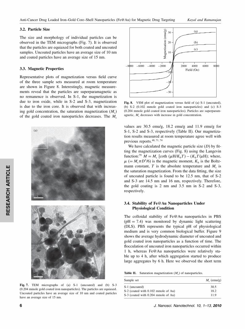
3.2. Particle Size
The size and morphology of individual particles can beobserved in the TEM micrographs (Fig. 7). It is observedthat the particles are equiaxed for both coated and uncoatedsamples. Uncoated particles have an average size of 10 nmand coated particles have an average size of 15 nm.
3.3. Magnetic Properties
Representative plots of magnetization versus field curveof the three sample sets measured at room temperatureare shown in Figure 8. Interestingly, magnetic measure-ments reveal that the particles are superparamagnetic asno remanence is observed. In S-1, the magnetization isdue to iron oxide, while in S-2 and S-3, magnetizationis due to the iron core. It is observed that with increas-ing gold concentration, the saturation magnetization (Ms)of the gold coated iron nanoparticles decreases. The Ms
(a)
(b)
10 nm
20 nm
Fig. 7. TEM micrographs of (a) S-1 (uncoated) and (b) S-3(0.204 mmole gold coated iron nanoparticles). The particles are equiaxed.Uncoated particles have an average size of 10 nm and coated particleshave an average size of 15 nm.
–8000 –6000 –4000 –2000 0 2000 4000 6000 8000
–30
–20
–10
0
10
20
30
Mag
netis
atio
n (e
mu/
g)
Field (Oe)
(a)
(b)
(c)
Fig. 8. VSM plot of magnetization versus field of (a) S-1 (uncoated),(b) S-2 (0.102 mmole gold coated iron nanoparticles) and (c) S-3(0.204 mmole gold coated iron nanoparticles). Particles are superparam-agnetic, Ms decreases with increase in gold concentration.
values are 30.5 emu/g, 18.2 emu/g and 11.9 emu/g forS-1, S-2 and S-3, respectively (Table II). Our magnetiza-tion results measured at room temperature agree well withprevious reports.48�71�74
We have calculated the magnetic particle size (D) by fit-ting the magnetization curves (Fig. 8) using the Langevinfunction:79 M =Ms [coth (�H/KbT �− �KbT /�H); where,� (=Ms�D3/6) is the magnetic moment, Kb is the Boltz-mann constant, T is the absolute temperature and Ms isthe saturation magnetization. From the data fitting, the sizeof uncoated particle is found to be 12.5 nm, that of S-2and S-3 are 14.5 nm and 16 nm, respectively. Therefore,the gold coating is 2 nm and 3.5 nm in S-2 and S-3,respectively.
3.4. Stability of Fe@Au Nanoparticles UnderPhysiological Condition
The colloidal stability of Fe@Au nanoparticles in PBS(pH = 7.4) was monitored by dynamic light scattering(DLS). PBS represents the typical pH of physiologicalmedium and is very common biological buffer. Figure 9shows the average hydrodynamic diameter of uncoated andgold coated iron nanoparticles as a function of time. Theflocculation of uncoated iron nanoparticles occurred within1 h, whereas Fe@Au nanoparticles were relatively sta-ble up to 4 h, after which aggregation started to producelarge aggregates by 6 h. Here we observed the short term
Table II. Saturation magnetization (Ms) of nanoparticles.
Sample set Ms (emu/g)
S-1 (uncoated) 30.5S-2 (coated with 0.102 mmole of Au) 18.2S-3 (coated with 0.204 mmole of Au) 11.9
6 J. Nanosci. Nanotechnol. 10, 1–13, 2010

RESEARCH
ARTIC
LE
Kayal and Ramanujan Anti-Cancer Drug Loaded Iron–Gold Core–Shell Nanoparticles (Fe@Au) for Magnetic Drug Targeting
Fig. 9. Average hydrodynamic diameter of S-1 (uncoated), S-2(0.102 mmole gold coated iron nanoparticles) and S-3 (0.204 mmole goldcoated iron nanoparticles) in PBS. Uncoated iron nanoparticles flocculatewithin 1 h, whereas Fe@Au nanoparticles are relatively stable up to 4 h.
stability of Fe@Au nanoparticles which are electrostati-cally stabilized. In order to get stability for several days,macromolecular stabilizers are used such as dextran,80
polyethylene glycol (PEG),81 Pluoronics82 which preventflocculation by steric stabilization.
3.5. Doxorubicin (DOX) Drug Loadingand Release Studies
We studied DOX loading and release profile of gold coatediron (Fe@Au) nanoparticles. The starting ratio by weightof Fe@Au naoparticles to DOX was 10. The drug loadingof the Fe@Au nanoparticles can be qualitatively moni-tored by a change in the color of DOX, which changedfrom deep orange (Fig. 10(a)) to a much lighter colorwith increasing time as DOX was adsorbed to the Fe@Aunanoparticles (Fig. 10(c)). We also used uncoated ironnanoparticles for drug loading, but we observed no changein the color and concentration of DOX which indicates thatthere is no drug adsorption on the uncoated iron nanopar-ticles (Fig. 10(b)). The DOX loading and release profilesof Fe@Au are shown in Figure 11. Initially there is rapidadsorption of DOX, then the adsorption rate slows downand finally reaches the saturation value (Fig. 11(a)). Ithas been found that the drug loading capacity increaseswith increase in gold content in the magnetic carriers. In26 h of loading, 83 �g and 94 �g of DOX are loadedper mg of S-2 and S-3, respectively. The drug releasebehavior of Fe@Au nanoparticle carriers was investigatedin PBS buffer at pH of 7 and temperature of 37 �Cto maintain the experimental conditions similar to bodyfluid. Figure 11(b) shows the release profile of DOXfrom the Fe@Au nanoparticle carriers. It is apparent fromFigure 11(b) that there is a continuous release of drug upto 10 h beyond which it slows down. A maximum of 18%and 25% of adsorbed drug is released in 80 h from S-2and S-3, respectively.
(a) (b) (c)
Fig. 10. The color of (a) pure DOX solution, (b) DOX solution after26 h mixing with uncoated iron nanoparticles and (c) DOX solution after26 h mixing with gold coated iron (Fe@Au) nanoparticles. The change incolor of DOX indicates that the DOX is attached onto Fe@Au nanopar-ticles only.
The binding of DOX with gold coated iron (Fe@Au)nanoparticles was studied by FTIR spectroscopy. Figure 12shows FTIR spectra of Fe@Au, pure DOX and DOX-attached Fe@Au nanoparticles. In case of Fe@Aunanoparticles, peaks at 3380 cm−1 (H–O stretching),1626 cm−1 (H–O–H bending) are due to adsorbed wateron the surface of nanoparticles and at 1392 cm−1 (C–Cstretching) from the surfactant. The FTIR spectrum ofpure DOX shows multiple peaks at 2932 cm−1 (C–Hstretching), 1730 cm−1 (C O stretching), 1618 cm−1
(N–H bending), 1414 cm−1 (C–C stretching), 1280 cm−1
(C–O–C stretching), 1070 cm−1 (C–O stretching),997 cm−1 (C–O–C stretching) and peaks at 870 and
(b)
(a)
Fig. 11. DOX (a) loading and (b) release profile of gold coated iron(Fe@Au) nanoparticles. The DOX loading and release increases withincrease in gold content in the magnetic carriers.
J. Nanosci. Nanotechnol. 10, 1–13, 2010 7

RESEARCH
ARTIC
LE
Anti-Cancer Drug Loaded Iron–Gold Core–Shell Nanoparticles (Fe@Au) for Magnetic Drug Targeting Kayal and Ramanujan
4000 3000 2000 1000
4000 3000 2000 1000
4000 3000 2000 1000
4756911392
1626
3380
(a)
(b)805
870
997
1070
12801618 1414
17302932
33303450
(c)
475
691
3340 2932
1730 99710701280
16181414
% T
rans
mis
sion
(a.
u)
Wave number (cm–1)
Fig. 12. FTIR spectra of (a) Fe@Au, (b) pure DOX and (c) DOX-attached Fe@Au nanoparticles. Amine (–NH2) group of DOX is involvedin attachment to the Fe@Au nanoparticles.
805 cm−1 corresponding to amine (N–H) wag.83 Inter-estingly for pure DOX, peaks at 3450 cm−1due to N–Hstretching vibrations for primary amine structure and at3330 cm−1due to O–H stretching vibrations are observed.However, for DOX-attached Fe@Au nanoparticles, peaksdue to N–H stretching vibrations and O–H stretchingvibrations overlap and are broadened (∼3340 cm−1). Thesharp peaks at 870 and 805 cm−1 observed in pure DOXdue to amine (N–H) wag also diminish in the FTIR spec-trum of DOX-attached Fe@Au nanoparticles. From thisFTIR analysis, it can be interpreted that –NH2 group ofDOX is the active site for the attachment to the [email protected] interaction of DOX with gold was further inves-
tigated by measuring the optical properties of gold col-loid, pure DOX solution and DOX-attached gold colloid
400 450 500 550 600 650 700
535
(c)
(b)Abs
orba
nce
(a.u
)
Wavelength (nm)
(a)
485
520
Fig. 13. UV-vis spectra of (a) pure DOX, (b) DOX-attached gold and(c) gold colloid. Peak is broadened and red shifted at 535 nm uponaddition of colloidal gold to DOX.
(Fig. 13). The absorption band observed for gold colloidat 520 nm in the UV-visible spectrum is due to surfaceplasmon resonance (SPR) which is characteristic of goldnanoparticles.49 Pure DOX shows an absorption maximumat 485 nm and it has been observed that with the addi-tion of colloidal gold to DOX, peak is broadened and redshifted at 535 nm due to interaction of DOX with theAu surface.84 This interaction is due to the attachment ofDOX with gold arising from large electrostatic attractionof active groups (–NH2) in DOX with gold nanoparticles.The zeta potential of Fe@Au nanoparticles and
DOX-attached Fe@Au nanoparticles were determined byMalvern Zetasizer. The average values of zeta potentialof Fe@Au and DOX-attached Fe@Au nanoparticles were−28.5 mV and −23.6 mV, respectively. In DOX-attachedFe@Au nanoparticles, a fraction of negative charges wasneutralized by complex formation, leading to change ofzeta potential from −28.5 mV to −23.6 mV.
3.6. In Vitro Targeting
The retention of Fe@Au nanoparticles (S-2 and S-3) atdistances 5, 10 and 15 mm away from the magnet sur-face with various flow rate of fluid is shown in Figure 14.
(a)
40
45
50
55
60
65
70
75
0 3 6 9 12
Flow rate (mm/s)
% R
eten
tion
5 mm distance (25 T/m)10 mm distance (15 T/m)15 mm distance (8.5 T/m)
(b)
30
35
40
45
50
55
60
0 3 6 9 12
Flow rate (mm/s)
% R
eten
tion
5 mm distance (25 T/m)10 mm distance (15 T/m)15 mm distance (8.5 T/m)
Fig. 14. Retention of (a) S-2 (0.1225 mmole gold coated iron nanoparti-cles) and (b) S-3 (0.245 mmole gold coated iron nanoparticles) at variousflow rate of fluid with various magnetic field gradient. The retention ismaximum at lower flow rate of fluid and higher magnetic field gradient.
8 J. Nanosci. Nanotechnol. 10, 1–13, 2010

RESEARCH
ARTIC
LE
Kayal and Ramanujan Anti-Cancer Drug Loaded Iron–Gold Core–Shell Nanoparticles (Fe@Au) for Magnetic Drug Targeting
This in vitro experimental study shows the capture of mag-netic carriers within fluidic system simulating the flowregime encountered in diseased capillary blood vessels(1.5–10 mm s−1�.85 It is evident from Figure 14 that theretention of Fe@Au nanoparticle carriers is maximum atlower fluid flow rate and higher magnetic field gradient.The maximum retention of S-2 at flow rate of 1.5 mm s−1
is about 70%, 65% and 58% (Fig. 14(a)) and that of S-3 isabout 58%, 53% and 46% (Fig. 14(b)) under the magneticfield gradient of 25, 15 and 8.5 Tm−1, respectively. Thedifference in retention of S-2 and S-3 is due to higher satu-ration magnetization of S-2 compared to S-3. As expected,retention of magnetic carriers decreases with increasingflow rate of fluid and decreasing magnetic field gradient.This in vitro study reveals that the capture of magneticcarriers within the tumor is influenced by magnetic fieldgradient, flow rate of fluid and saturation magnetizationof the magnetic carrier particles, the counterpart modelingstudies will be reported elsewhere.
4. DISCUSSION
Our results show that Fe@Au nanoparticles are promisingnovel agents for DOX loading, release and targeting. Wenow discuss some specific results in the context of drugtargeting.
4.1. Characterization of Fe@Au Nanoparticles
Due to its attractive combination of optical and magneticproperties, several groups have studied the structural, opti-cal and magnetic properties of gold coated iron (Fe@Au)nanopaticles. Lin et al.71 synthesized Fe@Au nanoparticlesof average size of 10 nm by the reverse micelle technique,the particles were superparamagnetic with Ms of 17 emu/gat 300 K. They reported the absorption bands of gold col-loid and Fe@Au colloid at 526 nm and 555 nm, respec-tively. Zhou et al. reported the reverse micelle synthesisof Fe–Au core–shell nanoparticles with 8 nm diameter andthe saturation magnetization (Ms), remanent magnetization(Mr) and coercivity (Hc) were 48 emu/g, 13.67 emu/g and400 Oe respectively56 at 2 K. Cho et al. also preparedFe core/Au shell of average size of 18 nm by inversemicelle technique and measured the magnetic propertiesat temperature of 5 K and 300 K.86 They reported Fe/Aunanoparticles having coercivity (Hc) of 400 Oe, remanentmagnetization (Mr) of 14 emu/g and saturation magnetiza-tion (Ms) of 43 emu/g at 5 K; whereas Fe/Au nanoparticleswere superparamagnetic with Ms of 17 emu/g at 300 K.The saturation magnetization (Ms) decreases with increas-ing temperature since thermal energy randomizes magneticmoment in different directions. Pana et al. reported a sat-uration magnetization of 4.4 emu/g for Fe–Au core–shellnanoparticles at room temperature, the nanoparticles hada broad size distribution with an average size of 25 nm.87
Kinoshita et al. reported gold–iron composite particlesof 5 nm, an absorption peak at 520 nm was observedin UV-Visible spectra due to surface plasmon resonance(SPR) of gold.88
Our results show that superparamagnetic Fe@Aunanoparticles with a narrow particle size distribution andan average size of 15 nm could be synthesized by reversemicelle technique. In the context of drug delivery, a nar-row particle size range such as those obtained (Fig. 7) inthe present work is useful since such particles offer equalprobability of magnetic capture of drug loaded nanopar-ticles and are characterized by similar drug content. Theuncoated particles are highly aggregated compared to goldcoated particles. The nanoparticles have a general tendencyto aggregate to reduce their surface energy regardless ofthe magnetic properties. When the particles are coated withsurfactant,89 polymer90 and dispersed in a carrier fluid,this aggregation is reduced. The uncoated particles (S-1)were oxidized to Fe3O4 (magnetite) due to exposure toair (Fig. 3) as nanoparticles have very large surface areato volume ratio, promoting oxidation to more stable state.By coating with the noble metal gold, this oxidation ofiron can be prevented. The gold coating is thin (2 nm and3.5 nm in S-2 and S-3, respectively), hence Fe as wellas Au peaks were observed in XPS spectra (Fig. 6). Thepositions of Fe and Au peaks (S-2 and S-3) in our XRDstudies agree well with previous reports.71�77�86�88�91
In this study, the observed saturation magnetization (Ms)of Fe@Au nanoparticles at room temperature is compara-ble to previous reports.56�71�77�86�87 However, the observedMs of Fe@Au nanoparticles is lower than that of bulk iron(220 emu/g)79 because of two effects:(a) gold is a diamagnetic material, interparticle couplingbetween iron and gold decreases the magnetic propertiesof the coated nanoparticles and(b) the Ms generally decreases with a decrease in mag-netic particle size.23
The reduced magnetization can also be attributed tothe spin disorder in the surface layer of magneticnanoparticles,92 their high surface area to volume ratiomagnifying this effect as the size decreases. Veranda et al.also reported a linear relationship between Ms and particlesize.93 In the context of drug delivery, the superparamag-netic nanoparticles we have obtained are useful becausethey do not retain magnetization before and after exposureto an external magnetic field, reducing the probability ofparticle aggregation due to magnetic dipole attraction.25�83
4.2. DOX Loading and Release
Our results show that a significant amount of DOX couldbe loaded on to the Fe@Au nanoparticles, this loadingcan be increased by increasing the gold coating. This issufficient to achieve DOX release in the range of �gwith conservative concentration of magnetic nanoparticles.
J. Nanosci. Nanotechnol. 10, 1–13, 2010 9

RESEARCH
ARTIC
LE
Anti-Cancer Drug Loaded Iron–Gold Core–Shell Nanoparticles (Fe@Au) for Magnetic Drug Targeting Kayal and Ramanujan
Considering breast cancer, the required dosage of anti-cancer drug is calculated based on Mosteller equation94 tofind the BSA (Body surface area). Mosteller equation isgiven by, BSA (m2�= [Height (cm)×Weight (kg)/3600]1/2.If a female patient of body weight of 55 kg and height
of 165 cm, then her BSA is 1.59 m2. Generally, the treat-ment of DOX requires a dose of 50 mg/m2.95 Therefore,the required dosage of DOX is (1.59 m2 × 50 mg/m2� =79.5 mg. The total body water (TBW) of woman is calcu-lated by Watson’s formula:96
Female TBW = �−2�097�+ 0�1069×height �cm�
+ 0�2466×weight �kg�
So the total body water is 29.1 L. Therefore, the DOXconcentration should be 2.73 �g/mL. In the present work,we have used 5 mg DOX loaded Fe@Au carriers in 5 mLPBS buffer for the drug release. We achieve the concentra-tion of DOX (2.73 �g/mL) in 2 h from S-2 and 1 h fromS-3, respectively (Fig. 11).The interaction of DOX to the Fe@Au nanoparticles
is due to the attachment of –NH2 group of DOX withthe gold shell, as evidenced by the peak broadening ofN–H stretching vibrations of DOX-conjugated Fe@Aunanoparticles in FTIR spectra (Fig. 12) and red shifting ofabsorption band of gold colloid with addition of DOX inUV-visible spectra (Fig. 13). Selvaraj et al. reported that–NH group of 5-Fluorouracil drug was involved in bindingthe drug onto the gold nanoparticle surface97 and Aslamet al. showed that gold has a strong affinity towards theamino group.98 The drug release can be explained bythe covalent conjugation model postulated by Ringsdorf,99
where the cleavage of Au-DOX coordinate linkers resultsin the release of attached drug. The DOX loading ofFe@Au nanoparticles is comparable to that of Arrueboet al.76 where zeolite-magnetite nanocomposite was loadedwith DOX. Kuznetsov et al. reported that a maximumof 62 �g of DOX was loaded per mg of ferro-carbonadsorbent and approximately 25% of adsorbed DOX wasreleased from iron–carbon adsorbent in 24 h.75
4.3. In Vitro Targeting
To date, there is limited study on the pharmacokinetic andbiodistribution of gold coated magnetic nanoparticles.100
In general, physicochemical properties of nanoparticlessuch as size, shape, morphology, charge, and surfacechemistry affect their pharmacokinetics and biodistribu-tion. The size of the nanoparticles should be small enoughto avoid immediate uptake by phagocytic cells of thereticulo-endothelial system (RES) and big enough to avoidrapid renal clearance. Very small particles can easily passthrough the capillary wall in the tumor but can eas-ily be pushed out from the tumor by blood flow.101 Asthe magnetic force acting on the magnetic particles is
proportional to the volume of the particles, fluidic dragforce can overcome the magnetic force experienced by thesmaller particles. Therefore, small particles may have goodpermeability but poor retention. On the other hand, largerparticles have higher magnetization and experience highermagnetic forces which offer better in vivo manipulabilityin the bloodstream by an external magnetic field for guid-ance to the tumor. This comes at the cost of circulationtime since larger particles are likely to be opsonized ear-lier. Various particle sizes have been successfully used inclinical trials and in vivo trials with animals, e.g., the aver-age size of 100 nm for magnetic drug targeting,102 and asize range of 100–200 nm in animal trials.103–105
In addition to size, surface charge plays a critical rolein blood circulation time of nanoparticles.106�107 Positivelycharged coatings nonspecifically stick to cells106 whereasthe negatively charged particle surface is easily taken up byliver due to sequestration by phagocytes.108 Therefore, it isgenerally agreed that nanoparticles with a neutral surfaceexperience extended blood circulation times.109
In previous studies, it was shown that a magnetic fieldof 0.8 T is sufficient to exceed linear blood flow in theintratumoral vasculature to localize 100% of magneticcarrier.110 Lubbe et al. studied the targeting of ferrofluidcontaining starch coated iron oxide nanoparticles (100 nm)loaded with mitoxantrone anti-cancer drug using a perma-nent magnet (0.5 T).111 Tumor bearing mice and rats wereused for the experiment which showed that ferrofluid com-plex was well tolerated by the animals and tumor remis-sion was achieved. Another major advantage is that theapplied dose of drug could be reduced to 20% of the regu-lar systemic dose.35 Clinical experiments in human patientsusing magnetic drug targeting were reported by Lubbeet al. who used anti-cancer drug epirubicin attached tostarch coated iron oxide nanoparticles in form of ferrofluid(100 nm) to concentrate at the breast tumor by means ofa permanent magnet (0.8 T) and demonstrated that theinfusion of ferrofluid was well tolerated in most of the14 patients studied without associated organ toxicity.34�102
Goodwin et al. used magnetic carriers to target at the liverand lungs in the swine model, a permanent magnet wasused (0.025–0.1 T), the depth of targeting was 8–12 cmand particle size was 0.5–5 �m.112 However, disadvan-tages in using large particles (∼5 �m) are that they mayclog the blood vessels prior to reaching the tumor andmay not reach the brain tumors because particles are toolarge to cross the endothelial barrier.113 Therefore, use ofa stronger magnetic field would be better choice to targetnanocarrier particles to the tumor located deep inside thebody. Preliminary investigations of the hydrodynamics ofdrug targeting suggest that a magnetic field of 0.2 T withfield gradient of 8 Tm−1 is sufficient to target magneticnanoparticle carriers in femoral arteries.114
In the present work, we used ferrofluids containingFe@Au nanoparticles for in vitro targeting. The mag-netic fields measured at 5, 10 and 15 mm distance
10 J. Nanosci. Nanotechnol. 10, 1–13, 2010

RESEARCH
ARTIC
LE
Kayal and Ramanujan Anti-Cancer Drug Loaded Iron–Gold Core–Shell Nanoparticles (Fe@Au) for Magnetic Drug Targeting
from the magnet surface were 0.25, 0.15 and 0.098 Twith field gradients of 25, 15 and 8.5 Tm−1, respec-tively. A significant percentage of Fe@Au carriers werecaptured for physiologically relevant flow speed of fluid(1.5–10 mm s−1) encountered in diseased capillary bloodvessels under these magnetic fields. Our in vitro target-ing results are comparable to that of Udrea et al. whoshowed the targeting of ferrofluid containing iron oxidenanoparticles under fluid flow rate of 1.5–9 mm s−1 at afield gradient of 15 Tm−1 generated by a C-shape bipolarmagnet.85
In summary, magnetic carriers comprising of Fe@Aunanoparticles were synthesized, characterized and studiedfor anticancer drug loading, drug release and drug target-ing applications. These studies show that Fe@Au nanopar-ticles are promising novel agents for magnetically targeteddrug delivery.
5. CONCLUSIONS
• Superparamagnetic gold coated iron (Fe@Au) nanopar-ticles were synthesized by the reverse micelle method andcharacterized by XRD, XPS, TEM and VSM.• Fe@Au nanoparticles show superparamagnetic charac-ter at room temperature, Ms of Fe@Au nanoparticlesdecreases with increasing gold concentration.• Fe@Au nanoparticles show great promise as potentialmagnetic drug carriers through binding with anti-cancerdrug DOX and a high percentage of carriers could beretained in the tumor when a suitable magnetic field ispresent.• Fe@Au nanoparticles are promising candidates as mag-netic drug carriers for tumor targeted drug delivery.
References and Notes
1. C. H. Stechell, J. Chem. Technol. Biotechnol. 35B, 175 (1985).2. O. Olsvik, T. Popovic, E. Skjerve, K. S. Cudjoe, E. Hornes,
J. Ugelstad, and M. Uhlen, Clin. Microbiol. Rev. 7, 43 (1994).3. M. Safarikova and I. Safarik, Lett. Appl. Microbiol. 33, 36
(2001).4. S. Deponte, J. Steingroewer, C. Loser, E. Boschke, and T. Bley,
Anal. Bioanal. Chem. 379, 419 (2004).5. M. Varshney, L. Yang, X. L. Su, and Y. Li, J. Food Prot. 68, 1804
(2005).6. I. Safarik and M. Safarikova, J. Chromatogr. B 722, 33 (1999).7. D. D. Stark, R. Weissleder, G. Elizondo, P. F. Hahn, S. Saini,
L. E. Todd, J. Wittenberg, and J. T. Ferucci, Radiology 168, 297(1988).
8. M. Suzuki, H. Honda, T. Kobayashi, T. Wakabayashi, J. Yoshida,and M. Takahashi, Brain Tumor Pathol. 13, 127 (1996).
9. R. Weissleder, H. C. Cheng, A. Bogdanova, and A. Bogdanov,J. Magn. Reson. Imaging 7, 258 (1997).
10. D. Pouliquen, R. Perdrisot, A. Ermias, S. Akoda, P. Jallet, and J. J.Jeune, Magn. Reson. Imaging 7, 619 (1989).
11. J. Lee, J. Yang, H. Ko, S. J. Oh, J. Kang, J. H. Son, K. Lee, S. W.Lee, H. G. Yoon, J. S. Suh, Y. M. Huh, and S. Haam, Adv. Funct.Mater. 18, 258 (2008).
12. Y. Wang, Y. W. Ng, Y. Chen, B. Shuter, J. Yi, J. Ding, S. C. Wang,and S. S. Feng, Adv. Funct. Mater. 18, 308 (2008).
13. C. Sun, J. S. H. Lee, and M. Q. Zhang, Adv. Drug Delivery Rev.60, 1252 (2008).
14. J. Kim, Y. Piao, and T. Hyeon, Chem. Soc. Rev. 38, 372(2009).
15. X. Q. Yang, S. Pilla, J. J. Grailer, D. A. Steeber, S. Q. Gong, Y. H.Chen, and G. H. Chen, J. Mater. Chem. 19, 5812 (2009).
16. R. Tietze, R. Jurgons, S. Lyer, E. Schreiber, F. Wiekhorst,D. Eberbeck, H. Richter, U. Steinhoff, L. Trahms, and C. Alexiou,J. Magn. Magn. Mater. 321, 1465 (2009).
17. K. J. Landmark, S. DiMaggio, J. Ward, C. Kelly, S. Vogt, S. Hong,A. Kotlyar, A. Myc, T. P. Thomas, J. E. Penner-Hahn, J. R. Baker,M. M. B. Holl, and B. G. Orr, ACS Nano 2, 773 (2008).
18. T. Y. Liu, S. H. Hu, K. H. Liu, R. S. Shaiu, D. M. Liu, and S. Y.Chen, Langmuir 24, 13306 (2008).
19. B. Chertok, B. A. Moffat, A. E. David, F. Q. Yu, C. Bergemann,B. D. Ross, and V. C. Yang, Biomaterials 29, 487 (2008).
20. P. Oswald, O. Clement, C. Chambon, E. S. Claeys, and G. Frija,Magn. Reson. Imaging 15, 1025 (1997).
21. D. K. Kim, Y. Zhang, J. Kehr, T. Klason, B. Bjelke, andM. Muhammed, J. Magn. Magn. Mater. 225, 256 (2001).
22. C. M. Niemeyer, Angew. Chem. Int. Ed. 40, 4128 (2001).23. S. J. Cho, B. R. Jarrett, A. Y. Louie, and S. M. Kauzlarich,
Nanotechnology 17, 640 (2006).24. T. Neuberger, B. Schöpf, H. Hofmann, M. Hofmann, and B. von
Rechenberg, J. Magn. Magn. Mater. 293, 483 (2005).25. P. Tartaj, M. P. Morales, T. Gonzalez-Carreno, S. Veintemillas-
Verdaguer, and C. J. Serna, J. Magn. Magn. Mater. 290–291, 28(2005).
26. J. Dobson, Drug Dev. Res. 67, 55 (2006).27. K. Park, S. Lee, E. Kang, K. Kim, K. Choi, and I. C. Kwon, Adv.
Funct. Mater. 19, 1553 (2009).28. B. Gaihre, M. S. Khil, D. R. Lee, and H. Y. Kim, Int. J. Pharm.
365, 180 (2009).29. M. Liong, J. Lu, M. Kovochich, T. Xia, S. G. Ruehm, A. E. Nel,
F. Tamanoi, and J. I. Zink, ACS Nano 2, 889 (2008).30. Y. Yang, J. S. Jiang, B. Du, Z. F. Gan, M. Qian, and P. Zhang,
J. Mater. Sci.—Mater. Med. 20, 301 (2009).31. C. Ross, Annu. Rev. Mater. Res. 31, 203 (2001).32. C. B. Murray, C. R. Kagan, and M. G. Bawendi, Annu. Rev. Mater.
Res. 30, 545 (2000).33. J. I. Martin, J. Nogues, K. Liu, J. L. Vicent, and I. K. Schuller,
J. Magn. Magn. Mater. 256, 449 (2003).34. A. S. Lubbe, C. Alexiou, and C. Bergemann, J. Surg. Res. 95, 200
(2001).35. C. Alexiou, W. Arnold, R. J. Klein, F. G. Parak, P. Hulin,
C. Bergemann, W. Erhardt, S. Wagenpfeil, and A. S. Lubbe, CancerRes. 60, 6641 (2000).
36. C. C. Berry and A. S. G. Curtis, J. Phys. D: Appl. Phys. 36, R198(2003).
37. A. K. Gupta and M. Gupta, Biomaterials 26, 3995 (2005).38. Q. A. Pankhurst, J. Connolly, S. K. Jones, and J. Dobson, J. Phys.
D: Appl. Phys. 36, R167 (2003).39. G. Ciofani, C. Riggio, V. Raffa, A. Menciassi, and A. Cuschieri,
Med. Hypotheses 73, 80 (2009).40. C. Alexiou, W. Arnold, R. J. Klein, F. G. Parak, P. Hulin,
C. Bergemann, W. Erhardt, S. Wagenpfeil, and A. S. Lubbe, CancerRes. 60, 6641 (2000).
41. D. Kim, S. Park, J. H. Lee, Y. Y. Jeong, and S. Jon, J. Am. Chem.Soc. 129, 7661 (2007).
42. R. K. Visaria, R. J. Griffin, B. W. Williams, E. S. Ebbini, G. F.Paciotti, C. W. Song, and J. C. Bischof, Mol. Cancer Ther. 5, 1014(2006).
43. D. P. O’Neal, L. R. Hirsch, N. J. Halas, J. D. Payne, and J. L. West,Cancer Lett. 209, 171 (2004).
J. Nanosci. Nanotechnol. 10, 1–13, 2010 11

RESEARCH
ARTIC
LE
Anti-Cancer Drug Loaded Iron–Gold Core–Shell Nanoparticles (Fe@Au) for Magnetic Drug Targeting Kayal and Ramanujan
44. T. B. Huff, L. Tong, Y. Zhao, M. N. Hansen, J. X. Cheng, andA. Wei, Nanomed. 2, 125 (2007).
45. A. P. Alivisatos, Science 271, 933 (1996).46. S. H. Koenig and K. E. Kellar, Magn. Reson. Med. 34, 227
(1995).47. F. Bodker, S. Morup, and S. Linderoth, Phys. Rev. Lett. 72, 282
(1994).48. S. J. Cho, J. C. Idrobo, J. Olamit, K. Liu, N. D. Browning, and
S. M. Kauzlarich, Chem. Mat. 17, 3181 (2005).49. I. Y. Goon, L. M. H. Lai, M. Lim, P. Munroe, J. J. Gooding, and
R. Amal, Chem. Mat. 21, 673 (2009).50. J. Zhang, M. Post, T. Veres, Z. J. Jakubek, J. Guan, D. Wang,
F. Normandin, Y. Deslandes, and B. Simard, J. Phys. Chem. B110, 7122 (2006).
51. Y. P. Bao, H. Calderon, and K. M. Krishnan, J. Phys. Chem. C111, 1941 (2007).
52. S. Pal, M. Morales, P. Mukherjee, and H. Srikanth, J. Appl. Phys.105, 07B504 (2009).
53. J. Lim, A. Eggeman, F. Lanni, R. D. Tilton, and S. A. Majetich,Adv. Mater. 20, 1721 (2008).
54. I. C. Chiang and D. H. Chen, Adv. Funct. Mater. 17, 1311(2007).
55. Y. H. Wei, R. Klajn, A. O. Pinchuk, and B. A. Grzybowski, Small4, 1635 (2008).
56. W. L. Zhou, E. E. Carpenter, J. Lin, A. Kumbhar, J. Sims, and C. J.O’Connor, Eur. Phys. J. D 16, 289 (2001).
57. E. E. Carpenter, J. Magn. Magn. Mater. 225, 17 (2001).58. Q. Sun, A. K. Kandalam, Q. Wang, P. Jena, Y. Kawazoe, and
M. Marquez, Phys. Rev. B: Condens. Matt. 73, 134409 (2006).59. A. Gole, J. W. Stone, W. R. Gemmill, H. C. zur Loye, and C. J.
Murphy, Langmuir 24, 6232 (2008).60. I. C. Chiang and D. H. Chen, Nanotechnology 20, 015602
(2009).61. M. C. Daniel and D. Astruc, Chem. Rev. 104, 293 (2004).62. Y. Xia, B. Gates, Y. Yin, and Y. Lu, Adv. Mater. 12, 693 (2000).63. D. S. Grubisha, R. J. Lipert, H. Y. Park, J. Driskell, and M. D.
Porter, Anal. Chem. 75, 5936 (2003).64. M. Brust, D. J. Schiffrin, D. Bethell, and C. J. Kiely, Adv. Mater.
7, 795 (1995).65. H. Y. Park, M. J. Schadt, Wang, I. I. S. Lim, P. N. Njoki, S. H.
Kim, M. Y. Jang, J. Luo, and C. J. Zhong, Langmuir 23, 9050(2007).
66. D. Gerion, F. Pinaud, S. C. Williams, W. J. Parak, D. Zanchet,S. Weiss, and A. P. Alivisatos, J. Phys. Chem. B 105, 8861 (2001).
67. C. M. Basquin, R. Kugler, N. N. Matsuzawa, and A. Yasuda, IEEEProc-Nanobiotechnol. 152, 97 (2005).
68. L. M. Demers, C. A. Mirkin, R. C. Mucic, R. A. Reynolds,R. L. Leitsinger, and G. Viswanadham, Anal. Chem. 72, 5535(2000).
69. C. E. Devita, Physician’s Cancer Chemotherapy Drug Manual,Jones and Barlett, Sudbury (2007).
70. D. Robyr, A. P. Wolfe, and W. Wahli, Mol. Endocrinol. 14, 329(2000).
71. J. Lin, W. Zhou, A. Kumbhar, J. Wiemann, J. Fang, E. E. Carpenter,and C. J. O’Connor, J. Solid State Chem. 159, 26 (2001).
72. M. P. Pileni, J. Phys. Chem. 97, 6961 (1993).73. M. Chen, S. Yamamuro, D. Farrell, and S. A. Majetich, J. Appl.
Phys. 93, 7551 (2003).74. C. T. Seip and C. J. O’Connor, Nanostruct. Mater. 12, 183 (1999).75. A. A. Kuznetsov, V. I. Filippov, O. A. Kuznetsov, V. G. Gerlivanov,
E. K. Dobrinsky, and S. I. Malashin, J. Magn. Magn. Mater. 194, 22(1999).
76. M. Arruebo, R. Fernandez-Pacheco, S. Irusta, J. Arbiol, M. R.Ibarra, and J. Santamaría, Nanotechnology 17, 4057 (2006).
77. S. J. Cho, S. M. Kauzlarich, J. Olamit, K. Liu, F. Grandjean,L. Rebbouh, and G. J. Long, J. Appl. Phys. 95, 6804 (2004).
78. V. Crist, Handbook of Monochromatic XPS Spectra, XPS Interna-tional LLC, Mountain View (2004).
79. B. D. Cullity, Introduction to Magnetic Materials, Addition Wesley,Reading, MA (1972).
80. S. Laurent, D. Forge, M. Port, A. Roch, C. Robic, L. Vander Elst,and R. N. Muller, Chem. Rev. 108, 2064 (2008).
81. Y. Zhang, N. Kohler, and M. Zhang, Biomaterials 23, 1553 (2002).82. T. K. Jain, M. A. Morales, S. K. Sahoo, D. L. Leslie-Pelecky, and
V. Labhasetwar, Mol. Pharmacol. 2, 194 (2005).83. S. Rana, A. Gallo, R. S. Srivastava, and R. D. K. Misra, Acta
Biomater. 3, 233 (2007).84. K. Yokoyama and D. R. Welchons, Nanotechnology 18, 105101
(2007).85. L. E. Udrea, N. J. C. Strachan, V. Badescu, and O. Rotariu, Phys.
Med. Biol. 51, 4869 (2006).86. S. J. Cho, J. C. Idrobo, J. Olamit, K. Liu, N. D. Browning, and
S. M. Kauzlarich, Chem. Mater. 17, 3181 (2005).87. O. Pana, C. M. Teodorescu, O. Chauvet, C. Payen, D. Macovei,
R. Turcu, M. L. Soran, N. Aldea, and L. Barbu, Surf. Sci. 601, 4352(2007).
88. T. Kinoshita, S. Seino, K. Okitsu, T. Nakayama, T. Nakagawa, andT. A. Yamamoto, J. Alloys Compd. 359, 46 (2003).
89. N. S. Kommareddi, M. Tata, V. T. John, G. L. McPherson, M. F.Herman, Y. S. Lee, C. J. O’Connor, J. A. Akkara, and D. L. Kaplan,Chem. Mater. 8, 801 (1996).
90. N. Fauconnier, J. N. Pons, J. Roger, and A. Bee, J. Colloid InterfaceSci. 194, 427 (1997).
91. E. E. Carpenter, C. Sangregorio, and C. J. O’Connor, IEEE Trans.Magn. 35, 3496 (1999).
92. T. Kim and M. Shima, J. Appl. Phys. 101, 09M516 (2007).93. L. C. Varanda, J. M. Jafelicci, P. Tartaj, K. O. Grady, T. Gonzalez-
Carreno, M. P. Morales, T. Munoz, and C. J. Serna, J. Appl. Phys.92, 2079 (2002).
94. D. S. Fischer, M. T. Knobf, H. Durivage, and N. Beaulieu, TheCancer Chemotherapy Handbook, Mosby, St. Louis (1997).
95. C. F. Lacy, L. L. Armstrong, M. P. Goldman, and L. L. Lance,Drug Information Handbook, Lexi-Comp, Hudson (2005).
96. P. E. Watson, I. D. Watson, and R. D. Batt, Am. J. Clin. Nutr. 33, 27(1980).
97. V. Selvaraj and A. Muthukaruppan, Int. J. Pharm. 337, 275(2007).
98. M. Aslam, L. Fu, M. Su, K. Vijayamohanan, and V. P. Dravid,J. Mater. Chem. 14, 1795 (2004).
99. H. Ringsdorf, J. Polym. Sci. Polym. Symp. 51, 135 (1975).100. M. P. Melancon, W. Lu, and C. Li, MRS Bull. 34, 415 (2009).101. H. S. Choi, W. Liu, P. Misra, E. Tanaka, J. P. Zimmer, B. I. Ipe,
M. G. Bawendi, and J. V. Frangioni, Nat. Biotechnol. 25, 1165(2007).
102. A. S. Lubbe, C. Bergemann, H. Riess, F. Schriever, P. Reichardt,K. Possinger, M. Matthias, B. Dorken, F. Herrmann, R. Gürtler,P. Hohenberger, N. Haas, R. Sohr, B. Sander, A. J. Lemke,D. Ohlendorf, W. Huhnt, and D. Huhn, Cancer Res. 56, 4686(1996).
103. C. Alexiou, W. Arnold, R. J. Klein, F. G. Parak, P. Hulin,C. Bergemann, W. Erhardt, S. Wagenpfeil, and A. S. Lubbe, CancerRes. 60, 6641 (2000).
104. P. Moroz, S. K. Jones, and B. N. Gray, J. Surg. Oncol. 80, 149(2002).
105. P. Moroz, S. K. Jones, and B. N. Gray, Int. J. Hyperthermia 18, 129(2002).
106. T. Fujita, M. Nishikawa, Y. Ohtsubo, J. Ohno, Y. Takakura,H. Sezaki, and M. Hashida, J. Drug Target. 2, 157 (1994).
107. M. I. Papisov, A. Bogdanov, Jr., B. Schaffer, N. Nossiff, T. Shen,R. Weissleder, and T. J. Brady, J. Magn. Magn. Mater. 122, 383(1993).
108. C. Choully, I. Pouliquen, J. J. Lucet, P. Jeune, and P. Jallet,J. Microencapsulation 13, 245 (1996).
12 J. Nanosci. Nanotechnol. 10, 1–13, 2010

RESEARCH
ARTIC
LE
Kayal and Ramanujan Anti-Cancer Drug Loaded Iron–Gold Core–Shell Nanoparticles (Fe@Au) for Magnetic Drug Targeting
109. C. Sun, J. S. H. Lee, and M. Zhang, Adv. Drug Delivery Rev.60, 1252 (2008).
110. A. Senyei, K. Widder, and G. Czerlinski, J. Appl. Phys. 49, 3578(1978).
111. A. S. Lubbe, C. Bergemann, W. Huhnt, T. Fricke, H. Riess, J. W.Brock, and D. Huhn, Cancer Res. 56, 4694 (1996).
112. S. Goodwin, C. Peterson, C. Hoh, and C. Bittner, J. Magn. Magn.Mater. 194, 132 (1999).
113. S. Jain, V. Mishra, P. Singh, P. K. Dubey, D. K. Saraf, and S. P.Vyas, Int. J. Pharm. 261, 43 (2003).
114. P. A. Voltairas, D. I. Fotiadis, and L. K. Michalis, J. Biomech.35, 813 (2002).
Received: 4 September 2009. Accepted: 12 October 2009.
J. Nanosci. Nanotechnol. 10, 1–13, 2010 13


![Core/Shell/Shell Nanomaterials of NaYF4: Yb, Er/Silica ...vibgyorpublishers.org/content/ijnn/ijnn-1-003.pdfand magnetic nanoparticles such as iron oxide [2,27] have been widely reported,](https://static.fdocuments.in/doc/165x107/5b1fbef37f8b9a112c8b5478/coreshellshell-nanomaterials-of-nayf4-yb-ersilica-magnetic-nanoparticles.jpg)


