Anterior Segment Surgery and Complications - Mosby · Anterior segment surgery ranges from routine...
68
10 Anterior Segment Surgery and Complications CATARACT EXTRACTION AND INTRAOCULAR LENS IMPLANTATION Complications PENETRATING KERATOPLASTY Complications Correction of Astigmatism in a Corneal Graft LAMELLAR KERATOPLASTY SUPERFICIAL KERATECTOMY EXCIMER LASER PHOTOTHERAPEUTIC KERATECTOMY CONJUNCTIVAL FLAP LIMBAL STEM CELL TRANSPLANTATION PTERYGIUM EXCISION AND CONJUNCTIVAL AUTOGRAFT CONJUNCTIVAL AND CORNEAL TUMOR EXCISION CORNEAL PERFORATION SURGERY PERMANENT KERATOPROSTHESIS REFRACTIVE SURGERY Radial Keratotomy Excimer Laser Photorefractive Keratectomy Laser In Situ Keratomileusis CONCLUSION Anterior segment surgery ranges from routine cataract extraction and lens implantation, one of the most common surgical operations in the United States, to rarely performed surgery such as permanent keratoprosthesis. It also encompasses surgery first performed centuries ago, such as rudimentary pterygium excision, to the latest in keratorefractive surgery.
Transcript of Anterior Segment Surgery and Complications - Mosby · Anterior segment surgery ranges from routine...
10
Anterior Segment Surgery and ComplicationsCATARACT EXTRACTION AND INTRAOCULAR LENS IMPLANTATION
Complications
PENETRATING KERATOPLASTY
Complications
Correction of Astigmatism in a Corneal Graft
LAMELLAR KERATOPLASTY
SUPERFICIAL KERATECTOMY
EXCIMER LASER PHOTOTHERAPEUTIC KERATECTOMY
CONJUNCTIVAL FLAP
LIMBAL STEM CELL TRANSPLANTATION
PTERYGIUM EXCISION AND CONJUNCTIVAL AUTOGRAFT
CONJUNCTIVAL AND CORNEAL TUMOR EXCISION
CORNEAL PERFORATION SURGERY
PERMANENT KERATOPROSTHESIS
REFRACTIVE SURGERY
Radial Keratotomy
Excimer Laser Photorefractive Keratectomy
Laser In Situ Keratomileusis
CONCLUSION
Anterior segment surgery ranges from routine cataract extraction and lens implantation, one of the most common
surgical operations in the United States, to rarely performed surgery such as permanent keratoprosthesis. It also
encompasses surgery first performed centuries ago, such as rudimentary pterygium excision, to the latest in
keratorefractive surgery.

CATARACT EXTRACTION AND INTRAOCULAR LENS IMPLANTATIONThe many reasons for the development of cataracts are discussed in detail in Chapter 8. Most cataracts are
acquired, but they can also be congenital. This section focuses primarily on the treatment of acquired cataracts in
adults. Cataracts in adults are generally age related, but some lens opacities may result from other causes such as
trauma, inflammation, systemic illness such as diabetes, or medications such as corticosteroids. Cataracts generally
advance slowly over years but can advance rapidly over months, or even faster in some patients.
The primary indication for cataract extraction is diminished vision caused by the cataract, significantly affecting
the patient's lifestyle. The exact point at which this hardship occurs depends on the patient. Certain patients require
little visual function and may delay cataract surgery for years or indefinitely. Other patients with high visual needs seek
cataract surgery with much smaller degrees of visual loss. Rarely, a cataract requires removal because it causes
inflammation (phacotoxic uveitis) or elevated intraocular pressure (phacolytic glaucoma). Equally as rare, a cataract
may expand to a size large enough to block the outflow of aqueous and cause the intraocular pressure to rise
dramatically (phacomorphic glaucoma), also requiring surgical removal.
When cataract surgery is performed, preoperative medications are used for antibiotic prophylaxis and pupillary
dilatation. Cycloplegic and mydriatic agents are used in combination to achieve maximal dilation. Nonsteroidal
antiinflammatory drugs (NSAIDs) can be used to maintain pupil dilatation during surgery by blocking prostaglandin-
induced miosis resulting from iris manipulation. Anesthesia may include a peribulbar or retrobulbar block and
occasionally a facial block. Newer techniques allow topical and intraocular anesthesia to be used in some patients. Such
techniques are especially useful in patients using systemic anticoagulants and those with significant systemic disease.
Just before surgery, an antiseptic preparation with povidone-iodine is applied to the eyelids and periocular skin as well
as to the conjunctival fornices. The operative eye is then draped in a sterile fashion, and an eyelid speculum is inserted.
The most common method to remove a cataract is with an extracapsular technique. This operation may be
performed through large or small incisions. The site of the incision is near the limbus, usually superiorly or temporally,
but it can be at any location. The basic steps are to make an incision in the eye, open the capsular bag, remove the
cataractous lens, and place an intraocular lens implant in the capsular bag. If required, the incision is then sutured
closed.
The planned extracapsular cataract extraction technique using a superior approach will be described (Fig. 10-1
and Box 10-1). It may involve placement of a bridle suture (e.g., 4-0 silk) around the superior rectus muscle to assist
with exposure of the surgical wound. A superior limbal conjunctival peritomy is performed for approximately 5 clock
hours. Hemostasis is achieved with light cautery. A partial-thickness scleral groove may be created just posterior to the
surgical limbus. A small scleral tunnel can be made into peripheral clear cornea. The anterior chamber is entered
through the scleral tunnel with a small blade. Viscoelastic is then injected into the anterior chamber to maintain its
depth. A cystotome (e.g., a bent 25-gauge needle) is placed in the eye and used to create an anterior capsulotomy. The
can-opener technique involves using the cystotome to make multiple small punctures in the anterior capsule, near the
edge of the pupil, in a circular area measuring approximately 5 to 6 mm in diameter. These punctures must be
connected so the central circle of anterior capsule is completely free and can be removed with forceps. The lens nucleus
may then be rocked gently with the cystotome to break the nucleus-cortex connections. The cystotome is removed, and
corneoscleral scissors or a blade is used to open the wound for approximately 4 to 5 clock hours. Once opened, gentle
external pressure is placed a few millimeters behind the limbus inferiorly and superiorly, and the nucleus is slowly
expressed from the eye. Once the nucleus is removed, the anterior chamber is reformed with balanced salt solution. The
wound is partially closed with sutures (e.g., 10-0 nylon). The residual cortex is removed with irrigation and aspiration.
This procedure may be performed using manual or automated techniques. Care must be taken to avoid breaking the
posterior capsule, which often results in vitreous prolapse and necessitates anterior vitrectomy. After the cortical
material is removed, the capsular bag is filled with viscoelastic, and a posterior chamber lens implant is inserted into
the eye and either placed or dialed into the bag. The diameter of the lens optic is generally 6 to 7 mm. The wound is
closed with additional sutures as needed, and the viscoelastic is removed with irrigation/aspiration. A miotic may be
instilled into the anterior chamber. The bridle suture is removed, and the conjunctiva is brought down to cover the
wound. Subconjunctival or topical antibiotics and corticosteroids are typically used at the conclusion of surgery (Fig.
10-2).
An alternative technique to remove the lens nucleus is phacoemulsification (Fig. 10-3). In this method an
ultrasonic probe is used to break up the lens nucleus into small fragments and to remove each piece individually.
Because the entire nucleus does not have to exit through the capsular opening in one piece, the opening can be small. A
tear capsulotomy technique (capsulorhexis) was developed to create a continuous circular opening without breaks; it
results in a lower tendency to tear posteriorly than the can-opener technique. In the tear capsulotomy method, a break is
created in the anterior capsule with a cystotome, and then the edge of the break is grasped and slowly moved in a
circular fashion, connecting at the initial break and creating a 4- to 6-mm diameter opening. The nucleus can then be

separated from its epinucleus (hydrodelineation), and the cortex can be separated from the capsule (hydrodissection) by
injecting saline through a blunt cannula tip into various portions of the cataractous lens.
Many effective methods to remove the lens nucleus have been developed, including bowling, divide-and-
conquer, chopping, stop-chop, and supracapsular techniques. Whichever technique is used, the phacoemulsification
procedure enables the surgeon to remove the cataract through a small (1.5- to 3-mm) incision. Before the advent of
foldable intraocular lenses, this small incision had to be enlarged to allow the lens implant to fit into the eye. Currently,
small incision intraocular lenses can be inserted through incisions as small as 2 to 3 mm. The optics of such lenses
typically enlarge to 5 to 6 mm in diameter. An incision this small is often self-sealing, averting the need for sutures.
Such small incisions have allowed surgeons to move the location of the incision to various clock hours around the
eye and also into clear cornea. Many surgeons find that a temporal clear cornea approach works best. With a clear
cornea technique, topical anesthetic drops alone can provide sufficient anesthesia for surgery in selected patients. One
percent nonpreserved lidocaine (0.75 ml) can be used intracamerally to supplement topical anesthesia (Box 10-2).
Another method to remove cataracts is the intracapsular technique. This operation, popular in the past, is
performed infrequently today. It is, however, extremely useful in certain conditions, namely those in which the
cataractous lens is subluxed because of broken zonular attachments. In this technique, large (approximately 200°)
conjunctival peritomies and limbal incisions are created, typically in the superior quadrant. An enzyme (e.g., alpha-
chymotrypsin) is instilled into the anterior chamber to dissolve the zonular adhesions. A suture is placed in the superior
lip of the cornea and it is elevated. The superior iris is retracted away from the lens. A cryotherapy probe is placed on
the lens, which is frozen and gently extracted from the eye. Ideally, the hyaloid face remains intact and no vitreous
prolapses into the anterior chamber. After the wound is partially closed and viscoelastic is placed in the anterior
chamber, an open-loop anterior chamber intraocular lens implant can be placed in the angle. A peripheral iridectomy is
performed, and the wound is sutured closed.
The lack of adequate capsular support does not completely prohibit the insertion of a lens implant in the posterior
chamber. A posterior chamber lens can be sutured to the iris or the sclera to secure fixation. A nonabsorbable suture,
such as 10-0 prolene, must be used. Additionally, complete anterior vitrectomy, with special attention to removing all
the vitreous in the areas of the lens haptics, is required. One advantage of fixation to the iris is no externalized sutures;
one disadvantage is significant iris-lens touch with potential increased inflammation. One advantage to scleral fixation
is the lack of need for adequate iris; one disadvantage is externalized sutures. Recent scleral fixation techniques require
the use of a lens with eyelets in the haptics through which a double-armed 10-0 prolene suture is placed (Fig. 10-4).
This newer technique allows the externalized suture knots to be rotated into the globe, minimizing the chance of suture
erosion through the conjunctiva (a major problem if the knot is left on the surface or even under a thin scleral flap.)
Postoperative medication regimens after cataract surgery vary but generally include topical antiinflammatory
medications (e.g., corticosteroids or NSAIDs) and antibiotics. These medications are continued for days to weeks,
depending on the clinical response.
COMPLICATIONS
Retrobulbar local anesthesia can result in a retrobulbar hemorrhage, which, though rare, may require lateral
canthotomy and cantholysis if it is severe to reduce intraocular pressure. Deep injections can directly injure the optic
nerve or cause an optic nerve sheath hemorrhage. Retrobulbar and peribulbar anesthesia injections are also associated
with ocular perforation (Box 10-3).
Pseudoexfoliation, a condition associated with mild to severe compromise of the lens zonule, which holds the
capsular bag in position, is associated with increased complications during cataract surgery (Figs. 10-5 and 10-6).
There is a greater likelihood that the zonular fibers will break during surgery, allowing vitreous to prolapse anteriorly
and necessitating anterior vitrectomy. Additionally, it is possible that the zonule will disrupt completely, permitting the
entire lens and capsular bag to exit the eye inadvertently in an intracapsular cataract extraction fashion.
When the lens zonular attachments break or the posterior capsule develops a rent, the chance is high that the
hyaloid face will also rupture and vitreous will enter the anterior chamber and even exit the wound. When such vitreous
prolapse does occur, it must be managed with mechanical anterior vitrectomy (Box 10-4). Once vitreous is in the
anterior chamber, the phacoemulsification must be stopped immediately. If a significant amount of lens nucleus
remains in the eye, the wound must be opened to allow nucleus expression. A lens loop or a thin plastic glide can be
extremely helpful in guiding the lens out of the eye and in preventing it from falling into the posterior segment. An
automated anterior vitrectomy must be performed to clear the anterior chamber and wound of all vitreous. Peripheral
iridectomy is needed to prevent pupillary block. Viscoelastic is then used to keep the vitreous out of the anterior
chamber, and a lens implant can usually be inserted. If enough capsule is still present, a posterior chamber lens can be
placed. Sometimes it can be placed in the capsular bag, but it is typically placed in the ciliary sulcus for maximal
support. When placed in the sulcus, the lens haptics should be oriented perpendicular to the axis of zonular dehiscence
or capsular tear. If minimal capsular support remains, an open-loop anterior chamber lens or a sutured posterior

chamber intraocular lens can be placed. The need for anterior vitrectomy during cataract surgery increases the chances
for postoperative infection (endophthalmitis), glaucoma, retinal detachment, and retinal swelling (cystoid macular
edema). The additional surgery is also associated with increased endothelial cell loss and temporary or permanent
corneal edema.
Inadequate capsular support is a common reason for decentered or dislocated posterior chamber intraocular lenses
(Figs. 10-7, 10-8, and 10-9). Occasionally, the surgeon who manages a ruptured posterior capsule appropriately with
anterior vitrectomy decides inappropriately to place a posterior chamber intraocular lens without sufficient capsular
support. In general, a posterior chamber intraocular lens may be placed in the ciliary sulcus if at least 66% to 75% of
capsular rim is present. Less than that amount of capsular support significantly increases the risk for lens decentration
or dislocation. If the lens is placed in the sulcus, the surgeon can perform a bounce test to help verify its stability. The
surgeon presses gently and lets go quickly just posterior to the limbus in the areas of the lens haptics to move the lens
slightly. If it bounces back to a stable central position, the chance is good that it will remain there after surgery (Fig.
10-10).
During surgery, several additional complications can occur. One is a phacoemulsification burn (Fig. 10-11). This
complication results from inadequate cooling of the phacoemulsification tip during surgery, causing a thermal burn of
the corneal or scleral entrance wound. It results from insufficient flow of irrigation fluid around the tip during
phacoemulsification. It may be caused by a tight wound that crimps the flow or by viscoelastic in the anterior chamber
that prevents irrigation flow. Newer sleeves for phacoemulsification handpieces may help reduce the chances for this
complication. Phacoemulsification burns range from minimal blanching of the wound to severe necrosis and loss of
tissue. They are usually managed with multiple sutures to close the wound but may require cyanoacrylate tissue
adhesive, or a patch graft with corneal, scleral, or alloplastic tissue, such as pericardium, if the wound cannot be closed
primarily. Necrotic tissue is also at higher risk for infection, and these eyes must be carefully monitored after surgery.
When the posterior capsule ruptures during phacoemulsification, the small size of the typical posterior capsule
break and the formed vitreous generally keep the lens material from falling posteriorly. However, with large posterior
capsular breaks or significant liquefied vitreous, portions of the lens nucleus or the entire lens nucleus can fall
posteriorly onto the retina. Should this complication occur, an anterior vitrectomy should be performed, and the
residual lens material in the anterior chamber should be cleaned up. If adequate capsular support is present, a posterior
chamber intraocular lens may be placed; otherwise, a sulcus sutured posterior chamber lens or an anterior chamber lens
is appropriate. The wound should then be closed and the patient referred within days to a vitreoretinal specialist to have
the large posteriorly dislocated lens fragments removed through pars plana vitrectomy. The anterior segment surgeon
should resist the temptation to remove lens fragments that are deep in the vitreous cavity. Managed appropriately, this
major complication can result in an excellent visual outcome.
Small detachments of Descemet's membrane are not uncommon near the cataract surgery entrance wound. Care
should be taken not to enlarge these small detachments during surgery. At the end of surgery a small air bubble can be
left in the anterior chamber to push a superior detachment back into position. Rarely large detachments occur. If
Descemet's membrane is detached, but not scrolled or shredded, it can be left alone; it often reattaches over several
weeks to months without additional surgery (Fig. 10-12). If it does not reattach spontaneously, surgical repair can then
be attempted. However, if the detached Descemet's membrane is scrolled, it is best to attempt to unscroll it and
reposition it against the posterior cornea (Fig. 10-13). This procedure can be performed using air or a cyclodialysis
spatula. The use of viscoelastic is not recommended because if it gets between the detachment and the stroma, it can
impede reattachment. Nonexpansile concentrations of intraocular gas, such as SF6 or C3F8, can be used to maintain the
detachment in proper position until it reattaches. Rarely, suturing of Descemet's detachments is required to reappose
multiple segments, but this procedure often results in severe damage to Descemet's membrane.
In the early postoperative period, the intraocular pressure can be too high or too low. Early elevated intraocular
pressure typically results from retained viscoelastic in the anterior chamber, impeding the outflow of aqueous through
the trabecular meshwork. It is managed with pressure-lowering medications, especially aqueous suppressants, and
generally resolves within a few days as the viscoelastic dissipates. Postoperative hyphema can also elevate intraocular
pressure, but it generally resolves over days to weeks. Pupillary block is another cause of elevated intraocular pressure
after cataract surgery. It is most common in patients with vitreous loss without a patent peripheral iridectomy. Laser
peripheral iridectomy is usually curative.
Abnormally low intraocular pressure is also worrisome. It may be due to a wound leak, through either the
primary incision (Figs. 10-14, 10-15, and 10-16) or a paracentesis site. If the anterior chamber is formed and the leak is
small, it can be managed medically with aqueous suppressants and a decrease in the antiinflammatory medications. If
the anterior chamber is flat or there is iris prolapse, the wound must be resutured. If the wounds are secure, low
intraocular pressure may result from a cyclodialysis cleft, choroidal or retinal detachment, or ciliary body shutdown

from severe inflammation or aqueous suppressant medication. These conditions must be investigated with gonioscopy
and dilated fundus examination and treated appropriately.
The two most dreaded complications of intraocular surgery are intraoperative expulsive hemorrhage and
postoperative endophthalmitis. Expulsive hemorrhage results from a broken choroidal blood vessel during surgery,
causing a hemorrhagic choroidal detachment that pushes the intraocular contents out of the surgical wound. Tell-tale
signs of an impending expulsive hemorrhage are darkening or loss of the red reflex, shallowing of the anterior chamber,
increasing posterior pressure, and firming of the globe. Once suspected, the surgical wound should be closed or at least
tamponade should be applied as rapidly as possible. The immediate goal is to prevent loss of intraocular structures. If
the wound can be closed, the intraocular pressure will rise to a point to stop the intraocular bleeding. Initially 8-0 silk
sutures may be required to close the wound. If the active bleeding stops, an automated vitrectomy can be performed to
clear vitreous from the wound and anterior chamber. Often the wound can then be resutured with 9-0 or 10-0 nylon
sutures. In general it is best to complete the surgery another day. Risk factors for choroidal hemorrhage include
increased age, systemic hypertension, cardiovascular disease, glaucoma, and preoperative elevated intraocular pressure.
Needless to say, the smaller the cataract wound, the easier it is to close and the less likely intraocular contents will be
lost. In fact, many small incision cataract surgery wounds are self-sealing, maximally protecting intraocular contents.
Postoperative endophthalmitis can be a devastating complication of any intraocular surgery. It can be caused by
less virulent organisms such as Staphylococcus epidermidis or more virulent organisms such as Staphylococcus aureus,
Streptococcus spp, and Pseudomonas aeruginosa. The typical presentation is acute onset of redness, pain, diminished
vision, hypopyon, and vitritis within days of surgery (Fig. 10-17). Once suspected, the immediate workup includes
aqueous and vitreous cultures, possible vitrectomy, and treatment with topical and intravitreal antibiotics. The
Endophthalmitis Vitrectomy Study found intravenous antibiotics of no use. They did find core vitrectomy beneficial in
patients with light perception vision or worse. Endophthalmitis, when diagnosed and treated early, is consistent with a
good visual outcome. However, when a virulent organism is responsible, even with rapid treatment, endophthalmitis
can result in grave visual loss. Late postoperative endophthalmitis is often caused by less virulent and slower growing
organisms such as Propionibacterium acnes and fungi. It generally presents with chronic anterior chamber and vitreous
inflammation (Fig. 10-18). A gray-white plaque involving the anterior or posterior capsule is often seen with P. acnes
infections (Figs. 10-19 and 10-20). Although it does not usually result in severe visual loss, such chronic inflammation
must be evaluated and treated. An anterior chamber or vitreous tap may be necessary. Topical and systemic
antiinfectives are occasionally curative in P. acnes and fungal infections, but intraocular medications are often required.
If inflammation persists despite aggressive antibiotic treatment, the intraocular lens and capsular bag may have to be
removed because the organism may be sequestered in the capsular remnants.
Another dreaded postoperative complication is epithelial downgrowth. It is extremely rare after cataract surgery,
but not quite as rare after penetrating trauma. It typically results from a penetrating wound (either surgical or traumatic)
that does not heal and seal appropriately, allowing epithelium to grow down the wound and into the eye. The
epithelium can grow onto the posterior cornea, iris, and retina, in addition to covering the angle. It is often recognized
as a gray-white membrane progressively growing on the posterior surface of the cornea and anterior surface of the iris.
It causes the iris to lose its normal crypts, and it can induce ectropion uveae. Although it is often associated with a
chronic wound fistula and low intraocular pressure, it can also produce severe glaucoma. When present on the iris, the
diagnosis can be confirmed by using an argon laser. On low power, the argon laser produces white laser burns in an
epithelial downgrowth membrane but not in normal iris. Once confirmed, epithelial downgrowth must be treated
aggressively with laser, cryotherapy, and surgical intervention to save the eye. Unfortunately, even with aggressive
treatment, the outcome is often poor.
Even after uncomplicated cataract surgery, eyes are at greater risk for corneal edema, cystoid macular edema, and
retinal detachment than they are before surgery. Corneal edema commonly occurs near the wound but can involve the
central cornea. It often resolves with medical treatment; if not, penetrating keratoplasty may be required. Cystoid
macular edema typically presents 4 to 8 weeks after cataract surgery with diminished vision. It generally responds to
medical treatment. When it does not, it can lead to chronic macular damage and diminished vision. Retinal detachment
occurs in approximately 1% to 2% of eyes after cataract surgery. Patients with high myopia, lattice retinal
degeneration, and a history of a retinal detachment in the fellow eye are at greatest risk.
Traumatic wound dehiscence can also occur after cataract surgery. In general, the larger the cataract wound, the
greater the risk for wound dehiscence with trauma. Intracapsular cataract surgery wounds open more readily than
extracapsular cataract surgery wounds, and phacoemulsification wounds are even less likely to open with trauma (Figs.
10-21 and 10-22). Emergency surgical repair is necessary.
A common event after cataract surgery is posterior capsular opacification (Fig. 10-23). Approximately 10% to
50% of the time within several years of cataract surgery, lens epithelial cells proliferate on the posterior capsule to such
a degree that it interferes with the clarity of vision. The rate of posterior capsular haze may depend on the intraocular

lens material used, with acrylic lenses tending to be associated with less haze than silicone or polymethylmethacrylate
lenses. Polishing the posterior capsule during cataract surgery may also decrease the risk of haze. When it develops,
posterior capsular haze can be rapidly and successfully treated with Nd:YAG laser capsulotomy. The risk is slightly
higher for cystoid macular edema, retinal detachment, and glaucoma after posterior capsulotomy. Rarely, after
continuous tear capsulotomy, especially in the presence of pseudoexfoliation, the anterior capsular opening shrinks and
becomes phimotic (Fig. 10-24). The opacified anterior capsule can then occlude the visual axis, leading to poor vision.
Anterior capsule phimosis is best detected early, before complete closure of the opening. An Nd:YAG anterior
capsulotomy, creating multiple radial cuts in the capsule, can be curative, though the anterior capsule is often dense and
requires significant laser power to open.
PENETRATING KERATOPLASTYCorneal transplantation, or penetrating keratoplasty, is performed for a wide variety of corneal conditions. The
most common reason for corneal transplantation is corneal edema, typically in the forms of pseudophakic or aphakic
corneal edema and Fuchs' dystrophy. Two other common indications for corneal transplantation are keratoconus and
failed corneal graft, and less common indications include corneal scars and other dystrophies. Most corneal transplants
are elective procedures, but emergency corneal transplantation may be required for severe corneal ulcers and
perforations.
When it is determined that corneal transplantation is required, donor tissue must be obtained from an eye bank.
Donors are routinely screened for dangerous conditions that may be transmitted to recipients, such as hepatitis, human
immunodeficiency virus, Creutzfeldt-Jakob disease, rabies, and sepsis. The surgeon should review the donor's
paperwork, paying close attention to the endothelial cell count as well as the cause of death, and should examine the
tissue personally before surgery.
Anesthesia for corneal transplantation generally consists of a facial block and a peribulbar or retrobulbar block.
General anesthesia may be used in selected patients. If the patient has a corneal perforation, general anesthesia is the
preferred method to eliminate the possibility of a retrobulbar hemorrhage causing expulsion of intraocular contents
through the perforation. Antibiotic prophylaxis, antiseptic prophylaxis, and sterile draping are used before surgery as
described for cataract surgery. Intraocular pressure is lowered with digital massage and occasionally intravenous
mannitol. Before the eye is entered, all instrumentation and intraocular lenses should be checked to prevent delays

during the critical open-sky time of surgery. For example, make certain all the required surgical instruments are ready,
the intraocular lens powers are appropriate, and the vitrector or irrigation/aspiration unit functions properly.
The surgical technique begins with an eyelid speculum that places minimal pressure on the globe (Box 10-5). A
Flieringa (or equivalent) ring is often placed to aid in maintaining the shape of the globe once the corneal button has
been removed. This ring is especially helpful in aphakic eyes, eyes after vitrectomy, and eyes undergoing combined
cataract extraction or anterior vitrectomy. Additionally, a ring is mandatory for corneal transplantation in infants and
children because their sclera is very contractile and causes significant posterior pressure (Fig. 10-25).
The size of the corneal transplant, which is most effectively determined at the slit lamp before surgery, is checked
with calipers. Once selected, the trephine is placed on the cornea, and the epithelium is marked as a final check for size
and centration. Typical trephination diameters range from 7.5 to 8.5 mm. The corneal donor button is then trephined,
usually 0.25 to 0.50 mm larger in diameter than the recipient trephination. Rarely, same size or 1.0 mm oversize grafts
are used. Most commonly, the donor button is trephined from the endothelial side, but artificial anterior chamber
systems are available that allow trephination from the epithelial side. Once the donor button is prepared, recipient
trephination can proceed using one of many techniques. Handheld trephines, with or without a guard, and vacuum
trephines (e.g., Barron-Hessburg and Hanna trephines) are available. It is extremely important that the trephine be
placed exactly where the surgeon intends (typically centered on the pupil, but occasionally decentered if the pathologic
lesion is decentered). The trephination should proceed slowly, allowing the surgeon to stop immediately if the anterior
chamber is entered. One technique is to trephine approximately 80% corneal depth and to enter the anterior chamber
with a blade; others prefer to enter carefully with the trephine. Once the anterior chamber is entered, corneal scissors
are used to remove the button. It is important not to damage the iris and lens during trephination and excision of the
corneal button. Care should to be taken to leave an even bevel of tissue for 360° because asymmetric bevels tend to
create significant astigmatism. Once the button is removed, the surgeon must make certain that Descemet's membrane
has not been left behind. Then all necessary planned intraocular surgery, such as cataract extraction, intraocular lens
implant, or anterior vitrectomy, should be performed without delay.
When cataract surgery is combined with penetrating keratoplasty, a relatively large (5- to 6-mm diameter) can-
opener or continuous circular capsulotomy is performed (Fig. 10-26). The lens nucleus can be hydrodissected with
saline, gently rocked out, or both. The cortical material is removed in a fashion similar to routine cataract surgery. The
primary difference is that in the open-sky technique, often posterior pressure pushes the posterior capsule forward,
increasing the risk for capsular rupture. Once the cortical material is removed, viscoelastic is used to attempt to open
the capsular bag, and a posterior chamber intraocular lens is carefully slipped under the anterior capsular flaps.
Viscoelastic is placed on the anterior surface of the lens, and the corneal button is sutured in place.
Alternatively, when combined cataract and corneal transplant surgery are required in an eye in which corneal
opacity is not severe, cataract extraction and lens implantation can be performed using a standard phacoemulsification
technique immediately before corneal transplantation. The advantages are that the cataract surgery is performed in a
closed system under more controlled circumstances and open-sky time is reduced. Disadvantages include separate
incisions, longer total surgical time, and compromised view of the lens.
A common indication for corneal transplantation is pseudophakic corneal edema. Many of these eyes have older
style anterior chamber or iris-fixated intraocular lenses, which are highly associated with progressive corneal edema.
Closed-loop anterior chamber intraocular lenses are especially harmful. When corneal transplantation is performed for
pseudophakic corneal edema, all closed-loop and iris-fixated anterior chamber lenses must be removed. Well-
positioned open-loop anterior chamber and posterior chamber lenses may be left in place if vitreous does not envelop
the lens. Once the lens is removed, the anterior chamber must be examined for evidence of vitreous prolapse. If the
hyaloid face is intact, vitrectomy is not necessary, and an open-loop anterior chamber intraocular lens should be placed.
If vitreous is present in the anterior chamber, an open-sky anterior vitrectomy should be performed. This procedure is
most effectively performed with an automated vitrector and a light pipe. Care should be taken to remove the core
vitreous so it does not prolapse through the pupil and to make sure the peripheral iridectomy is free of vitreous. After
the vitrectomy, a posterior chamber intraocular lens can be placed if adequate capsular support exists, or an anterior
chamber intraocular lens can be situated in the angle (Fig. 10-27). Alternatively, a posterior chamber intraocular lens
can be sutured to the sclera or the iris. As briefly described in the section on cataract surgery, a double-armed
nonabsorbable suture (e.g., 10-0 prolene) is placed through the eyelet in the haptic of a posterior chamber lens. The two
needles are then maneuvered under the iris, through the ciliary sulcus, and out the sclera approximately 1 mm apart.
The same procedure is performed 180° away. The lens is placed in the ciliary sulcus, and sutures are tied externally.
Because the sutures are single loops around the eyelets in the haptics, the knots can be buried in the globe. Previously
created conjunctival peritomies are closed to cover the externalized sutures.
Once all intraocular procedures have been performed, viscoelastic or balanced salt solution is placed in the
anterior chamber. The corneal donor button is carefully transferred to the surgical field and sutured into position. The
goals of corneal transplant suturing are to secure the wound, to reform the anterior chamber, to create a relatively
smooth corneal contour, and to minimize excessive flattening, steepening, and astigmatism. Numerous suturing
techniques, including all interrupted sutures, single or double running sutures, and combined interrupted and running
sutures, have been used successfully to achieve these goals (Fig. 10-28). The exact number of sutures depends on the
size of the graft and the suture technique used. For a typical 8-mm diameter graft, 16 interrupted sutures or
approximately a 24-bite running 10-0 nylon suture would generally be used. Running sutures are usually not used if
areas of the graft are likely to vascularize or heal before other areas, causing localized areas of suture to loosen. Should
differential healing and suture loosening occur with interrupted sutures in place, individual loose sutures can be
removed without ill effect. However, should part of a running suture loosen before other areas are well healed, early
removal of the running suture risks wound slippage or dehiscence. Such differential healing is more likely in infants
and children, in previously vascularized corneas, and in grafts that approach the corneal periphery. Once the sutures are
in place, the knots should be buried and the wound carefully checked to be watertight. An attempt should be made to
evaluate graft astigmatism at the end of surgery, preferably after the removal of the Flieringa ring. Any gross
irregularities should be corrected in the operating room. At the completion of surgery, subconjunctival antibiotics and
corticosteroids and topical antibiotics are given, and a pressure patch and protective shield are placed over the eye. In
eyes predisposed to postoperative intraocular pressure spikes, such as after combined cataract or anterior vitrectomy
surgery and eyes with glaucoma, the intraocular pressure should be checked several hours after surgery.
Relatively central corneal scars affecting vision are, on rare occasions, amenable to treatment with a rotating
autograft. This procedure, typically considered in children in whom the chance for graft rejection is greater, involves
trephining the cornea in such a way that the scar can be rotated outside the center of vision and the corneal button
sutured back into the same eye (Fig. 10-29). Because no donor tissue is involved, there is no chance for graft rejection.
Its usefulness is limited because of significant irregular astigmatism.
In eyes with retinal pathology requiring vitreoretinal surgery, in which the corneal opacity is too dense to allow
an adequate view of the posterior segment, a temporary keratoprosthesis can be used. This procedure involves
trephination of the pathologic cornea and placement of the keratoprosthesis. This small lens is sutured into the graft
opening and allows an excellent view of the posterior segment. Polymethylmethacrylate (e.g., Landers-Foulks) (Fig.
10-30) or silicone (e.g., Eckardt) (Fig. 10-31) lenses are available. At the conclusion of the posterior segment repair, the
keratoprosthesis is removed and a corneal graft is sutured into position.
After corneal transplantation surgery, subconjunctival and topical antibiotics and corticosteroids are used.
Because the corneal epithelium is often compromised in corneal grafts, topical medication toxicity should be
minimized. Topical corticosteroids are extremely important in preventing allograft rejection and should be continued
for months and often years. In eyes at high risk for graft rejection, high-dose topical corticosteroids and occasionally
systemic corticosteroids as well as topical or systemic cyclosporine are used.
COMPLICATIONS
Many postoperative complications, such as endophthalmitis, expulsive hemorrhage, and epithelial downgrowth
(Fig. 10-32) are more common after corneal transplantation than after cataract surgery. However, diagnosis and
management are similar and are discussed in the section on cataract complications (Table 10-1).
Epithelial defects in the graft are common immediately after penetrating keratoplasty (Fig. 10-33). In eyes
without significant ocular surface disease, the epithelial defect typically resolves within a few days to a week or two.
Whorl superficial punctate keratopathy is seen commonly after corneal transplant surgery (Figs. 10-34 and 10-35).
However, in certain eyes, there is delayed healing of the epithelium (Fig. 10-36). At high risk for this problem include
eyes with neurotrophic corneas, significant ocular surface disease, such as from ocular rosacea, and limbal stem cell
deficiencies. If delayed healing is suspected, toxic medications (e.g., aminoglycoside antibiotics) should be replaced
with less offensive agents (e.g., erythromycin ointment). Topical corticosteroids may need to be decreased temporarily
until the epithelial defect heals. Pressure patching and bandage soft contact lenses may be helpful. A temporary lateral
tarsorrhaphy is often extremely effective in healing chronic epithelial defects (Fig. 10-37 and Box 10-6).
As with cataract surgery, a wound leak can occur right after surgery. If the anterior chamber is shallow or flat,
surgical repair should be performed immediately to minimize trauma to the graft endothelium. If the anterior chamber
is well formed, adding aqueous suppressants and decreasing the topical corticosteroids temporarily helps seal the leak.
If a suture breaks early in the postoperative period causing the wound to gape or bulge, even with no wound leak, it
should be replaced to decrease the chance for severe astigmatism.
Graft rejection and failure are important aspects of corneal transplant management. Primary graft failure occurs
when severe graft edema is noted at the end of surgery or on the first postoperative day (Fig. 10-38). The graft
demonstrates marked thickening and has a gray-white opacified appearance, obscuring the view of the anterior segment
structures. Primary graft failure generally results from poor-quality graft tissue or significant endothelial damage at the
time of surgery. Occasionally, such grafts clear over 4 to 12 weeks with intensive topical corticosteroids, but if
progressive improvement is not noted in that time, corneal transplantation should be repeated to obtain good vision.
Evidence of epithelial and endothelial rejection should be sought on every postoperative visit. Symptoms include
redness, discomfort, photophobia, and diminished vision, but some patients are asymptomatic. Eyes with epithelial
rejection demonstrate an elevated epithelial ridge on the cornea that stains with fluorescein (Figs. 10-39 and 10-40).
Subepithelial infiltrates also signal graft rejection (Fig. 10-41). Signs of endothelial rejection include conjunctival
injection, anterior uveitis, keratic precipitates on the endothelium (Fig. 10-42) (occasionally in the form of a line) (Figs.
10-43 and 10-44), and corneal edema. Treatment is with intensive hourly topical corticosteroids and occasionally
subconjunctival or systemic corticosteroids. This condition is discussed in detail in Chapter 5.
Grafts may develop corneal edema without signs of endothelial rejection. Newly diagnosed graft edema is
typically treated with intensive topical corticosteroids in the hope for a reversible inflammatory component. When no
improvement is noted, the diagnosis of graft failure is made. It is often unclear whether the graft failure resulted from
previous undetected endothelial rejection or nonimmunologic progressive endothelial cell loss. In such eyes, the grafts
can often be repeated with good success. Other causes of graft failure include recurrence of the initial condition, such as
a corneal dystrophy, infectious keratitis (Fig. 10-45), chronic surface disease causing corneal scarring, and high
astigmatism.
Corneal transplant sutures are typically left in place for months to years. They are removed after 3 to 12 months
(depending on the surgical technique) if they are thought to be tight, causing astigmatism. Loose and exposed sutures as
well as all broken sutures should be removed at the time of discovery. Some surgeons leave in sutures for years if they
are not loose or tight and if the eye sees well, whereas others remove all sutures between 1 and 2 years after surgery.
The disadvantage to removing all sutures at a set time is that the corneal curvature can shift dramatically and
unpredictably, even years after surgery, causing the vision to change for the worse. The disadvantage to leaving sutures
in place for extended periods of time is suture loosening and breakage (Figs. 10-46, 10-47, and 10-48).
Exposed sutures cause pain and irritation. Even more important, they can lead to graft rejection, infectious
corneal infiltrates (Fig. 10-49), ulcers (suture abscesses) (Fig. 10-50), and rarely endophthalmitis (Fig. 10-51). Exposed
sutures should be removed and the eye treated with topical antibiotics, even in the absence of an infiltrate. When an
infiltrate or ulcer is present, the removed suture should be cultured, and intensive topical antibiotic treatment with close
follow-up should be initiated.

An unusual type of corneal infection can occur in patients who undergo corneal transplantation, especially those
who have remaining sutures and are still taking topical corticosteroids. Infectious crystalline keratopathy is an indolent
infection usually caused by Streptococcus viridans, but it can be caused by other streptococcal species, in addition to
other organisms, especially gram-positive bacteria and fungi. It usually arises adjacent to a corneal suture, often, but
not always, with an epithelial defect. The infiltrate is typically gray-white and is found in the superficial stroma,
seeming to track between corneal lamellae (Fig. 10-52). At the edges of the infiltrate is a characteristic crystalline
pattern with tiny protruding branches (Fig. 10-53). Because the organisms are intrastromal, they are often difficult to
identify on smears and cultures. Treatment is with frequent topical antibiotics, such as fortified vancomycin, until
culture results (it is hoped) isolate the precise organism. Patients are often not very symptomatic, and the infection
progresses slowly. Unfortunately, it also improves slowly, even with appropriate treatment.
Graft clarity, though important, does not guarantee excellent vision because transplant astigmatism can severely
limit visual outcome (Fig. 10-54). Astigmatism can be treated with single running suture adjustment (in the early
postoperative period) or selective interrupted suture removal. Mild to moderate astigmatism can be treated successfully
with glasses or soft toric contact lenses. Rigid gas-permeable contact lenses can treat higher degrees of astigmatism and
provide excellent visual acuity. Patients intolerant of contact lenses may be treated with refractive surgery to improve
visual outcome.
CORRECTION OF ASTIGMATISM
in a Corneal Graft
One of the most frustrating problems after corneal transplantation is astigmatism. Selective suture removal is
often useful to reduce postoperative astigmatism. Glasses and soft contact lenses can also be helpful with mild to
moderate degrees of astigmatism. Rigid contact lenses can correct high degrees of astigmatism but are not well
tolerated by some patients. Surgical correction of graft astigmatism can be very effective in selected patients. Relaxing
incisions with or without compression sutures and wedge resection are used to treat astigmatism after penetrating
keratoplasty. Most recently laser in situ keratomileusis (LASIK) has also been used to treat mild to moderate degrees of
myopic astigmatism (Box 10-7).
The preoperative evaluation should include a manifest refraction, standard keratometry readings, and computed
corneal topographic analysis. Using this information, the steep axes are identified and marked in the cornea. Regular
astigmatism responds best, but asymmetric astigmatism can also be treated successfully. The effect of the procedure

can often be monitored during and immediately after surgery to titrate the results using standard keratometry readings,
computed corneal topographic analysis, and operating room keratoscopy techniques.
Moderate degrees of astigmatism (typically 3 to 6 D) can be treated with relaxing incisions. Generally 2 to 3
clock hours are incised at each of the steep axes, depending on the degree of astigmatism. These incisions may be
performed with a preset diamond blade just central to the graft-host interface; the procedure is identical to the
astigmatic keratotomy technique (Fig. 10-55). It is usually performed under an operating microscope, but it can be
performed at the slit lamp. Alternatively, a cutdown technique in the graft-host interface can also be used, usually
performed at the slit lamp. Here, a metal blade (typically) is used to break the epithelium along the intended incisions
and cautiously cut into the cornea. The depth of the incision varies, depending on the degree of astigmatism and the
configuration of the graft-host interface scar (Fig. 10-56). As a rule, the incision should be approximately 60% to 80%
corneal depth. Microperforations and macroperforations can occur, even more commonly than in radial keratotomy
(RK), because of the nonuniform interface incised, but they are treated in the same manner. Vision generally improves
within 2 to 6 weeks.
Higher degrees of astigmatism (typically 5 to 10 D) can be treated with relaxing incisions and compression
sutures. Relaxing incision surgery is identical to that described above. Three to five sutures (e.g., 10-0 nylon or
Mersilene) are placed in the graft host interface 90° from the incisions at an operating microscope. These compression
sutures are tied tightly, and the knots are buried (Fig. 10-57). Immediately after surgery the astigmatism should be
100% overcorrected. These sutures must remain in place for several months and are then removed one at a time, if
necessary. It may take several months for vision to improve (Fig. 10-58).
Even higher degrees of astigmatism (approximately 10 to 15 D) can be treated with wedge resection (Fig. 10-59).
This procedure is performed on the flat axis using an operating microscope. A crescent 2 to 3 clock hours in length,
reaching almost full-thickness cornea, is typically removed. The width of the crescent depends on the amount of
astigmatism. The rule of thumb is 0.1 mm per diopter of astigmatism, not to exceed 1.5 mm, but it is approximate. One
incision is made in the graft-host interface and the other either from the donor cornea side or the host cornea side. Once
the wedge is excised, the wound is closed with five to eight sutures (e.g., 10-0 nylon or Mersilene). A paracentesis to
remove a small amount of aqueous can be helpful to close the wound. The sutures must remain in place for 6 to 12
months and are then removed one at a time, if necessary. A common problem after wedge resection is significant

irregular astigmatism. Visual recovery is slowest after wedge resection, and it may take 1 year or more for good vision
to be restored.
After all these procedures on corneal grafts, postoperative treatment with topical antibiotics and corticosteroids is
required. Eyes after penetrating keratoplasty are at greater risk for epithelial healing problems and infections than
nongrafted eyes. Additionally, the risk for graft wound problems and graft rejection is always there. Patients should be
reminded of the signs and symptoms of rejection (redness, pain, sensitivity to light, and decreased vision) and told to
return immediately if they develop. Refractive surgery after corneal transplantation can be extremely rewarding for
both the surgeon and the patient as the final step to achieving excellent vision after penetrating keratoplasty.
LAMELLAR KERATOPLASTYAs the success rate for penetrating keratoplasty has improved (because of better tissue quality and availability,
surgical techniques, and postoperative management, among other factors), the indications for lamellar keratoplasty
have diminished. Lamellar keratoplasty is technically more difficult and time consuming, and often it does not have the
same visual potential as penetrating keratoplasty, due to interface opacity. Consequently, it is not frequently performed.
However, lamellar keratoplasty does have certain advantages over full-thickness corneal transplantation. The anterior
chamber is generally not entered, significantly decreasing the risk for glaucoma, cataract, and especially
endophthalmitis. Because only the anterior cornea is transplanted, the chance for endothelial rejection does not exist.
Currently, lamellar keratoplasty is reserved for middle, and occasionally deep, stromal pathology in which the
endothelium is normal. Patients must realize that the best vision after lamellar keratoplasty is in the 20/40 range, not the
20/20 range achieved after successful penetrating keratoplasty. Occasionally, it is also used to prepare an extremely
thinned cornea for penetrating keratoplasty. In these cases, a large, often limbus-to-limbus, lamellar graft is performed,
and months later a smaller, central penetrating graft is performed for improved vision. Lamellar keratoplasty can also
be used when the stroma is thinned, such as after repeated pterygium excision, to stabilize the cornea (Fig. 10-60).
Lamellar keratoplasty is performed using similar anesthesia and preoperative antiseptic prophylaxis as used for
penetrating keratoplasty. The eye does not have to be softened as it does before penetrating keratoplasty. Lamellar
tissue is most easily fashioned from a whole donor globe; however, whole globes are becoming more and more difficult
to obtain. Techniques for creating lamellar grafts from corneoscleral rims (routinely available) can be effective.
The size of the graft must be determined at the slit lamp and checked in the operating room. The trephine guard is
set to the appropriate depth for the pathology, as determined before surgery at the slit lamp, often with the aid of

pachymetry and ultrasound biomicroscopy. The trephine blade is centered over the pupil or over the area of pathology,
and the host cornea is trephined partial thickness. A rounded-end sharp blade is used to begin the lamellar dissection,
after which a lamellar spreader (e.g., Martinez dissecting spatula) is used to dissect the lamellar graft. If the plane
dissected with the spreader is deeper than the trephination, corneal scissors are used to remove the button (Fig. 10-61).
The lamellar dissection should be carried out 0.5 mm peripheral to the trephination to facilitate suturing of the graft. If
the lamellar dissection is not deep enough to adequately remove the pathology, a deeper lamellar dissection can be
performed. Once the recipient bed is ready, the donor button can be prepared to match the thickness of the bed.
The donor button is typically fashioned from a donor globe. A limbal cutdown is performed with a rounded-end
sharp blade or a diamond blade to the desired depth, and 1 to 2 mm of lamellar dissection is begun. Then the lamellar
spreader is used in a sweeping motion to create a limbus-to-limbus dissection. A trephine of the same size or 0.25 mm
larger is then used to cut out the donor graft. When only corneoscleral rim tissue is available, it is possible to securely
wrap the tissue onto a sphere (such as a sterile ball implant or a hydroxyapatite sphere) and to perform a similar
dissection. The lamellar graft is transferred to the recipient and sutured into position using deep bites in the donor and
entering the peripheral lamellar dissection on the host. The interface should be carefully cleared of debris with
irrigation. Typically 16 interrupted 10-0 nylon sutures are used. The knots are buried. They are usually removed within
several months, though they can remain longer. Postoperative medications are similar to those used after penetrating
keratoplasty, but less corticosteroid medication is used for a shorter period of time.
Complications of lamellar keratoplasty are similar to full-thickness transplants, with the exception of endothelial
rejection. One additional complication is inadvertent entrance into the anterior chamber during the lamellar procedure.
This event can lead to aqueous fluid infiltrating the interface between the host cornea and the donor tissue, and
ultimately can cause graft failure. If the anterior chamber is entered, the full-thickness wound must be sutured closed to
prevent aqueous leakage. Should a large entrance into the anterior chamber be created, it is often best to convert the
surgery into full-thickness corneal transplantation. This adverse event is why many surgeons have penetrating
keratoplasty tissue available in the operating room.
SUPERFICIAL KERATECTOMYAnterior corneal pathology, especially elevated conditions, are often treatable with superficial keratectomy.
When corneal pathology extends into the middle or deep stroma, lamellar or penetrating keratoplasty is usually a better
option. Superficial keratectomy involves surgical excision of abnormal corneal tissue to clear or smooth the cornea, or

both. The advantages of this technique include the ability to perform it relatively rapidly, in a minor surgery suite
setting, often under topical anesthesia. It is extraocular surgery with no need for donor tissue. Disadvantages include
creating an epithelial defect and its limited indication for anterior pathology. Elevated lesions, such as Salzmann's
nodular degeneration and keratoconus nodules, respond extremely well to superficial keratectomy (Figs. 10-62 and 10-
63).
Sometimes superficial keratectomies are required for diagnostic purposes. In cases of chronic corneal ulceration,
in which superficial culture results are negative, a corneal biopsy can be performed; a portion of the specimen is sent
for histopathologic examination and a portion is sent for cultures.
The goal of superficial keratectomy is to remove anterior corneal opacity and to leave the remaining cornea with
a smooth, clear surface. Nodular lesions can generally be shaved off with a rounded-end sharp blade placed flat against
the cornea at the edge of the lesion. The blade is initially pushed forward and backward (not side to side) to create a
plane between the lesion and the cornea. Often a plane is easily achieved and the lesion can be peeled off, leaving a
smooth Bowman's layer underneath (Fig. 10-64). Occasionally, the lesion is strongly adherent to the underlying cornea,
and gentle side-to-side cutting is required to remove it. All attempts should be made not to cut deeply into the cornea
and to leave it as smooth as possible. At the end of the procedure, the eye is treated as a corneal abrasion with
antibiotics, cycloplegics, NSAIDs, and occasionally pressure patching. The specimen should be sent for histopathologic
examination.
Complications of superficial keratectomy include poor epithelial healing and chronic ulceration, infectious
keratitis, irregular astigmatism, scarring, and recurrent pathology. Many of these problems are best avoided by
meticulous surgical technique and close follow-up.
EXCIMER LASER PHOTOTHERAPEUTIC KERATECTOMYExcimer laser phototherapeutic keratectomy (PTK) was approved by the Food and Drug Administration (FDA) in
1995 for the removal of anterior corneal pathology. The laser should not remove greater than one third of the corneal
thickness and should leave at least 250 µm of tissue after the procedure. The advantages of PTK over superficial
keratectomy with a blade are that it can precisely remove tiny amounts of tissue at a time (0.25 µm per laser pulse) and
leave a very smooth surface. The disadvantage is that it does not discriminate between normal and abnormal tissue.
When abnormal elevated corneal tissue is easily peeled off the surface, leaving a smooth Bowman's layer, superficial
keratectomy is the procedure of choice. When an anterior stromal scar or opacity affects vision and a smooth plane

cannot be dissected, excimer laser PTK can be very effective to create a smooth surface. Typical indications for PTK
include epithelial, Bowman's, and anterior stromal corneal dystrophies (Figs. 10-65 and 10-66), and anterior corneal
scars without significant underlying thinning. Recurrent corneal dystrophies after penetrating keratoplasty can also
respond extremely well to PTK (Figs. 10-67, 10-68, 10-69, and 10-70). Elevated lesions that do not readily peel off the
cornea are also good candidates for PTK. Additionally, recurrent erosions, though not specifically FDA approved for
PTK, respond well to this treatment.
The surgical techniques for the removal of elevated corneal lesions, flat anterior stromal pathology, and recurrent
erosions are different (Box 10-8). Some corneas require a combination of techniques for the best results. Elevated
corneal lesions are treated by manually removing the epithelium from the tops of the nodules while leaving the
epithelium between. The epithelium acts as a protective layer and does not allow the laser to ablate the underlying
stroma (Fig. 10-71). Masking agents, in the form of artificial tears of varying viscosities, are extremely useful adjuncts
in PTK. The masking agent is applied in the "valleys," leaving the "mountain tops" bare. Excimer laser ablation is
applied to the mountain tops. Laser spot sizes smaller than the nodules are used and moved around the lesions to flatten
them. Masking agents are reapplied to the low points as needed. After a certain amount of treatment, the patient is
examined at the slit lamp and the surgeon determines how much more treatment is required, if any. This "ablate and
check" process is continued until the desired result is achieved (Figs. 10-72 and 10-73). The goal is a smoother, clearer
surface, but crystal clarity is not necessary. Often, removing the bulk of the pathology improves vision dramatically
without greatly changing the refraction. On the other hand, deep ablation may result in a beautifully clear cornea but
with severe corneal flattening and induced hyperopia.
Eyes with anterior stromal pathology often have relatively smooth epithelium. In these cases the epithelium is not
removed manually but with the laser. A large spot size (e.g., 6 to 6.5 mm in diameter) is selected (Fig. 10-74). The
depth of the pathology must be determined before surgery at the slit lamp with the aid of pachymetry and occasionally
of ultrasound biomicroscopy. Approximately 60% to 75% of the predetermined depth of treatment is programmed into
the laser. Ablation is performed centered over the pupil. The surgeon proceeds with the ablate and check sequence until
the desired result is achieved. Here, too, the cornea need not be perfectly clear, but it should be much clearer than
before surgery for a good effect (Figs. 10-75 and 10-76). If deep ablation is required to remove most of the opacity, an
antihyperopia ablation can be performed. A 2-mm diameter spot is marched around the periphery of the 6- to 6.5-mm
central ablation to deepen the periphery, theoretically steepening the central cornea to counteract the flattening effect of
the central treatment.
Recurrent erosions can often be successfully treated with medical regimens, anterior stromal micropuncture, and
epithelial debridement with or without diamond burr polishing of Bowman's membrane. However, occasionally these
treatments are not recommended or they fail, and excimer laser PTK can be attempted. The technique is to debride all
loose epithelium mechanically and treat all areas of exposed Bowman's membrane with a 5-µm ablation.
After PTK, the eye should be treated as a corneal abrasion with antibiotics, cycloplegics, NSAIDs, and
occasionally pressure patching or a bandage soft contact lens. Complications of PTK are similar to those for superficial
keratectomy and include poor epithelial healing, chronic ulceration, infectious keratitis, irregular astigmatism, scarring,
and recurrence of pathology. A common problem after PTK is induced hyperopia from deep central ablations. The best
way to manage induced hyperopia is to avoid it by ablating as little tissue as possible to achieve the desired effect.
Occasionally, a contact lens is required to treat severe induced hyperopia. If significant postexcimer haze occurs after
PTK, topical corticosteroids are often helpful. Excimer laser PTK is a promising procedure for a number of superficial
corneal problems and often delays or averts more extensive corneal surgery, such as penetrating keratoplasty.
CONJUNCTIVAL FLAPThe conjunctival flap procedure is an extremely effective and generally underutilized operation for the treatment
of certain corneal surface abnormalities. Conditions such as chronic sterile corneal ulcerations, painful bullous
keratopathy in eyes with poor visual potential, and, uncommonly, indolent corneal infections such as fungal or herpetic
keratitis, often respond well to this surgery (Figs. 10-77, 10-78, and 10-79). Contraindications include active infectious
ulcers and corneal perforations. The goal of surgery is to stabilize the corneal surface and to improve painful
symptoms, but it does not improve vision. The cosmetic appearance is typically good, but it is not the same as that of a
normal eye (Fig. 10-80).
The procedure is performed under local, or occasionally general, anesthesia (Fig. 10-81 and Box 10-9). The entire
corneal epithelium is debrided with a blunt or sharp instrument. A traction suture (e.g., 6-0 Vicryl or Mersilene) is
placed at the superior limbus and is used to pull the globe downward. Local anesthetic is injected into the superior
subconjunctival space to separate the conjunctiva from Tenon's fascia, and 12 to 14 mm superior to the limbus, a small
conjunctival incision is made. Blunt and sharp dissection between the conjunctiva and Tenon's capsule is then carried
down to the limbus superiorly and into the nasal and temporal quadrants. The goal should be a thin conjunctival flap

without buttonholes. It is better to have a slightly thicker flap with a small amount of Tenon's tissue than an extremely
thin, friable flap at risk for perforations. Once most of the flap has been dissected, the superior conjunctival incision is
extended several millimeters nasally and temporally parallel to the limbus.
After the flap is completely undermined, the dissection is carried through the limbal attachments onto the cornea.
This peritomy is continued for 360°. The result is a superior conjunctival flap with bridges nasally and temporally. The
flap is gently pulled down over the cornea, and the inferior edge is sutured to the inferior corneal limbus (using
episcleral bites) and into the inferior edge of conjunctiva. The superior edge of the flap is sutured to the superior
episclera. The flap should lie securely on the cornea without a great degree of tension. Absorbable (e.g., 8-0 Vicryl) or
nonabsorbable suture (e.g., 9-0 nylon) can be used. If a buttonhole arises in the flap, it should be closed with a fine
suture (e.g., 11-0 nylon). The superior Tenon's/scleral area is left bare to reepithelialize.
Postoperative medications consist of antibiotics and corticosteroids, often in ointment form. Complications
include poorly healing buttonholes (Fig. 10-82) that can lead to chronic epithelial defects and corneal melting or
infection. Large buttonholes or extreme tension of the flap can lead to flap retraction (Fig. 10-83). Infection is always a
risk, and these compromised eyes require close follow-up. The flap tends to become more transparent, and often
cosmetically pleasing, with time.
LIMBAL STEM CELL TRANSPLANTATIONSevere ocular surface disease can result from many conditions. Multiple ocular surgeries, contact lens-induced
keratopathy, and aniridia primarily affect the corneal limbal stem cells and cause corneal disease. Ocular cicatricial
pemphigoid, Stevens-Johnson syndrome, chronic medication toxicity, and chemical and thermal ocular injuries can
affect conjunctival and corneal limbal stem cells, resulting in more widespread ocular damage. Surgical procedures to
correct these deficiencies have been developed. They include the transplantation of conjunctiva and limbal tissue as
well as corneal and limbal tissue and are obtained from fellow eyes, blood relatives, and cadavers. The terminology for
these different procedures is outlined in Table 10-2. Donor material is obtained from the fellow eye in unilateral cases.
When a conjunctival deficiency exists and a living related donor is available, a living related conjunctival limbal
allograft is performed. When primarily corneal disease exists or a living related donor is unavailable, a keratolimbal
allograft is obtained from a cadaver. When allografts are used, systemic immunosuppression is required.
These procedures involve the removal of approximately 2 mm width of limbal conjunctiva for 360° in the
recipient. All abnormal corneal epithelium and fibrovascular pannus are excised from the recipient using blunt and

sharp dissection. For conjunctival limbal allografts, the donor material is fashioned superiorly and inferiorly for 3 clock
hours. For keratolimbal allografts, a superficial limbal and corneal specimen with intact epithelium is harvested. It may
be obtained in several, typically four, small circular grafts or in fewer large crescentic grafts. These grafts are secured
to the sclera and the limbus of the recipient with 10-0 nylon or 8-0 Vicryl sutures, or both.
Complications of these limbal stem cell transplant procedures include poor reepithelialization of the recipient
cornea and sterile or infectious keratitis. Most important, rejection can occur in allografts; therefore treatment with
topical and systemic immunosuppressive agents for 12 months or longer is required. Once the surface has stabilized,
corneal transplantation for vision may be attempted with improved prognosis. Amniotic membrane (the avascular inner
tissue of the placenta) transplantation has recently been investigated for ocular surface reconstruction because it appears
to promote reepithelialization. It has been used alone and in conjunction with limbal stem cell transplantation for
patients with severe ocular surface disease. Recently, amniotic membrane grafts have been used to cover the entire
cornea and limbus, on top of which the limbal stem cell transplants are sutured. This technique creates a smooth
basement membrane surface for better adherence of the migrating epithelial cells.
PTERYGIUM EXCISION AND CONJUNCTIVAL AUTOGRAFTPterygium excision is typically indicated for pterygia that progress centrally and threaten vision and pterygia that
cause significant discomfort, difficulty with contact lens wear, or cosmetic problems. These lesions have been
surgically excised for centuries with variable success. The primary complication of pterygium surgery is recurrence.
Multiple surgical procedures for pterygia have been devised over the years, mainly to decrease the chance for
recurrence. The following describes one option, a pterygium excision with a conjunctival autograft. Advantages to this
procedure include low recurrence rate, decades of follow-up, and straightforward surgical technique. The main
disadvantages are that it is moderately time consuming and that it violates the superior bulbar conjunctiva.
After local (and occasionally topical) anesthesia, a traction suture (e.g., 6-0 Vicryl or Mersilene) is placed at the
superior limbus (Fig. 10-84 and Box 10-10). The extent of the pterygium may be demarcated with superficial cautery.
Local anesthesia can be used to infiltrate the pterygium to balloon it off the sclera. Westcott scissors are used to excise
the conjunctival portion of the pterygium down to bare sclera. Care should be taken to avoid cutting the rectus muscle,
which is often adherent to recurrent pterygia. The corneal portion of the pterygium is removed with a rounded-end
sharp blade. After the head of the pterygium is grasped with forceps, the blade is placed flat against the cornea, just
central to the head of the pterygium. Using a forward-backward pushing motion, the pterygium is "peeled" off the
cornea. If it is very adherent, some side-to-side cutting is required, but the base should be left as smooth as possible.
This dissection is continued to the limbus, to connect with the conjunctival portion, and the pterygium is removed and
sent for pathologic evaluation. The cornea should be smoothed with the blade and, if available, a large-tipped (5-mm
ball) diamond burr. It is critical that the surface, especially near the limbus, be as smooth as possible to decrease the
chances for recurrence.
The conjunctival defect is measured with calipers to determine the size of the conjunctival autograft. The globe is
rotated downward, and light cautery or a marking pen is used to mark the four corners of the conjunctival graft on the
superior bulbar conjunctiva. Once marked, the superior subconjunctival space can be infiltrated with local anesthesia to
aid in creation of the graft. Westcott scissors are used to incise one corner of the graft. Careful blunt-and-sharp
dissection are used to create a thin graft (i.e., with minimal Tenon's removal). Although a thin conjunctival graft is
ideal, buttonholes should be avoided. Once it is completely undermined, the conjunctival edges are cut, using the
cautery marks as a guide. The graft is then slid over to the area of pterygium excision, making certain to keep the
epithelial side up. The cautery marks are helpful to establish that the correct side is up. Absorbable (e.g., 8-0 or 9-0
Vicryl) or nonabsorbable (e.g., 10-0 nylon) suture is used to secure the graft. The two limbal corners are anchored to
episclera and conjunctiva while other sutures attach conjunctiva to conjunctiva (Fig. 10-85). All large gapes should be
closed. Buttonholes should also be closed with fine suture (e.g., 11-0 nylon).
Postoperative treatment is with topical antibiotics and corticosteroids, often in ointment form. Once the epithelial
defects have healed, the antibiotic can be discontinued, but the corticosteroid medication should be continued for
several months to prevent recurrence. Intraocular pressure must be monitored while a patient takes corticosteroids.
Complications include delayed healing of the corneal epithelial defect, delle formation (Fig. 10-86), and
infection. Flap retraction can occur if the sutures release prematurely. With any large epithelial defect, such as with
severe flap retraction or with a bare sclera pterygium excision technique, pyogenic granuloma can develop (Fig. 10-87).
They generally respond to topical corticosteroids. The most common complication is recurrence of the pterygium that
is occasionally worse than the original (Figs. 10-88 and 10-89). Meticulous surgical technique and postoperative
corticosteroids are the best ways to avert this complication. Recently, the use of topical mitomycin C to prevent
recurrence, especially in reoperations, has been advocated. When used, it should be applied in the operating room, and
the area should be covered with conjunctiva. Rare but severe complications including scleritis, scleral melting (Fig. 10-
90), and perforation have been reported with the use of mitomycin C for pterygium surgery. Some surgeons have
advocated the use of a small subconjunctival injection of mitomycin C into the head of the pterygium several weeks to
months before pterygium excision as a method to reduce recurrences.
CONJUNCTIVAL AND CORNEAL TUMOR EXCISIONMost conjunctival tumors are superficial in that they do not involve the corneal stroma, episclera, or sclera.
Surgical treatment of squamous cell lesions is relatively straightforward. Conversely, surgeons experienced in
melanocytic tumor excision should treat pigmented lesions because these lesions are more difficult to eradicate and
they can metastasize.
Squamous cell lesions are treated with excisional biopsy and supplemental cryotherapy. Under local anesthesia,
the lesion is excised first from the conjunctival side leaving a wide, approximately 1- to 2-mm, margin. The lesion is
undermined to the limbus, leaving bare sclera, and then excised. Any abnormal corneal epithelium is removed with
debridement with a sharp rounded-end blade, taking care not to incise Bowman's membrane because it is a barrier to
invasive tumor (Fig. 10-91). Some surgeons apply absolute alcohol to the involved corneal surface. Double freeze-thaw
cryotherapy is then applied to the remaining conjunctival margin and affected limbus, theoretically to eradicate any
remaining tumor cells. Large areas of exposed sclera can be partially closed with adjacent conjunctiva using absorbable
(e.g., 8-0 Vicryl) sutures.
Postoperative treatment is with antibiotics and corticosteroids. Pyogenic granulomas often develop in large areas
of bare sclera (Figs. 10-92 and 10-93). They generally respond to topical corticosteroids and should not be confused
with recurrent tumor. The main complication is recurrence of the tumor, which, when it occurs, is typically at the
limbus (Figs. 10-94, 10-95, and 10-96). Recurrent tumors occasionally involve the episclera or sclera, and rarely the
corneal stroma. Deeper excisions, including sclerectomies and keratectomies, may be required to treat recurrent lesions.
Patients must be followed up regularly for recurrence.
CORNEAL PERFORATION SURGERYThe exact type and severity of the corneal perforation determine the mode of treatment. The treatment of
traumatic corneal lacerations is covered in Chapter 11. Perforations are generally caused by progressive loss of corneal
tissue. This ulcerative process may be infectious or sterile (primarily inflammatory). When an infectious corneal ulcer
is suspected, it requires a workup and treatment, as detailed in Chapter 4. The etiology and medical therapy for
sterile/inflammatory ulcers or melts are discussed in Chapter 5. The general goal in the treatment of progressive corneal
ulceration is to heal the epithelium. Once the epithelial defect is completely healed, the ulcer does not progress. A
temporary lateral tarsorrhaphy can be extremely effective is getting the epithelial defect to heal. Severe corneal
ulcerations can be followed up closely if the causative process is being adequately treated and the epithelial defect is
improving. However, when a corneal perforation is imminent or has occurred, surgical treatment is often indicated.
Various surgical options are available to manage an impending or completed corneal perforation (Box 10-11).
Medical-grade cyanoacrylate tissue adhesive (not obtained from the local hardware store) is very useful in treating
thinned or perforated corneal ulcers. The ulcers must be concave to allow space for the glue to maintain a foothold
(Figs. 10-97, 10-98, 10-99, 10-100, and 10-101). Bulging descemetoceles do not respond well to corneal glue. The
perforation must be small, ideally smaller than 1 mm. In addition, perforations near the limbus do not do as well with
this treatment as do more central ulcers because the glue adheres poorly to conjunctiva. Corneal glue can usually be
applied in a minor surgery setting under topical anesthesia. In the rare cases in which a large corneal perforation must
be treated with glue, all attempts should be made to keep the glue out of the anterior chamber (Fig. 10-102). A collagen
shield trimmed to the size of the base of the corneal perforation can be placed just before gluing to prevent the glue
from going into the eye.
Multiple techniques exist for the application of corneal glue (Box 10-12). All require debriding the epithelium
from the area to be glued. A small amount of glue is applied to an applicator, the area to be glued is dried with a
cellulose sponge, and the glue is placed on the dry ulceration site (Fig. 10-103). Many different applicators have been
used, including tiny plastic pipettes, 30-gauge syringes, and the broken flat end of a wooden sterile cotton-tipped
applicator. The least amount of glue to seal the perforation and remain securely in place is used. The glue may require
several minutes to polymerize before it is solid. A bandage soft contact lens is applied. The patient is examined at the
slit lamp 1 hour later and then 1 day later to make certain the contact lens and the glue are in place and that the anterior
chamber is forming. Do not expect the contact lens to move much with each blink. Postoperative treatment includes the
frequent application of preservative-free artificial tears, topical antibiotic drops, and occasionally aqueous suppressants.
The glue tends to remain in place for weeks to months, allowing time for the underlying cornea to heal. Bandage
contact lenses require careful replacement with a new lens (using an eyelid speculum to prevent the eyelid from
dislodging the glue during the exchange) every 1 to 3 months, depending on the amount of protein buildup on the lens.
Once reepithelialization has occurred, the glue falls off on its own.

A perforation too large to glue may require an onlay lamellar patch graft or penetrating keratoplasty. Small
peripheral ulcers often do well with lamellar patch grafts because the graft sutures stay outside the visual axis (Figs. 10-
104, 10-105, and 10-106). Large peripheral ulcers and central ulcers can be treated similarly; however, the visual result
is often poor because of irregularity from the wound edge or sutures centrally. In these cases, a central penetrating graft
is often required once the acute inflammation has resolved. It may be best to treat these ulcers initially with larger
penetrating grafts so the visual axis remains clear. The disadvantage is that these grafts have a greater tendency to fail
because of increased inflammation from the ulceration and may require repeat corneal transplantation. Peribulbar and
retrobulbar anesthesia are generally contraindicated in eyes with perforations because of the small chance that a
retrobulbar hemorrhage or the mass effect of the local block will cause the expulsion of intraocular contents through the
perforation site. Therefore, general anesthesia is typically used.
Corneal tissue is needed for lamellar patch and penetrating grafts. When a lamellar patch graft is performed, the
smallest trephine large enough to encompass the entire ulcerated area is selected. The trephine is used to perform
trephination of approximately 50% corneal thickness. A sharp blade is then used to slowly deepen the trephination
wound to approximately 90% corneal thickness and then to excise the ulcerated corneal button. A trephine of the same
size or 0.25 mm larger is used to fashion a donor button from a donor cornea. It is sutured into position with multiple
interrupted 10-0 nylon sutures. Wound security is checked after the anterior chamber is reformed through the graft
wound or a separate paracentesis. If corneal tissue is unavailable, prepared sclera, pericardium, dura mater, or fascia
lata may be used in an emergency.
The technique for central penetrating keratoplasty for a perforated corneal ulcer is similar to that for routine
corneal transplantation. Special care must be taken during trephination because the anterior chamber is usually flat and
the globe is soft. When a freehand trephination is performed, minimal pressure should be applied and the perforation
site monitored to ensure no protrusion of iris or lens occurs. It is extremely important for a 360° mark to be identifiable
so that a blade and scissors can complete the excision of the button. A suction trephine (e.g., Barron-Hessburg) may be
better than a freehand trephination because less pressure is required for tissue penetration. A shallow incision can be
deepened carefully with a sharp blade in a cutdown manner. Once the anterior chamber is entered, the trephination
mark is followed with the corneal scissors to complete the excision of the corneal button. Increased inflammation at the
time of surgery predisposes to posterior synechiae formation and pupillary block, so at least two surgical peripheral

iridectomies should be performed. A corneal donor button 0.5 mm larger than the recipient trephination is sutured in
place with multiple interrupted 10-0 nylon sutures.
Postoperative medications and complications are similar to those after routine penetrating keratoplasty.
Frequently severe postoperative inflammation requires a higher dose of topical corticosteroids than usual. Nonsteroidal
antiinflammatory drops can also be helpful in treating high degrees of inflammation. Additionally,
mydriatic/cycloplegic agents can aid in decreasing inflammation and preventing pupillary block. In patients with
significant ocular surface disease, punctal occlusion, lateral tarsorrhaphy, or both should be considered to decrease the
chance for recurrent corneal melting. Close follow-up is required, especially to evaluate for infection, which is more
common after grafts for perforations. Scleral ulcerations and perforations can be treated with scleral or corneal tissue
(Figs. 10-107 and 10-108). Tutoplast, preserved dura mater or pericardium, and fascia lata have also been used. If
enough adjacent conjunctiva is available, it can be used to cover the graft. Otherwise, an amniotic membrane graft can
be used (Figs. 10-109 and 10-110).
PERMANENT KERATOPROSTHESISIn the rare patient with severe bilateral corneal disease (or unilateral corneal disease with severe visual loss in the
fellow eye) not amenable to penetrating keratoplasty, a permanent keratoprosthesis can be considered. This implanted
"telescopic lens" can result in very good central visual acuity, but with a severely limited visual field. Before surgery
the eye should have good macular and optic nerve function. Typical candidates for permanent keratoprosthesis include
patients with bilateral chemical burns, ocular cicatricial pemphigoid, Stevens-Johnson syndrome, and other severe
corneal surface diseases.
The many different keratoprostheses and various techniques for implantation are usually performed under general
anesthesia. A typical procedure involves securing the keratoprosthesis with autologous tissue, such as periosteum,
which must be harvested from the patient at the beginning of the procedure. To minimize eyelid movement and prevent
extrusion, the entire bulbar and palpebral conjunctiva is excised, along with the bulk of the superior tarsus, and a
permanent tarsorrhaphy is performed at the conclusion of surgery. In addition, to decrease eye movement, the
horizontal recti are disinserted. The crystalline lens or pseudophakic lens is removed if present. Total iridectomy and
core vitrectomy are performed. A small trephine is used to remove a central corneal button, and the keratoprosthesis
and its supporting plate are inserted. They are secured with Dacron mesh and periosteum. The outer part of the

keratoprosthesis is placed through an incision in the upper eyelid (Fig. 10-111) or the palpebral fissure, and the total
permanent tarsorrhaphy is completed.
Postoperative medications include topical antibiotic ointment and systemic antibiotics and corticosteroids.
Complications include sterile and infectious endophthalmitis and glaucoma, which can be treated medically or
surgically. Retroprosthetic membrane formation can occur. It can be treated with surgical removal or Nd:YAG laser
therapy. If the Nd:YAG laser is used, care must be taken not to damage the posterior aspect of the keratoprosthesis. The
most common long-term complication is extrusion of the keratoprosthesis, which occurs in many of these eyes within 5
to 10 years.
REFRACTIVE SURGERYReducing people's dependence on optical correction, namely glasses and contact lenses, has been a goal of the
ophthalmic profession for decades. In the 1970s, modern methods of radial keratotomy (RK) were developed in the
Soviet Union and then became popular around the world. This operation involves incisions in the cornea that
permanently flatten the central curvature, correcting myopia. More recently, other methods of reshaping the cornea
have been devised, among them excimer laser ablation of the corneal surface (photorefractive keratectomy) and
creating a lamellar incision and performing the excimer laser ablation in the middle of the cornea (LASIK). Other
refractive surgery techniques are under investigation, including intracorneal ring implants, intrastromal ablation, and
phakic intraocular lens implants, and they may become integral to the refractive surgery arsenal. Recently, LASIK has
been used to treat high refractive errors after penetrating keratoplasty.
Although it is important for any surgical procedure, it is imperative that patients undergoing refractive surgery be
informed of the different refractive surgery options available and appropriate for their kind of refractive error (Box 10-
13). Benefits and risks must be discussed in detail and documented in the medical record. The patient must understand
the potential for undercorrection and overcorrection, glare, halos, corneal haze or scarring, corneal irregularity,
diminished best-corrected vision, and the need for distance or reading glasses. For surgery to be performed, the patient's
refraction must have been stable for several years and the eye free of significant pathology. Before examination the
patient should remove contact lenses for a period of time to allow the cornea to stabilize. An evaluation of corneal
curvature with computerized corneal topographic analysis should be obtained to make certain the cornea is regular.
Cycloplegic and manifest refractions should be performed. Additionally, the pupil size should be assessed in dim and
bright illumination because patients with large pupils tend to have more optical symptoms after keratorefractive
surgery. Ultrasound pachymetry measurements are important before RK and LASIK.
RADIAL KERATOTOMY
Originally, the indications for RK included moderate and high degrees of myopia, and the procedure involved 8,
16, and even 32 radial corneal incisions. It was later determined that more than 8 incisions and incisions that reached
very close to the visual axis (closer than a clear zone of 3.5 to 4 mm) were associated with more complications. It was
also learned that older patients responded more to the same incisions than younger patients. Consequently, the current
indications for RK are low degrees of myopia in young patients (generally <2 to 3 D) and slightly higher degrees of
myopia in older patients (generally <4 D). Astigmatic keratotomy can be performed alone or in combination with RK
to correct low to moderate degrees of astigmatism (generally <2 to 3 D) (Figs. 10-112 and 10-113).
A patient's age and the amount of intended myopic correction determine the number and lengths of RK incisions.
The nomogram for the particular RK technique lets the surgeon know how many incisions to perform and how large a
clear central zone to leave. Once all the numbers are double-checked, topical anesthesia and an eyelid speculum are
placed in the operative eye, and the fellow eye is covered with an opaque shield (Box 10-14). The operating
microscope is used, and ultrasound pachymetry is performed to determine the corneal thickness. The central clear
optical zone, centered on the pupil, and the number and placement of the incisions are marked on the epithelium. A
diamond blade is set to a percentage of the corneal thickness (typically 90% to 110%, depending on the technique). It is
used to incise along the marks, with extreme care taken not to cross into the central clear zone. With the "American"
technique the incision is begun at the central clear zone and proceeds peripherally. The main advantage is safety,
namely that it is extremely difficult to cross into the central clear zone. The disadvantage is that the incisions are of less
uniform depth. With the "Russian" technique the incision begins peripherally and ends at the central clear zone.
Although the incisions are of more uniform depth, it is not difficult to extend the incision into the central clear zone or
even across the visual axis. A "combined" technique uses a two-sided diamond blade. It is sharp on the entire surface of
the American side but only on the deepest 200 µm on the Russian side. The blade is inserted at the central clear zone
mark, and the American side of the blade is used to incise peripherally. Once the blade reaches close to the limbus, its
direction is reversed and the incision is recut with the deep Russian side of the blade (Fig. 10-114). The blade cannot
cut across the central clear zone because only the deep part of the blade is sharp, but the new incision depth is more
uniform. This technique combines the safety of the American incision with the efficacy of the Russian incision.

Arcuate or tangential astigmatic incisions are performed in a similar manner before the radial incisions. Care should be
taken not to cross any incisions because poor healing often results.
Surgical complications include microperforations (full-thickness perforations that do not leak spontaneously) and
macroperforations (full-thickness perforations that leak spontaneously or with minimal pressure) (Fig. 10-115 and Box
10-15). Microperforations may be observed, but macroperforations require suturing. Incisions that extend into the
central clear zone, and certainly those that cross the visual axis, are often associated with significant visual symptoms.
It is rare for infections of the incisions (Figs. 10-116 and 10-117), and even more rare for endophthalmitis, to occur.
When suspected, incision infections require aggressive treatment because the infection can quickly gain access to the
anterior chamber. Crossed incisions can result in significant corneal scarring and surface irregularity (Fig. 10-118).
Incisions that reach close to the limbus, especially when coupled with contact lens wear, can induce neovascularization
into the RK incisions (Fig. 10-119).
The primary long-term problem with RK is refractive instability. Certain patients, especially those with smaller
central clear zones, have symptomatic fluctuating vision and hyperopic drift. Patients with fluctuating vision can have
vastly different refractions in the morning than in the evening. Long-term follow-up of patients with RK has
demonstrated that 43% have at least 1 D of progressive hyperopia from 6 months to 10 years. Remarkably, this
hyperopic drift does not seem to abate with time. Rupture of radial and astigmatic keratotomy incisions can also occur
with trauma.
EXCIMER LASER PHOTOREFRACTIVE KERATECTOMY
Another method to treat myopia, astigmatism, and hyperopia is excimer laser photorefractive keratectomy (PRK).
In 1995 the FDA approved the excimer laser to treat up to 7 D of myopia at the corneal plane. It has since approved
PRK for up to 12 D of myopia and 4 D of astigmatism at the spectacle plane. It recently approved PRK for up to +6 D
of hyperopia at the spectacle plane. Advantages of PRK over RK include the wider range of treatable refractive errors,
less fluctuating vision, and more stable long-term refraction. Ablation reaches a maximal depth of only approximately
25% corneal thickness (and usually much less) compared with close to 100% corneal thickness for RK, reducing the
risk for traumatic rupture of the cornea. Disadvantages of PRK compared with RK include more postoperative pain,
slower visual recovery, and surgery in the visual axis, with greater potential for central corneal haze and scarring.
Additionally, more postoperative topical corticosteroids are generally used after PRK than RK, increasing the risk for
corticosteroid response glaucoma.

The exact technique for PRK varies by the specific laser and the surgeon (Box 10-16). The excimer laser machine
must be calibrated and the centration checked before use. Anesthetic drops are administered to both of the patient's
eyes, and the nonoperative eye is covered with an opaque shield. An eyelid speculum is placed in the operative eye,
which is centered under the laser unit's operating microscope. Care should be taken to make sure the eye and the head
are level with the floor. An epithelial mark the approximate size of the ablation zone is placed, centered on the pupil.
The epithelium is debrided with either sharp or blunt instrumentation. Alternatively, the laser can be used to ablate the
epithelium. A smooth Bowman's membrane with no residual epithelial debris in the ablation zone is critical. The laser
is then centered over the pupil and the ablation performed (Fig. 10-120). The patient and surgeon must maintain
excellent centration during the laser treatment. When astigmatism is being treated, the 3 and 9 o'clock axes are marked
at the slit lamp before surgery and lined up appropriately under the laser.
The postoperative regimen varies, but typically it includes a bandage soft contact lens and topical antibiotics,
corticosteroids, NSAIDs, and preservative-free artificial teardrops. The bandage contact lens, antibiotic drops, and
NSAID drops are discontinued after a few days, depending on the healing (Fig. 10-121). The corticosteroid drops are
slowly tapered over 1 to 4 months, depending on the refractive results.
Early postoperative complications include delayed reepithelialization and infectious keratitis. Later complications
include haze (Figs. 10-122 and 10-123), scarring (Figs. 10-124 and 10-125), irregular astigmatism, and decreased best-
corrected vision. Visual side effects include glare and halos, especially in patients with large pupils and those with high
degrees of myopia or astigmatism correction, or both. Decentered ablations can cause severe optical aberrations and
monocular diplopia (Fig. 10-126). Rarely, the laser ablation is well centered, but asymmetric healing can create a
functionally decentered ablation pattern. Consequently, it is recommended that computed corneal topographic analysis
be performed approximately 1 month after surgery to assess the surgical ablation centration. Intraocular pressure
elevations can result from prolonged corticosteroid use. Retreatment for haze, undercorrection, or overcorrection can be
performed on eyes with stable refractions 6 months after surgery at the earliest.
LASER IN SITU KERATOMILEUSIS
Building on the older techniques of myopic keratomileusis with a cryolathe and automated lamellar keratoplasty,
LASIK combines the advantages of intrastromal tissue removal with the precision of the excimer laser. In this surgery,
a microkeratome is used to create a lamellar corneal flap, which is moved to the side to allow excimer laser ablation of
the stroma. Once the laser ablation is complete, the flap is replaced and allowed to adhere without sutures (Fig. 10-

127). The advantages of LASIK over PRK include less postoperative pain and faster visual recovery because of the
minimal epithelial defect. Additionally, the risk for corneal haze is smaller, most likely because of the preservation of
Bowman's membrane centrally. In general LASIK involves less postoperative medication and fewer follow-up visits.
When it is compared with PRK, its disadvantages include the added risk for intraoperative and postoperative flap
complications ( Table 10-3).
The procedure involves similar laser calibration, patient positioning, and anesthesia to PRK (Box 10-17). The
microkeratome must be meticulously maintained and assembled to ensure a good result. Some surgeons drape the eye.
The eyelid speculum must create a wide opening to ensure unimpeded movement of the microkeratome. The
epithelium in the area of flap incision (temporally with a nasally hinged flap) is marked with dye. This mark allows
appropriate replacement of the flap, especially should a free cap occur. A suction ring is placed on the eye, increasing
the intraocular pressure to at least 65 to 70 mm Hg. The eye is checked for adequate suction, the microkeratome is
engaged in the suction ring, and the flap is created. After the suction ring is removed, the flap is lifted and moved to the
side. The stroma is cleared of epithelial debris and excess fluid, and the ablation is performed as in PRK. After the
ablation the flap is gently replaced and the interface irrigated. The flap is allowed to adhere for several minutes. Any
small epithelial tears should be replaced. If a significant epithelial defect is present, a bandage soft contact lens may be
helpful. Once the flap is secure, the eyelid speculum is cautiously removed with care taken to not dislodge the flap. The
patient is examined at the slit lamp to make sure the flap is in excellent position and that the interface is clear (Fig. 10-
128). If not, the flap must be repositioned or the interface cleared at that time.
Postoperative treatment is typically with antibiotic, corticosteroid, and preservative-free teardrops. The antibiotic
and corticosteroid drops are usually discontinued within a week, but the tears are continued for several weeks to
months. It is of prime importance that the patient not touch or rub the eye for several days because the flap may become
dislodged. The vision is generally good on the first postoperative day and improves over the ensuing several weeks.
Complications of LASIK include many of the complications of PRK (Table 10-4). Specific to LASIK are the flap
complications. During creation of the flap, the microkeratome may stop midway, leaving a partial flap, or it can create a
flap that is too thin or even buttonholed. In these instances, it is best simply to replace the flap without any laser
treatment. A new, slightly thicker flap can generally be created a minimum of 3 months later. The flap hinge can be
amputated, leaving a free cap. Laser ablation can proceed, but special attention must be given to cap replacement and
adherence. If the microkeratome is not appropriately assembled, the blade may enter the anterior chamber and even

damage the iris and lens. New microkeratome designs significantly decrease the risk for this dreadful complication.
After surgery the flap may dislodge and require replacement (Fig. 10-129). Infections under the flap are rare but may
develop (Fig. 10-130). Although mild interface debris is common and causes no symptoms, significant interface
opacities can affect vision. Diffuse lamellar keratitis (also called Sands of Sahara Syndrome) is an inflammatory
reaction at the level of the flap interface. It generally begins 1 to 5 days after surgery with diminished vision, redness,
pain, and photophobia. Slit-lamp examination reveals mild to severe granularity and haziness of the flap interface.
Patients with mild to moderate inflammation can be treated with intensive topical corticosteroids (e.g., 1% prednisolone
acetate every 1 to 2 hours), but those with severe inflammation may require lifting of the flap and irrigation of the
interface in addition to topical corticosteroids.
Prolonged epithelial defects involving the flap can also cause flap melt and scarring, most commonly in eyes with
significant anterior basement membrane dystrophy. Epithelial defects at the edge of the flap predispose to epithelial
ingrowth, especially after LASIK enhancements. Epithelium may grow under the flap, causing corneal surface
irregularity and diminished vision (Fig. 10-131). A thick area of epithelial ingrowth can impede vision and even cause
flap necrosis. Large and progressive areas of epithelial ingrowth should be removed by lifting the flap and scraping
both the stromal bed and the undersurface of the flap. Microstriae are commonly seen and do not appear to affect visual
acuity (Fig. 10-132). Macrostriae and other flap irregularities can cause irregular astigmatism. Reoperations for stable
undercorrection or overcorrection can be performed by lifting the flap and reablating. Reoperations ideally take place 2
to 6 months after the original surgery, when the refraction is usually stable and the flap is still easily lifted.
CONCLUSIONAnterior segment surgery encompasses a wide variety of techniques varying from straightforward to complex.
The principles of appropriate preoperative evaluation, informed consent, sterile preparation, excellent surgical
technique, and appropriate postoperative management apply to all of these surgeries. Although not all anterior segment
surgeons have to master all these procedures, they should be aware of the benefits and risks to give their patients the
best ophthalmic care.
Suggested ReadingsAldredge OC, Krachmer JH: Clinical types of corneal transplant rejection: Their manifestations, frequency,
preoperative correlates and treatment, Arch Ophthalmol 99:599-604, 1981.

American Academy of Ophthalmology: Excimer laser photorefractive keratectomy (PRK) for myopia and
astigmatism, Ophthalmology 106:422-437, 1999.
Chiou AG, Florakis GJ, Kazim M: Management of conjunctival cicatrizing diseases and severe ocular surface
dysfunction, Surv Ophthalmol 43:19-46, 1998.
Collaborative Corneal Transplantation Studies Research Group (CCTS): Effectiveness of histocompatibility
matching in high-risk corneal transplantation, Arch Ophthalmol 110:1392-1403, 1992.
Croasdale CR, Schwartz GS, Malling JV, Holland EJ: Keratolimbal allograft: recommendations for tissue
procurement and preparation by eye banks, and for standard surgical procedure, Cornea 18:52-58, 1999.
Dinh R, Rapuano CJ, Cohen EJ, Laibson PR: Recurrence of corneal dystrophy after excimer laser
phototherapeutic keratectomy, Ophthalmology 106:1490-1497, 1999.
Endophthalmitis Vitrectomy Study Group: Results of endophthalmitis vitrectomy study: a randomized trial of
immediate vitrectomy and of intravenous antibiotics for the treatment of postoperative bacterial endophthalmitis, Arch
Ophthalmol 113:1479-1496, 1995.
Gimbel HV, Penno EE, van Westenbrugge JA, et al: Incidence and management of intraoperative and early
postoperative complications in 1000 consecutive laser in situ keratomileusis cases, Ophthalmology 105:1839-1848,
1998.
Gimbel HV, van Westenbrugge JA, Penno EE, et al: Simultaneous bilateral laser in situ keratomileusis,
Ophthalmology 106:1461-1468, 1999.
Holland EJ, Schwartz GS: The evolution of epithelial transplantation for severe ocular surface disease and a
proposed classification system, Cornea 15:549-556, 1996.
Kenyon KR, Tseng SCG: Limbal autograft transplantation for ocular surface disease/disorders, Ophthalmology
96:709-723, 1989.
Musch DC, Farjo AA, Meyer RF, et al: Assessment of health-related quality of life after corneal transplantation,
Am J Ophthalmol 124:1-8, 1997.
Pérez-Santonja JJ, Sakla HF, Cardona C, et al: Corneal sensitivity after photorefractive keratectomy and laser in
situ keratomileusis for low myopia, Am J Ophthalmol 127:497-504, 1999.
Rapuano CJ: Excimer laser phototherapeutic keratectomy: long-term results and practical considerations, Cornea
16:151-157, 1997.
Rowsey JJ: Ten caveats in keratorefractive surgery, Ophthalmology 90:148-155, 1983.
Roy R, Des Marchais B, Bazin R, et al: Role of ABO and Lewis blood group antigens in donor-recipient
compatibility of corneal transplantation rejection, Ophthalmology 104:508-512, 1997.
Rozs Aaival P, Feuermannov Aaa A: Retreatment after photorefractive keratectomy for low myopia,
Ophthalmology 105:1189-1193, 1998.
Rubinfeld RS, Pfister RR, Stein RM, et al: Serious complications of topical mitomycin C after pterygium surgery,
Ophthalmology 99:1647-1654, 1992.
Seiler T, McDonnell PJ: Excimer laser photorefractive keratectomy, Surv Ophthalmol 40:89-118, 1995.
Starck T, Kenyon KR, Serrano F: Conjunctival autograft for primary and recurrent pterygia: surgical techniques
and problem management, Cornea 10:196-202, 1991.
Tseng SCG, Prabhasawat P, Lee SH: Amniotic membrane transplantation for conjunctival surface reconstruction,
Am J Ophthalmol 124:765-774, 1997.
Waring GO III, Carr JD, Stulting RD, et al: Prospective randomized comparison of simultaneous and sequential
bilateral laser in situ keratomileusis for the correction of myopia, Ophthalmology 106:732-738, 1999.
Waring GO III, Lynn MJ, McDonnell PJ, PERK Study Group: Results of the prospective evaluation of radial
keratotomy (PERK) study 10 years after surgery, Arch Ophthalmol 112:1298-1308, 1994.
10-1
Extracapsular cataract extraction and posterior intraocular lens implant procedure. A, After a superior
conjunctival peritomy and a limbal incision have been performed, viscoelastic material is placed into the anterior
chamber and a can-opener anterior capsulotomy is fashioned with a 25-gauge needle cystotome. B, Light pressure is
placed posterior to the limbus inferiorly and superiorly to gently express the lens nucleus. C, An irrigation/aspiration
machine is used to strip residual cortical lens material from the anterior and posterior capsules. D, After additional
viscoelastic is used to inflate the capsular bag, a posterior chamber intraocular lens is placed in the bag. ;B>;D>
10-2
Extracapsular cataract extraction and posterior intraocular lens implant, postoperative day 1. A superior limbal
incision was performed and was closed with 10 interrupted 10-0 nylon sutures. The superior conjunctiva partially
covers the wound.
10-3
Cataract extraction with phacoemulsification and posterior intraocular lens implant procedure. A, After a
localized conjunctival peritomy is made, a blade is used to create a small scleral tunnel incision into clear cornea. B,
After the anterior chamber is entered with a blade and viscoelastic material is injected into the anterior chamber, a
continuous circular capsulorhexis is created with a bent 25-gauge needle cystotome. C, The phacoemulsification probe
is used to remove the lens nucleus. Here a central groove is made. D, After a deep central groove is created, a lens
cracker is used to fracture the nucleus. ;B>;D>
10-4
Sulcus sutured posterior chamber intraocular lens procedure. A, Two small conjunctival peritomies are performed
180° apart just posterior to the limbus. A superior limbal incision is made. A complete anterior vitrectomy is necessary.
A double-armed 10-0 prolene suture is fed through each eyelet of the posterior chamber intraocular lens haptics. The
needles from each suture are passed through the wound and under the iris to exit through the sclera just posterior to the
limbus in the areas of the conjunctival peritomies. B, The lens is inserted into the eye, and the sutures are pulled up and
tied with no slack. The knots are rotated into the eye, and the conjunctival peritomies are sutured closed. ;B>
10-5
Pseudoexfoliation cataract. Direct illumination of the anterior lens surface after pupillary dilation, demonstrating
the classic double ring of pseudoexfoliation. The central clear annulus is where the iris has appeared to rub off the
white superficial flaky material on the anterior capsule.
10-6
Pseudoexfoliation cataract. Retroillumination off the iris through a dilated pupil highlights the double ring in this
condition.
10-7
Subluxed intraocular lens. This posterior chamber intraocular lens subluxed inferiorly soon after cataract surgery
complicated by a ruptured posterior capsule and vitreous loss. Note the peaked pupil superiorly caused by a vitreous
strand to the wound. Such inferior subluxation is often termed sunset syndrome.
10-8
Subluxed intraocular lens. Retroillumination off the retina of a subluxed posterior chamber intraocular lens.
Inferior capsular rupture and vitreous loss complicated the cataract surgery. Some posterior capsule is visible. The optic
size was only 5.5 mm in diameter.
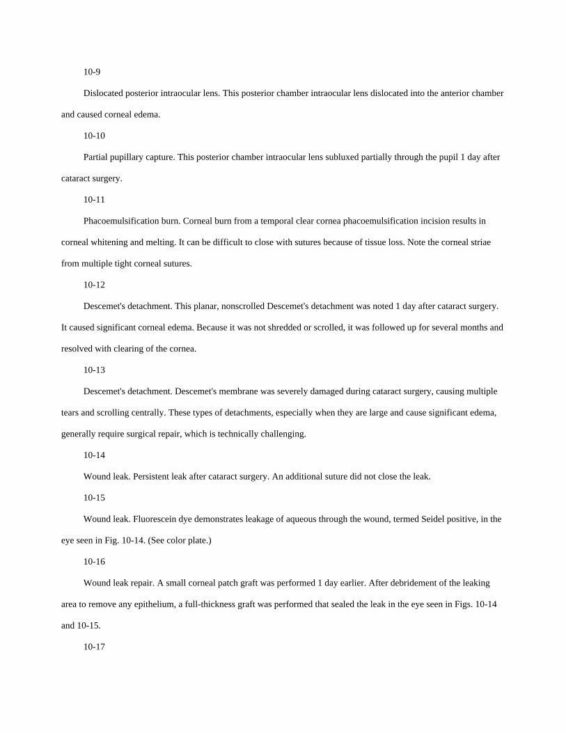
10-9
Dislocated posterior intraocular lens. This posterior chamber intraocular lens dislocated into the anterior chamber
and caused corneal edema.
10-10
Partial pupillary capture. This posterior chamber intraocular lens subluxed partially through the pupil 1 day after
cataract surgery.
10-11
Phacoemulsification burn. Corneal burn from a temporal clear cornea phacoemulsification incision results in
corneal whitening and melting. It can be difficult to close with sutures because of tissue loss. Note the corneal striae
from multiple tight corneal sutures.
10-12
Descemet's detachment. This planar, nonscrolled Descemet's detachment was noted 1 day after cataract surgery.
It caused significant corneal edema. Because it was not shredded or scrolled, it was followed up for several months and
resolved with clearing of the cornea.
10-13
Descemet's detachment. Descemet's membrane was severely damaged during cataract surgery, causing multiple
tears and scrolling centrally. These types of detachments, especially when they are large and cause significant edema,
generally require surgical repair, which is technically challenging.
10-14
Wound leak. Persistent leak after cataract surgery. An additional suture did not close the leak.
10-15
Wound leak. Fluorescein dye demonstrates leakage of aqueous through the wound, termed Seidel positive, in the
eye seen in Fig. 10-14. (See color plate.)
10-16
Wound leak repair. A small corneal patch graft was performed 1 day earlier. After debridement of the leaking
area to remove any epithelium, a full-thickness graft was performed that sealed the leak in the eye seen in Figs. 10-14
and 10-15.
10-17
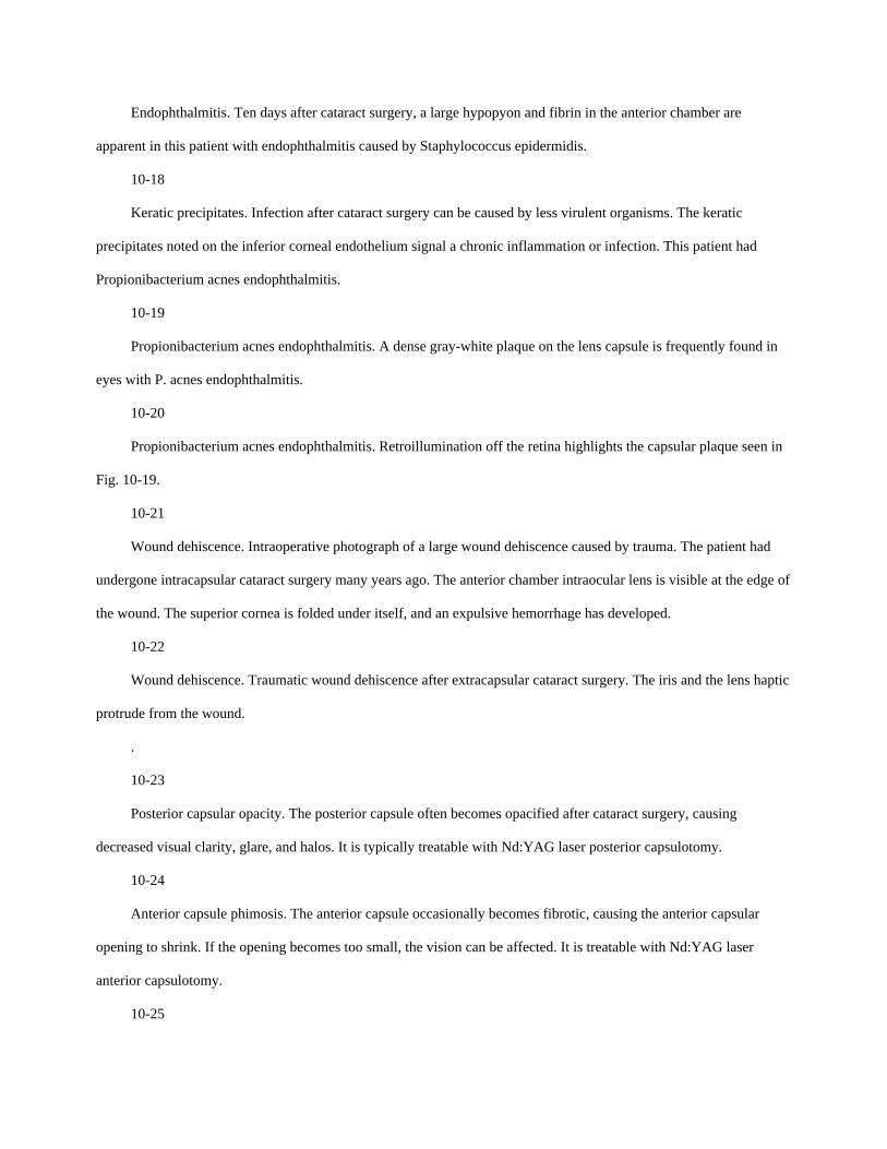
Endophthalmitis. Ten days after cataract surgery, a large hypopyon and fibrin in the anterior chamber are
apparent in this patient with endophthalmitis caused by Staphylococcus epidermidis.
10-18
Keratic precipitates. Infection after cataract surgery can be caused by less virulent organisms. The keratic
precipitates noted on the inferior corneal endothelium signal a chronic inflammation or infection. This patient had
Propionibacterium acnes endophthalmitis.
10-19
Propionibacterium acnes endophthalmitis. A dense gray-white plaque on the lens capsule is frequently found in
eyes with P. acnes endophthalmitis.
10-20
Propionibacterium acnes endophthalmitis. Retroillumination off the retina highlights the capsular plaque seen in
Fig. 10-19.
10-21
Wound dehiscence. Intraoperative photograph of a large wound dehiscence caused by trauma. The patient had
undergone intracapsular cataract surgery many years ago. The anterior chamber intraocular lens is visible at the edge of
the wound. The superior cornea is folded under itself, and an expulsive hemorrhage has developed.
10-22
Wound dehiscence. Traumatic wound dehiscence after extracapsular cataract surgery. The iris and the lens haptic
protrude from the wound.
.
10-23
Posterior capsular opacity. The posterior capsule often becomes opacified after cataract surgery, causing
decreased visual clarity, glare, and halos. It is typically treatable with Nd:YAG laser posterior capsulotomy.
10-24
Anterior capsule phimosis. The anterior capsule occasionally becomes fibrotic, causing the anterior capsular
opening to shrink. If the opening becomes too small, the vision can be affected. It is treatable with Nd:YAG laser
anterior capsulotomy.
10-25
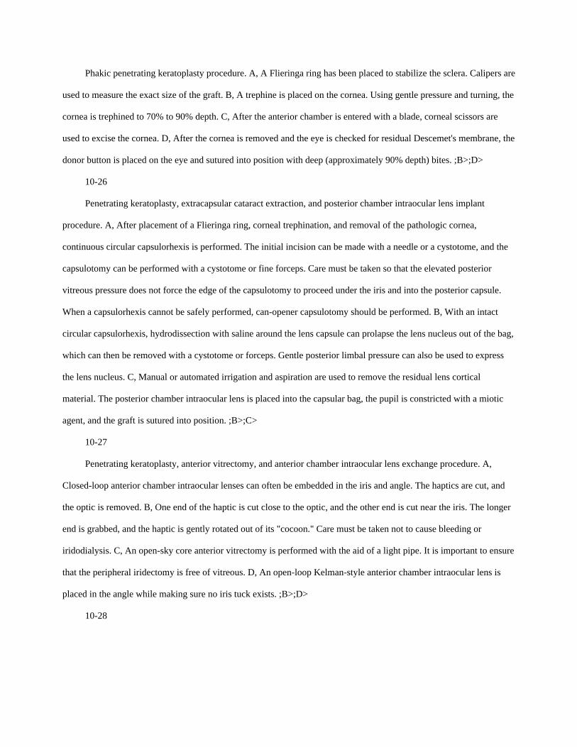
Phakic penetrating keratoplasty procedure. A, A Flieringa ring has been placed to stabilize the sclera. Calipers are
used to measure the exact size of the graft. B, A trephine is placed on the cornea. Using gentle pressure and turning, the
cornea is trephined to 70% to 90% depth. C, After the anterior chamber is entered with a blade, corneal scissors are
used to excise the cornea. D, After the cornea is removed and the eye is checked for residual Descemet's membrane, the
donor button is placed on the eye and sutured into position with deep (approximately 90% depth) bites. ;B>;D>
10-26
Penetrating keratoplasty, extracapsular cataract extraction, and posterior chamber intraocular lens implant
procedure. A, After placement of a Flieringa ring, corneal trephination, and removal of the pathologic cornea,
continuous circular capsulorhexis is performed. The initial incision can be made with a needle or a cystotome, and the
capsulotomy can be performed with a cystotome or fine forceps. Care must be taken so that the elevated posterior
vitreous pressure does not force the edge of the capsulotomy to proceed under the iris and into the posterior capsule.
When a capsulorhexis cannot be safely performed, can-opener capsulotomy should be performed. B, With an intact
circular capsulorhexis, hydrodissection with saline around the lens capsule can prolapse the lens nucleus out of the bag,
which can then be removed with a cystotome or forceps. Gentle posterior limbal pressure can also be used to express
the lens nucleus. C, Manual or automated irrigation and aspiration are used to remove the residual lens cortical
material. The posterior chamber intraocular lens is placed into the capsular bag, the pupil is constricted with a miotic
agent, and the graft is sutured into position. ;B>;C>
10-27
Penetrating keratoplasty, anterior vitrectomy, and anterior chamber intraocular lens exchange procedure. A,
Closed-loop anterior chamber intraocular lenses can often be embedded in the iris and angle. The haptics are cut, and
the optic is removed. B, One end of the haptic is cut close to the optic, and the other end is cut near the iris. The longer
end is grabbed, and the haptic is gently rotated out of its "cocoon." Care must be taken not to cause bleeding or
iridodialysis. C, An open-sky core anterior vitrectomy is performed with the aid of a light pipe. It is important to ensure
that the peripheral iridectomy is free of vitreous. D, An open-loop Kelman-style anterior chamber intraocular lens is
placed in the angle while making sure no iris tuck exists. ;B>;D>
10-28
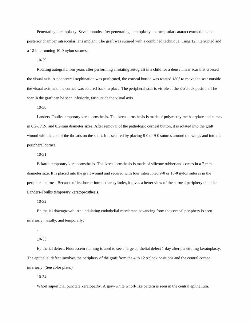
Penetrating keratoplasty. Seven months after penetrating keratoplasty, extracapsular cataract extraction, and
posterior chamber intraocular lens implant. The graft was sutured with a combined technique, using 12 interrupted and
a 12-bite running 10-0 nylon sutures.
10-29
Rotating autograft. Ten years after performing a rotating autograft in a child for a dense linear scar that crossed
the visual axis. A noncentral trephination was performed, the corneal button was rotated 180° to move the scar outside
the visual axis, and the cornea was sutured back in place. The peripheral scar is visible at the 3 o'clock position. The
scar in the graft can be seen inferiorly, far outside the visual axis.
10-30
Landers-Foulks temporary keratoprosthesis. This keratoprosthesis is made of polymethylmethacrylate and comes
in 6.2-, 7.2-, and 8.2-mm diameter sizes. After removal of the pathologic corneal button, it is rotated into the graft
wound with the aid of the threads on the shaft. It is secured by placing 8-0 or 9-0 sutures around the wings and into the
peripheral cornea.
10-31
Eckardt temporary keratoprosthesis. This keratoprosthesis is made of silicone rubber and comes in a 7-mm
diameter size. It is placed into the graft wound and secured with four interrupted 9-0 or 10-0 nylon sutures in the
peripheral cornea. Because of its shorter intraocular cylinder, it gives a better view of the corneal periphery than the
Landers-Foulks temporary keratoprosthesis.
10-32
Epithelial downgrowth. An undulating endothelial membrane advancing from the corneal periphery is seen
inferiorly, nasally, and temporally.
.
10-33
Epithelial defect. Fluorescein staining is used to see a large epithelial defect 1 day after penetrating keratoplasty.
The epithelial defect involves the periphery of the graft from the 4 to 12 o'clock positions and the central cornea
inferiorly. (See color plate.)
10-34
Whorl superficial punctate keratopathy. A gray-white whorl-like pattern is seen in the central epithelium.

10-35
Whorl superficial punctate keratopathy. Fluorescein dye demonstrates a whorl-like epithelial staining pattern,
common after penetrating keratoplasty. (See color plate.)
10-36
Chronic epithelial defect. Delayed epithelialization in this eye 6 weeks after penetrating keratoplasty results in
corneal haze and melting.
10-37
Temporary lateral tarsorrhaphy. A noncutting temporary lateral tarsorrhaphy is an extremely effective procedure
to aid in epithelialization after penetrating keratoplasty. It involves local anesthesia to the eyelids and placement of a
mattress suture through both tarsal plates. The suture is tied over bolsters to prevent "cheesewiring." It is generally kept
in place for 3 to 4 weeks and then removed.
10-38
Primary graft failure. Severe corneal edema with a poor view of the iris on postoperative day 1 suggests primary
graft failure.
10-39
Epithelial graft rejection. An elevated gray-white epithelial line is noted from the 9 to 3 o'clock positions in the
peripheral graft area. The superior cornea has significant stromal neovascularization.
10-40
Epithelial graft rejection. Fluorescein dye stains the irregular elevated epithelial rejection line seen in Fig. 10-39.
Serial examinations can demonstrate the line to move from the periphery to the central cornea. (See color plate.)
10-41
Subepithelial graft rejection. Multiple subepithelial infiltrates are seen in the graft. Although they typically do not
cause significantly diminished vision, they signal immune activity. They should be treated and the patient should be
followed up closely for the development of endothelial rejection.
10-42
Keratic precipitates. Clumps of white blood cells (keratic precipitates) are apparent on the endothelial surface.
They are a sign of early endothelial graft rejection and require treatment.
10-43

Endothelial graft rejection. An endothelial rejection line (Khodadoust line) can be seen stretching from the 3 to
the 6 o'clock positions of the cornea. The central aspect of the line appears slightly elevated, whereas the cornea
peripheral to the line is edematous.
10-44
Endothelial graft rejection. The slit beam demonstrates corneal edema peripheral to the endothelial rejection line.
10-45
Recurrent herpes simplex keratitis. A herpes simplex virus dendrite can be seen to originate from the host cornea
and progress onto the corneal graft. This eye had undergone a corneal transplant for herpes simplex corneal scarring.
10-46
Broken graft suture. A broken, protruding, interrupted 10-0 nylon suture. A windshield wiper effect around the
suture can be seen. Such broken, exposed sutures necessitate removal.
10-47
Broken graft suture. A broken, exposed, running 10-0 nylon suture. Assuming the wound is secure, the entire
running suture should be removed. If the wound is not entirely secure, the exposed portion of suture can be trimmed,
but the remaining running suture will continue to loosen and become exposed.
10-48
Loose graft suture. This running suture and several interrupted sutures have become severely loosened and
exposed to the point of causing corneal infiltration and melting. Removal and close follow-up are required.
10-49
Graft suture infiltrate. A dense corneal infiltrate is present related to a graft suture. Moderate surrounding edema
has developed. Suture removal and treatment with intensive topical antibiotics are required.
.
10-50
Graft suture abscess. A dense corneal ulcer can be seen at the 9 o'clock position, related to a broken, exposed
suture that was removed. A hypopyon is present inferiorly.
10-51
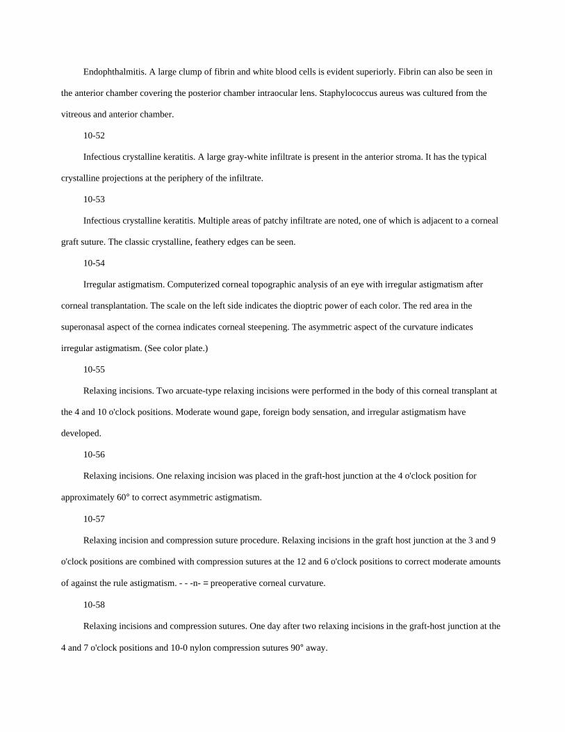
Endophthalmitis. A large clump of fibrin and white blood cells is evident superiorly. Fibrin can also be seen in
the anterior chamber covering the posterior chamber intraocular lens. Staphylococcus aureus was cultured from the
vitreous and anterior chamber.
10-52
Infectious crystalline keratitis. A large gray-white infiltrate is present in the anterior stroma. It has the typical
crystalline projections at the periphery of the infiltrate.
10-53
Infectious crystalline keratitis. Multiple areas of patchy infiltrate are noted, one of which is adjacent to a corneal
graft suture. The classic crystalline, feathery edges can be seen.
10-54
Irregular astigmatism. Computerized corneal topographic analysis of an eye with irregular astigmatism after
corneal transplantation. The scale on the left side indicates the dioptric power of each color. The red area in the
superonasal aspect of the cornea indicates corneal steepening. The asymmetric aspect of the curvature indicates
irregular astigmatism. (See color plate.)
10-55
Relaxing incisions. Two arcuate-type relaxing incisions were performed in the body of this corneal transplant at
the 4 and 10 o'clock positions. Moderate wound gape, foreign body sensation, and irregular astigmatism have
developed.
10-56
Relaxing incisions. One relaxing incision was placed in the graft-host junction at the 4 o'clock position for
approximately 60° to correct asymmetric astigmatism.
10-57
Relaxing incision and compression suture procedure. Relaxing incisions in the graft host junction at the 3 and 9
o'clock positions are combined with compression sutures at the 12 and 6 o'clock positions to correct moderate amounts
of against the rule astigmatism. - - -n- = preoperative corneal curvature.
10-58
Relaxing incisions and compression sutures. One day after two relaxing incisions in the graft-host junction at the
4 and 7 o'clock positions and 10-0 nylon compression sutures 90° away.
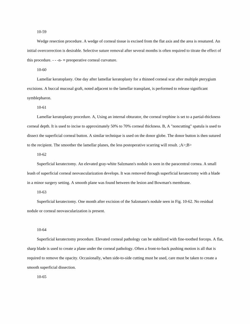
10-59
Wedge resection procedure. A wedge of corneal tissue is excised from the flat axis and the area is resutured. An
initial overcorrection is desirable. Selective suture removal after several months is often required to titrate the effect of
this procedure. - - -n- = preoperative corneal curvature.
10-60
Lamellar keratoplasty. One day after lamellar keratoplasty for a thinned corneal scar after multiple pterygium
excisions. A buccal mucosal graft, noted adjacent to the lamellar transplant, is performed to release significant
symblepharon.
10-61
Lamellar keratoplasty procedure. A, Using an internal obturator, the corneal trephine is set to a partial-thickness
corneal depth. It is used to incise to approximately 50% to 70% corneal thickness. B, A "noncutting" spatula is used to
dissect the superficial corneal button. A similar technique is used on the donor globe. The donor button is then sutured
to the recipient. The smoother the lamellar planes, the less postoperative scarring will result. ;A>;B>
10-62
Superficial keratectomy. An elevated gray-white Salzmann's nodule is seen in the paracentral cornea. A small
leash of superficial corneal neovascularization develops. It was removed through superficial keratectomy with a blade
in a minor surgery setting. A smooth plane was found between the lesion and Bowman's membrane.
10-63
Superficial keratectomy. One month after excision of the Salzmann's nodule seen in Fig. 10-62. No residual
nodule or corneal neovascularization is present.
10-64
Superficial keratectomy procedure. Elevated corneal pathology can be stabilized with fine-toothed forceps. A flat,
sharp blade is used to create a plane under the corneal pathology. Often a front-to-back pushing motion is all that is
required to remove the opacity. Occasionally, when side-to-side cutting must be used, care must be taken to create a
smooth superficial dissection.
10-65
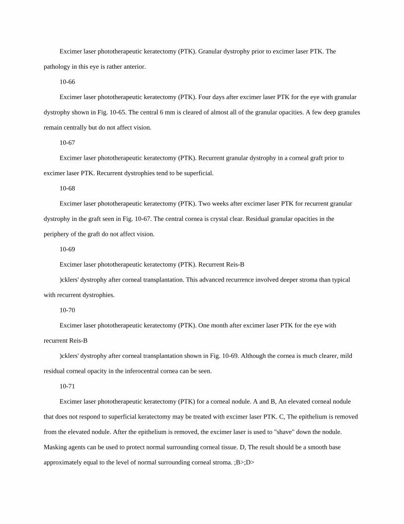
Excimer laser phototherapeutic keratectomy (PTK). Granular dystrophy prior to excimer laser PTK. The
pathology in this eye is rather anterior.
10-66
Excimer laser phototherapeutic keratectomy (PTK). Four days after excimer laser PTK for the eye with granular
dystrophy shown in Fig. 10-65. The central 6 mm is cleared of almost all of the granular opacities. A few deep granules
remain centrally but do not affect vision.
10-67
Excimer laser phototherapeutic keratectomy (PTK). Recurrent granular dystrophy in a corneal graft prior to
excimer laser PTK. Recurrent dystrophies tend to be superficial.
10-68
Excimer laser phototherapeutic keratectomy (PTK). Two weeks after excimer laser PTK for recurrent granular
dystrophy in the graft seen in Fig. 10-67. The central cornea is crystal clear. Residual granular opacities in the
periphery of the graft do not affect vision.
10-69
Excimer laser phototherapeutic keratectomy (PTK). Recurrent Reis-B
)cklers' dystrophy after corneal transplantation. This advanced recurrence involved deeper stroma than typical
with recurrent dystrophies.
10-70
Excimer laser phototherapeutic keratectomy (PTK). One month after excimer laser PTK for the eye with
recurrent Reis-B
)cklers' dystrophy after corneal transplantation shown in Fig. 10-69. Although the cornea is much clearer, mild
residual corneal opacity in the inferocentral cornea can be seen.
10-71
Excimer laser phototherapeutic keratectomy (PTK) for a corneal nodule. A and B, An elevated corneal nodule
that does not respond to superficial keratectomy may be treated with excimer laser PTK. C, The epithelium is removed
from the elevated nodule. After the epithelium is removed, the excimer laser is used to "shave" down the nodule.
Masking agents can be used to protect normal surrounding corneal tissue. D, The result should be a smooth base
approximately equal to the level of normal surrounding corneal stroma. ;B>;D>

10-72
Excimer laser phototherapeutic keratectomy (PTK). This eye with Salzmann's nodular degeneration did not
respond to lamellar keratectomy with a blade, so an excimer laser PTK was performed.
10-73
Excimer laser phototherapeutic keratectomy (PTK). Three months after excimer laser PTK for the eye with
Salzmann's nodular degeneration shown in Fig. 10-72. The cornea is much smoother and clearer.
10-74
Excimer laser phototherapeutic keratectomy (PTK) procedure for anterior stromal opacities. A, Confluent
anterior stromal opacities, such as corneal stromal dystrophies, are often amenable to excimer laser PTK. B, Because
the overlying epithelium is typically smooth, transepithelial treatment is performed through the epithelium and into the
anterior stroma. C, The result should be a smooth base with the bulk of the stromal opacity removed. Care should be
taken not to ablate too deeply because that would result in excessive corneal flattening and induced hyperopia. ;B>;C>
10-75
Excimer laser phototherapeutic keratectomy (PTK). Schnyder's crystalline dystrophy with significant central
corneal crystals. The patient had severe symptoms of glare before excimer laser PTK.
10-76
Excimer laser phototherapeutic keratectomy (PTK). Two weeks after excimer laser PTK for Schnyder's
crystalline dystrophy. The central crystals are almost, but not entirely, eliminated. The patient's symptoms of severe
glare, however, are totally eliminated. The small amount of remaining crystals could have been removed with the
excimer laser, but it would have required a significantly deeper ablation and risked causing more induced hyperopia.
10-77
Conjunctival flap. Sterile corneal melt with severe corneal thinning is present in an eye with poor visual potential.
Calcific degeneration is present in the cornea from the 8 to 9 o'clock positions, and a hypopyon can be seen inferiorly.
10-78
Conjunctival flap. Three weeks after a conjunctival flap in the eye in Fig. 10-77. A few residual Vicryl sutures
are still found inferiorly and superiorly.
10-79

Conjunctival flap. Three months after a conjunctival flap in the eye in Figs. 10-77 and 10-78. Note the gradual
thinning and clearing of the flap. The hypopyon is gone, but the calcific degeneration is still present.
10-80
Conjunctival flap. Six months after a conjunctival flap in a different eye. The flap is relatively clear, and the
cosmetic result is reasonable.
10-81
Conjunctival flap procedure A, A superior limbal traction suture is placed to pull the eye downward. A
conjunctival incision is made approximately 12 to 14 mm superior to the limbus. B, The conjunctiva is carefully
dissected downward to the limbus. C, Once the limbus is reached, a 360° conjunctival peritomy is performed. The
conjunctival flap is then moved from the superior sclera to cover the entire cornea. It is sutured to episclera and
conjunctiva inferiorly and to episclera superiorly. D, When the corneal pathology involves only the inferior cornea, an
inferior conjunctival flap can be performed. ;B>;D>
10-82
Buttonhole in a conjunctival flap. A conjunctival flap buttonhole, which stains with fluorescein, is present
inferiorly. Two 11-0 nylon sutures, which were used to close a buttonhole during surgery, can be seen superiorly.
10-83
Retraction of a conjunctival flap. The conjunctival flap has retracted and melted, but the central cornea has
epithelialized well.
10-84
Pterygium excision and conjunctival autograft procedure. A, A superior limbal traction suture is placed. The
pterygium is outlined with light cautery. B, The pterygium is removed from the cornea with a sharp, rounded blade
using a front-to-back pushing motion. The pterygium is removed from the scleral side with scissors and blunt-and-
sharp dissection. C, After the size of the conjunctival defect is measured, the superior conjunctiva is marked with
cautery and the conjunctival graft is dissected. D, The free conjunctival graft is carefully transferred to the area of
pterygium excision, taking care to keep the epithelial side up. It is sutured to episclera at the limbus and conjunctiva
elsewhere. ;B>;D>
10-85

Pterygium excision and conjunctival autograft. One day after pterygium excision and conjunctival autograft. The
corneal epithelial defect is healing. The conjunctival autograft is excellently positioned with multiple 8-0 Vicryl
sutures.
10-86
Delle after pterygium excision and conjunctival autograft. A delle, a distinct area of corneal drying and thinning,
can be seen adjacent to an elevated conjunctival autograft.
10-87
Pyogenic granuloma after pterygium excision with a bare sclera technique. A large elevated nodule of granulation
tissue may develop in extensive areas of bare sclera.
10-88
Recurrent pterygium. This pterygium recurred soon after excision. Note the symblepharon formation with
conjunctival adhesion to the upper eyelid.
10-89
Recurrent pterygium. Significant recurrent pterygium formation after three previous excisions with associated
pyogenic granuloma formation and symblepharon. This patient has diplopia on outward gaze in this eye because of
restricted ocular motility.
10-90
Scleral melting after pterygium surgery. A scleral melt developed 18 days after a bare sclera pterygium excision.
This complication, though rare, is more common with the use of topical mitomycin C after pterygium excision.
10-91
Conjunctival tumor excision procedure. A, Conjunctival tumors can extend onto the cornea. A wide excision on
both the conjunctival and the corneal sides must be performed. B, Scissors are used to excise the conjunctival portion,
and blunt dissection with a rounded-end blade is usually adequate to remove the corneal portion. Double freeze-thaw
cryotherapy is used to treat the remaining conjunctival edges and occasionally the limbus. ;B>
10-92
Conjunctival tumor excision. A large limbal conjunctival papillomatous tumor can be seen from the 4 toward the
10 o'clock positions before removal.
10-93

Conjunctival tumor excision. Soon after the surgical excision with supplemental double freeze-thaw cryotherapy
of the conjunctival tumor seen in Fig. 10-92, a pyogenic granuloma developed in a large area of bare sclera. It resolved
with topical corticosteroids, but recurrent conjunctival tumor must be considered.
10-94
Conjunctival tumor excision. A large leukoplakic, squamous cell carcinoma of the conjunctiva and cornea is seen
before excision.
10-95
Conjunctival tumor excision. One month after the mass seen in Fig. 10-94 was excised, no apparent residual or
recurrent tumor has developed.
10-96
Conjunctival tumor excision. Two months after surgical excision of the mass seen in Figs. 10-94 and 10-95, it has
recurred at the limbus, requiring more extensive excision.
10-97
Corneal perforation repair. A full-thickness corneal perforation is seen with a shallow anterior chamber. It was
caused by a metallic foreign body that was present for several weeks and then removed.
10-98
Corneal perforation repair. Cyanoacrylate corneal tissue adhesive was placed to seal the perforation seen in Fig.
10-97. A bandage soft contact lens is used over the glue.
10-99
Corneal perforation repair. A full-thickness corneal perforation is seen inferocentrally with a flat anterior
chamber. It occurred from a sterile corneal melt in an eye with severe dry eye syndrome.
10-100
Corneal perforation repair. A small amount of medical-grade corneal glue and a bandage soft contact lens are
placed to seal the perforation seen in Fig. 10-99.
10-101
Corneal perforation repair. Two months after the successful gluing of the perforation seen in Figs. 10-99 and 10-
100, the glue fell out spontaneously. The perforation was sealed, and the cornea was completely reepithelialized. A
moderate localized scar has formed in the area of the old perforation.

10-102
Corneal perforation repair. A large perforation was treated with corneal glue in a patient who could not undergo
corneal transplantation. The glue entered the anterior chamber before polymerizing and created a collar-button shape to
the glue. A small portion of collagen shield placed at the base of the perforation would likely have prevented this
occurrence.
10-103
Cyanoacrylate gluing of a corneal perforation procedure. A, A cotton-tipped applicator is broken to create a
beveled end. Ointment is placed 1 to 2 mm from the end (depending on the amount of glue required to seal the wound).
A drop of medical-grade cyanoacrylate glue is placed on the very end of the applicator. Corneal epithelium must be
removed from the area to be glued. B, A cellulose sponge is used in one hand to dry the perforation site. Once dry, the
glue is quickly applied. Just enough glue to seal the perforation should be used. It is allowed to polymerize for several
minutes. A bandage soft contact lens is placed over the glue. ;A>;B>
10-104
Corneal perforation repair. A large corneal perforation with significant iris prolapse cannot be treated with
corneal glue. This eye requires corneal transplantation.
10-105
Corneal perforation repair. One day after a lamellar corneal patch graft was performed in the eye seen in Fig. 10-
104. The epithelium was carefully peeled from the iris, which was then treated with cryotherapy to prevent epithelial
downgrowth. A trephine was placed to encompass the entire lesion, and a partial-thickness corneal and scleral incision
was created. A lamellar corneal transplant was placed and partially covered with conjunctiva.
10-106
Corneal perforation repair. Ten days after the lamellar corneal patch graft was performed in the eye seen in Figs.
10-104 and 10-105. The corneal graft is healing well, and the conjunctiva is retracting.
10-107
Scleral patch graft. Uveal prolapse is beginning to develop in this eye with progressive necrotizing scleritis. A
tectonic graft is required.
.
10-108
Scleral patch graft. Two weeks after a scleral patch graft for the eye with necrotizing scleritis seen in Fig. 10-107.
Adjacent conjunctiva was used to cover the sclera. A small delle is seen at the limbus.
10-109
Scleral patch graft. A large area of necrotizing scleritis with early uveal prolapse is seen. No healthy adjacent
conjunctiva is present.
10-110
Scleral patch graft. One day after a large scleral patch graft for necrotizing scleritis seen in Fig. 10-109. Because
no healthy adjacent conjunctiva was available to cover the graft, an amniotic membrane transplant was used. Two rows
of sutures can be seen, one securing the scleral graft and the other securing the amniotic membrane.
10-111
Permanent keratoprosthesis. This permanent keratoprosthesis, in a patient with a bilateral chemical burn, is
placed through the upper eyelid. The patient achieved 20/25 central visual acuity with a tunnel visual field.
10-112
Radial keratotomy (RK). Four years after an eight-incision RK for myopia and a two-incision T-cut for
astigmatism.
10-113
Radial keratotomy (RK). Retroillumination off the retina of the eye seen in Fig. 10-112 of an eight-incision RK
coupled with two T-cuts.
10-114
Radial keratotomy. A, In the "combined" cutting technique, the initial incision ("American" cut) begins at the
optical zone and proceeds toward the limbus. B, Without removing the blade from the cornea, the direction is reversed
and the incision proceeds toward the optical zone ("Russian" cut). It stops at the optical zone because only the bottom
of the blade cuts in this direction.
10-115
Radial keratotomy macroperforation. Seidel-positive leak from the 6 o'clock radial incision.
10-116
Corneal infiltrates after radial keratotomy (RK). Corneal infiltrates are seen at the central ends of two RK
incisions. The special concern is that organisms can easily progress through the incision to deep cornea or even the
anterior chamber and cause a vision-threatening infection.
10-117
Corneal ulceration after radial keratotomy (RK). Infectious keratitis can develop after RK. The 9 o'clock radial
and adjacent T-cuts are both ulcerated with significant adjacent deep corneal infiltration. This infection requires
intensive topical antibiotic treatment.
10-118
Crossed incisions after radial and astigmatic keratotomy. This eye with 16 radial incisions has several intersecting
T-cuts. When corneal incisions intersect, wound gaping and significant scarring often result.
10-119
Neovascularization into radial keratotomy (RK) incisions. Corneal neovascularization to multiple peripheral
incisions in this eye after a 16-incision RK. This finding is more common in association with contact lens wear. This
patient was undercorrected and wore extended wear soft contact lenses.
10-120
Excimer laser photorefractive keratectomy (PRK) procedure. A, PRK for myopia. After epithelial removal, the
excimer laser ablates more tissue centrally than peripherally to flatten the central cornea. B, PRK for hyperopia. After
epithelial removal the excimer laser is used to ablate more tissue peripherally than centrally to steepen the central
cornea. ;B>
10-121
Excimer laser photorefractive keratectomy (PRK). Postoperative day 1 after PRK. An approximately 3 × 4 mm
central epithelial defect is covered with a bandage soft contact lens.
10-122
Excimer laser photorefractive keratectomy (PRK) haze. Mild granular anterior stromal haze 1 month after PRK.
10-123
Excimer laser photorefractive keratectomy (PRK) haze. Moderate reticular anterior stromal haze from the 6 to 12
o'clock positions 6 months after PRK. The central cornea is relatively clear, and the patient has excellent uncorrected
vision and minimal visual symptoms.

.
10-124
Excimer laser photorefractive keratectomy (PRK) scar. Dense reticular corneal scar in the mid-periphery of the
ablation 18 months after PRK. Mild irregular astigmatism is induced.
10-125
Excimer laser photorefractive keratectomy (PRK) haze. Moderate central corneal haze over the entire 6 mm
ablation zone 6 months after PRK. Significant associated regression and decreased uncorrected and best-corrected
vision improved moderately with topical corticosteroids.
.
10-126
Excimer laser photorefractive keratectomy (PRK) decentered ablation. Computed corneal topographic analysis of
an eye after PRK. The scale on the left side indicates the dioptric power of each color. The large circular blue area
nasally corresponds to the area of excimer laser ablation. The ablation is significantly decentered. The patient has
monocular diplopia and triplopia. (See color plate.)
10-127
Laser in situ keratomileusis (LASIK) procedure. A, After a suction ring is placed to pressurize the eye, a
microkeratome is used to fashion a flap of superficial cornea attached by a small hinge. B, The flap is reflected, and the
excimer laser is used to treat the corneal stroma. C, The flap is replaced, the interface irrigated, and the flap is allowed
to adhere without sutures.
;B>;C>
10-128
Laser in situ keratomileusis (LASIK). Two weeks after uncomplicated LASIK with a nasal hinge in this right
eye. The semicircular edge of the LASIK flap is evident temporally. The flap is in excellent position and is healing
well.
10-129
Dislodged laser in situ keratomileusis (LASIK) flap. One day after uncomplicated LASIK, the patient felt pain
after rubbing the right eye. A dislodged flap can be seen folded upon itself but still attached at the nasal hinge. The
central cornea demonstrates an irregular light reflex. The flap was successfully reapproximated and adhered well.

10-130
Laser in situ keratomileusis (LASIK) infection. A small corneal ulcer developed after LASIK. Mild surrounding
corneal edema was treated with intensive topical antibiotics and resolved.
10-131
Epithelial ingrowth after laser in situ keratomileusis (LASIK). A gray-white opacity of epithelial ingrowth is seen
in the LASIK interface. It slowly enlarged and caused irregular astigmatism. The flap was lifted, and the epithelium
was removed from the stromal side and the flap side and did not recur.
10-132
Microstriae after laser in situ keratomileusis (LASIK). Multiple horizontal microstriae are seen in this eye 3
months after uncomplicated LASIK. Microstriae do not appear to affect vision, whereas macrostriae are associated with
irregular astigmatism.
MAJOR POINTS
Although cataract surgery is generally extremely successful, it is associated with potentially serious
complications.
Certain complications, such as cystoid macular edema or retinal detachment, may develop after uncomplicated
cataract surgery.
Cataract surgery (or any intraocular surgery) may rarely result in blindness of the eye if a serious complication
such as expulsive hemorrhage or endophthalmitis develops.
A cataractous lens may be removed in toto (intracapsular method) or the lens nucleus may be removed in one
piece (planned extracapsular method) or the lens nucleus may be fragmented and removed in small pieces
(phacoemulsification technique).
When inadequate capsular support exists for safe routine implantation of a posterior chamber intraocular lens, an
anterior chamber intraocular lens or a sutured (to iris or sclera) posterior chamber intraocular lens may alternatively be
implanted.
Due to loose zonular attachments, pseudoexfoliation is associated with increased complications during cataract
surgery including vitreous loss and inadequate capsular support for a posterior chamber intraocular lens.
When vitreous loss is encountered during cataract surgery, an anterior vitrectomy is required to clear the vitreous
from the anterior chamber and wound.

Significant anterior chamber and vitreal inflammation after cataract surgery may signal early endophthalmitis and
requires immediate workup and possible treatment.
Corneal donor information needs to be reviewed and the tissue examined preoperatively by the surgeon.
Flieringa ring (or equivalent) placement prior to penetrating keratoplasty is extremely helpful in aphakic eyes,
eyes after vitrectomy, and eyes undergoing cataract extraction, and is mandatory in infants and children.
All iris-fixated and closed-loop anterior chamber intraocular lenses and all unstable lenses should be removed
during corneal transplantation for bullous keratopathy.
Postoperative epithelial defects in a corneal transplant not responding to medical treatment should undergo a
lateral tarsorrhaphy before any stromal melting occurs.
The primary symptoms of corneal graft rejection are redness, discomfort, photophobia, and decreased vision, but
patients may be asymptomatic.
Signs of corneal graft rejection, including subepithelial infiltrates, new anterior chamber reaction, keratic
precipitates, and corneal edema should be checked for on each postoperative visit.
All exposed corneal transplant sutures should be removed at the time of discovery.
Excimer laser phototherapeutic keratectomy can be extremely effective for elevated and anterior stromal corneal
pathology, especially Reis-Bücklers', granular, and lattice corneal dystrophies.
Conjunctival flap surgery is an effective and often underutilized procedure for chronic corneal ulcerations and
painful bullous keratopathy in eyes with poor visual potential that have not perforated.
Keys to decreasing the risk of recurrence of pterygia after excision and conjunctival autograft are the creation of a
smooth cornea and limbus in the area of excision and the generous use of postoperative topical corticosteroids.
Conjunctival flaps and autografts need to be relatively thin, but without buttonholes, and sutured flat to the
surface without excessive tension.
When squamous cell tumors of the conjunctiva recur, it is usually at the limbus.
Cyanoacrylate corneal glue can be extremely effective in the treatment of small impending and completed
corneal perforations. A bandage soft contact lens must be used over the glue.
Refractive surgery patients should be out of contact lenses prior to evaluation. They should have stable
refractions, essentially normal eyes, and regular corneas. They must have an understanding of the various refractive
surgery options and their respective benefits and risks, in addition to realistic expectations regarding the outcomes of
surgery.
Prior to PRK and LASIK, the surgeon needs to check the calibration and centration of the excimer laser, and for
LASIK, the assembly and function of the microkeratome.
10-1
Major Steps in Planned Extracapsular Cataract Extraction
Place a superior rectus bridle suture (optional)
Perform a superior conjunctival peritomy and obtain hemostasis
Create a scleral groove for approximately 5 clock hours
Create a small incision into the anterior chamber
Fill the anterior chamber with viscoelastic
Perform an anterior capsulotomy
Open the wound for approximately 5 clock hours
Prolapse the lens nucleus
Partially close the wound
Remove the residual lens cortex with irrigation and aspiration
Open the capsular bag with viscoelastic
Place a posterior chamber intraocular lens in the capsular bag
Close the wound and remove the viscoelastic
10-1
General complications of intraocular surgery
Complication Cataract
surgery
Corneal
transplant
Increased intraocular pressure+ ++
Wound leak + +
Expulsive hemorrhage + ++
Endophthalmitis + ++
Epithelial downgrowth + ++
Retinal detachment + ++
Cystoid macular edema + +
10-17
Major Steps of Laser In Situ Keratomileusis (LASIK)
Laser setup and patient positioning are identical to those for photorefractive keratectomy (PRK)
Adjust the attempted correction per the LASIK nomogram
Open the eyelid speculum wide to expose the inferior and superior sclera
Mark the edge of the cornea where the flap will begin
Secure the suction ring and check the intraocular pressure
Engage the microkeratome and create the flap
Release suction
Move the flap to the side
Make sure all epithelium and debris are off the ablation zone
Ablate as per PRK
Replace the flap
Irrigate the interface
Let the flap adhere
Carefully remove the eyelid speculum, and check the flap and interface
10-16
Major Steps of Excimer Laser Photorefractive Keratectomy
Check the calibration and centration of the excimer laser unit and amount of refractive error to be treated
Mark the 3 and 9 o'clock axes at the slit lamp if astigmatism is also being treated
Make sure the head is level, center the eye under the laser, and tilt the head so the 3 and 9 o'clock axes are lined
up with the reticule
Mark and remove the epithelium over the ablation area
Make sure all epithelium and debris are off Bowman's layer in the ablation zone

Center the ablation over the pupil
Ablate while maintaining centration over the pupil
10-15
Complications of Radial and Astigmatic Keratotomy
Microperforation
Macroperforation
Incision into the clear zone or across the visual axis
Infection of incisions and, rarely, endophthalmitis
Traumatic rupture of incisions
Late hyperopic drift (radial keratotomy)
10-14
Major Steps in the Radial Keratotomy Procedure
Check the diamond blade for flaws
Select the number and length of the incisions from the nomogram
Mark the 3 and 9 o'clock axes at the slit lamp if astigmatic incisions are planned
Check the corneal thickness with ultrasound pachymetry
Set the diamond blade depth
Mark the central clear zone and the planned radial and astigmatic incisions
Perform the astigmatic and radial incisions
10-13
General Patient Requirements for Refractive Surgery
Age greater than 18 to 21 years
Stable manifest and cycloplegic refractions, out of contact lenses
Complete ocular examination with minimal to no ocular disease
Normal corneal curvature (typically evaluated by computerized topography)
Normal corneal thickness (typically evaluated by ultrasound pachymetry and slit-lamp evaluation)
Normal pupil size
Complete understanding of various refractive surgery options and their benefits and risks
10-12
Major Steps for the Application of Cyanoacrylate Corneal Glue
Insert an eyelid speculum
Debride the epithelium off the area to be glued
Use a cellulose sponge to dry the area to be glued
Apply a small amount of glue to the perforation site
Allow the glue to polymerize
Place a bandage soft contact lens
Remove the eyelid speculum
Check the anterior chamber formation and stability of the glue and the contact lens 1 hour and 1 day later
10-11
Treatment Options for Corneal Perforations
Cyanoacrylate corneal glue and bandage soft contact lens
Lamellar patch graft
Penetrating corneal graft
Scleral, dura mater, pericardium, or fascia lata patch graft
10-10
Major Steps in the Pterygium Excision and Conjunctival Autograft Procedure
Place a superior limbal traction suture
Mark the extent of the pterygium
Balloon the pterygium with local anesthetic
Dissect the conjunctival and corneal portions of the pterygium, connecting at the limbus, and send for pathologic
evaluation
Smooth the cornea and limbus with a diamond burr
Measure the conjunctival defect and mark the superior bulbar conjunctiva
Balloon the superior conjunctiva with local anesthetic
Dissect the conjunctival graft
Slide the graft over the area of pterygium excision and secure in place

10-9
Major Steps in the Total Conjunctival Flap Procedure
Manually debride the entire corneal epithelium
Place a superior limbal traction suture
Balloon the superior conjunctiva with local anesthetic
Beginning 12 to 14 mm superior to the limbus, dissect a conjunctival flap
Create a 360° peritomy
Move the conjunctival flap over the cornea and secure it to the inferior and superior limbus
10-8
Major Steps in Phototherapeutic
Keratectomy
Treatment of elevated corneal lesions
Debride epithelium from the elevated areas
Apply masking agents to the "valleys"
Use varying size ablation spots to shave down the "mountain tops," leaving the valleys untouched
Check at the slit lamp frequently and reablate as needed
Treatment of anterior stromal pathology
For regular epithelium, use a large laser spot to ablate through the epithelium into the stroma
For irregular epithelium, remove the epithelium manually and ablate the stroma directly
Ablate approximately 60% to 75% intended depth
Check at the slit lamp frequently and reablate as needed
Consider an antihyperopia treatment if a deep stromal ablation is performed
Treatment of recurrent erosions
Manually debride all loose epithelium
Ablate all exposed Bowman's membrane for 5 µm, taking care not to overlap ablations
10-7
Surgical Correction of Astigmatism after Penetrating Keratoplasty
3-6 D

Relaxing incisions
5-10 D
Relaxing incisions and compression sutures
10-15 D
Wedge resection
Note: For <4 D myopic astigmatism, consider laser in situ keratomileusis (LASIK).
10-6
Complications of Penetrating Keratoplasty
More common
Elevated intraocular pressure (short-term and long-term)
Wound leak
Chronic epithelial defect
Broken and loose sutures
Graft rejection
Cystoid macular edema
Cataract
Significant astigmatism
Recurrence of disease
Endothelial decompensation
Less common
Retrobulbar hemorrhage
Primary graft failure
Suture-related corneal ulcer
Endophthalmitis
Retinal detachment
Expulsive hemorrhage
Wound dehiscence
Epithelial downgrowth

10-5
Major Steps in the Penetrating Keratoplasty Procedure
Obtain and examine the donor tissue
Place an eyelid speculum that minimizes external pressure on the globe
Place a Flieringa ring (optional)
Determine the trephine size and mark the epithelium
Fashion the donor corneal button
Trephine the recipient cornea
Enter the anterior chamber
Remove the recipient corneal button
Perform intraocular surgery as needed
Suture the donor cornea in position, intermittently reforming the anterior chamber
Bury the knots
Check astigmatism and adjust sutures as necessary
Inspect the wound and make certain it is watertight
10-4
Major Steps in Managing Vitreous Loss During Cataract Extraction
Stop the phacoemulsification
If residual nucleus is present, support the lens with viscoelastic, enlarge the wound, and gently deliver the lens
remnant(s), often with the aid of a lens glide or lens loop
Perform an automated anterior vitrectomy to clear the anterior chamber of vitreous
Remove the bulk of residual cortical material with the vitrector or with manual or automated irrigation/aspiration
Make sure the wound and anterior chamber are clear of vitreous
Assess the degree of capsular support
Use viscoelastic to keep the remaining vitreous back
If at least two thirds to three fourths of the capsular rim remains, consider placement of a posterior chamber
intraocular lens in the ciliary sulcus; once in position, perform a "bounce test" to check lens stability
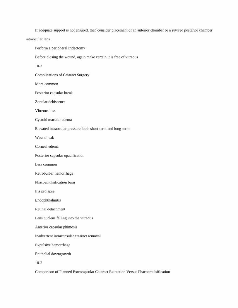
If adequate support is not ensured, then consider placement of an anterior chamber or a sutured posterior chamber
intraocular lens
Perform a peripheral iridectomy
Before closing the wound, again make certain it is free of vitreous
10-3
Complications of Cataract Surgery
More common
Posterior capsular break
Zonular dehiscence
Vitreous loss
Cystoid macular edema
Elevated intraocular pressure, both short-term and long-term
Wound leak
Corneal edema
Posterior capsular opacification
Less common
Retrobulbar hemorrhage
Phacoemulsification burn
Iris prolapse
Endophthalmitis
Retinal detachment
Lens nucleus falling into the vitreous
Anterior capsular phimosis
Inadvertent intracapsular cataract removal
Expulsive hemorrhage
Epithelial downgrowth
10-2
Comparison of Planned Extracapsular Cataract Extraction Versus Phacoemulsification

Advantages of phacoemulsification
Faster surgical technique
Smaller incision size with:
Faster visual recovery
Less induced astigmatism
Less chance of expulsive hemorrhage
Less chance of wound dehiscence after surgery
More stable refraction
Can perform under topical anesthesia
Can perform using a clear cornea technique
Advantages of planned extracapsular cataract extraction
Technically less difficult
Can perform with a less clear view through the cornea
Easier to perform with a small pupil and with a very dense nucleus
Less chance of capsular rupture and vitreous loss during the learning curve
Less chance of lens nucleus falling into the vitreous
No chance of "phacoemulsification burn"
Less dependence on expensive, high technology equipment
10-2
Classification of epithelial transplantation procedures for ocular surface disease
Procedure Abbreviation Donor Transplanted tissue
Conjunctival transplantation
Conjunctival autograft CAU Fellow eye Conjunctiva
Cadaveric conjunctival allograft c-CAL Cadaver Conjunctiva
Living-related conjunctival allograft lr-CAL Living relative Conjunctiva
Limbal transplantation
Conjunctival limbal autograftCLAU Fellow eye Limbus/conjunctiva
Cadaveric conjunctival limbal allograft c-CLAL Cadaver Limbus/conjunctiva
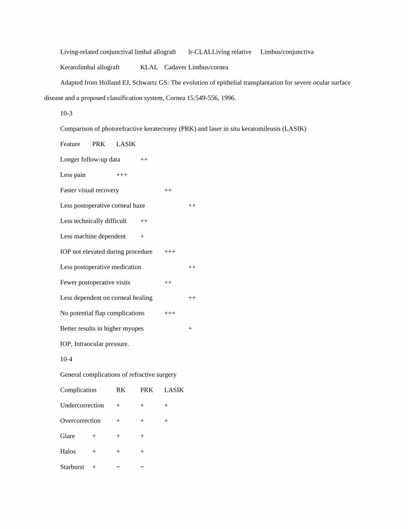
Living-related conjunctival limbal allograft lr-CLALLiving relative Limbus/conjunctiva
Keratolimbal allograft KLAL Cadaver Limbus/cornea
Adapted from Holland EJ, Schwartz GS: The evolution of epithelial transplantation for severe ocular surface
disease and a proposed classification system, Cornea 15:549-556, 1996.
10-3
Comparison of photorefractive keratectomy (PRK) and laser in situ keratomileusis (LASIK)
Feature PRK LASIK
Longer follow-up data ++
Less pain +++
Faster visual recovery ++
Less postoperative corneal haze ++
Less technically difficult ++
Less machine dependent +
IOP not elevated during procedure +++
Less postoperative medication ++
Fewer postoperative visits ++
Less dependent on corneal healing ++
No potential flap complications +++
Better results in higher myopes +
IOP, Intraocular pressure.
10-4
General complications of refractive surgery
Complication RK PRK LASIK
Undercorrection + + +
Overcorrection + + +
Glare + + +
Halos + + +
Starburst + − −

Monocular diplopia + + +
Decreased best corrected vision + + +
Fluctuating vision ++ − −
Progression of effect++ − −
Regression + + +
Induced astigmatism+ + +
Central corneal haze − + −