
Growth Characteristics and Tracing Antagonistic Properties ...

Antagonistic Regulationof PIN Phosphorylation by PP2Aand PINOID Directs Auxin FluxMarta Michniewicz,1,9 Marcelo K. Zago,2 Lindy Abas,3 Dolf Weijers,4 Alois Schweighofer,5 Irute Meskiene,5
Marcus G. Heisler,6 Carolyn Ohno,6 Jing Zhang,1 Fang Huang,2 Rebecca Schwab,7,10 Detlef Weigel,7
Elliot M. Meyerowitz,6 Christian Luschnig,3 Remko Offringa,2 and Jirı Friml1,8,*1Zentrum fur Molekularbiologie der Pflanzen, Universitat Tubingen, Tubingen, Germany2Institute of Biology Leiden, Leiden University, Leiden, the Netherlands3Institute for Applied Genetics and Cell Biology, University of Natural Resources and Applied Life Sciences – BOKU, Wien, Austria4Laboratory of Biochemistry, Wageningen University, Wageningen, the Netherlands5Max F. Perutz Laboratories, University of Vienna, Wien, Austria6Division of Biology, California Institute of Technology, Pasadena, CA, USA7Department of Molecular Biology, Max Planck Institute for Developmental Biology, Tubingen, Germany8Albrecht-von-Haller-Institut fur Pflanzenwissenschaften, Universitat Gottingen, Gottingen, Germany9Present address: Department of Biological Sciences, Stanford University, Stanford, CA, USA.10Present address: Cold Spring Harbor Laboratory, Cold Spring Harbor, NY 11724, USA.
*Correspondence: [email protected]
DOI 10.1016/j.cell.2007.07.033
SUMMARY
In plants, cell polarity and tissue patterning areconnected by intercellular flow of the phyto-hormone auxin, whose directional signalingdepends on polar subcellular localization ofPIN auxin transport proteins. The mechanismof polar targeting of PINs or other cargos inplants is largely unidentified, with the PINOIDkinase being the only known molecular compo-nent. Here, we identify PP2A phosphatase as animportant regulator of PIN apical-basal target-ing and auxin distribution. Genetic analysis,localization, and phosphorylation studies dem-onstrate that PP2A and PINOID both partiallycolocalize with PINs and act antagonisticallyon the phosphorylation state of their centralhydrophilic loop, hence mediating PIN apical-basal polar targeting. Thus, in plants, polar sort-ing by the reversible phosphorylation of cargosallows for their conditional delivery to specificintracellular destinations. In the case of PINproteins, this mechanism enables switches inthe direction of intercellular auxin fluxes, whichmediate differential growth, tissue patterning,and organogenesis.
INTRODUCTION
Polarity is one of the elementary properties of eukaryotic
cells and is inseparable from other fundamental processes
1044 Cell 130, 1044–1056, September 21, 2007 ª2007 Elsevie
such as division, differentiation, and cellular signaling. The
intimate relation between cell polarity and patterning is
particularly prominent in plants, since even fully differenti-
ated cells often retain the potential to redefine their
polarity, enabling crucial adaptation processes such as
directional growth, tissue regeneration or de novo organ
formation (Sauer et al., 2006a). The molecular mecha-
nisms of polarized cargo traffic are conserved from yeast
to humans and have been extensively characterized in
animal epithelial cells, which exhibit a clearly discernible
asymmetry between the apical and the basolateral plasma
membrane domains (Mostov et al., 2003).
Substantially less is known about the mechanism(s) of
cell polarity establishment in plants. Much of our knowl-
edge has been acquired by studying the asymmetric
targeting of plant-specific plasma membrane-resident
PIN proteins, which show distinct polar subcellular locali-
zations. PIN proteins have emerged as key regulators of
a plethora of developmental processes including axis
formation in embryogenesis (Friml et al., 2003), postem-
bryonic organogenesis (Okada et al., 1991; Benkova
et al., 2003; Reinhardt et al., 2003), root meristem organi-
zation (Friml et al., 2002a; Blilou et al., 2005) and tropisms
(Luschnig et al., 1998; Friml et al., 2002b). PIN proteins
facilitate the polar efflux of the plant growth regulator
auxin from cells (Petrasek et al., 2006) and their polar
localization determines the direction of local intercellular
auxin transport (Wi�sniewska et al., 2006).
Distinct polar localizations of PIN proteins in different
cell types depend on so far unidentified cell type-specific
and PIN sequence-based signals (Wi�sniewska et al.,
2006). Furthermore, PIN localization can be modulated
by environmental (e.g., gravity) or developmental cues
(Friml et al., 2002b, 2003; Benkova et al., 2003; Reinhardt
r Inc.

et al., 2003; Scarpella et al., 2006). The rapid retargeting of
PINs to different polar domains is possibly related to the
constitutive endocytosis and recycling of PIN proteins
(Dhonukshe et al., 2007). Such flexible regulation of PIN
polarity provides a way to integrate multiple signals at
the level of single cells, translating them into intercellular
auxin fluxes, relevant for diverse developmental pro-
cesses (reviewed in Friml, 2003).
Despite the importance of PIN polarity control for plant
development, the underlying mechanisms are still not well
understood. Genetic and pharmacological studies have
indicated involvement of (de)phosphorylation processes
in regulation of PIN-dependent auxin transport (Garbers
et al., 1996; Benjamins et al., 2001; Friml et al., 2004;
Shin et al., 2005). The protein serine/threonine (Ser/Thr)
kinase PINOID (Christensen et al., 2000; Benjamins et al.,
2001) is the only as yet identified molecular component
directly involved in the regulation of polar delivery of PIN
proteins. Loss of PINOID (PID) function causes an api-
cal-to-basal shift in PIN polarity, correlating with defects
in embryo and shoot organogenesis. On the other hand,
PID gain-of-function results in an opposite basal-to-apical
PIN polarity shift, which leads to auxin depletion from the
root meristem, ultimately leading to its collapse (Friml
et al., 2004). These results indicate that PID-dependent
phosphorylation leads to preferentially apical PIN localiza-
tion, whereas low phosphorylation levels result in basal
PIN targeting. In such a scenario, reversible phosphoryla-
tion of components of the apical versus the basal targeting
machinery could regulate their activities, which are deci-
sive for polar targeting of membrane proteins. Alterna-
tively, direct phosphorylation of cargo proteins, such as
PINs, could determine their intracellular targeting.
Here, we identify a phosphatase activity required for
apical-basal PIN targeting and auxin transport-dependent
development. We show that protein phosphatase 2A
(PP2A) and PID act antagonistically on phosphorylation
of PIN proteins. Our findings demonstrate that decisions
about apical or basal targeting in plants require reversible
phosphorylation of cargo proteins.
RESULTS
PP2AAs Are Required for Auxin-Related SeedlingDevelopmentTo identify a possible phosphatase component of the
mechanism responsible for PIN polar targeting, we ana-
lyzed the three closely related regulatory A subunits of
the protein phosphatase 2A (PP2A) complex in Arabidop-
sis - PP2AA1, PP2AA2 and PP2AA3. We focused preferen-
tially on the PP2A class since a loss-of-function mutation in
one of the A regulatory subunits (PP2AA1), called ROOT
CURLING ON NPA1 (RCN1) causes various developmen-
tal defects, some of which are in processes governed by
auxin transport (Garbers et al., 1996; Rashotte et al., 2001).
We analyzed the expression pattern of PP2AA genes us-
ing PP2AA1::GUS, PP2AA2::GUS, PP2AA3::GUS and
PP2AA1::PP2AA1:GFP fusions. Analysis of seedlings 4
Cell 1
and 8 days after germination (dag) revealed high and over-
lapping transcriptional activity for all three genes (Figures
S1A–S1D in the Supplemental Data available with this
article online). In the root, PP2AA1 was expressed in the
whole root tip, PP2AA2 prominently in the elongation zone
and columella root cap, and PP2AA3 more restricted to the
columella root cap (Figure 1A). These overlapping expres-
sion patterns of PP2AAs have been confirmed by anal-
ysis of global transcription data (http://www.weigelworld.
org/resources/microarray/AtGenExpress) and are in ac-
cordance with previous reports (Zhou et al., 2004).
To test the requirement of PP2AA activities for plant
development, we isolated and analyzed phenotypes of
pp2aa1, pp2aa2 and pp2aa3 double and triple mutant
combinations. In line with previous observations (Zhou
et al., 2004), the pp2aa2 and pp2aa3 single mutants, and
pp2aa2 pp2aa3 double mutants showed largely normal
development (data not shown), but double-mutant combi-
nations that included the pp2aa1 suffered from increas-
ingly severe developmental aberrations (Figure 1B). Both
pp2aa1 pp2aa2/+ and pp2aa1 pp2aa3/+ mutants dis-
played defects in root growth and root gravity response
(n = 90) (Figures S2A and S2B). Also homozygous seed-
lings for pp2aa1 pp2aa2 and pp2aa1 pp2aa3 showed
identical but more severe defects (Figures S2A and 1C).
Primary root meristems typically collapsed at 6-8 dag
(76% for pp2aa1 pp2aa2, n = 40; 80% for pp2aa1
pp2aa3, n = 50) as evident from the disturbance of proper
patterning and lack of columella-specific lugol staining
(Figure 1L). During subsequent development, initiation of
numerous lateral roots substituted for a defective primary
root (Figure 1D). About 30% (n = 90) of pp2aa1-containing
double-mutant seedlings showed defects in cotyledon de-
velopment. These included collar-shaped or fused cotyle-
dons (10%) as well as aberrant positioning and irregular
numbers of cotyledons (19%) (Zhou et al., 2004; Figure 1C).
Because pp2aa1 pp2aa2 pp2aa3 triple mutant seed-
lings were not recovered among progeny of pp2aa1
pp2aa2 pp2aa3/+ plants (n = 87), we constructed trans-
genic lines with estrogen (4-hydroxytamoxifen)-inducible
overexpression of two different artificial microRNAs
(amiRNAs) (Schwab et al., 2006) that simultaneously
target all three PP2AA genes. In both amiRNA lines, we
observed identical defects that were similar to but more
severe than the phenotypes observed in pp2aa1 double
mutants. These included root and cotyledon defects
(Figure 1E), which resembled phenotypes seen in mutants
compromised in auxin signaling (monopteros, Hardtke
and Berleth, 1998) or auxin transport (pins, Friml et al.,
2003). Functional primary and lateral root meristems
were not established (n = 120), causing arrest of further
growth at the seedling stage (Figure 1E).
These results extend previous findings (Zhou et al.,
2004) that PP2A activity (as assessed by downregulation
of its regulatory A subunits) is important for seedling
development. Importantly, our additional analysis high-
lighted that some features of loss of PP2AA function
phenotypes including root agravitropism, cotyledon
30, 1044–1056, September 21, 2007 ª2007 Elsevier Inc. 1045
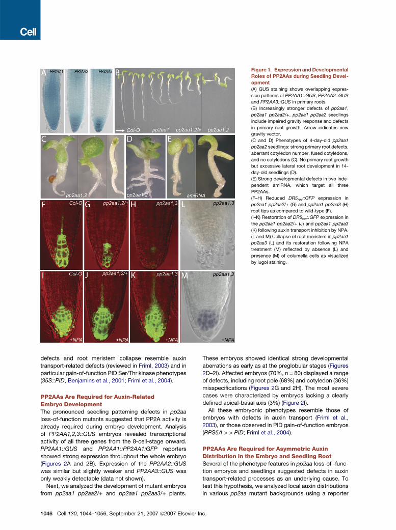
Figure 1. Expression and Developmental
Roles of PP2AAs during Seedling Devel-
opment
(A) GUS staining shows overlapping expres-
sion patterns of PP2AA1::GUS, PP2AA2::GUS
and PP2AA3::GUS in primary roots.
(B) Increasingly stronger defects of pp2aa1,
pp2aa1 pp2aa2/+, pp2aa1 pp2aa2 seedlings
include impaired gravity response and defects
in primary root growth. Arrow indicates new
gravity vector.
(C and D) Phenotypes of 4-day-old pp2aa1
pp2aa2 seedlings: strong primary root defects,
aberrant cotyledon number, fused cotyledons,
and no cotyledons (C). No primary root growth
but excessive lateral root development in 14-
day-old seedlings (D).
(E) Strong developmental defects in two inde-
pendent amiRNA, which target all three
PP2AAs.
(F–H) Reduced DR5rev::GFP expression in
pp2aa1 pp2aa2/+ (G) and pp2aa1 pp2aa3 (H)
root tips as compared to wild-type (F).
(I–K) Restoration of DR5rev::GFP expression in
the pp2aa1 pp2aa2/+ (J) and pp2aa1 pp2aa3
(K) following auxin transport inhibition by NPA.
(L and M) Collapse of root meristem in pp2aa1
pp2aa3 (L) and its restoration following NPA
treatment (M) reflected by absence (L) and
presence (M) of columella cells as visualized
by lugol staining.
defects and root meristem collapse resemble auxin
transport-related defects (reviewed in Friml, 2003) and in
particular gain-of-function PID Ser/Thr kinase phenotypes
(35S::PID, Benjamins et al., 2001; Friml et al., 2004).
PP2AAs Are Required for Auxin-RelatedEmbryo DevelopmentThe pronounced seedling patterning defects in pp2aa
loss-of-function mutants suggested that PP2A activity is
already required during embryo development. Analysis
of PP2AA1,2,3::GUS embryos revealed transcriptional
activity of all three genes from the 8-cell-stage onward.
PP2AA1::GUS and PP2AA1::PP2AA1:GFP reporters
showed strong expression throughout the whole embryo
(Figures 2A and 2B). Expression of the PP2AA2::GUS
was similar but slightly weaker and PP2AA3::GUS was
only weakly detectable (data not shown).
Next, we analyzed the development of mutant embryos
from pp2aa1 pp2aa2/+ and pp2aa1 pp2aa3/+ plants.
1046 Cell 130, 1044–1056, September 21, 2007 ª2007 Elsevier
These embryos showed identical strong developmental
aberrations as early as at the preglobular stages (Figures
2D–2I). Affected embryos (70%, n = 80) displayed a range
of defects, including root pole (68%) and cotyledon (36%)
misspecifications (Figures 2G and 2H). The most severe
cases were characterized by embryos lacking a clearly
defined apical-basal axis (3%) (Figure 2I).
All these embryonic phenotypes resemble those of
embryos with defects in auxin transport (Friml et al.,
2003), or those observed in PID gain-of-function embryos
(RPS5A > > PID; Friml et al., 2004).
PP2AAs Are Required for Asymmetric AuxinDistribution in the Embryo and Seedling RootSeveral of the phenotype features in pp2aa loss-of -func-
tion embryos and seedlings suggested defects in auxin
transport-related processes as an underlying cause. To
test this hypothesis, we analyzed local auxin distributions
in various pp2aa mutant backgrounds using a reporter
Inc.
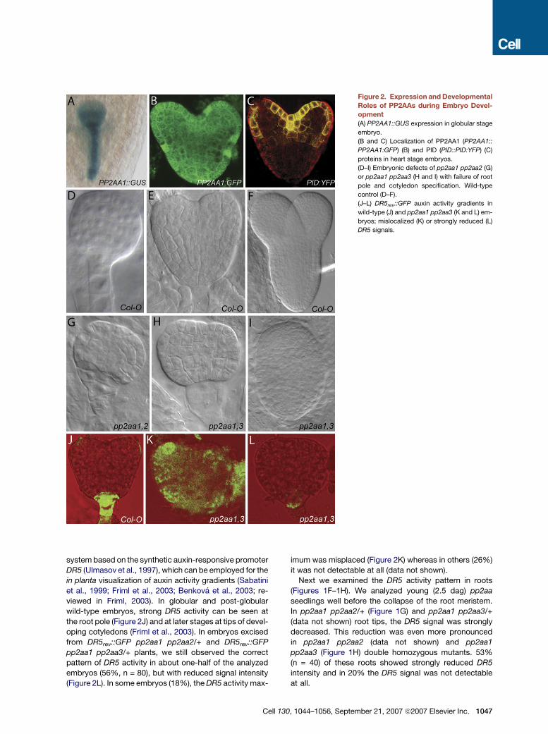
Figure 2. Expression and Developmental
Roles of PP2AAs during Embryo Devel-
opment
(A) PP2AA1::GUS expression in globular stage
embryo.
(B and C) Localization of PP2AA1 (PP2AA1::
PP2AA1:GFP) (B) and PID (PID::PID:YFP) (C)
proteins in heart stage embryos.
(D–I) Embryonic defects of pp2aa1 pp2aa2 (G)
or pp2aa1 pp2aa3 (H and I) with failure of root
pole and cotyledon specification. Wild-type
control (D–F).
(J–L) DR5rev::GFP auxin activity gradients in
wild-type (J) and pp2aa1 pp2aa3 (K and L) em-
bryos; mislocalized (K) or strongly reduced (L)
DR5 signals.
system based on the synthetic auxin-responsive promoter
DR5 (Ulmasov et al., 1997), which can be employed for the
in planta visualization of auxin activity gradients (Sabatini
et al., 1999; Friml et al., 2003; Benkova et al., 2003; re-
viewed in Friml, 2003). In globular and post-globular
wild-type embryos, strong DR5 activity can be seen at
the root pole (Figure 2J) and at later stages at tips of devel-
oping cotyledons (Friml et al., 2003). In embryos excised
from DR5rev::GFP pp2aa1 pp2aa2/+ and DR5rev::GFP
pp2aa1 pp2aa3/+ plants, we still observed the correct
pattern of DR5 activity in about one-half of the analyzed
embryos (56%, n = 80), but with reduced signal intensity
(Figure 2L). In some embryos (18%), the DR5 activity max-
Cell 1
imum was misplaced (Figure 2K) whereas in others (26%)
it was not detectable at all (data not shown).
Next we examined the DR5 activity pattern in roots
(Figures 1F–1H). We analyzed young (2.5 dag) pp2aa
seedlings well before the collapse of the root meristem.
In pp2aa1 pp2aa2/+ (Figure 1G) and pp2aa1 pp2aa3/+
(data not shown) root tips, the DR5 signal was strongly
decreased. This reduction was even more pronounced
in pp2aa1 pp2aa2 (data not shown) and pp2aa1
pp2aa3 (Figure 1H) double homozygous mutants. 53%
(n = 40) of these roots showed strongly reduced DR5
intensity and in 20% the DR5 signal was not detectable
at all.
30, 1044–1056, September 21, 2007 ª2007 Elsevier Inc. 1047

Figure 3. PP2AAs Act Antagonistically to PID on Root Development
(A) Comparison of frequency of primary roots collapse in 4-, 6-, and 8-day-old 35S::PID, pp2aa1 pp2aa3/+ and pp2aa1 pp2aa3 seedlings grown in the
presence or absence of NPA; in all mutant combinations, inhibition of auxin transport by NPA rescues the root collapse.
(B and C) Strongly enhanced phenotypes of pp2aa1 pp2aa3 35S::PID seedlings as compared to pp2aa1 pp2aa3 double mutants of the same age.
(D) Additive shoot phenotypes of an adult pp2aa1 pp2aa3 pid plant in comparison to pp2aa1 pp2aa3 and pid.
(E) Partial rescue of pp2aa1 pp2aa3 double-mutant phenotype in pp2aa1 pp2aa3 pid seedlings; rescue of root growth and root meristem activity.
Arrow indicates new gravity vector.
(F and G) As in case of pp2aa mutants (see Figure 1), reduced DR5 activity (F) and resulting root collapse (G) in 35S::PID root tips is rescued following
auxin transport inhibition by NPA.
(H) Collapsing root meristem of pp2aa1 pp2aa3 is rescued in pp2aa1 pp2aa3 pid triple mutant.
The observed changes in DR5 activity could be either
due to alterations in auxin response or auxin distribution.
To address this question, we treated pp2aa1 pp2aa2
and pp2aa1 pp2aa3 roots with the synthetic auxin 2,4-di-
chlorophenoxyacetic acid (2,4-D) or the auxin transport in-
hibitor 1-N-naphthylphthalamic acid (NPA). 2,4-D caused
a comparable induction of DR5 in both wild-type and
mutant roots, suggesting that auxin response is unaf-
fected (Figures S2G–S2J). Notably, NPA did rescue the
decrease in the DR5 expression and the root meristem
collapse of the pp2aa1 double mutants (Figures 1F–1M),
as previously observed for 35S::PID seedlings (Figures
3A, 3F, and 3G; Benjamins et al., 2001), indicating that
the mutants are affected in auxin transport. To substanti-
ate these observations, we assessed a possible defect in
auxin transport of pp2aa mutants by monitoring auxin
redistribution during gravity response. It is well established
1048 Cell 130, 1044–1056, September 21, 2007 ª2007 Elsevie
that the root gravitropic response is mediated by trans-
port-dependent redistribution of auxin to the lower side
of the responding root tip (e.g., Luschnig et al., 1998). In
contrast to wild-type roots, which all responded (100%,
n = 25), only about 23% of pp2aa1 pp2aa2/+ and pp2aa1
pp2aa3/+ roots (total n = 48) showed weak relocation of
the DR5 signal following gravistimulation (Figures S2C–
S2F; not shown).
In summary, these data suggest that PP2A activity is
required for transport-dependent auxin distribution in
embryos and seedling roots.
PP2AA Acts Antagonistically to PINOIDon Seedling DevelopmentThe same phenotypes as those caused by loss of PP2AA
function, including embryo patterning defects, agravi-
tropic root growth and decreased DR5 activity in roots
r Inc.
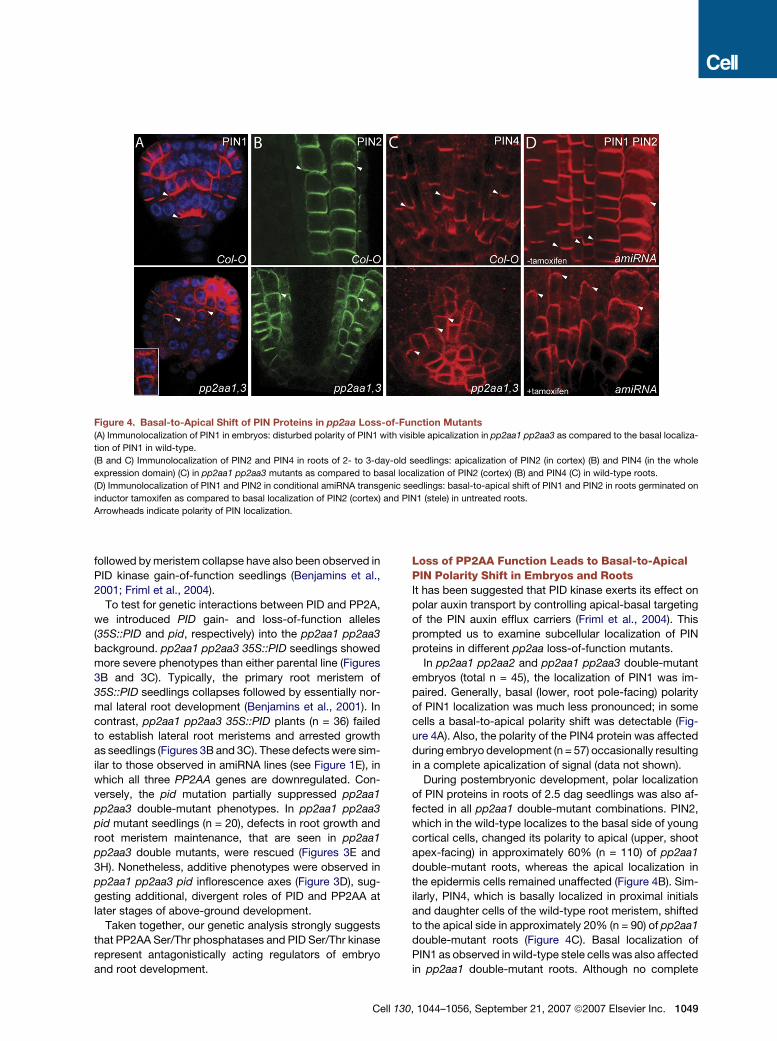
Figure 4. Basal-to-Apical Shift of PIN Proteins in pp2aa Loss-of-Function Mutants
(A) Immunolocalization of PIN1 in embryos: disturbed polarity of PIN1 with visible apicalization in pp2aa1 pp2aa3 as compared to the basal localiza-
tion of PIN1 in wild-type.
(B and C) Immunolocalization of PIN2 and PIN4 in roots of 2- to 3-day-old seedlings: apicalization of PIN2 (in cortex) (B) and PIN4 (in the whole
expression domain) (C) in pp2aa1 pp2aa3 mutants as compared to basal localization of PIN2 (cortex) (B) and PIN4 (C) in wild-type roots.
(D) Immunolocalization of PIN1 and PIN2 in conditional amiRNA transgenic seedlings: basal-to-apical shift of PIN1 and PIN2 in roots germinated on
inductor tamoxifen as compared to basal localization of PIN2 (cortex) and PIN1 (stele) in untreated roots.
Arrowheads indicate polarity of PIN localization.
followed by meristem collapse have also been observed in
PID kinase gain-of-function seedlings (Benjamins et al.,
2001; Friml et al., 2004).
To test for genetic interactions between PID and PP2A,
we introduced PID gain- and loss-of-function alleles
(35S::PID and pid, respectively) into the pp2aa1 pp2aa3
background. pp2aa1 pp2aa3 35S::PID seedlings showed
more severe phenotypes than either parental line (Figures
3B and 3C). Typically, the primary root meristem of
35S::PID seedlings collapses followed by essentially nor-
mal lateral root development (Benjamins et al., 2001). In
contrast, pp2aa1 pp2aa3 35S::PID plants (n = 36) failed
to establish lateral root meristems and arrested growth
as seedlings (Figures 3B and 3C). These defects were sim-
ilar to those observed in amiRNA lines (see Figure 1E), in
which all three PP2AA genes are downregulated. Con-
versely, the pid mutation partially suppressed pp2aa1
pp2aa3 double-mutant phenotypes. In pp2aa1 pp2aa3
pid mutant seedlings (n = 20), defects in root growth and
root meristem maintenance, that are seen in pp2aa1
pp2aa3 double mutants, were rescued (Figures 3E and
3H). Nonetheless, additive phenotypes were observed in
pp2aa1 pp2aa3 pid inflorescence axes (Figure 3D), sug-
gesting additional, divergent roles of PID and PP2AA at
later stages of above-ground development.
Taken together, our genetic analysis strongly suggests
that PP2AA Ser/Thr phosphatases and PID Ser/Thr kinase
represent antagonistically acting regulators of embryo
and root development.
Cell 1
Loss of PP2AA Function Leads to Basal-to-ApicalPIN Polarity Shift in Embryos and RootsIt has been suggested that PID kinase exerts its effect on
polar auxin transport by controlling apical-basal targeting
of the PIN auxin efflux carriers (Friml et al., 2004). This
prompted us to examine subcellular localization of PIN
proteins in different pp2aa loss-of-function mutants.
In pp2aa1 pp2aa2 and pp2aa1 pp2aa3 double-mutant
embryos (total n = 45), the localization of PIN1 was im-
paired. Generally, basal (lower, root pole-facing) polarity
of PIN1 localization was much less pronounced; in some
cells a basal-to-apical polarity shift was detectable (Fig-
ure 4A). Also, the polarity of the PIN4 protein was affected
during embryo development (n = 57) occasionally resulting
in a complete apicalization of signal (data not shown).
During postembryonic development, polar localization
of PIN proteins in roots of 2.5 dag seedlings was also af-
fected in all pp2aa1 double-mutant combinations. PIN2,
which in the wild-type localizes to the basal side of young
cortical cells, changed its polarity to apical (upper, shoot
apex-facing) in approximately 60% (n = 110) of pp2aa1
double-mutant roots, whereas the apical localization in
the epidermis cells remained unaffected (Figure 4B). Sim-
ilarly, PIN4, which is basally localized in proximal initials
and daughter cells of the wild-type root meristem, shifted
to the apical side in approximately 20% (n = 90) of pp2aa1
double-mutant roots (Figure 4C). Basal localization of
PIN1 as observed in wild-type stele cells was also affected
in pp2aa1 double-mutant roots. Although no complete
30, 1044–1056, September 21, 2007 ª2007 Elsevier Inc. 1049
apicalization of PIN1 localization was detectable, the sub-
cellular polarity of PIN1 was less pronounced in these
double mutants (data not shown).
When activity of all three PP2AAs was decreased in
tamoxifen-inducible amiRNA lines, a pronounced basal-
to-apical shift of PIN1, PIN2 and PIN4 polarity could be
observed following tamoxifen treatment (Figure 4D and
data not shown). In control experiments, the same trans-
genic seedlings grown without tamoxifen (Figure 4D) as
well as tamoxifen-treated transgenics harboring the
empty T-DNA vector (data not shown) did not show
changes in PIN polarity (Figure 4D).
These data collectively show that loss of PP2AA func-
tion leads to a basal-to-apical shift in PIN polarity, a phe-
nomenon identical to PIN polarity changes in PID gain-
of-function plants. Furthermore, the observed reversal of
basal PIN localization fully explains all observed auxin
transport-related aspects of pp2aa mutant phenotypes.
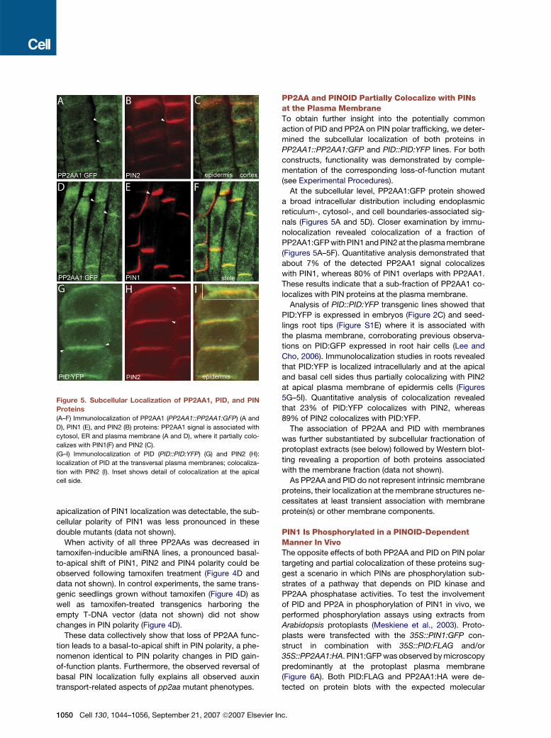
Figure 5. Subcellular Localization of PP2AA1, PID, and PIN
Proteins
(A–F) Immunolocalization of PP2AA1 (PP2AA1::PP2AA1:GFP) (A and
D), PIN1 (E), and PIN2 (B) proteins: PP2AA1 signal is associated with
cytosol, ER and plasma membrane (A and D), where it partially colo-
calizes with PIN1(F) and PIN2 (C).
(G–I) Immunolocalization of PID (PID::PID:YFP) (G) and PIN2 (H):
localization of PID at the transversal plasma membranes; colocaliza-
tion with PIN2 (I). Inset shows detail of colocalization at the apical
cell side.
1050 Cell 130, 1044–1056, September 21, 2007 ª2007 Elsevier
PP2AA and PINOID Partially Colocalize with PINsat the Plasma MembraneTo obtain further insight into the potentially common
action of PID and PP2A on PIN polar trafficking, we deter-
mined the subcellular localization of both proteins in
PP2AA1::PP2AA1:GFP and PID::PID:YFP lines. For both
constructs, functionality was demonstrated by comple-
mentation of the corresponding loss-of-function mutant
(see Experimental Procedures).
At the subcellular level, PP2AA1:GFP protein showed
a broad intracellular distribution including endoplasmic
reticulum-, cytosol-, and cell boundaries-associated sig-
nals (Figures 5A and 5D). Closer examination by immu-
nolocalization revealed colocalization of a fraction of
PP2AA1:GFP with PIN1 and PIN2 at the plasma membrane
(Figures 5A–5F). Quantitative analysis demonstrated that
about 7% of the detected PP2AA1 signal colocalizes
with PIN1, whereas 80% of PIN1 overlaps with PP2AA1.
These results indicate that a sub-fraction of PP2AA1 co-
localizes with PIN proteins at the plasma membrane.
Analysis of PID::PID:YFP transgenic lines showed that
PID:YFP is expressed in embryos (Figure 2C) and seed-
lings root tips (Figure S1E) where it is associated with
the plasma membrane, corroborating previous observa-
tions on PID:GFP expressed in root hair cells (Lee and
Cho, 2006). Immunolocalization studies in roots revealed
that PID:YFP is localized intracellularly and at the apical
and basal cell sides thus partially colocalizing with PIN2
at apical plasma membrane of epidermis cells (Figures
5G–5I). Quantitative analysis of colocalization revealed
that 23% of PID:YFP colocalizes with PIN2, whereas
89% of PIN2 colocalizes with PID:YFP.
The association of PP2AA and PID with membranes
was further substantiated by subcellular fractionation of
protoplast extracts (see below) followed by Western blot-
ting revealing a proportion of both proteins associated
with the membrane fraction (data not shown).
As PP2AA and PID do not represent intrinsic membrane
proteins, their localization at the membrane structures ne-
cessitates at least transient association with membrane
protein(s) or other membrane components.
PIN1 Is Phosphorylated in a PINOID-DependentManner In VivoThe opposite effects of both PP2AA and PID on PIN polar
targeting and partial colocalization of these proteins sug-
gest a scenario in which PINs are phosphorylation sub-
strates of a pathway that depends on PID kinase and
PP2AA phosphatase activities. To test the involvement
of PID and PP2A in phosphorylation of PIN1 in vivo, we
performed phosphorylation assays using extracts from
Arabidopsis protoplasts (Meskiene et al., 2003). Proto-
plasts were transfected with the 35S::PIN1:GFP con-
struct in combination with 35S::PID:FLAG and/or
35S::PP2AA1:HA. PIN1:GFP was observed by microscopy
predominantly at the protoplast plasma membrane
(Figure 6A). Both PID:FLAG and PP2AA1:HA were de-
tected on protein blots with the expected molecular
Inc.
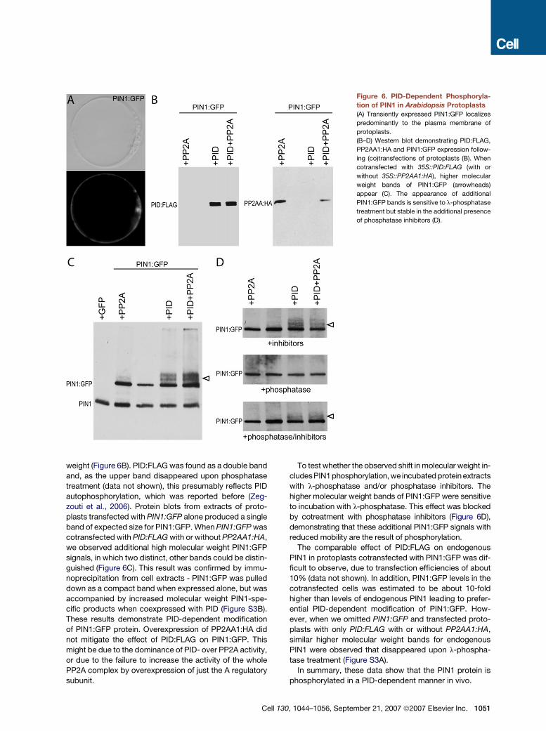
Figure 6. PID-Dependent Phosphoryla-
tion of PIN1 in Arabidopsis Protoplasts
(A) Transiently expressed PIN1:GFP localizes
predominantly to the plasma membrane of
protoplasts.
(B–D) Western blot demonstrating PID:FLAG,
PP2AA1:HA and PIN1:GFP expression follow-
ing (co)transfections of protoplasts (B). When
cotransfected with 35S::PID:FLAG (with or
without 35S::PP2AA1:HA), higher molecular
weight bands of PIN1:GFP (arrowheads)
appear (C). The appearance of additional
PIN1:GFP bands is sensitive to l-phosphatase
treatment but stable in the additional presence
of phosphatase inhibitors (D).
weight (Figure 6B). PID:FLAG was found as a double band
and, as the upper band disappeared upon phosphatase
treatment (data not shown), this presumably reflects PID
autophosphorylation, which was reported before (Zeg-
zouti et al., 2006). Protein blots from extracts of proto-
plasts transfected with PIN1:GFP alone produced a single
band of expected size for PIN1:GFP. When PIN1:GFP was
cotransfected with PID:FLAG with or without PP2AA1:HA,
we observed additional high molecular weight PIN1:GFP
signals, in which two distinct, other bands could be distin-
guished (Figure 6C). This result was confirmed by immu-
noprecipitation from cell extracts - PIN1:GFP was pulled
down as a compact band when expressed alone, but was
accompanied by increased molecular weight PIN1-spe-
cific products when coexpressed with PID (Figure S3B).
These results demonstrate PID-dependent modification
of PIN1:GFP protein. Overexpression of PP2AA1:HA did
not mitigate the effect of PID:FLAG on PIN1:GFP. This
might be due to the dominance of PID- over PP2A activity,
or due to the failure to increase the activity of the whole
PP2A complex by overexpression of just the A regulatory
subunit.
Cell 1
To test whether the observed shift in molecular weight in-
cludesPIN1phosphorylation, we incubated protein extracts
with l-phosphatase and/or phosphatase inhibitors. The
higher molecular weight bands of PIN1:GFP were sensitive
to incubation with l-phosphatase. This effect was blocked
by cotreatment with phosphatase inhibitors (Figure 6D),
demonstrating that these additional PIN1:GFP signals with
reduced mobility are the result of phosphorylation.
The comparable effect of PID:FLAG on endogenous
PIN1 in protoplasts cotransfected with PIN1:GFP was dif-
ficult to observe, due to transfection efficiencies of about
10% (data not shown). In addition, PIN1:GFP levels in the
cotransfected cells was estimated to be about 10-fold
higher than levels of endogenous PIN1 leading to prefer-
ential PID-dependent modification of PIN1:GFP. How-
ever, when we omitted PIN1:GFP and transfected proto-
plasts with only PID:FLAG with or without PP2AA1:HA,
similar higher molecular weight bands for endogenous
PIN1 were observed that disappeared upon l-phospha-
tase treatment (Figure S3A).
In summary, these data show that the PIN1 protein is
phosphorylated in a PID-dependent manner in vivo.
30, 1044–1056, September 21, 2007 ª2007 Elsevier Inc. 1051

Figure 7. PID and PP2A Activities Mediate Phosphorylation of PIN Proteins in Their Hydrophilic Loop
(A) MS/MS spectrum of phosphorylated peptide derived from PIN1 hydrophilic loop. The sequence of the precursor peptide. Asterisks indicate Serin
and Threonin, one of which is phosphorylated. The major peak in the spectrum corresponds to the precursor peptide (Mass: 1163.2 Da) with neutral
loss of a single phosphate group (1130.6 Da). A second major peak is consistent with the precursor peptide losing both a phosphate group and a water
molecule (1124.6 Da).
(B and C) GST:PID or HIS:PID autophosphorylates and efficiently phosphorylates the large hydrophilic loop of PIN1 (HIS:PIN1HL) and Myelin Basic
Protein (MBP) (B) or the loop of PIN2 (HIS:PIN2HL) (C) in vitro. GST (C) and the myosin-like protein (HIS:MLP) (B) are not phosphorylated by PID.
(D) Increased phosphorylation of the HIS:PIN2HL by protein extracts of 35S::PID (P), pp2aa1 pp2aa2 (p12) and pp2aa1 pp2aa3 (p13) as compared to
wild-type (w) extracts. Lane 1 contains wild-type extract without HIS:PIN2HL.
PINOID and PP2A Antagonistically Act onPhosphorylation of PIN Proteins in Their CentralHydrophilic LoopTo test whether PINs are present in a phosphorylated state
in planta, we immunoprecipitated PIN1:GFP protein from
seedling roots and performed Mass Spectrometry (nLC-
MS/MS) on tryptic peptides. Seven different PIN1 peptides
were identified, all falling within the large central hydro-
philic loop (Supplementary Table). One of these peptides
was found both in a nonphosphorylated and phosphory-
1052 Cell 130, 1044–1056, September 21, 2007 ª2007 Elsevie
lated state (Figure 7A; Supplementary Table). From the
MS/MS spectrum, it cannot be determined whether the
phosphorylation occurs at Serine 337 or at Threonine 340,
both of which are conserved in several members of the PIN
gene family. These results together with previous reports
(Nuhse et al., 2004; Benschop et al., 2007) demonstrate
that PIN1 and possibly also other PIN proteins are phos-
phorylated in their central hydrophilic loop in Arabidopsis.
Next we tested whether the central hydrophilic loop of
PIN proteins can be directly phosphorylated by PID. The
r Inc.

HIS-tagged hydrophilic loop (HL) of PIN1 and GST-tagged
PID were heterologously expressed in E. coli, and coincu-
bated in an in vitro phosphorylation reaction. Following
electrophoretic separation of the proteins, clear PID-
dependent phosphorylation of HIS:PIN1HL was detected
(Figure 7B). In addition, PID autophosphorylation and
phosphorylation of the standard Ser/Thr kinase substrate
Myelin Basic Protein (MBP), but not of an unrelated
myosin-like protein (MLP) was detected. In another exper-
iment, both GST:PID and HIS:PID were able to phos-
phorylate HIS:PIN2HL as well (Figure 7C). These results
demonstrate that the PID kinase is able to phosphorylate
the hydrophilic loop of PIN proteins in vitro.
Furthermore, we tested the ability of protein extracts
derived from wild-type, 35S::PID and pp2aa mutants to
phosphorylate HIS:PIN2HL. The phosphorylation of HIS:
PIN2HL was enhanced upon incubation with 35S::PID
protein extract when compared to extracts from wild-
type (Figure 7D). Similarly, protein extracts derived from
plant material lacking PP2AA1 and either PP2AA2 or
PP2AA3 had increased ability to phosphorylate PIN2HL
(Figure 7D). These data confirm that PID activity positively
regulates phosphorylation of PIN hydrophilic loops,
whereas PP2A activity has a negative effect.
When taken together, the results obtained in in vitro
and the in vivo phosphorylation assays corroborate the
genetic and cell biological studies and provide strong
support for a scenario in which PID kinase and PP2A
phosphatase activities antagonistically regulate phos-
phorylation of PIN proteins in their middle, hydrophilic
loop.
DISCUSSION
PP2A Phosphatase and PINOID Kinase ActAntagonistically on Auxin Transport-DependentDevelopment and PIN Apical-Basal TargetingFunctional characterization of the Ser/Thr protein kinase
PINOID revealed a role for protein phosphorylation in
PIN polar targeting, auxin transport and auxin-related
development (Christensen et al., 2000; Benjamins et al.,
2001; Friml et al., 2004). Moreover, loss-of-function of
the A regulatory subunits of PP2A was shown to cause
severe developmental defects (Garbers et al., 1996;
Rashotte et al., 2001; Zhou et al., 2004), many of which
correlate with defects in auxin distributions. Our detailed
observations on pp2aa mutants showed that the loss-of-
function phenotypes affecting root and embryo develop-
ment are strikingly similar to PID gain-of-function pheno-
types (Benjamins et al., 2001; Friml et al., 2004), and that
pid and pp2aa show antagonistic genetic interactions.
The antagonistic activities of PID and PP2A were also
apparent at the cellular level, since both, PP2A and PID
activities influence the apical versus basal polar targeting
of PIN proteins. In wild-type plants, basal polarity of PIN
localization in the inner embryo and root tissues mediates
auxin flow toward the root pole, thereby triggering and,
later, maintaining the activity of the root meristem (Friml
Cell 1
et al., 2002a, 2003; Blilou et al., 2005). In the PID
gain-of-function or pp2aa loss-of-function alleles, PIN
localization is to a large extent apicalized, causing auxin
depletion in the root pole, and resulting in meristem col-
lapse. These results imply that PP2A phosphatase and
PID kinase act on the same auxin transport-related devel-
opmental processes, by antagonistically regulating PIN
polar targeting.
PP2A phosphatase is a heterotrimeric protein consist-
ing of a C catalytic subunit together with A and B regula-
tory subunits. 5 Arabidopsis loci encode C subunits, 17
loci were found to encode B subunits, whereas 3 loci
code for A subunits. Different combinations of these sub-
units form holoenzymes with distinct properties, which in
animals are known to regulate a wide range of develop-
mental processes (reviewed in Janssens and Goris,
2001). Considering this, it is quite surprising that a general
decrease of PP2A activity in Arabidopsis roots and
embryos primarily affects auxin transport-related pattern-
ing. This may reflect both the specificity of PP2A action in
auxin transport-related processes as well as high suscep-
tibility of root and embryos to perturbations in the auxin
distribution. On the other hand, in inflorescence and flower
development, PP2A have been reported to mediate addi-
tional auxin-unrelated processes (Zhou et al., 2004) and
show additive rather than antagonistic effects to PID.
This suggests a broader specificity of PP2A action as
compared to PID at these developmental stages.
PINOID and PP2A Act Antagonisticallyon Phosphorylation of PIN ProteinsAntagonistic action of PID and PP2A implies that they
might act as a kinase/phosphatase pair on common
substrates. As expected for soluble proteins, PID and in
particular PP2AA show a broad intracellular distribution.
Nevertheless, a fraction of both proteins was detected
together with PIN proteins associated with the plasma
membrane. Similar localization of a kinase was found in
mammalian epithelial cells, where the atypical protein
kinase C, which mediates apicalization of early blasto-
meres, localizes to the apical plasma membrane (Chalm-
ers et al., 2005). The demonstration of close subcellular
association of PID, PP2AA and PINs favors a scenario in
which PID/PP2AA pairs directly control phosphorylation
of PIN proteins. This model is further reinforced by obser-
vations that the hydrophilic parts of PIN proteins are phos-
phorylated in vitro and in vivo in PID-dependent manner. In
addition, protein extracts from both PID gain-of-function
and pp2aa loss-of-function plants show increased ability
to phosphorylate PIN proteins. There are two scenarios
consistent with these data: (1) Both PP2A and PID act
directly on (de)phosphorylation of PINs or (2) PP2A acts
on dephosphorylation of PID, thus downregulating its
kinase activity on PINs. In either case, the data clearly
show that the PID kinase and PP2A trimeric phosphatases
act antagonistically on reversible phosphorylation of PIN
proteins, which in turn determines the apical-basal target-
ing of these auxin efflux carriers.
30, 1044–1056, September 21, 2007 ª2007 Elsevier Inc. 1053

Reversible Phosphorylation of Cargos as a Meansfor Conditional Apical-Basal Targeting in PlantsWe propose a model for conditional apical or basal deliv-
ery of polarly localized cargos in plants. Cargos such as
PIN proteins can be targeted either to the apical or to
the basal side of cells, depending on their phosphorylation
status. Conditions in which PID kinase activities are rela-
tively high would result in predominantly phosphorylated
PIN proteins, causing their targeting to the apical side of
cells. In the converse situation, when PID activities are
lower than those of PP2A phosphatase, PIN proteins will
be dephosphorylated and targeted preferentially to the
basal side of the cell.
Regulation of polar delivery of membrane components
in mammalian and plant cells may share important
features. In mammalian epithelial cells, phosphorylation
of cargos has been shown to influence their delivery. For
example, delivery of the immunoglobulin receptor to the
apical cell surface largely depends on its phosphorylation
status (Casanova et al., 1990). Our demonstration of the
kinase/phosphatase regulation of PIN1 polarity shows
that similar processes occur in plants. The suggested
mechanism might also help to answer outstanding ques-
tions about the regulation of auxin flow, such as how
differences between distinct PIN proteins and cell types
together contribute to the decision of apical or basal PIN
delivery. Variations in phosphorylation of different PIN
proteins could, for example, arise as a consequence of
divergent phosphorylation sites, some of which would
be phosphorylated more efficiently, whereas others might
represent rather poor substrates for PID. In parallel, rela-
tive expression levels of PID and/or PP2A in different cell
types could play a decisive role. Furthermore, activities
of PID and PP2A may be downstream of different signaling
pathways, which by this mechanism could redirect auxin
flow through modulation of PIN polar targeting. In this sce-
nario, a combination of constitutive endocytosis of PINs
(Dhonukshe et al., 2007) and their reversible phosphoryla-
tion would allow for flexible retargeting of PINs to different
sub-cellular destinations in response to various signals.
Overall, control of PIN protein phosphorylation appears
to represent a hitherto unappreciated level of regulation
of directional auxin fluxes, which are causal in plant pat-
tern formation, organogenesis and tropisms.
EXPERIMENTAL PROCEDURES
Material
Columbia ecotype (Col-O) plants were used for all experiments. The
details on mutants, transgenic plants and constructs can be found in
the supplementary material. Double mutants were generated by
crosses and F2 progeny from five independent crosses for each com-
bination of mutants were screened for phenotypes and confirmed by
PCR genotyping. In an attempt to obtain pp2aa1 pp2aa2 pp2aa3 triple
mutants among 120 genotyped F2 seedlings (all revealing strong
phenotypes) no triple homozygous plants were found.
Growth Conditions and Phenotypic Analysis
Seeds were grown as described (Benkova et al., 2003). Short-time
incubation of 3–5 dag seedlings with 5 mM 2,4-dichlorophenoxyacetic
1054 Cell 130, 1044–1056, September 21, 2007 ª2007 Elsevier
acid (2,4-D; Sigma) was performed in 24-well cell-culture plates in liq-
uid AM medium with 1% sucrose for 4 hr. Long-time treatment was
done by growing seedling on AM medium supplemented with 0.3 mM
NPA (Sigma). Seeds carrying inducible amiRNA system were germi-
nated on AM medium supplemented with 1 or 5 mM tamoxifen (Sigma).
Root gravitropic assays (Paciorek et al., 2005) and embryo analyses
(Friml et al., 2003) were performed as described. Seedlings were ana-
lyzed at 3, 4, 6, 8, and 14 dag. Nine independent amiRNA lines were
analyzed (four for amiRNA-1 and five for amiRNA-2). For pp2aas pid
and pp2aas 35S::PID triple mutants five independent segregating
lines of (n > 500 individuals) were examined. Microscopy was done
with a Zeiss Axiophot equipped with Axiocam HR CCD camera.
In Situ Expression and Localization Analysis
Histochemical stainings for GUS activity and whole-mount immuno-
localization were performed as described (Friml et al., 2003; Sauer
et al., 2006b). Each experiment was done on three to ten independent
lines with minimum of two repetitions. Antibodies were diluted as
follows: anti-PIN1 (1:1000; Paciorek et al., 2005), anti-PIN2 (1:1000;
Abas et al., 2006), anti-PIN4 (1:400; Friml et al., 2002a), anti-GFP
(1:500; Molecular Probes); FITC- and CY3-conjugated anti-rabbit sec-
ondary antibodies (Dianova) were both diluted 1:500. For in vivo GFP
inspections, plant material was mounted in 5% glycerol. Analysis
was done using a Leica TCS SP2 confocal laser scanning microscope.
Images were processed in Adobe Photoshop and assembled in Adobe
Illustrator. The colocalizations were quantitatively analyzed using
MetaMorph (Molecular Devices).
Immunoprecipitation and Mass Spectrometry
PIN1:GFP seedlings were grown vertically for 5 days and crude protein
extract was prepared from excised roots (Karlova et al., 2006). Follow-
ing preclearing protein extract with Tris-conjugated Microlink agarose
matrix (Pierce), PIN1:GFP was precipitated by overnight incubation
with anti-YFP antibody-coupled Microlink agarose matrix (R. Karlova,
W. van Dongen, and S. de Vries, personal communication; details
available upon request). The immunocomplexes were washed and
Trypsin-digested as in Karlova et al. (2006). After Trypsin digestion,
peptides were separated by nano-Liquid Chromatography and sub-
jected to tandem Mass Spectrometry using an LCQ Classic (Thermo
Electron, San Jose, CA). Spectra were compared with a custom-
made database encompassing the Arabidopsis proteome using Bio-
works 3.2 (Thermo Electron) software. The experiments were per-
formed with three biological replicas giving comparable results.
In Vivo Phosphorylation Assays
Arabidopsis protoplasts were isolated from suspension culture, trans-
formed according to Meskiene et al. (2003) and harvested after 10–
22 hr. Cell pellets were lysed by freeze-thaw cycles followed by
a Dounce-type homogenizer. The extraction buffer used was based
on Abas et al. (2006) except that PVPP was excluded, and 20% sorbi-
tol was used instead of glycerol.
Phosphorylation assays were performed using protoplasts
extracted as above, but omitting the phosphatase inhibitors. Total
cell extracts or membrane fractions were solubilised with 0.1%
Brij35 and preheated at 65�C for 10 min to inactivate endogenous
enzymes. After l-phosphatase buffer (Sigma P9614) was added,
four treatments were performed in a final volume of 30 or 50 ml: (a)
sample plus 3 mM MnCl2; (b) sample plus 3 mM MnCl2 and 100 U
l-phosphatase (Sigma P9614); (c) sample plus phosphatase inhibitors
(20 mM EDTA, 13 mM EGTA, 40 mM betaglycerolphosphate, 0.5 mM
sodium orthovanadate, 1 mM sodium molybdate, 5 nM okadaic acid,
50 mM sodium fluoride); (d) sample plus 100 U l-phosphatase and
phosphatase inhibitors as in (c). All samples were incubated at 30�C
for 5–20 min. Reactions were stopped by adding phosphatase inhibi-
tors to (a) and (b) and freezing all samples. Protoplasts transformed
with GFP and subjected to phosphatase treatment as above showed
no change in GFP specific bands as detected by Western blotting,
Inc.

indicating that GFP itself was not phosphorylated when overexpressed
in protoplasts (data not shown). Samples were separated as described
(Abas et al., 2006) and probed with the following antibodies: affinity
purified rabbit polyclonal anti-PIN1 (Paciorek et al., 2005), mouse
monoclonal anti-FLAG (Sigma, clone M2, used at 2.5 mg/ml), mouse
monoclonal anti-GFP (Roche, clones 7.1 and 13.1, used at 0.4 mg/ml),
rat monoclonal anti-HA (Roche, clone 3F10, used at 0.2 mg/ml).
Secondary antibodies (HRP-conjugated goat anti-rabbit, anti-mouse
or anti-rat IgG, all from Jackson) were used at 0.02–0.08 mg/ml. Detec-
tion was performed using enhanced chemiluminescence (Pierce Super
Signal).
In Vitro Phosphorylation Assays
Approximately 1 mg of purified protein expressed in E. coli (PID and
substrates) were added to kinase reaction mix (20 ml total volume),
containing 1x kinase buffer (25 mM Tris-HCl [pH 7.5]; 1 mM DTT;
5 mM MgCl2) and 13 ATP solution (100 mM MgCl2/ATP; 1 mCi 32P-g-
ATP). For the assays with seedling extracts, 3–4 dag seedlings were
harvested in aliquots of 50 seedlings and stored at �80�C. Approxi-
mately 4 mg of the PIN2HL was incubated with 50 mg total protein
extracted from seedlings, 13 kinase buffer and 13 ATP solution (see
above) in a total volume of 150 ml. Reactions were incubated at 30�C
for 30 min. and stopped by the addition of respectively 5 or 40 ml of
5 3 protein loading buffer (310 mM Tris-HCl [pH 6.8]; 10% SDS;
50% Glycerol; 750 mM b-Mercaptoethanol; 0,125% Bromophenol
Blue) and 5 min. boiling. Reactions were subsequently separated
over 12,5% acrylamide gels, which were washed 3 times for 30 min.
with kinase gel wash buffer (5% TCA [trichoroacetic acid]; 1%
Na2H2P2O7), Coomassie stained, destained, dried, and exposed to
X-ray films for 24 to 48 hr at �80�C using intensifier screens.
Supplemental Data
Supplemental Data include Supplemental Experimental Procedures,
Supplemental References, three figures, and one table and can be
found with this article online at http://www.cell.com/cgi/content/full/
130/6/1044/DC1/.
ACKNOWLEDGMENTS
We thank S. Boeren, R. Karlova, J. Kleine-Vehn, F. Maraschin and H.
Robert for technical help and advice; S. Peck for helpful discussions;
J. Strauss and L. Mach for sharing reagents. We gratefully acknowl-
edge financial support from the VolkswagenStiftung and EMBO Young
Investigator Program (J.F.), the Deutsche Forschungsgemeinschaft -
SFB 446 (M.M.), Brazilian Funding Agency for Post-Graduation Educa-
tion-CAPES (M.K.Z.), the Austrian Research Fund - P16311; P18440
(L.A., C.L.), - P16409; P19005 (A.S., I.M.), the Netherlands Proteomic
Center (NPC; Grant to S. de Vries; D.W.), the China Scholarship Coun-
cil (F.H.), the National Science Foundation FIBR 0330786 and Depart-
ment of Energy FG02-88ER13873 (M.G.H., C.O. and E.M.M.).
Received: August 9, 2006
Revised: April 6, 2007
Accepted: July 23, 2007
Published: September 20, 2007
REFERENCES
Abas, L., Benjamins, R., Malenica, N., Paciorek, T., Wi�sniewska, J.,
Moulinier-Anzola, J.C., Sieberer, T., Friml, J., and Luschnig, C.
(2006). Intracellular trafficking and proteolysis of the Arabidopsis
auxin-efflux facilitator PIN2 are involved in root gravitropism. Nat.
Cell Biol. 8, 249–256.
Benjamins, R., Quint, A., Weijers, D., Hooykaas, P., and Offringa, R.
(2001). The PINOID protein kinase regulates organ development in
Cell 1
Arabidopsis by enhancing polar auxin transport. Development 128,
4057–4067.
Benkova, E., Michniewicz, M., Sauer, M., Teichmann, T., Seifertova,
D., Jurgens, G., and Friml, J. (2003). Local, efflux-dependent auxin
gradients as a common module for plant organ formation. Cell 115,
591–602.
Benschop, J.J., Mohammed, S., O’flaherty, M., Heck, A.J., Slijper, M.,
and Menke, F.L. (2007). Quantitative phospho-proteomics of early
elicitor signalling in Arabidopsis. Mol. Cell. Proteomics 6, 1198–1214.
Published online on February 21, 2007.
Blilou, I., Xu, J., Wildwater, M., Willemsen, V., Paponov, I., Friml, J.,
Heidstra, R., Aida, M., Palme, K., and Scheres, B. (2005). The PIN auxin
efflux facilitator network controls growth and patterning in Arabidopsis
roots. Nature 443, 39–44.
Casanova, J.E., Breitfeld, P.P., Ross, S.A., and Mostov, K.E. (1990).
Phosphorylation of the polymeric immunoglobulin receptor required
for its efficient transcytosis. Science 248, 742–745.
Chalmers, A.D., Pambos, M., Mason, J., Lang, S., Wylie, C., and
Papalopulu, N. (2005). aPKC, Crumbs3 and Lgl2 control apicobasal
polarity in early vertebrate development. Development 132, 977–986.
Christensen, S.K., Dagenais, N., Chory, J., and Weigel, D. (2000). Reg-
ulation of auxin response by the protein kinase PINOID. Cell 100,
469–478.
Dhonukshe, P., Aniento, F., Hwang, I., Robinson, D., Mravec, J., Stier-
hof, Y.-D., and Friml, J. (2007). Clathrin-mediated constitutive endocy-
tosis of PIN auxin efflux carriers in Arabidopsis. Curr. Biol. 17, 520–527.
Friml, J. (2003). Auxin transport—shaping the plant. Curr. Opin. Plant
Biol. 6, 7–12.
Friml, J., Benkova, E., Blilou, I., Wi�sniewska, J., Hamann, T., Ljung, K.,
Woody, S., Sandberg, G., Scheres, B., Jurgens, G., and Palme, K.
(2002a). AtPIN4 mediates sink-driven auxin gradients and root pattern-
ing in Arabidopsis. Cell 108, 661–673.
Friml, J., Vieten, A., Sauer, M., Weijers, D., Schwarz, H., Hamann, T.,
Offringa, R., and Jurgens, G. (2003). Efflux-dependent auxin gradients
establish the apical-basal axis of Arabidopsis. Nature 426, 147–153.
Friml, J., Wi�sniewska, J., Benkova, E., Mendgen, K., and Palme, K.
(2002b). Lateral relocation of auxin efflux regulator PIN3 mediates
tropism in Arabidopsis. Nature 415, 806–809.
Friml, J., Yang, X., Michniewicz, M., Weijers, D., Quint, A., Tietz, O.,
Benjamins, R., Ouwerkerk, P.B., Ljung, K., Sandberg, G., et al.
(2004). A PINOID-dependent binary switch in apical-basal PIN polar
targeting directs auxin efflux. Science 306, 862–865.
Garbers, Ch., DeLong, A., Deruere, J., Bernasconi, P., and Soll, D.
(1996). A mutation in protein phosphatase 2A regulatory subunit A
affects auxin transport in Arabidopsis. EMBO J. 15, 2115–2124.
Hardtke, C.S., and Berleth, T. (1998). The Arabidopsis gene MONOP-
TEROS encodes a transcription factor mediating embryo axis forma-
tion and vascular development. EMBO J. 17, 1405–1411.
Janssens, V., and Goris, J. (2001). Protein phosphatase 2A: a highly
regulated family of serine/threonine phosphatases implicated in cell
growth and signalling. Biochem. J. 353, 417–439.
Karlova, R., Boeren, S., Russinova, E., Aker, J., Vervoort, J., and de
Vries, S.C. (2006). The Arabidopsis SOMATIC EMBRYOGENESIS
RECEPTOR-LIKE KINASE1 protein complex includes BRASSINOSTE-
ROID-INSENSITIVE1. Plant Cell 18, 626–638.
Lee, S.H., and Cho, H.T. (2006). PINOID positively regulates auxin ef-
flux in Arabidopsis root hair cells and tobacco cells. Plant Cell 18,
1604–1616.
Luschnig, C., Gaxiola, R.A., Grisafi, P., and Fink, G.R. (1998). EIR1,
a root-specific protein involved in auxin transport, is required for grav-
itropism in Arabidopsis thaliana. Genes Dev. 12, 2175–2187.
Meskiene, I., Baudouin, E., Schweighofer, A., Liwosz, A., Jonak, C.,
Rodriguez, P.L., Jelinek, H., and Hirt, H. (2003). The Stress-induced
30, 1044–1056, September 21, 2007 ª2007 Elsevier Inc. 1055

protein phosphatase 2C is a negative regulator of a mitogen-activated
protein kinase. J. Biol. Chem. 278, 18945–18952.
Mostov, K., Su, T., and ter Beest, M. (2003). Polarized epithelial mem-
brane traffic: conservation and plasticity. Nat. Cell Biol. 5, 287–293.
Nuhse, T.S., Stensballe, A., Jensen, O.N., and Peck, S.C. (2004).
Phosphoproteomics of the Arabidopsis plasma membrane and a
new phosphorylation site database. Plant Cell 16, 2394–2405.
Okada, K., Ueda, J., Komaki, M.K., Bell, C.J., and Shimura, Y. (1991).
Requirement of the Auxin Polar Transport System in Early Stages of
Arabidopsis Floral Bud Formation. Plant Cell 3, 677–684.
Paciorek, T., Zazimalova, E., Ruthardt, N., Petrasek, J., Stierhof, Y.D.,
Kleine-Vehn, J., Morris, D.A., Emans, N., Jurgens, G., Geldner, N., and
Friml, J. (2005). Auxin inhibits endocytosis and promotes its own efflux
from cells. Nature 435, 1251–1256.
Petrasek, J., Mravec, J., Bouchard, R., Blakeslee, J.J., Abas, M.,
Seifertova, D., Wi�sniewska, J., Tadele, Z., Kubes, M., Covanova, M.,
et al. (2006). PIN proteins perform a rate-limiting function in cellular
auxin efflux. Science 312, 914–918.
Rashotte, A.M., DeLong, A., and Muday, G.K. (2001). Genetic and
chemical reductions in protein phosphatase activity alter auxin trans-
port, gravity response, and lateral root growth. Plant Cell 13, 1683–
1697.
Reinhardt, D., Pesce, E.R., Stieger, P., Mandel, T., Baltensperger, K.,
Bennett, M., Traas, J., Friml, J., and Kuhlemeier, C. (2003). Regulation
of phyllotaxis by polar auxin transport. Nature 426, 255–260.
Sauer, M., Balla, J., Luschnig, C., Wi�sniewska, J., Reinohl, V., Friml, J.,
and Benkova, E. (2006a). Canalization of auxin flow by Aux/IAA-ARF-
dependent feed-back regulation of PIN polarity. Genes Dev. 20,
2902–2911.
1056 Cell 130, 1044–1056, September 21, 2007 ª2007 Elsevie
Sauer, M., Paciorek, T., Benkova, E., and Friml, J. (2006b). Immunocy-
tochemical techniques for whole-mount in situ protein localization in
plants. Nat. Protoc. 1, 98–103.
Scarpella, E., Marcos, D., Friml, J., and Berleth, T. (2006). Control of
leaf vascular patterning by polar auxin transport. Genes Dev. 20,
1015–1027.
Schwab, R., Ossowski, S., Riester, M., Warthmann, N., and Weigel, D.
(2006). Highly specific gene silencing by artificial microRNAs in Arabi-
dopsis. Plant Cell 18, 1121–1133.
Shin, H., Shin, H.S., Guo, Z., Blancaflor, E.B., Masson, P.H., and Chen,
R. (2005). Complex regulation of Arabidopsis AGR1/PIN2-mediated
root gravitropic response and basipetal auxin transport by canthari-
din-sensitive protein phosphatases. Plant J. 42, 188–200.
Ulmasov, T., Murfett, J., Hagen, G., and Guilfoyle, T.J. (1997). Aux/IAA
proteins repress expression of reporter genes containing natural and
highly active synthetic auxin response elements. Plant Cell 9, 1963–
1971.
Wi�sniewska, J., Xu, J., Seifertova, D., Brewer, P.B., Ruzicka, K., Blilou,
I., Rouquie, D., Benkova, E., Scheres, B., and Friml, J. (2006). Polar PIN
localization directs auxin flow in plants. Science 312, 883.
Zegzouti, H., Anthony, R.G., Jahchan, N., Bogre, L., and Christensen,
S.K. (2006). Phosphorylation and activation of PINOID by the phospho-
lipid signaling kinase 3-phosphoinositide-dependent protein kinase 1
(PDK1) in Arabidopsis. Proc. Natl. Acad. Sci. USA 103, 6404–6409.
Zhou, H.W., Nussbaumer, C., Chao, Y., and DeLong, A. (2004). Dispa-
rate roles for the regulatory A subunit isoforms in Arabidopsis protein
phosphatase 2A. Plant Cell 16, 709–722.
r Inc.


![PhosphoTyrosyl Phosphatase Activator of Plasmodium ......PTPA, known also as PP2A activator protein, in the activation loop of PP2A and in cell growth and survival [12–16]. PTPA](https://static.fdocuments.in/doc/165x107/5ed57526513fbd300d330366/phosphotyrosyl-phosphatase-activator-of-plasmodium-ptpa-known-also-as-pp2a.jpg)


