ANoncatalyticDomainofGlycogenSynthaseKinase-3 (GSK-3 ... · GSK-3 inhibition through...
8
A Noncatalytic Domain of Glycogen Synthase Kinase-3 (GSK-3) Is Essential for Activity * □ S Received for publication, December 3, 2009, and in revised form, January 8, 2010 Published, JBC Papers in Press, January 15, 2010, DOI 10.1074/jbc.M109.091603 Jessica L. Buescher ‡ and Christopher J. Phiel ‡§1 From the ‡ Integrated Biomedical Science Graduate Program, College of Medicine, The Ohio State University, Columbus, Ohio 43210 and the § Center for Molecular and Human Genetics, The Research Institute at Nationwide Children’s Hospital, Columbus, Ohio 43205 Glycogen synthase kinase-3 (GSK-3) isoforms, GSK-3 and GSK-3, are serine/threonine kinases involved in numerous cel- lular processes and diverse diseases, including Alzheimer dis- ease, cancer, and diabetes. GSK-3 isoforms function redun- dantly in some settings, while, in others, they exhibit distinct activities. Despite intensive investigation into the physiological roles of GSK-3 isoforms, the basis for their differential activities remains unresolved. A more comprehensive understanding of the mechanistic basis for GSK-3 isoform-specific functions could lead to the development of isoform-specific inhibitors. Here, we describe a structure-function analysis of GSK-3 and GSK-3 in mammalian cells. We deleted the noncatalytic N and C termini in both GSK-3 isoforms and generated point muta- tions of key regulatory residues. We examined the effect of these mutations on GSK-3 activity toward Tau, activity in Wnt signal- ing, interaction with Axin, and GSK-3/ Tyr 279/216 phosphor- ylation. We found that the N termini of both GSK-3 isoforms were dispensable, whereas progressive C-terminal deletions resulted in protein misfolding exhibited by deficient activity, impaired ability to interact with Axin, and a loss of Tyr 279/216 phosphorylation. Our data predict that small molecules target- ing the divergent C terminus may lead to isoform-specific GSK-3 inhibition through destabilization of the GSK-3 structure. Glycogen synthase kinase-3 (GSK-3) 2 enzymes are serine/ threonine kinases originally identified based on their activity toward glycogen synthase (1). Two mammalian GSK-3 iso- forms exist as products of distinct genes, GSK-3 and GSK-3 (2), which are highly homologous within their internal kinase domains but diverge in sequence outside this region. Aberrant regulation and subsequent hyperactivity of GSK-3 has been linked to diseases such as Alzheimer disease, cancer, and dia- betes (3). Thus, tight regulation of ubiquitous (4) and constitu- tive (5) GSK-3 activity is important for maintaining normal cel- lular function. Control of GSK-3 activity occurs by a combination of mechanisms, including prerequisite phosphor- ylation of GSK-3 substrates (priming), phosphorylation of GSK-3 itself, and localization of GSK-3 activity through protein interactions. With 40 putative substrates (6, 7), GSK-3 isoforms are broadly influential, and their activity toward substrates is com- monly influenced by a priming phosphorylation mechanism (8). Recognition of a primed substrate by GSK-3 requires a triad of conserved residues, Arg 159/96 , Arg 243/180 , and Lys 268/205 of GSK-3/, which form a phosphate-binding pocket and facili- tate optimal alignment of GSK-3 into a catalytically active con- formation (9 –11). Although priming enhances the efficiency of GSK-3 phosphorylation, it is not a strict requirement. For example, the microtubule-associated protein Tau has both primed and unprimed sites targeted by GSK-3 activity (12). Phosphorylation of Tyr 279 in GSK-3 and Tyr 216 in GSK-3 has been reported to facilitate GSK-3 activity by inducing rota- tion of the tyrosine side chain, which moves out of the sub- strate-binding groove, promoting substrate accessibility (9, 11, 13). Though other mechanisms have been reported (7, 14, 15), phosphorylation of GSK-3 at Tyr 216 is believed to occur through a post-translational and intramolecular autophosphor- ylation event during protein folding in an Hsp90-dependent manner (5, 16, 17). Inhibition of GSK-3 occurs as a downstream event in several signaling cascades, including insulin and Wnt signaling. Insulin signaling inhibits GSK-3 through N-terminal serine phosphor- ylation, Ser 21 in GSK-3 and Ser 9 in GSK-3 (7, 14, 15), which causes a conformational transition of GSK-3 to a pseudosub- strate structure and precludes substrate binding (9, 10). Wnt signaling regulates GSK-3 activity by controlling GSK-3 local- ization and thus its accessibility to the primed substrate -cate- nin (18 –21). Interestingly, regulation of GSK-3 via Wnt signal- ing does not involve Ser 21/9 phosphorylation, and likewise, Ser 21/9 phosphorylation of GSK-3 does not affect Wnt signaling (22), indicating unique mechanisms of regulation likely due to insulated subcellular pools of GSK-3. Recently, a new mechanism of GSK-3 regulation was reported. Persistent activation of p38 MAPK was shown to mediate inhibitory phosphorylation of Thr 390 in GSK-3, resulting in accumulation of -catenin and increased expres- sion of Wnt target genes (23). Interestingly, p38 MAPK specif- ically phosphorylated GSK-3 but not GSK-3 (23). Consis- tently, other evidence has also suggested that GSK-3 isoforms * This work was supported in part by National Institutes of Health Grant R01AG031883 (to C. J. P.) from NIA. □ S The on-line version of this article (available at http://www.jbc.org) contains supplemental Table 1 and Figs. 1–3. 1 To whom correspondence should be addressed: Center for Molecular and Human Genetics, The Research Institute at Nationwide Children’s Hospital, 700 Children’s Drive W432, Columbus, OH 43205. E-mail: [email protected]. 2 The abbreviations used are: GSK-3, glycogen synthase kinase-3; MAPK, mito- gen-activated protein kinase; HEK, human embryonic kidney; GST, gluta- thione S-transferase; GID, GSK-3 interaction domain; EGFP, enhanced green fluorescent protein; PHF, paired helical filament; Lef, lymphoid enhancer factor; Tricine, N-[2-hydroxy-1,1-bis(hydroxymethyl)ethyl]gly- cine; TCF, T-cell factor; WT, wild type. THE JOURNAL OF BIOLOGICAL CHEMISTRY VOL. 285, NO. 11, pp. 7957–7963, March 12, 2010 © 2010 by The American Society for Biochemistry and Molecular Biology, Inc. Printed in the U.S.A. MARCH 12, 2010 • VOLUME 285 • NUMBER 11 JOURNAL OF BIOLOGICAL CHEMISTRY 7957 by guest on March 19, 2020 http://www.jbc.org/ Downloaded from
Transcript of ANoncatalyticDomainofGlycogenSynthaseKinase-3 (GSK-3 ... · GSK-3 inhibition through...
A Noncatalytic Domain of Glycogen Synthase Kinase-3(GSK-3) Is Essential for Activity*□S
Received for publication, December 3, 2009, and in revised form, January 8, 2010 Published, JBC Papers in Press, January 15, 2010, DOI 10.1074/jbc.M109.091603
Jessica L. Buescher‡ and Christopher J. Phiel‡§1
From the ‡Integrated Biomedical Science Graduate Program, College of Medicine, The Ohio State University, Columbus, Ohio 43210 andthe §Center for Molecular and Human Genetics, The Research Institute at Nationwide Children’s Hospital, Columbus, Ohio 43205
Glycogen synthase kinase-3 (GSK-3) isoforms, GSK-3� andGSK-3�, are serine/threoninekinases involved innumerous cel-lular processes and diverse diseases, including Alzheimer dis-ease, cancer, and diabetes. GSK-3 isoforms function redun-dantly in some settings, while, in others, they exhibit distinctactivities. Despite intensive investigation into the physiologicalroles of GSK-3 isoforms, the basis for their differential activitiesremains unresolved. A more comprehensive understanding ofthe mechanistic basis for GSK-3 isoform-specific functionscould lead to the development of isoform-specific inhibitors.Here, we describe a structure-function analysis of GSK-3� andGSK-3� inmammalian cells.We deleted the noncatalytic N andC termini in both GSK-3 isoforms and generated point muta-tions of key regulatory residues.We examined the effect of thesemutations onGSK-3 activity toward Tau, activity inWnt signal-ing, interaction with Axin, and GSK-3�/� Tyr279/216 phosphor-ylation. We found that the N termini of both GSK-3 isoformswere dispensable, whereas progressive C-terminal deletionsresulted in protein misfolding exhibited by deficient activity,impaired ability to interact with Axin, and a loss of Tyr279/216
phosphorylation. Our data predict that small molecules target-ing the divergent C terminus may lead to isoform-specificGSK-3 inhibition through destabilization of the GSK-3structure.
Glycogen synthase kinase-3 (GSK-3)2 enzymes are serine/threonine kinases originally identified based on their activitytoward glycogen synthase (1). Two mammalian GSK-3 iso-forms exist as products of distinct genes, GSK-3� and GSK-3�(2), which are highly homologous within their internal kinasedomains but diverge in sequence outside this region. Aberrantregulation and subsequent hyperactivity of GSK-3 has beenlinked to diseases such as Alzheimer disease, cancer, and dia-betes (3). Thus, tight regulation of ubiquitous (4) and constitu-
tive (5) GSK-3 activity is important formaintaining normal cel-lular function. Control of GSK-3 activity occurs by acombination of mechanisms, including prerequisite phosphor-ylation of GSK-3 substrates (priming), phosphorylation ofGSK-3 itself, and localization of GSK-3 activity through proteininteractions.With �40 putative substrates (6, 7), GSK-3 isoforms are
broadly influential, and their activity toward substrates is com-monly influenced by a priming phosphorylation mechanism(8). Recognition of a primed substrate byGSK-3 requires a triadof conserved residues, Arg159/96, Arg243/180, and Lys268/205 ofGSK-3�/�, which form a phosphate-binding pocket and facili-tate optimal alignment of GSK-3 into a catalytically active con-formation (9–11). Although priming enhances the efficiency ofGSK-3 phosphorylation, it is not a strict requirement. Forexample, the microtubule-associated protein Tau has bothprimed and unprimed sites targeted by GSK-3 activity (12).Phosphorylation of Tyr279 in GSK-3� and Tyr216 in GSK-3�
has been reported to facilitate GSK-3 activity by inducing rota-tion of the tyrosine side chain, which moves out of the sub-strate-binding groove, promoting substrate accessibility (9, 11,13). Though other mechanisms have been reported (7, 14, 15),phosphorylation of GSK-3� at Tyr216 is believed to occurthroughapost-translational and intramolecular autophosphor-ylation event during protein folding in an Hsp90-dependentmanner (5, 16, 17).Inhibition of GSK-3 occurs as a downstream event in several
signaling cascades, including insulin andWnt signaling. Insulinsignaling inhibits GSK-3 through N-terminal serine phosphor-ylation, Ser21 in GSK-3� and Ser9 in GSK-3� (7, 14, 15), whichcauses a conformational transition of GSK-3 to a pseudosub-strate structure and precludes substrate binding (9, 10). Wntsignaling regulates GSK-3 activity by controlling GSK-3 local-ization and thus its accessibility to the primed substrate�-cate-nin (18–21). Interestingly, regulation of GSK-3 viaWnt signal-ing does not involve Ser21/9 phosphorylation, and likewise,Ser21/9 phosphorylation ofGSK-3 does not affectWnt signaling(22), indicating unique mechanisms of regulation likely due toinsulated subcellular pools of GSK-3.Recently, a new mechanism of GSK-3 regulation was
reported. Persistent activation of p38 MAPK was shown tomediate inhibitory phosphorylation of Thr390 in GSK-3�,resulting in accumulation of �-catenin and increased expres-sion of Wnt target genes (23). Interestingly, p38 MAPK specif-ically phosphorylated GSK-3� but not GSK-3� (23). Consis-tently, other evidence has also suggested that GSK-3 isoforms
* This work was supported in part by National Institutes of Health GrantR01AG031883 (to C. J. P.) from NIA.
□S The on-line version of this article (available at http://www.jbc.org) containssupplemental Table 1 and Figs. 1–3.
1 To whom correspondence should be addressed: Center for Molecular andHuman Genetics, The Research Institute at Nationwide Children’s Hospital,700 Children’s Drive W432, Columbus, OH 43205. E-mail: [email protected].
2 The abbreviations used are: GSK-3, glycogen synthase kinase-3; MAPK, mito-gen-activated protein kinase; HEK, human embryonic kidney; GST, gluta-thione S-transferase; GID, GSK-3 interaction domain; EGFP, enhancedgreen fluorescent protein; PHF, paired helical filament; Lef, lymphoidenhancer factor; Tricine, N-[2-hydroxy-1,1-bis(hydroxymethyl)ethyl]gly-cine; TCF, T-cell factor; WT, wild type.
THE JOURNAL OF BIOLOGICAL CHEMISTRY VOL. 285, NO. 11, pp. 7957–7963, March 12, 2010© 2010 by The American Society for Biochemistry and Molecular Biology, Inc. Printed in the U.S.A.
MARCH 12, 2010 • VOLUME 285 • NUMBER 11 JOURNAL OF BIOLOGICAL CHEMISTRY 7957
by guest on March 19, 2020
http://ww
w.jbc.org/
Dow
nloaded from
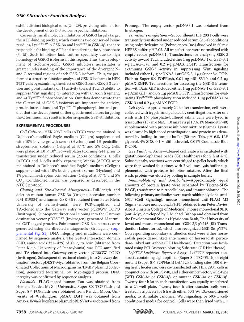
exhibit distinct biological roles (24–29), providing rationale forthe development of GSK-3 isoform-specific inhibitors.Currently, small molecule inhibitors of GSK-3 largely target
the ATP-binding pocket, which contains two conserved lysineresidues, Lys148/149 in GSK-3� and Lys85/86 in GSK-3�, that areresponsible for binding ATP and transferring the �-phosphate(3, 15). Such inhibitors lack isoform specificity due to highhomology of GSK-3 isoforms in this region. Thus, the develop-ment of isoform-specific GSK-3 inhibitors necessitates agreater understanding of the importance of the divergent N-and C-terminal regions of each GSK-3 isoform. Thus, we per-formed a structure-function analysis ofGSK-3 isoforms inHEK293T cells by examining the effect ofGSK-3� andGSK-3�dele-tion and point mutants on 1) activity toward Tau, 2) ability tosuppress Wnt signaling, 3) interaction with an Axin fragment,and 4) Tyr279/216 phosphorylation. Our data demonstrate thatthe C termini of GSK-3 isoforms are important for activity,protein interactions, and Tyr279/216 phosphorylation and pre-dict that the development of therapeutic modulators targetingtheC terminusmay result in isoform-specificGSK-3 inhibition.
EXPERIMENTAL PROCEDURES
Cell Culture—HEK 293T cells (ATCC) were maintained inDulbecco’s modified Eagle medium (Cellgro) supplementedwith 10% bovine growth serum (Hyclone) and 1% penicillin-streptomycin solution (Cellgro) at 37 °C and 5% CO2. Cellswere plated at 1.0 � 106 in 6-well plates (Corning) 24 h prior totransfection under reduced serum (2.5%) conditions. L cells(ATCC) and L cells stably expressing Wnt3a (ATCC) weremaintained in Dulbecco’s modified Eagle’s medium (Cellgro)supplemented with 10% bovine growth serum (Hyclone) and1% penicillin-streptomycin solution (Cellgro) at 37 °C and 5%CO2. Conditioned media was prepared as described in theATCC protocol.Cloning and Site-directed Mutagenesis—Full-length and
deletion mutant human GSK-3� (Origene, accession numberNM_019884) and human GSK-3� (obtained from Peter Klein,University of Pennsylvania) were PCR-amplified andTA-cloned into the Gateway entry vector pCR8GW TOPO(Invitrogen). Subsequent directional cloning into the Gatewaydestination vector pDEST27 (Invitrogen) generated N-termi-nal GST-tagged proteins. Point mutant GSK-3 constructs weregenerated using site-directed mutagenesis (Stratagene) (sup-plemental Fig. S1). DNA integrity and mutations were con-firmed by sequence analysis. The GSK-3 interaction domain(GID, amino acids 321–429) of Xenopus Axin (obtained fromPeter Klein, University of Pennsylvania) was PCR-amplifiedand TA-cloned into Gateway entry vector pCR8GW TOPO(Invitrogen). Subsequent directional cloning into Gateway des-tination vector, pDEST-Myc (obtained from the Belgian Coor-dinated Collections of Microorganisms/LMBP plasmid collec-tion), generated N-terminal 6� Myc-tagged protein. DNAintegrity was confirmed by sequence analysis.Plasmids—FLAG-tagged human Tau was obtained from
Hemant Paudel, McGill University. Super 8� TOPFlash andSuper 8� FOPFlash were obtained from Randall Moon, Uni-versity of Washington. pMAX EGFP was obtained fromAmaxa.Renilla luciferase plasmid pRLSV40was obtained from
Promega. The empty vector pcDNA3.1 was obtained fromInvitrogen.Transient Transfections—SubconfluentHEK 293T cells were
transiently transfected under reduced serum (2.5%) conditionsusing polyethylenimine (Polysciences, Inc.) dissolved in 50 mM
HEPES buffer, pH 7.05. All transfections were normalized withempty vector pcDNA3.1. Transfections for analyzing GSK-3activity towardTau included either 1�g pcDNA3.1 orGSK-3, 1�g FLAG-Tau, and 0.2 �g pMAX EGFP. Transfections forexamining GSK-3 activity in suppressing Wnt signalingincluded either 1�g pcDNA3.1 or GSK-3, 1�g Super 8�TOP-Flash or Super 8� FOPFlash, 0.01 �g pRL SV40, and 0.2 �gpMAX EGFP. Transfections for assessing the GSK-3 interac-tionwithAxinGID included either 1�g pcDNA3.1 orGSK-3, 1�g Axin GID, and 0.2 �g pMAX EGFP. Transfections for eval-uating Tyr279/216 phosphorylation included 1 �g pcDNA3.1 orGSK-3 and 0.2 �g pMAX EGFP.Cell Lysis—Approximately 24 h after transfection, cells were
collected with trypsin and pelleted by centrifugation. After onewash with 1� phosphate-buffered saline, cells were lysed inlysis buffer (137mMNaCl, 10mMTris pH7.4, 1%Nonidet P-40)supplemented with protease inhibitor mixture (Sigma). Lysatewas cleared by high speed centrifugation, and proteinwas dena-tured by boiling in sample buffer (50 mM Tris, pH 6.8, 12%glycerol, 4% SDS, 0.1 M dithiothreitol, 0.01% Coomassie BlueR-250).GST PulldownAssay—Cleared cell lysate was incubated with
glutathione-Sepharose beads (GE Healthcare) for 2 h at 4 °C.Subsequently, reactions were centrifuged to pellet beads, whichwere then washed four times with 2 volumes lysis buffer sup-plemented with protease inhibitor mixture. After the finalwash, protein was eluted by boiling in sample buffer.Immunoblotting and Antibodies—Approximately equal
amounts of protein lysate were separated by Tricine-SDS-PAGE, transferred to nitrocellulose, and immunoblotted. Thefollowing primary antibodies were used: rabbit polyclonal anti-GST (Cell Signaling), mouse monoclonal anti-FLAG M2(Sigma),mousemonoclonal PHF1 (obtained fromPeterDavies,Albert Einstein College of Medicine), mouse monoclonal 9E10(anti-Myc, developed by J. Michael Bishop and obtained fromtheDevelopmental StudiesHybridoma Bank, TheUniversity ofIowa) and mouse monoclonal anti-GSK-3� pY216 (BD Trans-duction Laboratories), which also recognized GSK-3� pY279.Corresponding secondary antibodies used were either horse-radish peroxidase-linked anti-mouse or horseradish peroxi-dase-linked anti-rabbit (GE Healthcare). Detection was facili-tated using ECLWestern blotting Substrate (GE Healthcare).Lef/TCF Luciferase Reporter Assay—Lef/TCF reporter con-
structs containing eight optimal (Super 8� TOPFlash) or eightmutant (Super 8� FOPFlash) Lef/TCF binding sites (30) driv-ing firefly luciferase, were co-transfected intoHEK293T cells inconjunctionwith pRL SV40, and either empty vector, wild-type(WT) GSK-3� or GSK-3�, or mutant GSK-3� or GSK-3�.Twenty-four h later, each transfection was equally transferredto a 24-well plate. Twenty-four h after transfer, cells weretreated in triplicate for 6 h with either 50%Wnt3a-conditionedmedia, to stimulate canonical Wnt signaling, or 50% L cell-conditioned media for control. Cells were then lysed with 1�
GSK-3 Structure-Function Analysis
7958 JOURNAL OF BIOLOGICAL CHEMISTRY VOLUME 285 • NUMBER 11 • MARCH 12, 2010
by guest on March 19, 2020
http://ww
w.jbc.org/
Dow
nloaded from
passive lysis buffer and Firefly and Renilla luciferase activitieswere measured using Firefly and Renilla Luciferase Assay kit(Biotium) in a Veritas microplate luminometer (TurnerBiosystems).Densitometry—Chemiluminescencewas quantified bymeas-
uring area density using UltraLum documentation system andsoftware (UltraLum Inc., Claremont, CA).
RESULTS
To investigate the functional significance of divergentregions and regulatory residues ofGSK-3 isoforms,mammalianexpression constructs containing N-terminal deletions, C-ter-minal deletions, or pointmutations of humanGSK-3� (Fig. 1A)and human GSK-3� (Fig. 1B) were generated. Deletion andpoint mutant GSK-3 expression constructs were tagged at theN terminus with GST.Deletion of GSK-3 C Termini Impairs Tau Phosphorylation—
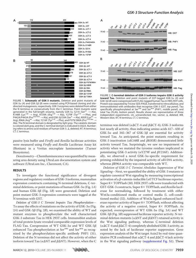
To assay the effects ofmutations on the activity of GSK-3� (Fig.2A) and GSK-3� (Fig. 2B), we examined the ability of WT andmutant enzymes to phosphorylate the well characterizedGSK-3 substrate Tau in HEK 293T cells. Immunoblot analysisof total protein lysate revealed comparable expression levels ofFLAG-Tau. Coexpression of WT GSK-3� and WT GSK-3�enhanced Tau phosphorylation at Ser396 and Ser404 as recog-nized by the phosphorylation-specific antibody PHF1 (31).Deletion of the N terminus did not affect the activity of eitherisoform toward Tau (��NT and ��NT). However, when the C
terminus was deleted (��CT-4 and ��CT-4), GSK-3 isoformslost nearly all activity, thus indicating amino acids 417–430 ofGSK-3� and 345–367 of GSK-3� are essential for activitytoward Tau. As anticipated, the point mutants resulting inGSK-3 inactivation (�K148R and �K85R) demonstrated littleactivity toward Tau. Surprisingly, we saw no impairment ofactivity when we mutated the tyrosine residues implicated inpotentiating GSK-3 activity (�Y279F and �Y216F). Addition-ally, we observed a novel GSK-3�-specific requirement forpriming exhibited by the impaired activity of �R159A activity,whereas �R96A activity was comparable with WT.Deletion of GSK-3 C Termini Abolishes Suppression of Wnt
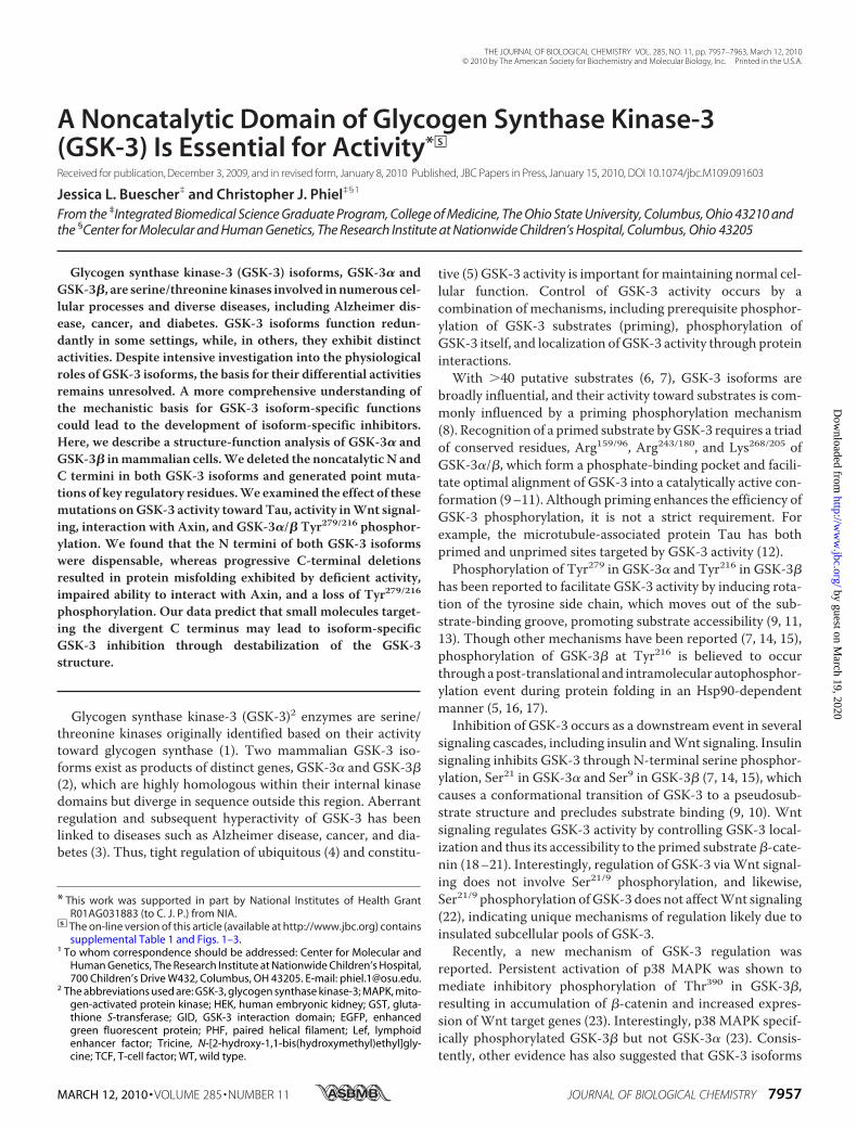
Signaling—Next, we quantified the ability of GSK-3mutants toregulate canonical Wnt signaling by measuring transcriptionalactivation of a�-catenin-inducible Lef/TCF luciferase reporter,Super 8�TOPFlash (30). HEK293T cells were transfectedwithGST-GSK-3 constructs, Super 8�TOPFlash, and Renilla lucif-erase for normalizing, followed by treatment with eitherWnt3a-conditioned media or control media (L cell-condi-tioned media) (32). Addition of Wnt3a ligand-enhanced lucif-erase reporter activity of Super 8�TOPFlash, without affectingthe activity of a negative control, Super 8� FOPFlash. Asexpected, overexpression of WT GSK-3� (Fig. 3A) and WTGSK-3� (Fig. 3B) suppressed luciferase reporter activity. N-ter-minal deletion mutants (��NT and ��NT) retained activity inthe Wnt signaling pathway, whereas C-terminal deletion(��CT-4 and��CT-4) completely abolishedGSK-3 activity, asnoted by the lack of luciferase reporter suppression. Geneexpression analysis of theWnt target Axin2 by real-time quan-titative PCR confirmed that ��CT-4 and ��CT-4 lack activityin the Wnt signaling pathway (supplemental Fig. S1). These
FIGURE 1. Schematic of GSK-3 mutants. Deletion and point mutants ofGSK-3� (A) and GSK-3� (B) were created using PCR-based cloning and site-directed mutagenesis, respectively. GSK-3 enzymes were deleted from eitherthe N terminus or consecutively from the C terminus. Point mutants weregenerated in the full-length enzyme, namely GSK-3� S21A (Ser21 3 Ala),K148R (Lys148 3 Arg), R159A (Arg159 3 Ala), Y279F (Tyr279 3 Phe), andP442A/P443A (Pro442/4433Ala), and GSK-3� S9A (Ser93Ala), K85R (Lys853Arg), R96A (Arg963Ala), Y216F (Tyr2163 Phe), and P379/380A (Pro379/3803Ala). The N-terminal domain is designated by light gray. The catalytic domainis colored dark gray, and the C-terminal domain is indicated in black. Number-ing refers to amino acid residues of human GSK-3. �, deleted; NT, N terminus;CT, C terminus.
FIGURE 2. C-terminal deletion of GSK-3 isoforms impairs GSK-3 activitytoward Tau. Deletion and point mutants of GST-tagged GSK-3� (A) andGSK-3� (B) were coexpressed with FLAG-tagged human Tau in HEK 293T cells.Protein was separated by Tricine-SDS-PAGE, transferred to nitrocellulose, andimmunoblotted with antibodies that recognize GSK-3 (GST, top panel), Tauspecifically phosphorylated at Ser396 and Ser404 (PHF1, middle panel), andtotal Tau (FLAG, bottom panel). Results shown are representative of threeindependent experiments. Un, untransfected; Vec, vector; �, deleted; WB,Western blot; NT, N terminus; CT, C terminus.
GSK-3 Structure-Function Analysis
MARCH 12, 2010 • VOLUME 285 • NUMBER 11 JOURNAL OF BIOLOGICAL CHEMISTRY 7959
by guest on March 19, 2020
http://ww
w.jbc.org/
Dow
nloaded from
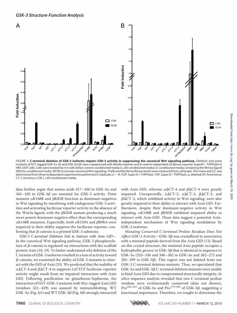
data further argue that amino acids 417–430 in GSK-3� and345–420 in GSK-3� are essential for GSK-3 activity. Pointmutants �K148R and �K85R function as dominant-negativesin Wnt signaling by interfering with endogenous GSK-3 activ-ities and activating luciferase reporter activity in the absence ofthe Wnt3a ligand, with the �K85R mutant producing a muchmore potent dominant-negative effect than the corresponding�K148R mutation. Expectedly, both �R159A and �R96A wereimpaired in their ability suppress the luciferase reporter, con-firming that �-catenin is a primed GSK-3 substrate.GSK-3 C-terminal Deletions Fail to Interact with Axin GID—
In the canonical Wnt signaling pathway, GSK-3 phosphoryla-tion of �-catenin is regulated via interactions with the scaffoldprotein Axin (18, 19). To better understand why deletion of theC termini ofGSK-3 isoforms resulted in a loss of activity toward�-catenin, we examined the ability of GSK-3 mutants to inter-act with the GID of Axin (33).We predicted that the inability of��CT-4 and ��CT-4 to suppress Lef/TCF luciferase reporteractivity might result from an impaired interaction with AxinGID. Following purification on glutathione-Sepharose, theinteraction of GST-GSK-3mutants withMyc-taggedAxinGID(residues 321–429) was assayed by immunoblotting. WTGSK-3� (Fig. 4A) andWTGSK-3� (Fig. 4B) strongly interacted
with Axin GID, whereas ��CT-4 and ��CT-4 were greatlyimpaired. Unexpectedly, ��CT-2, ��CT-3, ��CT-2, and��CT-3, which exhibited activity in Wnt signaling, were alsogreatly impaired in their ability to interact with Axin GID. Fur-thermore, despite their dominant-negative activity in Wntsignaling, �K148R and �K85R exhibited impaired ability tointeract with Axin GID. These data suggest a potential Axin-independent mechanism of Wnt signaling modulation byGSK-3 isoforms.Mutating Conserved C-terminal Proline Residues Does Not
Affect GSK-3 Activity—GSK-3� was crystallized in associationwith a minimal peptide derived from the Axin GID (13). Basedon this crystal structure, the minimal Axin peptide occupies ahydrophobic groove in GSK-3� that is identical in sequence toGSK-3� (325–336 and 348–362 in GSK-3� and 262–273 and285–299 in GSK-3�). This region was not deleted from ourGSK-3 C-terminal deletion mutants. Thus, we speculated thatGSK-3� andGSK-3�C-terminal deletionmutants were unableto bind Axin GID due to compromised structurally integrity. Insilico sequence analysis revealed that two C-terminal prolineresidues were evolutionarily conserved (data not shown),Pro442/443 of GSK-3� and Pro379/380 of GSK-3�, suggesting afunctional importance. Therefore, we sought to determine the
FIGURE 3. C-terminal deletion of GSK-3 isoforms impairs GSK-3 activity in suppressing the canonical Wnt signaling pathway. Deletion and pointmutants of GST-tagged GSK-3� (A) and GSK-3� (B) were coexpressed with Renilla reporter and �-catenin-dependent luciferase reporter Super8� TOPFlash inHEK 293T cells. Cells were treated for 6 h with either control-conditioned media (L cell-conditioned media) or conditioned media containing the Wnt3a ligand(Wnt3a-conditioned media, WCM) to activate canonical Wnt signaling. Firefly and Renilla luciferase levels were measured from cell lysate. The mean and S.E. wasdetermined from three independent experiments performed in triplicate (n � 9). FOP, Super 8� FOPFlash; TOP, Super 8� TOPFlash; �, deleted; NT, N terminus;CT, C terminus; LCM, L cell-conditioned media.
GSK-3 Structure-Function Analysis
7960 JOURNAL OF BIOLOGICAL CHEMISTRY VOLUME 285 • NUMBER 11 • MARCH 12, 2010
by guest on March 19, 2020
http://ww
w.jbc.org/
Dow
nloaded from
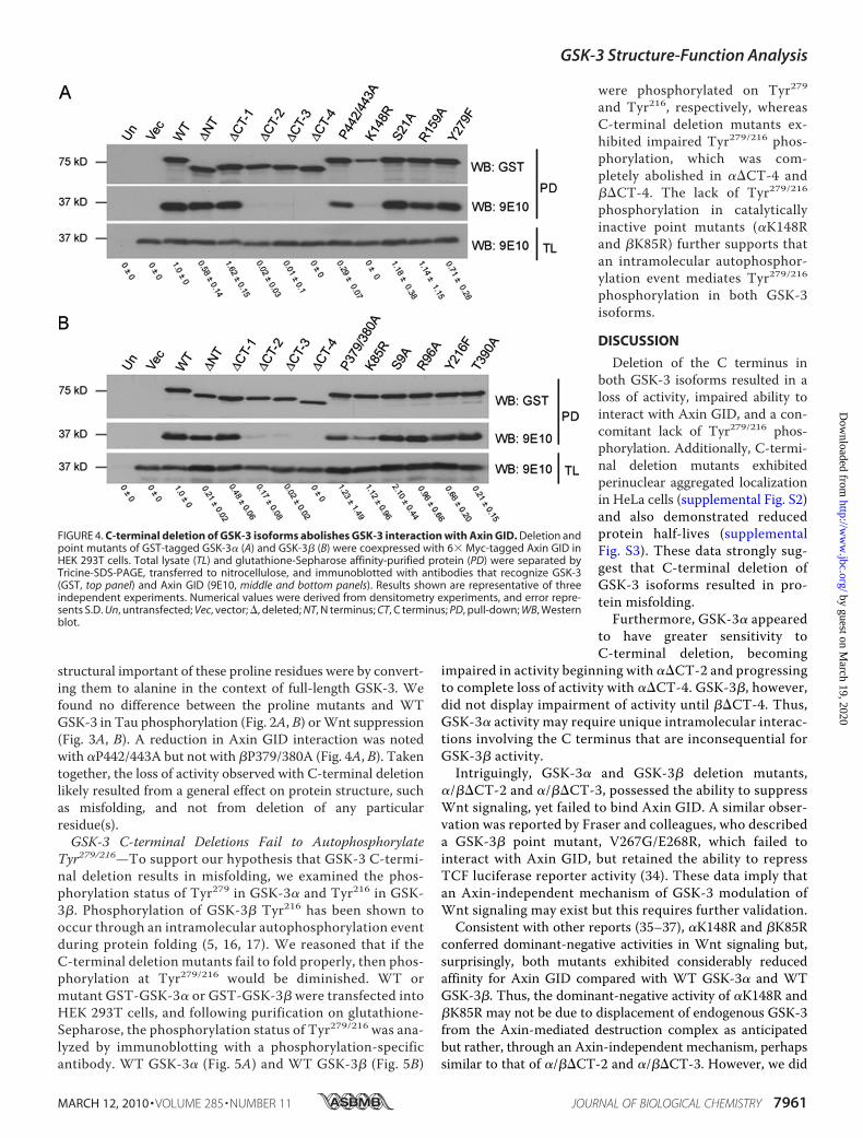
structural important of these proline residues were by convert-ing them to alanine in the context of full-length GSK-3. Wefound no difference between the proline mutants and WTGSK-3 in Tau phosphorylation (Fig. 2A, B) orWnt suppression(Fig. 3A, B). A reduction in Axin GID interaction was notedwith �P442/443A but not with �P379/380A (Fig. 4A, B). Takentogether, the loss of activity observed with C-terminal deletionlikely resulted from a general effect on protein structure, suchas misfolding, and not from deletion of any particularresidue(s).GSK-3 C-terminal Deletions Fail to Autophosphorylate
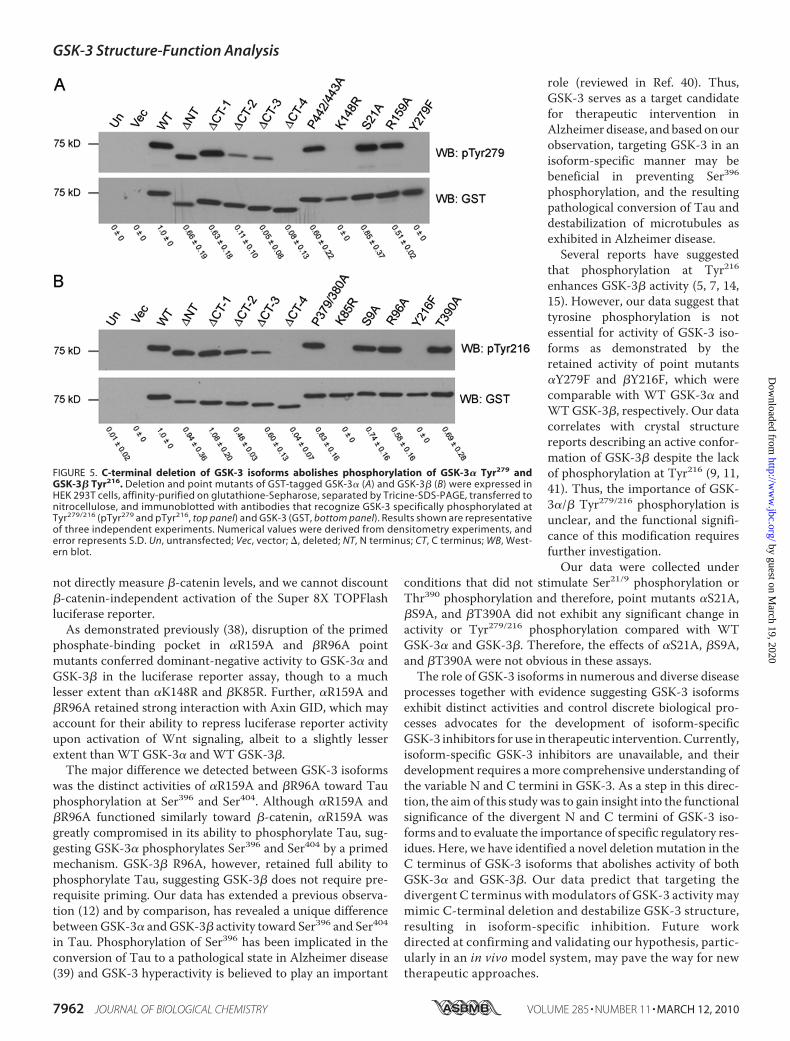
Tyr279/216—To support our hypothesis that GSK-3 C-termi-nal deletion results in misfolding, we examined the phos-phorylation status of Tyr279 in GSK-3� and Tyr216 in GSK-3�. Phosphorylation of GSK-3� Tyr216 has been shown tooccur through an intramolecular autophosphorylation eventduring protein folding (5, 16, 17). We reasoned that if theC-terminal deletion mutants fail to fold properly, then phos-phorylation at Tyr279/216 would be diminished. WT ormutant GST-GSK-3� or GST-GSK-3� were transfected intoHEK 293T cells, and following purification on glutathione-Sepharose, the phosphorylation status of Tyr279/216 was ana-lyzed by immunoblotting with a phosphorylation-specificantibody. WT GSK-3� (Fig. 5A) and WT GSK-3� (Fig. 5B)
were phosphorylated on Tyr279and Tyr216, respectively, whereasC-terminal deletion mutants ex-hibited impaired Tyr279/216 phos-phorylation, which was com-pletely abolished in ��CT-4 and��CT-4. The lack of Tyr279/216phosphorylation in catalyticallyinactive point mutants (�K148Rand �K85R) further supports thatan intramolecular autophosphor-ylation event mediates Tyr279/216phosphorylation in both GSK-3isoforms.
DISCUSSION
Deletion of the C terminus inboth GSK-3 isoforms resulted in aloss of activity, impaired ability tointeract with Axin GID, and a con-comitant lack of Tyr279/216 phos-phorylation. Additionally, C-termi-nal deletion mutants exhibitedperinuclear aggregated localizationin HeLa cells (supplemental Fig. S2)and also demonstrated reducedprotein half-lives (supplementalFig. S3). These data strongly sug-gest that C-terminal deletion ofGSK-3 isoforms resulted in pro-tein misfolding.Furthermore, GSK-3� appeared
to have greater sensitivity toC-terminal deletion, becoming
impaired in activity beginning with ��CT-2 and progressingto complete loss of activity with ��CT-4. GSK-3�, however,did not display impairment of activity until ��CT-4. Thus,GSK-3� activity may require unique intramolecular interac-tions involving the C terminus that are inconsequential forGSK-3� activity.
Intriguingly, GSK-3� and GSK-3� deletion mutants,�/��CT-2 and �/��CT-3, possessed the ability to suppressWnt signaling, yet failed to bind Axin GID. A similar obser-vation was reported by Fraser and colleagues, who describeda GSK-3� point mutant, V267G/E268R, which failed tointeract with Axin GID, but retained the ability to repressTCF luciferase reporter activity (34). These data imply thatan Axin-independent mechanism of GSK-3 modulation ofWnt signaling may exist but this requires further validation.Consistent with other reports (35–37), �K148R and �K85R
conferred dominant-negative activities in Wnt signaling but,surprisingly, both mutants exhibited considerably reducedaffinity for Axin GID compared with WT GSK-3� and WTGSK-3�. Thus, the dominant-negative activity of �K148R and�K85R may not be due to displacement of endogenous GSK-3from the Axin-mediated destruction complex as anticipatedbut rather, through an Axin-independent mechanism, perhapssimilar to that of �/��CT-2 and �/��CT-3. However, we did
FIGURE 4. C-terminal deletion of GSK-3 isoforms abolishes GSK-3 interaction with Axin GID. Deletion andpoint mutants of GST-tagged GSK-3� (A) and GSK-3� (B) were coexpressed with 6� Myc-tagged Axin GID inHEK 293T cells. Total lysate (TL) and glutathione-Sepharose affinity-purified protein (PD) were separated byTricine-SDS-PAGE, transferred to nitrocellulose, and immunoblotted with antibodies that recognize GSK-3(GST, top panel) and Axin GID (9E10, middle and bottom panels). Results shown are representative of threeindependent experiments. Numerical values were derived from densitometry experiments, and error repre-sents S.D. Un, untransfected; Vec, vector; �, deleted; NT, N terminus; CT, C terminus; PD, pull-down; WB, Westernblot.
GSK-3 Structure-Function Analysis
MARCH 12, 2010 • VOLUME 285 • NUMBER 11 JOURNAL OF BIOLOGICAL CHEMISTRY 7961
by guest on March 19, 2020
http://ww
w.jbc.org/
Dow
nloaded from
not directly measure �-catenin levels, and we cannot discount�-catenin-independent activation of the Super 8X TOPFlashluciferase reporter.As demonstrated previously (38), disruption of the primed
phosphate-binding pocket in �R159A and �R96A pointmutants conferred dominant-negative activity to GSK-3� andGSK-3� in the luciferase reporter assay, though to a muchlesser extent than �K148R and �K85R. Further, �R159A and�R96A retained strong interaction with Axin GID, which mayaccount for their ability to repress luciferase reporter activityupon activation of Wnt signaling, albeit to a slightly lesserextent than WT GSK-3� and WT GSK-3�.The major difference we detected between GSK-3 isoforms
was the distinct activities of �R159A and �R96A toward Tauphosphorylation at Ser396 and Ser404. Although �R159A and�R96A functioned similarly toward �-catenin, �R159A wasgreatly compromised in its ability to phosphorylate Tau, sug-gesting GSK-3� phosphorylates Ser396 and Ser404 by a primedmechanism. GSK-3� R96A, however, retained full ability tophosphorylate Tau, suggesting GSK-3� does not require pre-requisite priming. Our data has extended a previous observa-tion (12) and by comparison, has revealed a unique differencebetweenGSK-3� andGSK-3� activity toward Ser396 and Ser404in Tau. Phosphorylation of Ser396 has been implicated in theconversion of Tau to a pathological state in Alzheimer disease(39) and GSK-3 hyperactivity is believed to play an important
role (reviewed in Ref. 40). Thus,GSK-3 serves as a target candidatefor therapeutic intervention inAlzheimer disease, and based onourobservation, targeting GSK-3 in anisoform-specific manner may bebeneficial in preventing Ser396phosphorylation, and the resultingpathological conversion of Tau anddestabilization of microtubules asexhibited in Alzheimer disease.Several reports have suggested
that phosphorylation at Tyr216enhances GSK-3� activity (5, 7, 14,15). However, our data suggest thattyrosine phosphorylation is notessential for activity of GSK-3 iso-forms as demonstrated by theretained activity of point mutants�Y279F and �Y216F, which werecomparable with WT GSK-3� andWTGSK-3�, respectively. Our datacorrelates with crystal structurereports describing an active confor-mation of GSK-3� despite the lackof phosphorylation at Tyr216 (9, 11,41). Thus, the importance of GSK-3�/� Tyr279/216 phosphorylation isunclear, and the functional signifi-cance of this modification requiresfurther investigation.Our data were collected under
conditions that did not stimulate Ser21/9 phosphorylation orThr390 phosphorylation and therefore, point mutants �S21A,�S9A, and �T390A did not exhibit any significant change inactivity or Tyr279/216 phosphorylation compared with WTGSK-3� and GSK-3�. Therefore, the effects of �S21A, �S9A,and �T390A were not obvious in these assays.The role of GSK-3 isoforms in numerous and diverse disease
processes together with evidence suggesting GSK-3 isoformsexhibit distinct activities and control discrete biological pro-cesses advocates for the development of isoform-specificGSK-3 inhibitors for use in therapeutic intervention. Currently,isoform-specific GSK-3 inhibitors are unavailable, and theirdevelopment requires a more comprehensive understanding ofthe variable N and C termini in GSK-3. As a step in this direc-tion, the aim of this study was to gain insight into the functionalsignificance of the divergent N and C termini of GSK-3 iso-forms and to evaluate the importance of specific regulatory res-idues. Here, we have identified a novel deletionmutation in theC terminus of GSK-3 isoforms that abolishes activity of bothGSK-3� and GSK-3�. Our data predict that targeting thedivergent C terminus with modulators of GSK-3 activity maymimic C-terminal deletion and destabilize GSK-3 structure,resulting in isoform-specific inhibition. Future workdirected at confirming and validating our hypothesis, partic-ularly in an in vivo model system, may pave the way for newtherapeutic approaches.
FIGURE 5. C-terminal deletion of GSK-3 isoforms abolishes phosphorylation of GSK-3� Tyr279 andGSK-3� Tyr216. Deletion and point mutants of GST-tagged GSK-3� (A) and GSK-3� (B) were expressed inHEK 293T cells, affinity-purified on glutathione-Sepharose, separated by Tricine-SDS-PAGE, transferred tonitrocellulose, and immunoblotted with antibodies that recognize GSK-3 specifically phosphorylated atTyr279/216 (pTyr279 and pTyr216, top panel) and GSK-3 (GST, bottom panel). Results shown are representativeof three independent experiments. Numerical values were derived from densitometry experiments, anderror represents S.D. Un, untransfected; Vec, vector; �, deleted; NT, N terminus; CT, C terminus; WB, West-ern blot.
GSK-3 Structure-Function Analysis
7962 JOURNAL OF BIOLOGICAL CHEMISTRY VOLUME 285 • NUMBER 11 • MARCH 12, 2010
by guest on March 19, 2020
http://ww
w.jbc.org/
Dow
nloaded from
Acknowledgments—We thank Drs. Jing Yang, Yusen Liu, ScottHarper, and Jeffrey Kuret for helpful suggestions and discussions andDrs. Joanna Groden and Kirk McHugh for critical reading of themanuscript. We are also grateful to Dr. Peter Klein for the GSK-3�andAxin constructs, Dr.Hemant Paudel for the FLAG-Tau construct,Dr. Randall Moon for the Super 8� TOPFlash and Super 8� FOP-Flash constructs, Dr. Nathan Lawson for the pCS Cherry plasmid,Dr. Peter Davies for the PHF1 antibody, and Drs. Brad Doble andJames Woodgett for GSK-3��/� ��/� embryonic stem cells.
REFERENCES1. Embi, N., Rylatt, D. B., andCohen, P. (1980) Eur. J. Biochem. 107, 519–5272. Woodgett, J. R. (1990) EMBO J. 9, 2431–24383. Meijer, L., Flajolet, M., and Greengard, P. (2004) Trends Pharmacol. Sci.
25, 471–4804. Lau, K. F.,Miller, C. C., Anderton, B. H., and Shaw, P. C. (1999) J. Pept. Res.
54, 85–915. Hughes, K., Nikolakaki, E., Plyte, S. E., Totty, N. F., and Woodgett, J. R.
(1993) EMBO J. 12, 803–8086. Jope, R. S., and Johnson, G. V. (2004) Trends Biochem. Sci. 29, 95–1027. Grimes, C. A., and Jope, R. S. (2001) Prog. Neurobiol. 65, 391–4268. Fiol, C. J., Wang, A., Roeske, R. W., and Roach, P. J. (1990) J. Biol. Chem.
265, 6061–60659. Dajani, R., Fraser, E., Roe, S.M., Young,N., Good, V., Dale, T. C., and Pearl,
L. H. (2001) Cell 105, 721–73210. Frame, S., Cohen, P., and Biondi, R. M. (2001)Mol. Cell 7, 1321–132711. ter Haar, E., Coll, J. T., Austen, D. A., Hsiao, H. M., Swenson, L., and Jain,
J. (2001) Nat. Struct. Biol. 8, 593–59612. Cho, J. H., and Johnson, G. V. (2003) J. Biol. Chem. 278, 187–19313. Dajani, R., Fraser, E., Roe, S. M., Yeo, M., Good, V. M., Thompson, V.,
Dale, T. C., and Pearl, L. H. (2003) EMBO J. 22, 494–50114. Frame, S., and Cohen, P. (2001) Biochem. J. 359, 1–1615. Doble, B. W., and Woodgett, J. R. (2003) J. Cell Sci. 116, 1175–118616. Cole, A., Frame, S., and Cohen, P. (2004) Biochem. J. 377, 249–25517. Lochhead, P. A., Kinstrie, R., Sibbet, G., Rawjee, T., Morrice, N., and Cleg-
hon, V. (2006)Mol. Cell 24, 627–63318. Logan, C. Y., and Nusse, R. (2004) Annu. Rev. Cell Dev. Biol. 20, 781–81019. Clevers, H. (2006) Cell 127, 469–48020. MacDonald, B. T., Yokota, C., Tamai, K., Zeng, X., andHe,X. (2008) J. Biol.
Chem. 283, 16115–16123
21. Wu, G., Huang, H., Garcia Abreu, J., andHe, X. (2009) PLoSONE 4, e492622. Ding, V. W., Chen, R. H., and McCormick, F. (2000) J. Biol. Chem. 275,
32475–3248123. Thornton, T.M., Pedraza-Alva, G., Deng, B.,Wood, C. D., Aronshtam, A.,
Clements, J. L., Sabio, G., Davis, R. J., Matthews, D. E., Doble, B., andRincon, M. (2008) Science 320, 667–670
24. Goode, N., Hughes, K., Woodgett, J. R., and Parker, P. J. (1992) J. Biol.Chem. 267, 16878–16882
25. Hoeflich, K. P., Luo, J., Rubie, E. A., Tsao,M. S., Jin, O., andWoodgett, J. R.(2000) Nature 406, 86–90
26. Phiel, C. J., Wilson, C. A., Lee, V. M., and Klein, P. S. (2003) Nature 423,435–439
27. Liang, M. H., and Chuang, D. M. (2006) J. Biol. Chem. 281, 30479–3048428. Koivisto, L., Jiang, G., Hakkinen, L., Chan, B., and Larjava, H. (2006) Exp.
Cell Res. 312, 2791–280529. MacAulay, K., Doble, B.W., Patel, S., Hansotia, T., Sinclair, E.M., Drucker,
D. J., Nagy, A., and Woodgett, J. R. (2007) Cell Metab. 6, 329–33730. Veeman, M. T., Slusarski, D. C., Kaykas, A., Louie, S. H., and Moon, R. T.
(2003) Curr. Biol. 13, 680–68531. Otvos, L., Jr., Feiner, L., Lang, E., Szendrei, G. I., Goedert, M., and Lee,
V. M. (1994) J. Neurosci. Res. 39, 669–67332. Willert, K., Brown, J. D., Danenberg, E., Duncan, A. W., Weissman, I. L.,
Reya, T., Yates, J. R., 3rd, and Nusse, R. (2003) Nature 423, 448–45233. Hedgepeth, C.M., Deardorff,M.A., Rankin, K., andKlein, P. S. (1999)Mol.
Cell Biol. 19, 7147–715734. Fraser, E., Young, N., Dajani, R., Franca-Koh, J., Ryves, J., Williams, R. S.,
Yeo, M., Webster, M. T., Richardson, C., Smalley, M. J., Pearl, L. H., Har-wood, A., and Dale, T. C. (2002) J. Biol. Chem. 277, 2176–2185
35. He, X., Saint-Jeannet, J. P., Woodgett, J. R., Varmus, H. E., and Dawid, I. B.(1995) Nature 374, 617–622
36. Dominguez, I., Itoh, K., and Sokol, S. Y. (1995) Proc. Natl. Acad. Sci. U.S.A.92, 8498–8502
37. Pierce, S. B., and Kimelman, D. (1995) Development 121, 755–76538. Hagen, T., Di Daniel, E., Culbert, A. A., and Reith, A. D. (2002) J. Biol.
Chem. 277, 23330–2333539. Bramblett, G. T., Goedert, M., Jakes, R., Merrick, S. E., Trojanowski, J. Q.,
and Lee, V. M. (1993) Neuron 10, 1089–109940. Avila, J. (2006) FEBS Lett. 580, 2922–292741. Aoki, M., Yokota, T., Sugiura, I., Sasaki, C., Hasegawa, T., Okumura, C.,
Ishiguro, K., Kohno, T., Sugio, S., and Matsuzaki, T. (2004) Acta Crystal-logr. D Biol. Crystallogr. 60, 439–446
42. Deleted in proof
GSK-3 Structure-Function Analysis
MARCH 12, 2010 • VOLUME 285 • NUMBER 11 JOURNAL OF BIOLOGICAL CHEMISTRY 7963
by guest on March 19, 2020
http://ww
w.jbc.org/
Dow
nloaded from
Jessica L. Buescher and Christopher J. PhielActivity
A Noncatalytic Domain of Glycogen Synthase Kinase-3 (GSK-3) Is Essential for
doi: 10.1074/jbc.M109.091603 originally published online January 15, 20102010, 285:7957-7963.J. Biol. Chem.
10.1074/jbc.M109.091603Access the most updated version of this article at doi:
Alerts:
When a correction for this article is posted•
When this article is cited•
to choose from all of JBC's e-mail alertsClick here
Supplemental material:
http://www.jbc.org/content/suppl/2010/01/15/M109.091603.DC1
http://www.jbc.org/content/285/11/7957.full.html#ref-list-1
This article cites 41 references, 14 of which can be accessed free at
by guest on March 19, 2020
http://ww
w.jbc.org/
Dow
nloaded from