ANNALS OF SAUDI MEDICINE - kau.edu.sa · PDF fileANNALS OF SAUDI MEDICINE VOLUME 31 NO. 1 ......
8
ANNALS OF SAUDI MEDICINE VOLUME 31 NO. 1 JANUARY-FEBRUARY 2011 PAGES 1-110 Published by the King Faisal Specialist Hospital and Research Centre riyadh, Saudi Arabia Editorial 1 Obstructive Sleep Apnea: From Simple Upper Airway Obstruction to Systemic Inflammation Ahmad Bahammam Review 3 18 F-FDG PET/CT Imaging In Oncology Ahmad Almuhaideb, Nikolaos Papathanasiou, Jamshed Bomanj Original Articles 14 Consequences of hypoxia-reoxygenation phenomena in patients with obstructive sleep apnea syndrome Tansu Ulukavak Ciftci, Oguz Kokturk, Senay Demirtas, Özlem Gülbahar, Neslihan Bukan 19 Prevalence of diabetes mellitus in a Saudi community Khalid A. Alqurashi, Khalid S. Aljabri, Samia A. Bokhari 24 Outcome of a newborn hearing screening program in a tertiary hospital in Malaysia: the first five years Amirozi Ahmad, Irfan Mohamad, Suzana Mansor, Mohd Khairi Daud, Dinsuhaimi Sidek 29 OX40/OX40L in systemic lupus erythematosus: Association with disease activity and lupus nephritis Mohamed N. Farres, Dina S. Al-Zifzaf, Alaa A. Aly, Nermine M. Abd Raboh 35 Prevalence of the Pro12Ala missense mutation in the PPARG2 gene in Kuwaiti patients with primary knee osteoarthritis Khaled F. Al-Jarallah, Diaa K. Shehab, Mohammad Z. Haider 40 -174G>C interleukin-6 gene polymorphism in Tunisian patients with coronary artery disease Lakhdar Ghazouani, Nesrine Abboud, Sonia Ben Hadj Khalifa, Faouzi Added, Ali Ben Khalfallah, Brahim Nsiri, Mounira Mediouni, Touhami Mahjoub 45 Detection of nucleophosmin and FMS-like tyrosine kinase-3 gene mutations in acute myeloid leukemia Vahid Pazhakh, Farhad Zaker, Kamran Alimoghaddam, Farzaneh Atashrazm 51 Celiac disease in children and adolescents at a singe center in Saudi Arabia Omar I. Saadah 58 How do medical students in their clinical years perceive basic sciences courses at King Saud University? Awatif Alam Reviews 62 A Reappraisal of Saphenous Vein Grafting Shi-Min Yuan, Hua Jing 72 Getting Your Paper Published: An Editor’s Perspective Peter A. Hall Brief Report 77 A cytogenetic study of couples with repeated spontaneous abortions Shirin Niroumanesh, Parvin Mehdipour, Ali Farajpour, Soodabeh Darvish
Transcript of ANNALS OF SAUDI MEDICINE - kau.edu.sa · PDF fileANNALS OF SAUDI MEDICINE VOLUME 31 NO. 1 ......

AN
NA
LS OF SAU
DI M
EDIC
INE
VO
LUM
E 31 NO
. 1 JAN
UA
RY-FEBRUA
RY 2011 PAG
ES 1-110
Published by the King Faisal Specialist Hospital and Research Centreriyadh, Saudi Arabia
Editorial1 Obstructive Sleep Apnea: From Simple Upper AirwayObstructiontoSystemicInflammation
Ahmad Bahammam
Review3 18F-FDG PET/CT Imaging In Oncology
Ahmad Almuhaideb, Nikolaos Papathanasiou, Jamshed Bomanj
Original Articles14 Consequences of hypoxia-reoxygenation phenomena in patients with obstructive sleep apnea syndrome
Tansu Ulukavak Ciftci, Oguz Kokturk, Senay Demirtas, Özlem Gülbahar, Neslihan Bukan
19 Prevalence of diabetes mellitus in a Saudi community
Khalid A. Alqurashi, Khalid S. Aljabri, Samia A. Bokhari
24 Outcome of a newborn hearing screening programinatertiaryhospitalinMalaysia:thefirst fiveyears
Amirozi Ahmad, Irfan Mohamad, Suzana Mansor, Mohd Khairi Daud, Dinsuhaimi Sidek
29 OX40/OX40L in systemic lupus erythematosus: Association with disease activity and lupus nephritis
Mohamed N. Farres, Dina S. Al-Zifzaf, Alaa A. Aly, Nermine M. Abd Raboh
35 Prevalence of the Pro12Ala missense mutation in the PPARG2 gene in Kuwaiti patients with primary knee osteoarthritis
Khaled F. Al-Jarallah, Diaa K. Shehab, Mohammad Z. Haider
40 -174G>C interleukin-6 gene polymorphism in Tunisian patients with coronary artery disease
Lakhdar Ghazouani, Nesrine Abboud, Sonia Ben Hadj Khalifa, Faouzi Added, Ali Ben Khalfallah, Brahim Nsiri, Mounira Mediouni, Touhami Mahjoub
45 Detection of nucleophosmin and FMS-like tyrosine kinase-3 gene mutations in acute myeloid leukemia
Vahid Pazhakh, Farhad Zaker, Kamran Alimoghaddam, Farzaneh Atashrazm
51 Celiac disease in children and adolescents at a singe center in Saudi Arabia
Omar I. Saadah
58 How do medical students in their clinical years perceive basic sciences courses at King Saud University?
Awatif Alam
Reviews62 A Reappraisal of Saphenous Vein Grafting
Shi-Min Yuan, Hua Jing
72 Getting Your Paper Published: An Editor’s Perspective
Peter A. Hall
Brief Report77 A cytogenetic study of couples with repeated spontaneous abortions
Shirin Niroumanesh, Parvin Mehdipour, Ali Farajpour, Soodabeh Darvish

original article
51Ann Saudi Med 31(1) January-February 2011 www.saudiannals.net
Celiac disease (CD) is an immune-mediated en-teropathy, caused by a permanent sensitivity to ingested gluten in genetically susceptible indi-
viduals. The disorder is common, occurring in 0.5% to 1% of the general population in most European coun-tries.1 In the past, CD was thought to exclusively affect people of European origin. New, simple, very sensitive and specific serological tests have now become available, and these have shown that CD is common, not only in Europe, but also in developing countries where the ma-jor staple diet is wheat.2 In developing countries, both serological screening in the general population and se-rological testing in groups at risk are necessary for early identification of CD patients. Reports of a high preva-lence of CD in Egypt3 and Tunisia4 indicate that the disease is also common in the Arab population.
There are no reported national epidemiological
Celiac disease in children and adolescents at a singe center in Saudi ArabiaOmar I. Saadah
From the Department of Pediatrics, Faculty of Medicine, King Abdulaziz University Hospital, King Abdulaziz University, Jeddah, Saudi Arabia
Correspondence: Dr. Omar I. Saadah · Assistant Professor of Pediatrics and Pediatric Gastroenterology, Department of Pediatrics, Faculty of Medicine, King Abdulaziz University Hospital, PO Box 80215 Jeddah 21589 · Saudi Arabia. [email protected] · Accepted: June 2010
Ann Saudi Med 2011; 31(1): 51-57
PMID: **** DOI: 10.4103/0256-4947.75779
BACKGROUND AND OBJECTIVES: Celiac disease (CD) is an immune-mediated enteropathy, induced by glu-ten in genetically susceptible individuals. The objective of this study was to describe the clinical pattern of CD in children from the western region of Saudi Arabia.DESIGN AND SETTING: Retrospective, hospital-based.PATIENTS AND METHODS: This study included children with a biopsy-proven diagnosis of CD made between September 2002 and July 2007. Children were admitted to the endoscopy unit for a small-bowel biopsy if they had gastrointestinal symptoms suggestive of CD or if they were positive for a CD-antibody screen performed for the high-risk groups.RESULTS: Eighty children were identified with a diagnosis of CD. Their mean (SD) age was 9.6 (4.9) years (range, 0.5-18 years). There were 44 (55%) female patients. Forty-one (51%) patients were detected during screening of high-risk groups, while 39 (49%) patients had classical symptoms of malabsorption. The screening also de-tected asymptomatic patients. Of 65 patients tested, 11 (17%) had elevated liver function tests, which reverted to normal after introduction of a gluten-free diet (GFD) except in one case. Seventy-three (91%) patients were positive for anti-tissue transglutaminase antibodies, 18 (23%), for IgG anti-gliadin antibodies; and 46 (58%), for IgA anti-gliadin antibodies. Forty-one (56%) patients showed good adherence to GFD as assessed by dietary history and the decline in anti-tTG level. CONCLUSION: CD may present with classical symptoms or be identified through screening programs. Growth and laboratory abnormalities usually improve after introduction of a GFD. Adherence to a GFD remains a problem; therefore, thorough assessment and counseling at the time of diagnosis and ongoing care are crucial.
studies of mass screening for CD in children in Saudi Arabia. However, Al Attas5 has reported a seropreva-lence for CD of 7.6% in a reference laboratory setting among the 145 patients with clinically suspected dis-ease and 2.5% among 18 patients with various autoim-mune diseases; none of her patients with inflammatory bowel disease or healthy blood donors were seroposi-tive for CD.
Implementation of a gluten-free diet (GFD) poses a challenging public health problem in developing coun-tries such as Saudi Arabia, since commercial gluten-free products are not widely available. The diagnosis can be obtained through demonstration of the characteris-tic histological changes (including villous atrophy) on small intestinal biopsy and the resolution of the mu-cosal lesions and symptoms upon withdrawal of glu-ten-containing foods.5 CD may present with classical
Avinash K
Rectangle
original article CELIAC DISEASE IN SAUDI ARABIA
52 Ann Saudi Med 31(1) January-February 2011 www.saudiannals.net
symptoms of malabsorption, such as chronic diarrhea, abdominal distension and growth failure, or it can be identified through screening of high-risk groups.6,7
The aim of this retrospective study was to describe the clinical picture, anthropometric changes and labo-ratory abnormalities of a group of children diagnosed with CD and to discuss the challenges faced in manage-ment, namely, adherence to GFD and the availability of commercial GFD products.
METHODSWe identified retrospectively all patients who had been diagnosed with CD at King Abdulaziz University Hospital, Jeddah, Saudi Arabia, in the period between September 2002 and July 2007. Children were admitted to the endoscopy unit for a small-bowel biopsy if they had gastrointestinal symptoms suggestive of CD or if they were positive for a CD-antibody screen performed for the high-risk groups. Small bowel biopsy specimens were obtained by upper gastrointestinal endoscopy performed by the author. Two to four specimens from the distal duodenum were sent for histopathology. The diagnosis of CD was based on compatible serologic tests, small bowel biopsy and response to a GFD. At the time of diagnosis, all patients received education about a GFD. Patients attended the gastroenterology clinic every 4 months for follow-up. Serial measurements of weight, height, triceps skin fold thickness and mid-arm circumference were obtained immediately before the diagnosis of CD and during the clinic visits in the first 12 months after the introduction of GFD. The z scores for ‘weight for age’ and ‘height for age’ were cal-culated by using an anthropometric software program (EpiInfo, Centers for Disease Control and Prevention, Atlanta, GA, USA). During the follow-up visits, di-etary awareness and adherence to GFD were assessed by taking a detailed dietary history from older children and/or their guardians. Measurements of celiac-specific antibodies (tissue transglutaminase or anti-gliadin an-tibodies) were done at 6 and 12 months after beginning of a GFD. A decrease of more than 50% in the antibody titer, with eventual disappearance in most children, is taken as an indirect indicator of dietary adherence and recovery.8
Statistical analysis was performed using Stata Statistical Software (Stata Corporation, Release 6.0, College Station, TX, USA). Results are expressed as percentage of the total, or as median with interquartile range. Repeated-measure ANOVA on rank was per-formed to test for changes during the follow-up period. Statistical significance was accepted if P value was less than .05.
RESULTSEighty children were identified with a diagnosis of CD. Their mean (SD) age at diagnosis was 9.6 (4.9) years (range, 0.5-18 years). There were 44 (55%) female pa-tients. Forty-one (51%) patients were detected during screening of high-risk individuals, while 39 (49%) had presented with classical symptoms of malabsorption. The characteristics of each group are presented in Table 1. The clinical presentation of the 39 children classified in the classical group included chronic diarrhea in 32 (82%), anorexia in 32 (82%), abdominal distension in 30 (77%), poor weight gain in 30 (77%), and abdominal pain in 12 (31%) patients. One patient presented at the age of 8 years with the rare presentation of celiac crisis, which included severe diarrhea and dehydration associ-ated with metabolic acidosis, hypokalemia, hypocalcae-mia, hypomagnesemia and hypoproteinemia.
Forty-one patients were detected during screening of high-risk groups. Twenty-three (56%) patients were identified during routine screening of children with type 1 diabetes mellitus (DM). The mean age at diag-nosis was 9 (4.2) years (range, 2-18 years), and there were 11 (48%) female patients. Four had occasional ab-dominal pain and 1 patient had diarrhea. Another 14 (35%) patients were identified during screening of chil-dren with isolated short stature; 9 (64%) were females. The mean age at diagnosis was 11.2 (4.5) years (range, 3-16 years), with mean ‘height for age’ z score at diagno-sis of –3.07±1. Only two of them reported occasional nonspecific abdominal pain. Four (10%) additional pa-tients were identified during screening of the families of affected individuals.
In this series, CD was associated with other disor-ders, including autoimmune disorders, as well as other disorders of nonimmune origin (Table 2). Two patients presented with seizure disorders; of these, one was a 16-year-old boy who had a history of repeated seizures for 1 year prior to the diagnosis of CD and was treated with sodium valproate, but without control. His brain CT scan was unremarkable, with no occipital calcifica-tions. The other patient was an 18-year-old girl with history of repeated seizures for 5 years prior to the diagnosis, treated with topiramate with some improve-ment. Her CT scan was unremarkable, but her MRI brain showed a scattered high signal intensity in both cerebral hemispheres interpreted as subcortical edema, post-epilepsy status. Both patients had better seizure control following the diagnosis of CD and institution of GFD.
Anthropometric parametersTwenty-eight (35%) patients had weight below the third

original articleCELIAC DISEASE IN SAUDI ARABIA
53Ann Saudi Med 31(1) January-February 2011 www.saudiannals.net
Table 1. Characteristics of screening-detected and classical-symptoms patients with celiac disease.
Screening-detected CDn=41
Classical CDn=39 P
Age (mean [SD]) in years 9.7 (4.2) 9.4 (5.6)
Male: female ratio 21:20 15:24
Saudis/non-Saudis 22/19 19/20
Nutritional status
Weight for age z score (mean [SD]) −1.44 (1.2) −1.5 (1.9) .9
Height for age z score (mean [SD]) −1.69 (1.3) −1.5 (1.35) .5
BMI z score (mean±SD) −0.69 (1.28) −0.68 (1.3) .9
Skin fold thickness (cm) 4 (1.09) 3.4 (1.1) .15
Mid-arm circumference (cm) 20 (3.7) 20 (4.5) .8
Albumin (g/L) 37.6 (4.4) 32.8 (8.4) .002
Hemoglobin (g/dL) 11.48 (1.7) 10 (1.8) .001
Calcium (mmol/L) 2.2 (0.37) 2.1 (0.37) .7
Phosphate (mmol/L) 1.5 (0.28) 1.47 (0.37) .8
Alkaline phosphatase (IU) 350 (275) 295 (174) .34
BMI: Body mass index
Table 2. Associated comorbidity in 80 patients with celiac disease at diagnosis.
Number Percentage
Autoimmune disorders
Type 1 DM 23 29
Autoimmune thyroiditis 6 7
Autoimmune hepatitis 1 1
Systemic lupus erythematosus
1 1
Vitiligo 1 1
Other disorders
Osteomalacia 3 4
Seizure disorders 2 2
Down syndrome 2 2
IgA deficiency 1 1
percentile at presentation, with the mean ‘weight for age’ z score (SD) of –3 (1.3) (range, –5.8 to –2.2). Thirty (38%) patients had short stature with height below the third percentile and mean ‘height for age’ z score (SD) of –2.9 (0.75) (range, –4.7 to –2.11). Eighteen (23%) patients had body mass index z score less than –2 with the mean (SD) of –2.35 (0.25) (range, –2.7 to –2). Following the introduction of a GFD, serial measure-ments of weight, height and body mass index for the whole group showed significant improvement over a period of 12 months (ANOVA, P<.001) (Figures 1-3). Nutritional status as indicated by serial measurements of subcutaneous fat thickness and triceps mid-arm circum-ference was also found to be improved after the introduc-tion of a GFD for the whole group (ANOVA, P<.001). In subgroup analysis according to adherence, patients with good adherence to the GFD demonstrated signifi-cant difference when compared with the non-adherent group in the respective mean values of weight for age z scores, BMI and skin fold thickness (ANOVA, P<.001) but not in the respective mean values of height for age z scores and triceps mid-arm circumference (ANOVA, P=.58 and ANOVA, P=.84, respectively).
Laboratory parametersAt presentation, albumin level was low (less than 35 g/L) in 28 (35%) patients, with a mean (SD) value of 27.6 (6) g/L (range, 14 to 34 g/L). Fifty-three (66%)
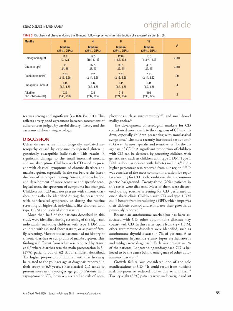
patients were anemic with a hemoglobin less than 12 g/dL with a mean (SD) of 9.8 (1.4) g/dL (range, 4.2 to 11.9 g/dL). Only 3 patients had corrected calcium level less than 2.2 mmol/L, with a mean value of 1.94 mmol/L. Repeated measurements of hemoglobin and albumin levels during the 12-month follow-up period showed significant improvement following the intro-duction of GFD (ANOVA, P<.001); however, repeat-ed measurements of calcium, phosphate and alkaline phosphatase showed no significant changes (ANOVA,

original article CELIAC DISEASE IN SAUDI ARABIA
54 Ann Saudi Med 31(1) January-February 2011 www.saudiannals.net
P=.09, 0.4, 0.2, respectively) (Table 3).Measurements of liver enzymes alanine amino-
transferase (ALT) and aspartate aminotransferase (AST) were performed in 65 patients at diagnosis. Eleven (17%) patients had elevated ALT and AST levels, with a mean (SD) ALT of 115 (95) IU and a mean (SD) AST of 118 (196) IU. Following the in-troduction of a GFD, the levels of both ALT and AST were normalized in 10 patients. One patient failed to respond to GFD and required corticosteroids after be-ing subsequently confirmed as an autoimmune hepati-tis (type 1) patient.
Serological testing for celiac-related antibodiesSerological testing was done in all 80 patients at di-agnosis. Most patients had more than one serologi-cal testing performed. Seventy-three (91%) patients had positive anti-tissue transglutaminase antibodies (anti-tTG) at diagnosis (more than 20 units), with a mean (SD) value of 118 (84) units. IgG anti-gliadin antibodies were positive (more than 20 units) in 18 (23%) patients with a mean (SD) value of 125 (102) units, while IgA anti-gliadin antibodies were positive in 46 (58%) patients with a mean (SD) value of 74 (71) units. Seven (9%) patients were not tested for anti-tTG and tested positive for IgA anti-gliadin an-tibodies.
HistopathologyThe initial small-bowel biopsy in all patients showed severe enteropathy. All except 1 (98%) patient had to-tal or subtotal villous atrophy with either completely flat villi or with clearly atrophic but recognizable villi. Only 1 patient had partial villous atrophy, in which the villi were blunt and shortened. Villous atrophy in all patients was associated with compensatory crypt hyperplasia and increased prominence of intraepithe-lial lymphocytes.
Introduction of GFD and assessment of adherenceGFD was started in all patients at diagnosis. Adherence to GFD was followed using detailed di-etary history and repeated measurements of anti-tTG, which were readily available for follow-up as per hospital policy, to gradually replace anti-gliadin anti-bodies. Adherence was tested only in the 73 patients who were positive for anti-tTG at diagnosis. Forty-one (56%) patients showed significant decline in their initial level of anti-tTG during the first 6 months following introduction of GFD. The correlation be-tween adherence as judged by the detailed dietary history and as assessed by the decline of anti-tTG ti-
Figure 1. Weight changes in 80 patients with celiac disease following the introduction of a gluten-free diet. Dots are oulying values. Floor and ceiling of box are 25th and 75th percentile. Center line is median.
Figure 3. Changes in body mass index following the introduction of a gluten-free diet in 80 children with celiac disease. Dots are oulying values. Floor and ceiling of box are 25th and 75th percentile. Center line is median.
Figure 2. Height changes in 80 patients with celiac disease following the introduction of a gluten-free diet. Dots are oulying values. Floor and ceiling of box are 25th and 75th percentile. Center line is median.
original articleCELIAC DISEASE IN SAUDI ARABIA
55Ann Saudi Med 31(1) January-February 2011 www.saudiannals.net
ter was strong and significant (r= 0.8, P=.001). This reflects a very good agreement between assessment of adherence as judged by careful dietary history and the assessment done using serology.
DISCUSSIONCeliac disease is an immunologically mediated en-teropathy caused by exposure to ingested gluten in genetically susceptible individuals.1 This results in significant damage to the small intestinal mucosa and malabsorption. Children with CD used to pres-ent with classical symptoms of chronic diarrhea and malabsorption, especially in the era before the intro-duction of serological testing. Since the introduction and development of more sensitive and specific sero-logical tests, the spectrum of symptoms has changed. Children with CD may not present with chronic diar-rhea, but rather be identified during the presentation with nonclassical symptoms, or during the routine screening of high-risk individuals, like children with type 1 DM and isolated short stature.
More than half of the patients described in this study were identified during screening of the high-risk individuals, including children with type 1 DM and children with isolated short stature; or as part of fam-ily screening. Most of those patients had no history of chronic diarrhea or symptoms of malabsorption. This finding is different from what was reported by Assiri et al,9 where diarrhea was the main presentation in 34 (57%) patients out of 62 Saudi children described. The higher proportion of children with diarrhea may be related to the younger age at diagnosis reported in their study of 6.5 years, since classical CD tends to present more in the younger age group. Patients with asymptomatic CD, however, are still at risk of com-
Table 3. Biochemical changes during the 12-month follow-up period after introduction of a gluten-free diet (n= 80).
Months 0 4 8 12PMedian
(25%, 75%)Median
(25%, 75%)Median
(25%, 75%)Median
(25%, 75%)
Hemoglobin (g/dL) 11.8(10, 12.8)
12.5(10.75, 13)
12.85(11.6, 13.5)
13.3(11.97, 13.9) <.001
Albumin (g/L) 35(34, 38)
37.5(36, 40)
38.5(37, 41)
40.5(39, 43) <.001
Calcium (mmol/L) 2.23(2.15, 2.29)
2.2(2.14, 2.28)
2.23(2.16, 2.28)
2.19(2.14, 2.22) .09
Phosphate (mmol/L) 1.46(1.3, 1.6)
1.44(1.3, 1.6)
1.45(1.3, 1.6)
1.41(1.2, 1.6) .4
Alkaline phosphatase (IU)
229(140, 298)
233(137, 305)
213(124, 294)
193(133, 275) .2
plications such as autoimmunity10,11 and small-bowel malignancies.12
The development of serological markers for CD contributed enormously to the diagnosis of CD in chil-dren, especially children presenting with nonclassical symptoms.7 The most recently introduced test of anti-tTG was the most specific and sensitive test for the di-agnosis of CD.13 A significant proportion of children with CD can be detected by screening children with genetic risk, such as children with type 1 DM. Type 1 DM has been associated with diabetes mellitus,14 and a higher percentage was reported from our region.15,16 It was considered the most common indication for regu-lar screening for CD. Both conditions share a common genetic background. Twenty-three (29%) patients in this series were diabetics. Most of them were discov-ered during routine screening for CD performed at our diabetic clinic. Children with CD and type 1 DM could benefit from introducing a GFD, which improves their diabetic control and stimulates their growth, as previously reported.17
Because an autoimmune mechanism has been as-sociated with CD, other autoimmune diseases may coexist with CD. In this series, apart from type 1 DM, other autoimmune disorders were identified, such as autoimmune thyroid disease in 7% of patients. Also autoimmune hepatitis, systemic lupus erythematosus and vitiligo were diagnosed. Each was present in 1% of the patients. Longstanding undiagnosed CD is be-lieved to be the cause behind emergence of other auto-immune diseases.11
Growth failure was considered one of the sole manifestations of CD.18 It could result from nutrient malabsorption or reduced intake due to anorexia.19 Twenty-eight (35%) patients were underweight and 30
original article CELIAC DISEASE IN SAUDI ARABIA
56 Ann Saudi Med 31(1) January-February 2011 www.saudiannals.net
(38%) patients had short stature. Significant improve-ment in growth parameters was observed following the introduction of GFD, indicating improvement in intes-tinal absorption due to healing of the damaged intesti-nal mucosa. This growth improvement was associated with improvement in muscle bulk and subcutaneous fat as measured by mid-arm circumference and skin fold thickness, respectively.
Furthermore, 53 (66%) patients were anemic and 28 (35%) patients had low albumin. The presence of ane-mia and low albumin was more pronounced in the clas-sical group than the screening-detected group (Table 1). This indicates that malabsorption of nutrients may be the main underlying cause; however, nutritional de-ficiencies alone may not explain this phenomenon in all cases.20 Recovery is usually possible with GFD alone.21
The loss of albumin through the damaged mucosa may contribute to the low serum albumin observed in such patients. The level of albumin usually improves follow-ing mucosal recovery.
The two main forms of liver damage (cryptogenic and autoimmune) appear to be related to CD.22 The GFD normalizes the cryptogenic form, but most likely not the autoimmune hepatitis.23 Nonalcoholic fatty in-filtration of the liver causing steatohepatitis may be as-sociated with elevated liver enzymes in CD patients.24,25 In this series, 65 patients were tested for abnormal liver enzymes. ALT and AST were elevated in 11 (17%) patients. Ten patients responded to a GFD with nor-malization of both ALT and AST levels. One patient did not respond to GFD alone and was further tested for the possibility of autoimmune hepatitis. He tested strongly positive for both ANA and anti–smooth mus-cle antibodies. This patient responded dramatically to corticosteroid treatment with eventual normalization of his liver enzymes. None of the patients in this study had evidence of fatty infiltration of the liver on ultrasound examination.
The severity of intestinal mucosal damage was grad-ed by Marsh26,27 from I to III. All patients but one re-ported in this series had subtotal and total villous atro-phy, representing the severe form of enteropathy, which corresponds to Marsh IIIB and IIIC, respectively. Only 1 patient had partial villous atrophy corresponding to Marsh IIIA. The severe grades of villous atrophy in the
studied patients may reflect the long duration of gluten exposure prior to the diagnosis and the possible delay in seeking medical care.
The cornerstone of treatment for CD is elimination of gluten from the diet. In most patients diagnosed with CD, a strict GFD alone results in complete resolution of symptoms and histological recovery, with subsequent reduction in the risk of complications. Noncompliance with GFD is the leading cause of failure of response in patients with CD. The reported adherence rate to GFD in children and adolescents in the Western literature was 56% to 81%.28-30 These variations may be due to differ-ent methods used to assess adherence in different stud-ies. In this study, the combination of dietary history and the repeated measures of serological testing (anti-tTG) was used to assess adherence, as previously reported.8,31 Forty-one (56%) patients demonstrated a decline in the antibody level during the first 6 months of follow-up.
The noncompliance rate of 44% in this series was high as a greater proportion of our patients were older children and adolescents. This indicates that the adher-ence rate in this study was comparable to that in similar reports from Europe and North America. The lack of availability of commercial GFD products was an ob-stacle shared by many children in this study. They did not have alternatives to substitute for the food items they liked but had to rely only on exclusion of gluten-containing food. The lack of availability of GFD prod-ucts could adversely influence the rate of adherence and compliance.
In conclusion, a significant proportion of children with CD may present with nonclassical symptoms. Most have severe mucosal damage, reflecting the delay in their presentation and diagnosis. Adherence to GFD remains a problem; therefore, a thorough assessment and counseling at the time of diagnosis and ongoing care are crucial.
AcknowledgmentsThe author thanks Dr. Jameel Al Mughales, Immunology Laboratory, King Abdulaziz University Hospital; Hunida Al Ghamdi, Master’s degree student in Dietetics, King Abdulaziz University; and the Department of Anatomical Pathology at King Abdulaziz University Hospital for their support and collaboration.

original articleCELIAC DISEASE IN SAUDI ARABIA
57Ann Saudi Med 31(1) January-February 2011 www.saudiannals.net
1. Hill ID, Dirks MH, Liptak GS, Colletti RB, Fasano A, Guandalini S, et al. Guideline for the diagnosis and treatment of celiac disease in children: Rec-ommendations of the North American Society for Pediatric Gastroenterology, Hepatology and Nutri-tion. J Pediatr Gastroenterol Nutr 2005;40:1-19.2. Sher KS, Fraser RC, Wicks AC, Mayberry JF. High risk of coeliac disease in Punjabis. Epide-miological study in the South Asian and Euro-pean populations of Leicestershire. Digestion 1993;54:178-82. 3. Abu-Zekry M, Kryszak D, Diab M, Catassi C, Fasano A. Prevalence of celiac disease in Egyp-tian children disputes the east-west agriculture-dependent spread of the disease. J Pediatr Gas-troenterol Nutr 2008;47:136-40.4. Ben Hariz M, Kallel-Sellami M, Kallel L, Lah-mer A, Halioui S, Bouraoui S, et al. Prevalence of celiac disease in Tunisia: Mass-screening study in schoolchildren. Eur J Gastroenterol Hepatol 2007;19:687-94.5. Al Attas RA. How common is celiac dis-ease in Eastern Saudi Arabia? Ann Saudi Med 2002;22:315-9. 6. Fasano A, Araya M, Bhatnagar S, Cameron D, Catassi C, Dirks M, et al. Federation of Inter-national Societies of Pediatric Gastroenterol-ogy, Hepatology, and Nutrition Consensus report on celiac disease. J Pediatr Gastroenterol Nutr 2008;47:214-9.7. Bottaro G, Cataldo F, Rotolo N, Spina M, Corazza GR. The clinical pattern of subclinical/silent celiac disease: An analysis on 1026 consecutive cases. Am J Gastroenterol 1999;94:691-6.8. Sharma A, Poddar U, Yachha SK. Time to rec-ognize atypical celiac disease in Indian children. Indian J Gastroenterol 2007;26:269-73.9. Hansson T, Dahlbom I, Rogberg S, Dannaeus A, Hopfl P, Gut H, et al. Recombinant human tis-sue transglutaminase for diagnosis and follow-up of childhood coeliac disease. Pediatr Res 2002;51:700-5.10. Assiri AM, El Mouzan MI, Al Sanie A, Al Ju-rayyan N, Al Herbish AS, Bakr AA. Pattern of ce-liac disease in infants and children. Trop Gastro-
enterol 2008;29:217-20.11. Not T, Tommasini A, Tonini G, Buratti E, Po-cecco M, Tortul C, et al. Undiagnosed coeliac disease and risk of autoimmune disorders in sub-jects with Type I diabetes mellitus. Diabetologia 2001;44:151-5.12. Ventura A, Magazzù G, Greco L. Duration of exposure to gluten and risk for autoimmune disor-ders in patients with celiac disease. SIGEP Study Group for Autoimmune Disorders in Celiac Dis-ease. Gastroenterology 1999;117:297-303.13. Askling J, Linet M, Gridley G, Halstensen TS, Ekström K, Ekbom A. Cancer incidence in a pop-ulation-based cohort of individuals hospitalized with celiac disease or dermatitis herpetiformis. Gastroenterology 2002;123:1428-35.14. Murray JA, Van Dyke C, Plevak MF, Dierkhising RA, Zinsmeister AR, Melton LJ 3rd. Trends in the identification and clinical features of celiac dis-ease in a North American community, 1950-2001. Clin Gastroenterol Hepatol 2003;1:19-27.15. Collin P, Kaukinen K, Valimaki M, Salmi J. Endo-crinological disorders and celiac disease. Endocr Rev 2002;23:464-83.16. Al-Ashwal AA, Shabib SM, Sakati NA, Attia NA. Prevalence and characteristics of celiac dis-ease in type I diabetes mellitus in Saudi Arabia. Saudi Med J 2003;24:1113-5.17. Saadah OI, Al Agha AE, Albokhari SM, Almo-ghales JA. Prevalence of celiac disease in Saudi children with type 1 diabetes mellitus. J Pediatr Gastroenol Nutr 2004;39:S211.18. Saadah OI, Zacharin M, O’Callaghan A, Oliver MR, Catto-Smith AG. Effect of gluten-free diet and adherence on growth and diabetic control in diabetics with coeliac disease. Arch Dis Child 2004;89:871-6.19. Catassi C, Fasano A. Celiac disease as a cause of growth retardation in childhood. Curr Opin Pedi-atr 2004;16:445-9.20. Fasano A. Clinical presentation of celiac dis-ease in the pediatric population. Gastroenterology 2005;128:S68-73. 21. Harper JW, Holleran SF, Ramakrishnan R, Bhagat G, Green PH. Anemia in celiac dis-
ease is multifactorial in etiology. Am J Hematol 2007;82:996-1000.22. Annibale B, Severi C, Chistolini A, Antonelli G, Lahner E, Marcheggiano A, et al. Efficacy of gluten-free diet alone on recovery from iron defi-ciency anemia in adult celiac patients. Am J Gas-troenterol 2001;96:132-723. Volta U. Liver dysfunction in celiac disease. Minerva Med 2008;99:619-29.24. Di Biase AR, Colecchia A, Scaioli E, Berri R, Viola L, Vestito A, et al. Autoimmune liver diseases in a paediatric population with coeliac disease - a 10-year single-centre experience. Aliment Phar-macol Ther 2010;31:253-60.25. Bardella MT, Valenti L, Pagliari C, Peracchi M, Farè M, Fracanzani AL, et al. Searching for coeliac disease in patients with non-alcoholic fatty liver disease. Dig Liver Dis 2004;36:333-6.26. Grieco A, Miele L, Pignatoro G, Pompili M, Rapaccini GL, Gasbarrini G. Is coeliac disease a confounding factor in the diagnosis of NASH? Gut 2001;49:596.27. Marsh MN. The immunopathology of the small intestinal reaction in gluten sensitivity. Immunol Invest 1989;18:509-31.28. Marsh MN. Gluten, major histocompatibility complex and the small intestine: A molecular and immunobiologic approach to the spectrum of glu-ten sensitivity (“celiac sprue”). Gastroenterology 1992;102:330-54.29. Mayer M, Greco L, Troncone R, Auricchio S, Marsh MN. Compliance of adolescents with coeliac disease with a gluten free diet. Gut 1991;32:881-5.30. Kumar PJ, Walker-Smith J, Milla P, Har-ris G, Colyer J, Halliday R. The teenage coeliac: Follow up study of 102 patients. Arch Dis Child 1988;63:916-20.31. Ljungman G, Myrdal U. Compliance in teen-agers with coeliac disease--a Swedish follow-up study. Acta Paediatrica 1993;82:235-8.32. Hoffenberg EJ, Bao F, Eisenbarth GS, Uhlhorn C, Haas JE, Sokol RJ, et al. Transglutaminase an-tibodies in children with a genetic risk for celiac disease. J Pediatr 2000;137:356-60.
REFERENCES