
Ankylosed Primary Molar and Eruption Guidance of Succeeded ...
9
J Korean Acad Pediatr Dent 44(1) 2017 ISSN (print) 1226-8496 ISSN (online) 2288-3819 99 http://dx.doi.org/10.5933/JKAPD.2017.44.1.99 Ankylosed Primary Molar and Eruption Guidance of Succeeded Permanent Premolar : Case Reports Hayoung Jang, Sohee Oh Department of Pediatric Dentistry, Dental Clinic Center, Hallym University Sacred Heart Hospital In the management of ankylosed primary molars, early diagnosis, proper treatment, and thorough follow-ups are very important. Untreated infraocclusion due to ankylosis has a negative impact on normal occlusal develop- ment, and may cause problems. There are many treatment options on infraoccluded deciduous molars, such as periodic observation, conservative method, restoration, and space regaining via extraction of the teeth. In this case report, two 6-year-old girls were diagnosed with ankylosed maxillary second primary molar and displaced tooth germ of the second premolar. Early surgical removal of the ankylosed primary molar was considered as a treatment approach. The long-term follow-up shows normal eruption of a succeeded permanent premolar. Key words : Ankylosis, Infraocclusion, Tooth germ displacement, Eruption guidance Abstract Ⅰ. Introduction An ankylosed tooth is defined as an anatomical fusion of the tooth cementum with the alveolar bone as a result of some disturbance in their periodontal ligament 1,2) . In this situation, the teeth have failed to undergo a normal process of eruption, and this will result in an under growth around the teeth. Andlaw and Rock 3) defined in- fraocclusion as “a tooth that has failed to maintain its position relative to the adjacent teeth in the developing dentition and is, therefore, submerged below the occlusal level” . Many studies showed that the ankylosis is considered as a typical etiology in infraocclusion. Many authors 4-7) reported that there was a hereditary component of anky- losis based on the observation of infraocclusion of the second maxillary primary molars in several members of the same family. The high incidence of ankylosed prima- ry teeth was seen in children between ages seven and eleven 4,8-10) . In the management of ankylosed primary molars, early diagnosis, proper treatment, and thorough follow-ups are very important. Extensive bony ankylosed primary molars interfere with the exfoliation and eruption of the permanent successors 11-15) . Several authors 11,15,16) have re- ported complications when the condition has remained untreated. Untreated infraocclusion has a negative im- pact on normal occlusal development, and may cause problems, such as prolonged retention of infraoccluded teeth, extrusion of apposed teeth, destruction of peri- odontal tissues via occlusal force and food packing, in- creased susceptibility to dental caries, and disturbances on the eruption pathway of the permanent successors. Over time, the progressive infraocclusion may cause Corresponding author : Sohee Oh Department of Pediatric Dentistry, Dental Clinic Center, Hallym University Sacred Heart Hospital, 896, Pyengchon-dong, Dongan-gu, Anyang, 14068, Republic of Korea Tel: +82-31-380-5973 / FAX : +82-31-380-1726 / E-mail: [email protected] Received April 28, 2016 / Revised July 1, 2016 / Accepted June 15, 2016
Transcript of Ankylosed Primary Molar and Eruption Guidance of Succeeded ...
J Korean Acad Pediatr Dent 44(1) 2017ISSN (print) 1226-8496 ISSN (online) 2288-3819
99
http://dx.doi.org/10.5933/JKAPD.2017.44.1.99
Ankylosed Primary Molar and Eruption Guidance of Succeeded Permanent Premolar : Case Reports
Hayoung Jang, Sohee Oh
Department of Pediatric Dentistry, Dental Clinic Center, Hallym University Sacred Heart Hospital
In the management of ankylosed primary molars, early diagnosis, proper treatment, and thorough follow-ups
are very important. Untreated infraocclusion due to ankylosis has a negative impact on normal occlusal develop-
ment, and may cause problems. There are many treatment options on infraoccluded deciduous molars, such as
periodic observation, conservative method, restoration, and space regaining via extraction of the teeth. In this
case report, two 6-year-old girls were diagnosed with ankylosed maxillary second primary molar and displaced
tooth germ of the second premolar. Early surgical removal of the ankylosed primary molar was considered as a
treatment approach. The long-term follow-up shows normal eruption of a succeeded permanent premolar.
Key words : Ankylosis, Infraocclusion, Tooth germ displacement, Eruption guidance
Abstract
Ⅰ. Introduction
An ankylosed tooth is defined as an anatomical fusion
of the tooth cementum with the alveolar bone as a result
of some disturbance in their periodontal ligament1,2). In
this situation, the teeth have failed to undergo a normal
process of eruption, and this will result in an under
growth around the teeth. Andlaw and Rock3) defined in-
fraocclusion as “a tooth that has failed to maintain its
position relative to the adjacent teeth in the developing
dentition and is, therefore, submerged below the occlusal
level”.
Many studies showed that the ankylosis is considered
as a typical etiology in infraocclusion. Many authors4-7)
reported that there was a hereditary component of anky-
losis based on the observation of infraocclusion of the
second maxillary primary molars in several members of
the same family. The high incidence of ankylosed prima-
ry teeth was seen in children between ages seven and
eleven4,8-10).
In the management of ankylosed primary molars, early
diagnosis, proper treatment, and thorough follow-ups
are very important. Extensive bony ankylosed primary
molars interfere with the exfoliation and eruption of the
permanent successors11-15). Several authors11,15,16) have re-
ported complications when the condition has remained
untreated. Untreated infraocclusion has a negative im-
pact on normal occlusal development, and may cause
problems, such as prolonged retention of infraoccluded
teeth, extrusion of apposed teeth, destruction of peri-
odontal tissues via occlusal force and food packing, in-
creased susceptibility to dental caries, and disturbances
on the eruption pathway of the permanent successors.
Over time, the progressive infraocclusion may cause
Corresponding author : Sohee OhDepartment of Pediatric Dentistry, Dental Clinic Center, Hallym University Sacred Heart Hospital, 896, Pyengchon-dong, Dongan-gu, Anyang, 14068,Republic of KoreaTel: +82-31-380-5973 / FAX : +82-31-380-1726 / E-mail: [email protected] April 28, 2016 / Revised July 1, 2016 / Accepted June 15, 2016
100
tipped adjacent teeth, bone defects, and hindered or de-
layed eruption of the permanent successors17). As a re-
sult, periodic check-ups and proper treatments are re-
quired.
Treatment should be based on the evaluation of the
growth potential that has been jeopardized by the condi-
tion. There are many treatment options on infraoccluded
deciduous molars, such as periodic observation, conserv-
ative method, restoration, and space regaining via ex-
traction of the teeth. The choice of treatment may de-
pend on the presence of succedaneous teeth, time of di-
agnosis, and degree of infraocclusion16). The general
treatment recommendation is to await normal exfoliation
of the infraoccluded primary molars. Henderson18) has
reported the possibility that ankylosed molars can break
loose and subsequently become aligned in a normal
manner. A constant supervision of the occlusal develop-
ment and periodic radiographic control of the normal
root resorption are recommended. Biederman and
Skolnick19,20) introduced a locking technique in an at-
tempt to break the bony union and allow the affected
tooth to resume eruption. A restorative procedure to
maintain the ankylosed tooth’s mesiodistal dimension
and occlusal contact is also taken into consideration14).
However, there are special cases accompanied by delete-
rious effects (occlusal disturbances with severe tipping of
the neighbouring teeth and space loss, malposition of the
permanent successor that prevents normal root resorp-
tion, and severe infraocclusion, among others that re-
quire an extraction. Most authorities17,21) have recom-
mended early extraction of the ankylosed primary teeth
and subsequent space management, particularly on a
younger patient.
Early developing ankylosis is viewed as a condition
which progressively worsens. Postponement of the re-
moval of an early ankylosed primary molar is not fre-
quently recommended because a subsequent removal will
be inevitable, and the surgical procedure may be more
difficult if the teeth mesial or distal to the ankylosed
tooth tip into its intended space.
In this case report, two 6-year-old girls were diag-
nosed with ankylosed maxillary second primary molar
and displaced tooth germ of the second premolar. Early
surgical removal of the ankylosed primary molar was
considered as a treatment approach. After a year, there
was evidence of further development of a permanent
successor premolar in radiographic evaluation. The long-
term follow-up shows normal eruption of a succeeded
permanent premolar and any other complications.
Ⅱ. Case Reports
1. Case 1
A six-year-old girl was referred to the Pediatric
Dentistry Department of Hallym University Sacred
Heart Hospital by general dentist to assess her anky-
losed maxillary left second primary molar. The patient
does not have a relevant medical history of any systemic
disease, and she has a normal development for her age.
Intraoral examination showed the patient to be in the
early mixed dentition with the entire primary teeth, ex-
cept for the maxillary left second primary molar. The
ankylosed maxillary left second primary molar was found
to be severely infraoccluded and the adjacent teeth had
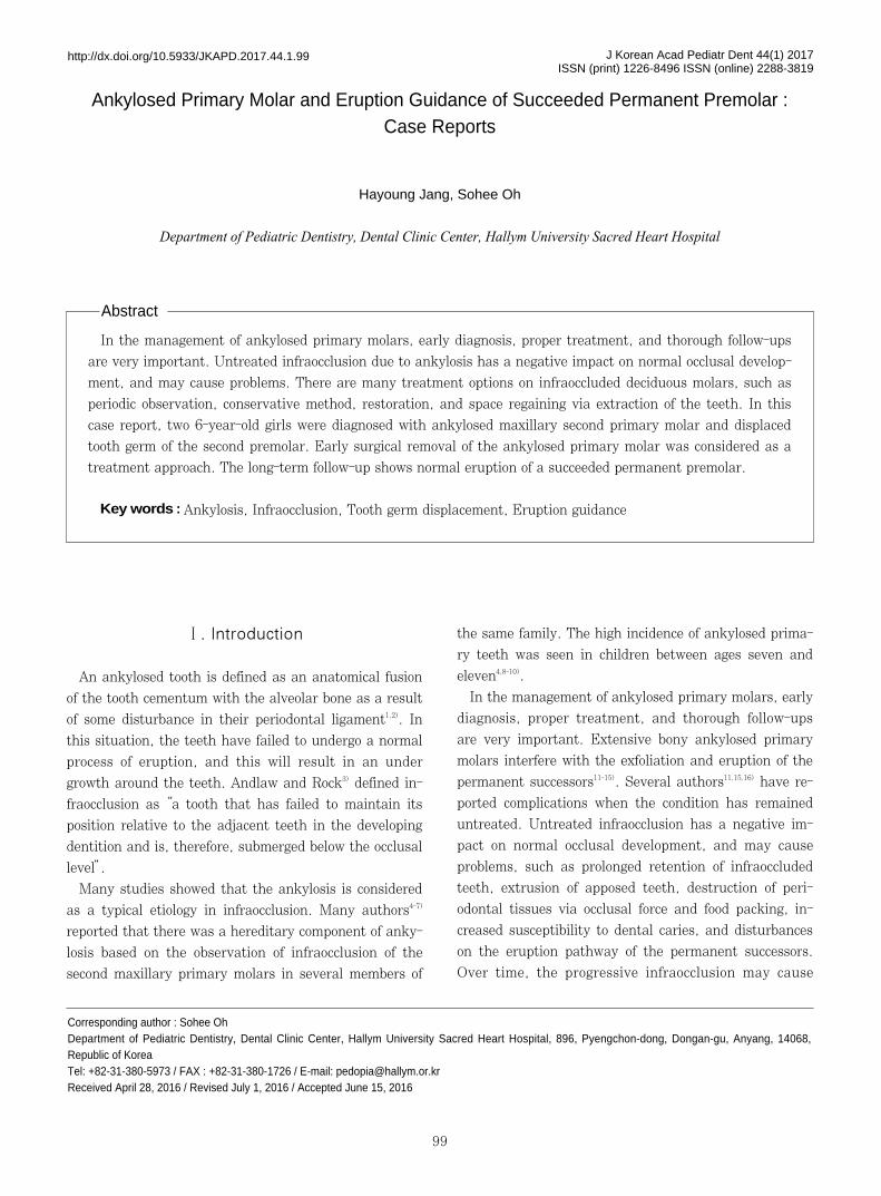
tilted toward the infraoccluded tooth (Fig. 1A).
Radiographic examination by means of a panoramic
radiographic showed the presence of a completely sub-
merged maxillary left second primary molar with an un-
derlying maxillary left second premolar tooth germ (Fig.
1B). The 3-dimensional cone-beam computed tomogra-
phy (CBCT) view revealed an ankylosed left second pri-
mary molar with displacement of the successor tooth
germ (Fig. 1C-1E). The underlying left second premolar
was in an abnormal relationship with the roots of the
primary molar and compared to right second premolar.
The left second premolar tooth germ was completely dis-
placed mesiopalatally.
An emergency intervention seemed logical when con-
sidering the loss of arch length and abnormal develop-
ment of the patient’s permanent successor tooth germ.
For this reason, surgical extraction was the preferred
treatment approach.
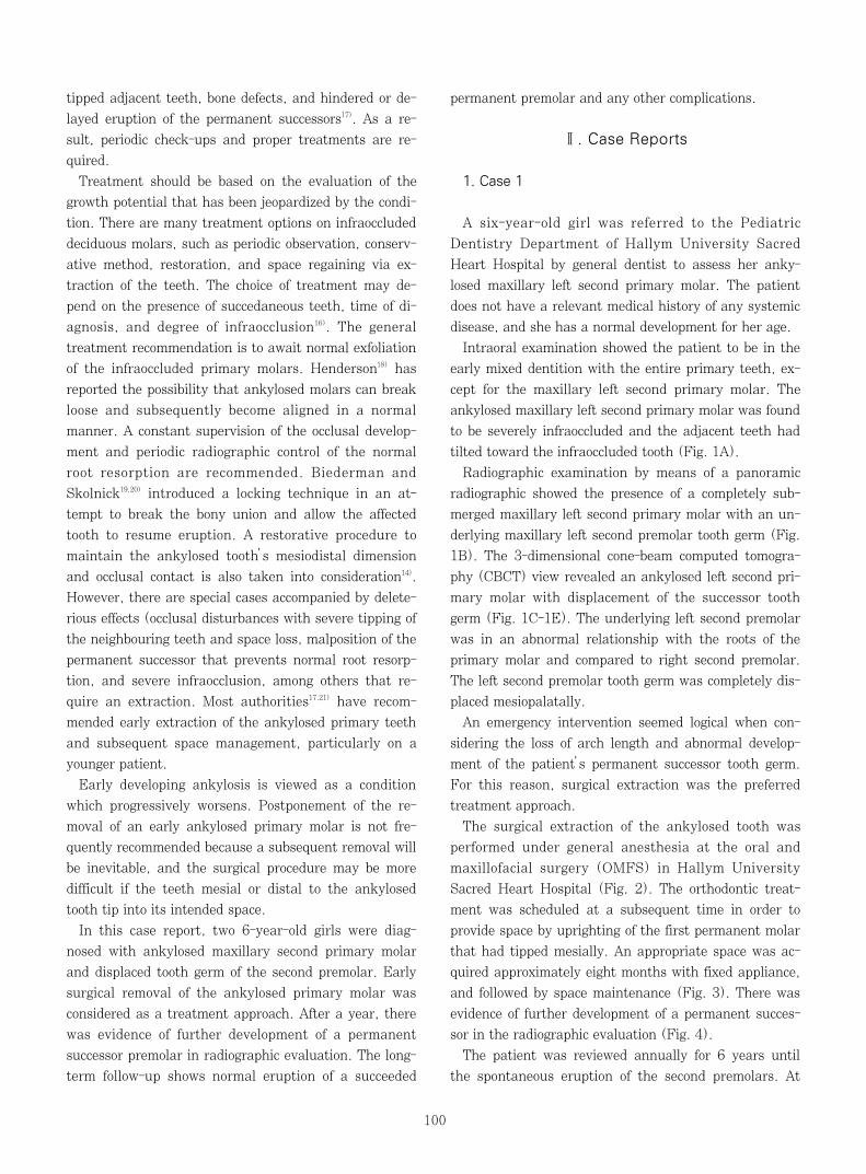
The surgical extraction of the ankylosed tooth was
performed under general anesthesia at the oral and
maxillofacial surgery (OMFS) in Hallym University
Sacred Heart Hospital (Fig. 2). The orthodontic treat-
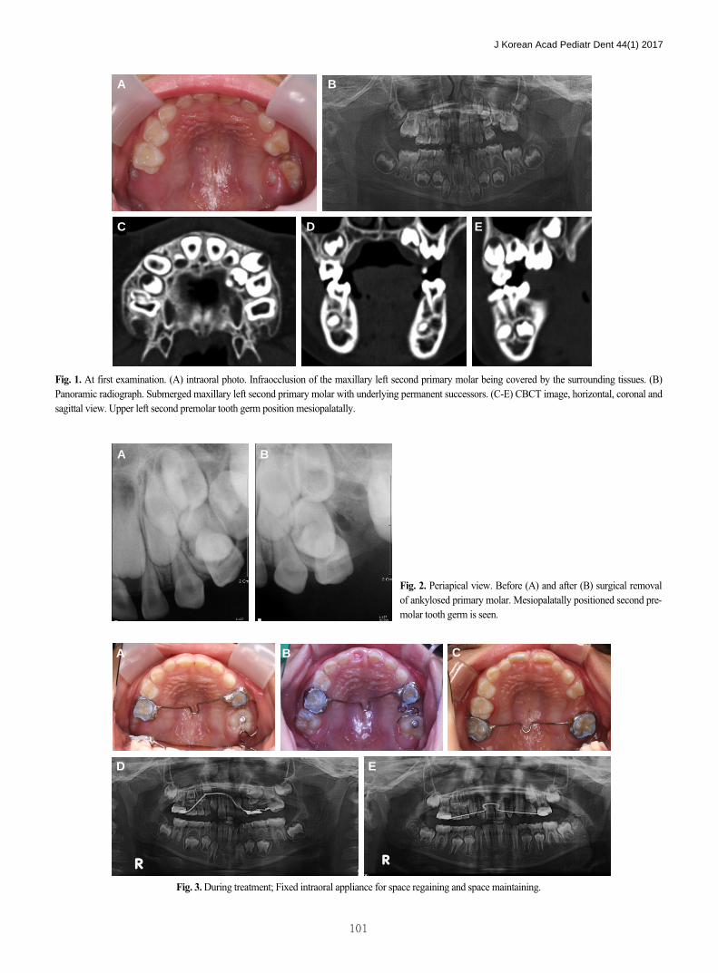
ment was scheduled at a subsequent time in order to
provide space by uprighting of the first permanent molar
that had tipped mesially. An appropriate space was ac-
quired approximately eight months with fixed appliance,
and followed by space maintenance (Fig. 3). There was
evidence of further development of a permanent succes-
sor in the radiographic evaluation (Fig. 4).
The patient was reviewed annually for 6 years until
the spontaneous eruption of the second premolars. At
J Korean Acad Pediatr Dent 44(1) 2017
101
Fig. 1. At first examination. (A) intraoral photo. Infraocclusion of the maxillary left second primary molar being covered by the surrounding tissues. (B)Panoramic radiograph. Submerged maxillary left second primary molar with underlying permanent successors. (C-E) CBCT image, horizontal, coronal andsagittal view. Upper left second premolar tooth germ position mesiopalatally.
Fig. 2. Periapical view. Before (A) and after (B) surgical removalof ankylosed primary molar. Mesiopalatally positioned second pre-molar tooth germ is seen.
Fig. 3. During treatment; Fixed intraoral appliance for space regaining and space maintaining.
A B
C D E
A B
A B C
D E

the age of 12, a comprehensive orthodontic treatment
was performed to align the whole dentition for approxi-
mately 18 months (Fig. 5). At the follow-up appoint-
ment, a favorable result was found on her dentition and
well-retained occlusion (Fig. 6).
J Korean Acad Pediatr Dent 44(1) 2017
102
Fig. 4. At 30 months after surgical removal of ankylosed tooth; Periapicalview shows the movement of permanent successor tooth germ in alveolarbone.
Fig. 5. Sign of spontaneously eruption (A) and complete eruption (B, C) of upper left second premolar.
Fig. 6. Follow-up examination of 8 years.
A B
C
A B

J Korean Acad Pediatr Dent 44(1) 2017
103
2. Case 2
A six-year-old girl was referred to the Pediatric
Dentistry Department of Hallym University Sacred
Heart Hospital by general dentist for the correction of a
partial eruption of the maxillary right second primary
molar. The patient had no relevant medical history of
any systemic disease, and her family reported no drug
use.
Intraoral examination showed the patient to be in the
early mixed dentition with a severely infraoccluded max-
illary right second primary molar. The maxillary left sec-
ond primary molar is ankylosed, partially erupted, and
covered by the surrounding tissues. Adjacent teeth,
maxillary first primary molar and first permanent molar
has tilted toward the infraoccluded tooth (Fig. 7A).
Radiographic examination by means of a panoramic
radiograph showed the presence of a infraoccluded max-
illary left second primary molar with an underlying max-
illary left second premolar tooth germ (Fig. 7B). The 3-
dimensional cone-beam computed tomography (CBCT)
view revealed an ankylosed right second primary molar,
and a displaced underlying permanent successor tooth
germ mesiopalatally located above the roots of the first
primary molar (Fig. 7C, 7D).
Surgical extraction of the ankylosed maxillary left sec-
ond primary molar was carried out under general anes-
thesia at the oral and maxillofacial surgery (OMFS) in
Hallym University Sacred Heart Hospital (Fig. 8). A
subsequent treatment involved regaining the loss of
space with a removable and fixed space regainer appli-
ance for approximately 6 months (Fig. 9). The space for
the permanent successor second premolar was acquired.
After a year, the development of maxillary right second
premolar germ was observed to its normal position (Fig.
10). In the follow-up examination, we observed a normal
and spontaneous eruption of the maxillary right second
premolar (Fig. 11).
Fig. 7. At first examination. (A) intraoral photo. Infraocclusion of the maxillary right second primary molar being covered by the surrounding tissues. (B)Panoramic radiograph. Submerged maxillary right second primary molar with underlying permanent successors. (C, D) CBCT image, horizontal and coro-nal view. Upper right second premolar tooth germ position mesiopalatally.
A B
C D
Fig. 8. After surgical removal of ankylosed second primary molar.

Ⅲ. Discussion
In this case report, the ankylosed maxillary primary
second molars are surgically removed, and the space is
regained for the spontaneous eruption of the displaced
tooth germ of the second premolar.
Normally, ankylosed primary molars with permanent
successor teeth are physiologically resorpted and exfoli-
ated. However, there are unknown factors that induce
extensive bony ankylosis. It may interrupt with a nor-
mal tooth resorption and induce eruption problems, such
as delayed eruption, ectopic eruption, and tooth im-
paction.
Ankylosis may occasionally occur even before the erup-
tion and complete root formation of the primary tooth.
Infraocclusion induced by ankylosis is diagnosed in the
early mixed dentition, between ages 8-9, at the peak of
facial growth9). The tooth remained in its ankylosed con-
dition until the adjacent first permanent molar began to
erupt through the gingival tissue. However, previous
studies have shown an infraocclusion of the primary mo-
lar that has been observed from a 3-year-old child4,10).
The ankylosed primary molar should undergo a period-
ic follow-up until the proper time for treatment interven-
tion, such as extraction and space management, for
guiding the eruption of the permanent successor. The
J Korean Acad Pediatr Dent 44(1) 2017
104
Fig. 9. During treatment. Fixed and removable intraoral appliance for Space regaining.
Fig. 10. At 24 months after surgical removal of ankylosed tooth; space maintaining appliance (A) and panoramic view (B) shows the movement of perma-nent successor tooth germ in alveolar bone.
Fig. 11. follow-up examination of 2 years; Normal eruption of Upper right second premolar is observed.
A B
A B
A B
J Korean Acad Pediatr Dent 44(1) 2017
105
decision, as to whether or not to instigate treatment, is
based on the age of the patient, the degree and extent of
the infraocclusion, the amount of root resorption, the
severity of the tilting of neighboring teeth, and the pres-
ence and location of the permanent successor.
The ankylosed maxillary primary molar has the ten-
dency to become severely submerged, and progressively
worse with age22). Infraocclusion in the maxillary primary
molar with permanent tooth germ displacement and loss
of arch length caused by tipping of the adjacent teeth
are predicted to delay the eruption or impaction of the
permanent successor. It should be surgically removed
and a space maintaining appliance should be used.
An extraction should be deferred if possible until the
first permanent molar erupts in order to allow space
maintenance. Extraction is recommended as soon as
possible by many authors. Meanwhile, other authors
recommended early treatment only when there is a se-
verely affected primary molar, and if there is evidence of
possible future crowding. According to Kurol and
Thilander23), early extraction is only necessary when
there is a combination of occlusal disturbance, severe
tipping of neighbouring teeth, space loss, malposition of
the permanent successor with irregular primary root re-
sorption, and severe infraocclusion. Early extraction may
be technically difficult and may result in fracture of the
roots, or possible disturbance of the succeeded tooth bud.
In addition, early extraction and long-term space
maintenance would have been simple solutions in these
cases, and may have prevented the tilting, loss of space
for the successor, and the significant centre line shift,
which can be difficult to correct. The marginal ridge of
the ankylosed tooth that is on the gingival level or below
must be extracted. The tendency of ankylosed maxillary
molars to be severely infraoccluded usually indicates
early extraction24).
Severe tipping of the neighbouring teeth toward the
infraoccluded teeth may induce arch length problems
that should be surgically extracted. Mesial tilting of the
first permanent molar makes surgical extraction difficult.
In this situation, space regaining is preceded until surgi-
cal extraction due to the severe loss of arch length that
required ankylosed teeth extraction. Altay and Cengiz25)
recommended space regaining prior to the extraction of a
submerged tooth.
Space regaining of the competent spaces and extrac-
tion were performed with minimum possibility of trauma
toward the subsequent permanent molar. These surgical
sequences are considered much easier and more comfort-
able for both the surgeon and the patient.
The sequence is not affected by the treatment progno-
sis or determined by the state of infraocclusion at the
initial diagnosis of its removal. Jung26) reported two cas-
es with space management and a case extraction prior to
space regaining. It was determined upon first examina-
tion where it was easy to extract. Both methods induced
the subsequent permanent premolar to erupt sponta-
neously, indicating that the treatment result is not af-
fected by the sequence.
Cozza17) reported three cases of early extraction of in-
fraoccluded ankylosed primary molars, accompanied by
orthodontic treatment until the spontaneous eruption of
the second premolar.
In this case report, ankylosed primary molars are ex-
tracted early, and space management is performed.
Surgical extraction of the primary molar should be care-
fully approached for the possibility of affecting the suc-
cessor permanent tooth germ through iatrogenic damage
or complication during operation. We achieved a sponta-
neous eruption of the successor permanent teeth via ear-
ly removal of the ankylosed teeth, and performed treat-
ment with space management. Periodic follow-up
showed proper vertical alveolar bone height on the whole
dentition.
In the management of an ankylosed tooth, early recog-
nition and diagnosis are extremely important. In an
ankylosed tooth, the obstruction is removed, and the
displaced permanent tooth will normally erupt. If there
is a severe infraocclusion that requires a space regaining
procedure, it should be considered before treatment due
to the possibility of re-evaluation of the orthodontic
treatment after the eruption of the second premolar.
Ⅳ. Summary
In this case, the maxillary second premolar tooth
germs were displaced due to the ankylosis of the maxil-
lary second primary molar. The ankylosed tooth was ex-
tracted, and the space regaining was performed, thereby
leading to the normal eruption of the subsequent perma-
nent tooth buds.
In two cases presented here, the early extraction of
the infraoccluded primary molars and the active treat-
ment of space regaining allowed the spontaneous erup-
tion of the second premolars, thereby obtaining a normal
vertical relationship and normal bone height.
References
1. Owen TL : Ankylosis of teeth. J Mich State Dent
Assoc, 47:347-350, 1965.
2. Pilo R, Littner MM, Marshak B, Aviv I : Severe
infraclusion ankylosis: report of three cases. ASDC J
Dent Child, 56:144-146, 1989.
3. Kurol J, Magnusson BC : Infraocclusion of primary
molars: a histologic study. Scand J Dent Res, 92:
564-576, 1984.
4. Kurol J : Infraocclusion of primary molars: an epi-
demiologic and familial study. Community Dent Oral
Epidemiol, 9:94-102, 1981.
5. Via WF : Submerged Deciduous Molars : Familial
Tendencies. J Am Dent Assoc, 69:127-129, 1964.
6. Helpin ML, Duncan WK : Ankylosis in monozygotic
twins. ASDC J Dent Child, 53:135-9, 1986.
7. Douglass J, Tinanoff N : The etiology, prevelance
and sequelae of infraclusion of primary molars.
ASDC J Dent Child, 58:481-483, 1991.
8. Noble J, Karaiskos N, Wiltshire WA : Diagnosis and
management of the infraerupted primary molar. Br
Dent J, 203:632-634, 2007.
9. Rune B, Sarnos KV : Root resorption and submer-
gence in retained deciduous second molars. Eur J
Orthod, 6:123-131, 1984.
10. Steigman S, Koyoumdjisky-Kaye E, Matrai Y :
Submerged deciduous molars in the preschool chil-
dren: an epidemiological survey. J Dent Res, 52:
322-326, 1973.
11. Biederman W : The problem of the ankylosed tooth.
Dent Clin North Am, 12:409-424, 1968.
12. Andlaw RJ : Submerged deciduous molars. A review
with special reference to the rationale of treatment.
J Int Assoc Dent Child, 5:59-66, 1974.
13. Darling AI, Levers BG : Submerged human decidu-
ous molars and ankylosis. Arch Oral Biol, 18:1021-
1040, 1973.
14. Krakowiak FJ : Ankylosed primary molars. ASDC J
Dent Child, 45:288-292, 1978.
15. Konstat MM, White GE : Ankylosed teeth: a review
of the literature. J Mass Dent Soc, 24:74-78, 1975.
16. Jenkins FR, Nichol RE : Atypical retention of
infraoccluded primary molars with permanent suc-
cessor teeth. Eur Arch Paediatr Dent, 9:51-55,
2008.
17. Cozza P, Gatto R, Mucedero M, et al. : Case report:
severe infraocclusion ankylosis occurring in siblings.
Eur J Paediatr Dent, 5:174-178, 2004.
18. Henderson HZ : Ankylosis of primary molars: a clin-
ical, radiographic and histologic study. ASDC J Dent
Child, 46:117-122, 1979.
19. Biederman W : Etiology and treatment of tooth
ankylosis. Am J Orthod, 48:670-684, 1962.
20. Skolnick IM : Ankylosis of Maxillary Permanent
First Molar. J Am Dent Assoc, 100:558-560, 1980.
21. Belanger GK, Strange M, Sexton JR : Early ankylo-
sis of a primary molar with self-correction: case
report. Am Academy Pediatr Dent, 8:37-40, 1986.
22. Messer LB, Cline JT : Ankylosed primary molars :
results and treatment recommendations from an
eight-year longitudinal study. Pediatr Dent, 2:37-
47, 1980.
23. Kurol J, Thilander B : Infraocclusion of primary
molars and the effect on occlusal development, a
longitudinal study. Eur J Orthod, 6:277-293, 1984.
24. Kurol J, Koch G : The effect of extraction of infraoc-
cluded deciduous molars : A longitudinal study. Am
J Orthod, 87:46-55, 1985.
25. Altay N, Cengiz SB : Space-regaining treatment for
a submerged primary molar: a case report. Int J
Paediatr Dent, 12:286-289, 2002.
26. Jung JH, Kim YJ, Kim HJ, Nam SH : Eruption
guidance for tooth germ of premolar displaced by
infraoccluded upper deciduous molar. J Korean Acad
Pediatr Dent, 39:390-396, 2012.
J Korean Acad Pediatr Dent 44(1) 2017
106

J Korean Acad Pediatr Dent 44(1) 2017
107
주요어:유착, 저위교합, 치배 변위, 맹출 유도
유착된 유구치와 후속 구 소구치의 맹출 유도 : 증례보고
장하 ∙오소희
한림 학교성심병원 치과진료센터 소아치과
유착된 유구치의 치료에 있어 조기 진단이 매우 중요하다. 유착으로 인하여 발생된 저위교합을 방치하는 경우 교합적인 문
제가 생길 수 있으므로 정기검진을 통한 진단과 이에 따른 적절한 치료가 필요하다. 저위교합 된 유구치의 치료 방법으로 정
기검진, 보존적 접근, 보철적 수복, 발치를 수반한 공간획득 등이 있다. 본 증례에서는 상악 제2유구치의 유착으로 인하여 제
2소구치 치배의 변위를 보이는 두 명의 환아를 상으로 이환치의 발거를 먼저 시행하고 고정성 혹은 가철성 교정장치를 이
용하여 공간확장술을 함으로써 변위 되었던 제2소구치 치배의 정상 맹출을 유도하 으며 장기간에 걸쳐 부작용이 없었음을
보고하는 바이다.
국문초록


![Ankylosed permanent teeth: incidence, etiology and ... · probably due to tongue thrusting [23]. Alternatively, supra-eruption of opposing teeth might occur. Diagnosis Overall, the](https://static.fdocuments.in/doc/165x107/5e230360f3eb8d36a1454f20/ankylosed-permanent-teeth-incidence-etiology-and-probably-due-to-tongue-thrusting.jpg)


